Article

Molecular Cloning and Functional Analysis of the Duck TLR4 Gene

Jiangsu Key Laboratory for Animal Genetic, Breeding and Molecular Design, Yangzhou University, Yangzhou 225009, Jiangsu, China
*
Authors to whom correspondence should be addressed.
These authors contributed equally to this work.
Int. J. Mol. Sci. 2013, 14(9), 18615-18628; https://doi.org/10.3390/ijms140918615
Submission received: 10 August 2013 / Revised: 26 August 2013 / Accepted: 29 August 2013 / Published: 10 September 2013
(This article belongs to the Section Biochemistry)

Abstract

:
Toll-like receptor 4 (TLR4) recognizes pathogen-associated molecular patterns in some animals and has been shown to be closely associated with several diseases such as tumors, atherosclerosis, and asthma. However, its function in ducks is not clear. Alternative splicing of the TLR4 gene has been identified in pigs, sheep, mice, and other species, but has not yet been reported in the duck. In this study, alternative splicing of the duck TLR4 gene was investigated using reverse transcription-polymerase chain reaction (RT-PCR). Duck TLR4 gene (duTLR4, accession number: KF278109) was found to consist of 3367 nucleotides of coding sequence. An alternative splice form, TLR4-b, was identified and shown by alignment to retain the intron between exons 1 and 2. Real-time quantitative polymerase chain reaction (qPCR) analyses suggested that duTLR4-a (wild-type) mRNA is widely expressed in various healthy tissues, whereas TLR4-b is expressed at only low levels. Following stimulation of normal duck embryo fibroblasts with lipopolysaccharide, the expression of both isoforms initially increased and then decreased. Expression of the wild-type isoform subsequently increased again, while that of the variant remained low. The expression levels of wild-type TLR4 were further analyzed by transient transfection of a pcDNA3.1(+)-TLR4-a overexpression vector into duck embryo fibroblasts. qRT-PCR analyses showed that after stimulation with LPS and poly(I:C) the expression levels of IL-1β, IL6, and MHC II increased with a response-efficacy relationship. Our experimental results indicate that TLR4 plays an important role in resistance to both bacterial and viral infections in the duck.

1. Introduction

Toll-like receptors (TLRs) and pathogen pattern recognition receptors (PRRs) were discovered in the last century and subsequently shown to be involved in innate immunity [1]. TLRs represent a major component of the vertebrate pattern-recognition receptor system, which plays a role in detecting invading microorganisms and distinguishing between them [2,3]. To date, 13 members of the TLR family have been investigated in mammals, among which TLR4 is the most extensively studied. The various TLRs exhibit different patterns of expression, and TLR4 is most abundantly expressed in placenta and in the myelomonocytic subpopulation of leukocytes.
TLR4 recognizes the specific and highly conserved molecular patterns expressed on pathogenic microorganisms, helping to initiate and regulate the body’s immune response [47]. TLR4 is associated with various diseases in animals, such as tumors, atherosclerosis, and asthma. Transcript variants of TLR4 have been found in pigs, sheep, mice, and other species, but the protein-coding potential of most variants is uncertain [810]. Alternative splicing of TLR4 in the duck has not yet been reported, and its function in ducks is not clear.
In this study, we identified an alternative splicing product of the duTLR4 gene (TLR4-b) but found only low expression of this variant in various healthy tissues. Next, we investigated TLR4 expression levels using a pcDNA3.1(+)-TLR4-a overexpression vector that was transiently transfected into duck embryo fibroblasts. The expression of wild-type TLR4-a increased after stimulation with polyinosinic:polycytidylic acid [poly(I:C)]. In addition, qRT-PCR analyses showed that expression levels of interleukin IL-1β, IL-6, and major histocompatibility complex II (MHCII) increased after stimulation with lipopolysaccharide (LPS) or poly(I:C), revealing a response-efficacy relationship. Our experimental results confirm that TLR4 plays an important role in resistance to both bacterial and viral infections in the duck.

2. Results

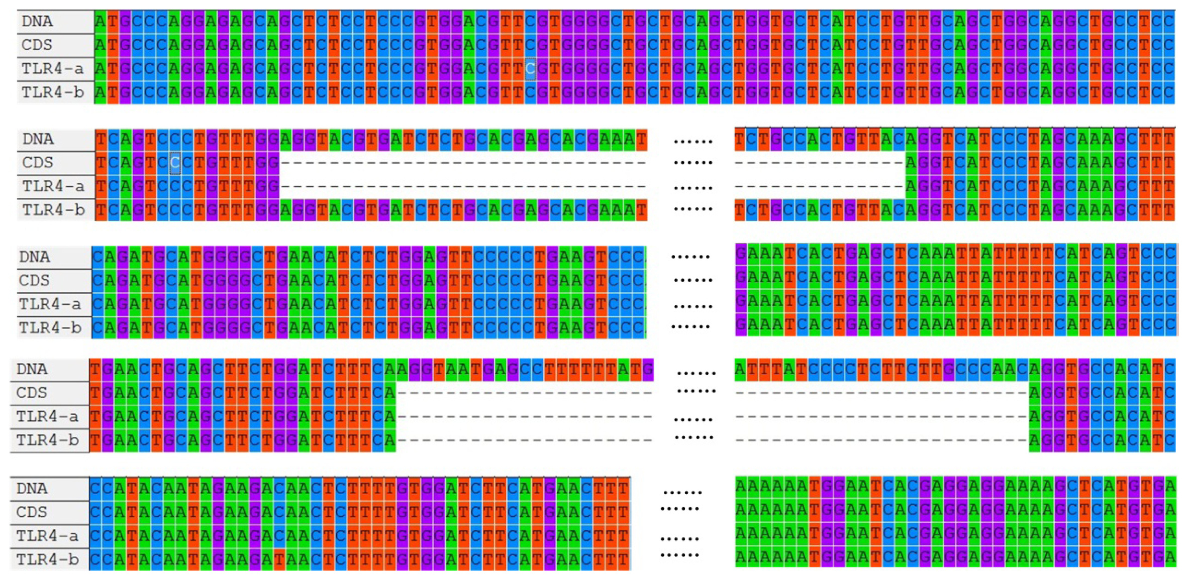
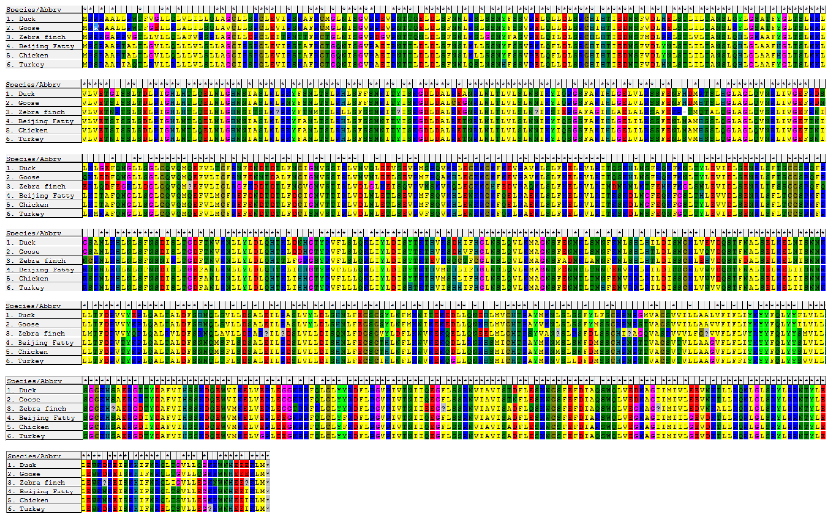
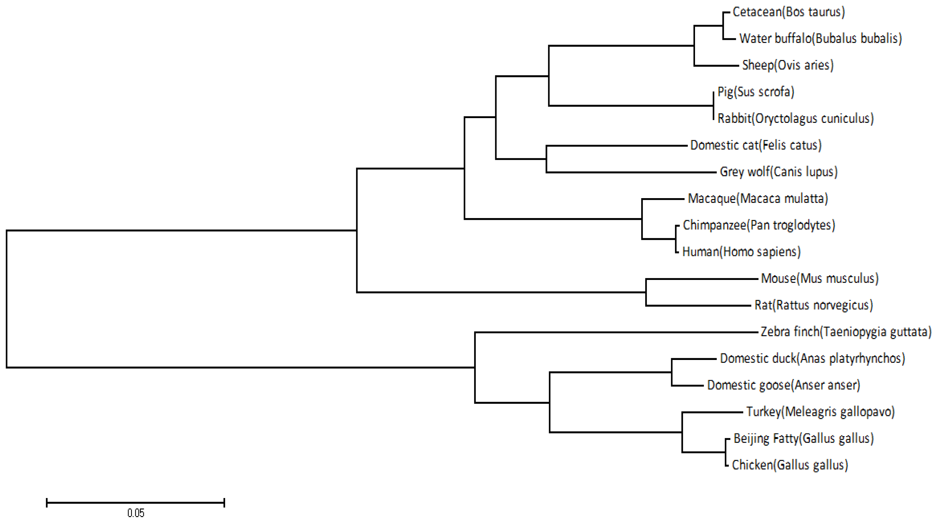
2.1. Cloning and Sequence Analysis of duTLR4

The coding sequence (CDS) of wild-type duTLR4 (TLR4-a) of the Jinding duck (Anas platyrhynchos), determined by full-length RT-PCR and long distance (LD) PCR, was 2532 bp in length and contained three exons and two introns. Alignment of duTLR4 mRNA (accession number: JN048668.1) and genomic DNA (accession number: JQ839148) revealed two variants (a and b) with an alternative 835 bp region between the first and second exons (Figure 1).
The entire open reading frame (ORF) of duck TLR4 is 2532 bp (GenBank ID: JN048668.1) and encodes 833 amino acid residues (Figure 2). Not surprisingly, the nucleotide sequence of TLR4 that we obtained is nearly identical to that previously reported for duck. The nucleotide sequence shows 96.8%, 86.5%, 86.3%, and 80.5% homology with the TLR4 sequences of red jungle fowl, goose, turkey, zebra finch, and chicken, respectively.
An alignment of TLR4 amino acid sequences of the above species is shown in Figure 2. A phylogenetic tree for TLR4 based on 18 species is shown in Figure 3.
The duck TLR4 sequence shows the highest sequence homology with goose TLR4 (96.8%, Figure 3). Homology between chicken, turkey, and red jungle fowl sequences is also high, but the sequence homology between zebra finches and the other species is lower. As shown in Figure 3, Beijing fatty chicken and red jungle fowl are tightly clustered into one class, and then further clustered with turkey, duck, and goose, and more distantly with zebra finch. This phylogenetic tree is consistent with the genetic relationship of those species. Thus, the TLR4 gene is conserved within the evolutionary process.

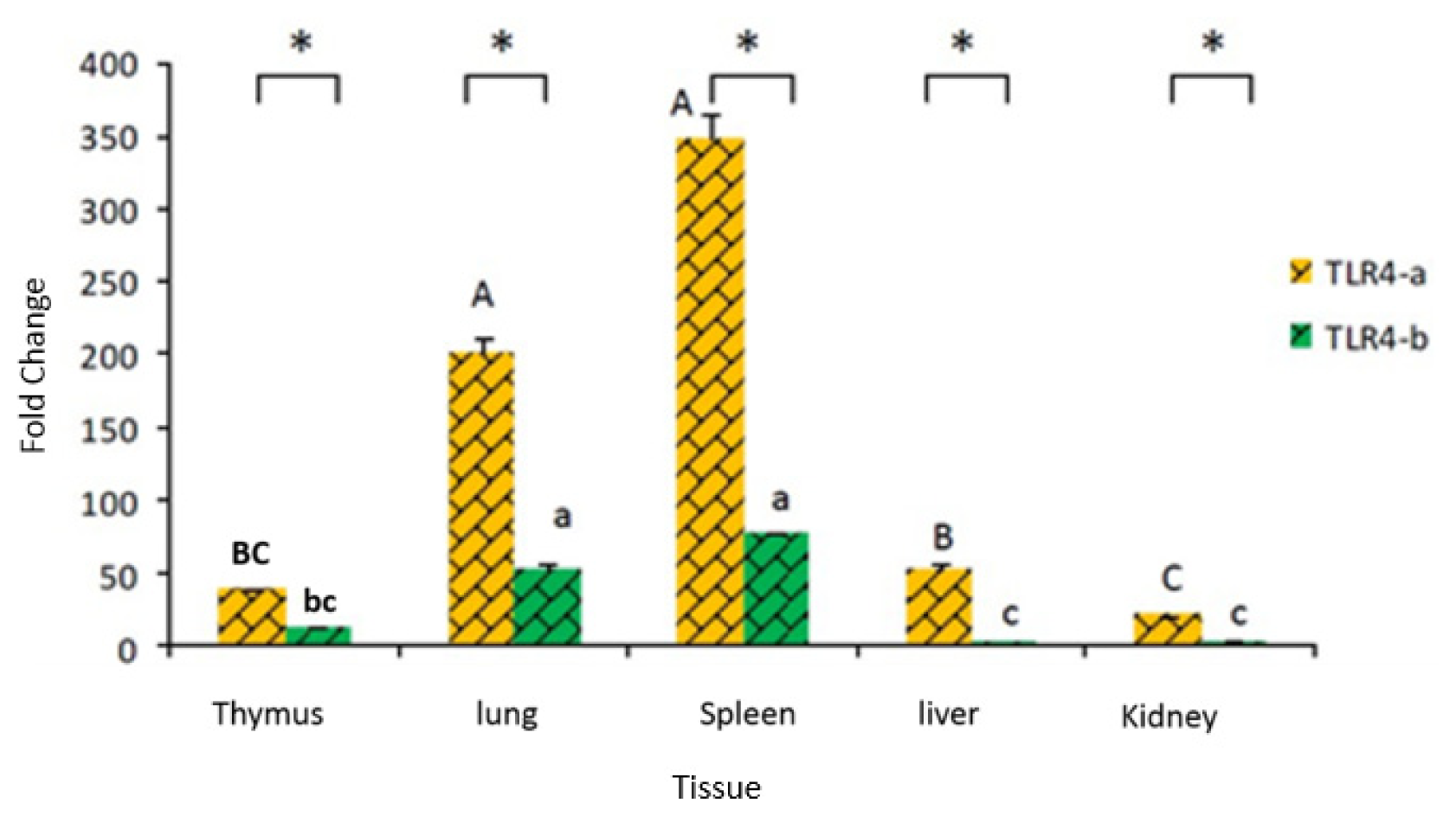
2.2. Tissue Distribution of Duck TLR4 mRNA

In healthy tissue, the highest expression of TLR4-a was seen in the spleen and lung. The expression of wild-type TLR4-a was significantly higher than that of the variant form TLR4-b (p < 0.05) in all tissues tested and especially in the liver and kidney (Figure 4).

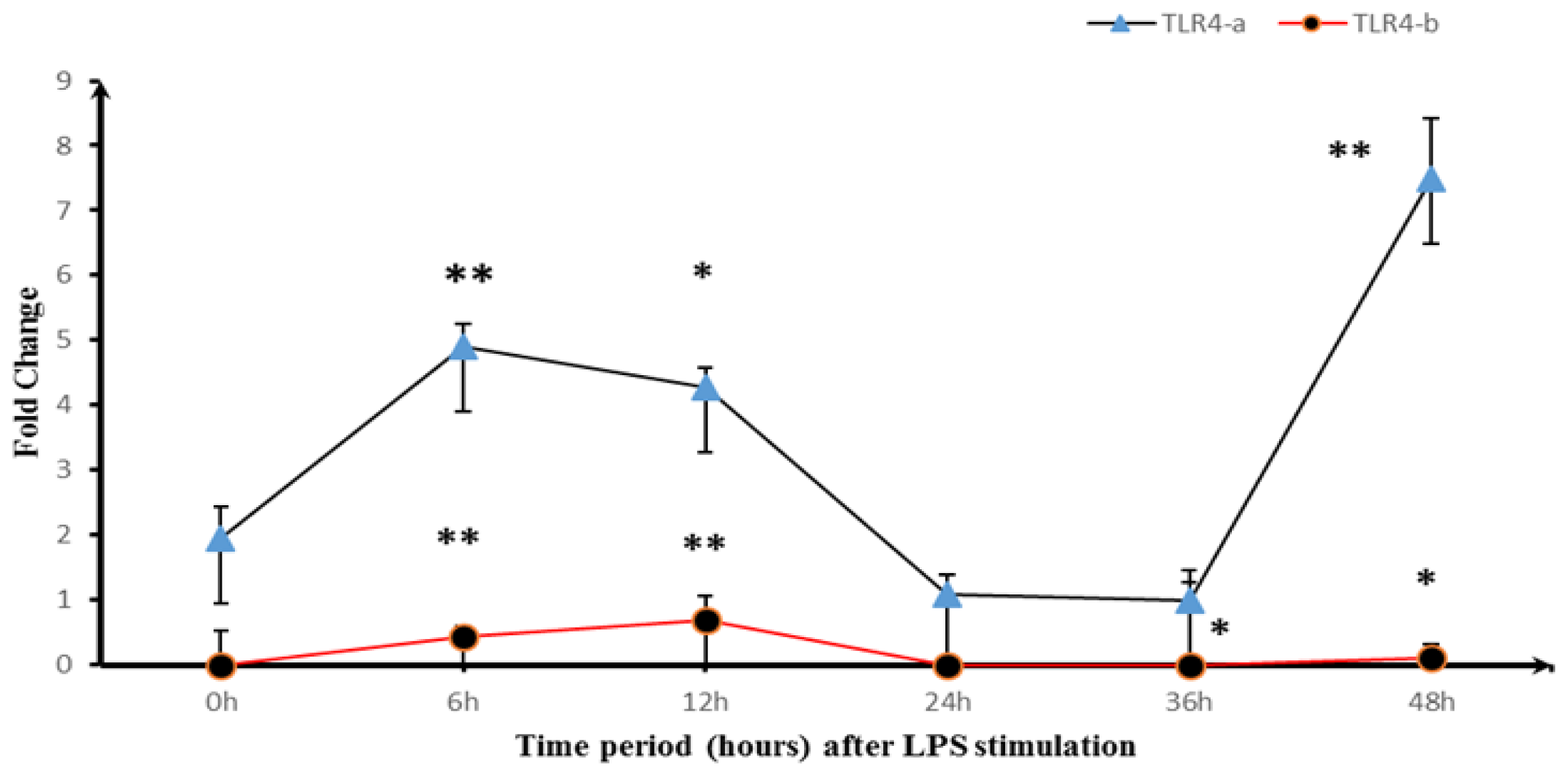
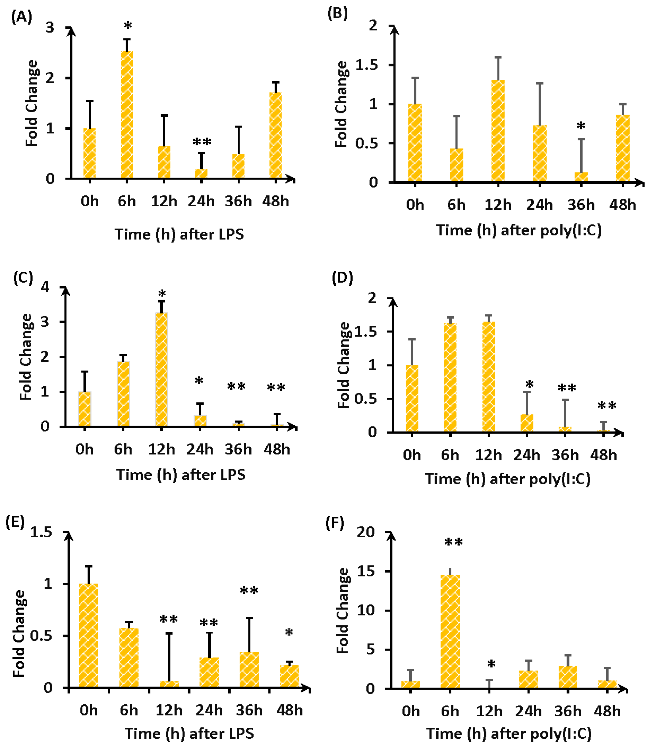
2.3. Expression of TLR4-a and TLR4-b Following Stimulation with LPS

Normal duck embryo fibroblasts (DEFs) were stimulated with LPS and cells were collected at 0 h, 6 h, 12 h, 24 h, 36 h, and 48 h. RNA was extracted and reverse transcribed, and the expression level of the two alternative splicing isoforms was compared (Figure 5). DEFs stimulated with LPS showed enhanced expression of both TLR4-a and TLR4-b, reaching a peak at 6 h and 12 h, followed by a sharp decline over time. Expression of TLR4-a subsequently increased again at 36 h, whereas expression of TLR4-b remained very low.
DEFs were transiently transfected with a pcDNA3.1(+)-TLR4 expression vector and stimulated with LPS or Poly(I:C) (a dsRNA mimetic) to simulate infection with Gram-negative bacteria or viruses, respectively.
After stimulation with LPS or poly(I:C), cells were collected and RNA was extracted at 0 h, 6 h, 12 h, 24 h, 36 h, and 48 h. Quantitative fluorescence was used to detect expression changes in genes encoding the immune modulators IL-1β (Figure 6A,B), IL-6 (Figure 6C,D), and MHC II (Figure 6E,F). Either LPS or poly (I:C) stimulation induced TLR4 gene overexpression in fibroblasts, IL-1β expression initially increased and then decreased, followed by a second increase in LPS-stimulated cells only. IL-6 expression showed a sharp increase in both LPS- or poly (I:C)-stimulated samples within 12 h, and then showed a sharp decline in expression. MHC II expression increased within 12 h after poly(I:C) stimulation and then decreased (12–48 h).

3. Discussion

TLR 4 is a specific receptor that can detect LPS present in most Gram-negative bacteria and regulate the signal transduction events induced by LPS, and therefore plays an important role in pathogen recognition and activation of the innate immune system [11]. TLR4 has been shown to play a significant role in the susceptibility of mammals and chickens to systemic salmonellosis [12].
Bacterial infections cause worldwide diseases each year [13], dramatically influencing the performance of livestock. The TLR4-mediated immune response is the first line of defense against bacterial infection. Several earlier studies have addressed expression of the duck TLR4 gene and protein during pathogenic challenge [4]. In most of these studies, activation of TLR4 was monitored by measuring the level of a transcript harboring the third exon of a previously reported transcript variant. A recent study has shown that more than 40% of human gene transcripts undergo alternative splicing, notably many of the genes involved in immunological responses [14,15]. Alternatively spliced proteins probably function in regulation of the immune response. This study focused on TLR4 splice variants in the duck and their function.
We identified a splice variant in duck, TLR4-b, which retains the first intron of 835 bp in length. There are two mechanisms by which this may affect the translation process: (A) introduction of a premature termination codon at 291 bp, leading to translation of only 76 amino acid residues; or (B) a reading frame shift (RFS) that starts translation from 967 bp, resulting in a 76-amino acid deletion and leading to removal of the first transmembrane domain. Previous studies have reported four alternative splice isoforms in humans [16], two in mice [17], three in sheep [7], and three in pigs [18]. Whether there are additional alternatively spliced forms in the duck requires further investigation. These findings strongly suggest that alternative TLR4 splice isoforms have a large impact on TLR4 function.
In our experiment, we found a constitutive expression of TLR4 gene in various tissues. Besides, we also detected that TLR4-a mRNA was significantly increased after LPS challenge, while a slight change of TLR4-b was observed. It is evident from these findings that duck TLR4’s function is dependent on TLR4-a.
A reliable method was developed to monitor TLR4 transcript variants individually after LPS stimulation of duck fibroblasts. Total TLR4 expression was rapidly down-regulated within the first 6 h following LPS stimulation, subsequently increased (8–12 h) and declined thereafter. It has been shown previously that expression of TNF-α, a marker of LPS-derived activation, was dramatically elevated within 0.5 h, indicating that LPS-induced immune responses are immediately triggered [19]. It is known that TLR4 shows distinct patterns of expression in different type of cells. For example, the degradation of TLR4 observed in monocytes is associated with LPS tolerance, helping the animal survive bacterial infection [20]. It is notable that fibroblasts are a kind of antigen-presenting cell that can trigger immunological responses through the release of large amounts of pro-inflammatory cytokines. Because fibroblasts are the most common cell type within the body, it is reasonable to assume that TLR4 expression in fibroblasts is tightly regulated.
The expression of TLR4-a was consistent with that of total TLR4, indicating that TLR4-a is the functional transcript variant. It is likely that these transcript variants have biological relevance because alternative splicing is a powerful and efficient regulatory mechanism known to regulate gene expression and protein production [21]. It has frequently been observed that the immune system regulates its own response during pathogen infection.
Emerging results indicate that TLR4-a is an important immune-related gene in the duck, in part because it is expressed preferentially in immune or immune-related organs under normal conditions. The duck TLR4 gene shows the same organization as TLR4 genes from sheep, mouse, chicken, goose, and pig, with three exons and two introns, demonstrating that this gene has been conserved during evolution. Conserved structural features are also present within the duck homologs. Mutations in TLR4 have been associated with differences in LPS responsiveness. According to the current model, TLR4 interacts with three different extracellular proteins: LPS binding protein (LBP), CD14, and myeloid differentiation protein 2 (MD-2), leading to LPS-mediated NF-κB activation and the production of proinflammatory cytokines [22,23]. Thus, TLR4 plays an important role in the defense against infections by Gram-negative bacteria that release LPS.
In this study, we used LPS to stimulate duck embryo fibroblasts and monitored expression of the effector genes IL1β, IL6, and MHC II in the TLR4 pathway [24]. Expression of these genes generally showed an upward trend immediately after stimulation, followed by a decrease and then a second increase. TLR4 is part of a complex immune system network and plays a key role in protection as an important regulator of components present in many infectious diseases. Sheng et al. found that LPS-induced expression of TLR4 is rapidly elevated and maintained at a high level during the entire TLR4 process [25,26]. In T lymphocytes, thymus, and spleen tissue macrophages, LPS can significantly increase expression of TLR4 without affecting gene expression. In human polymorphonuclear leukocytes and monocytes, TLR4 expression is significantly increased upon stimulation.
Cell and tissue distribution are important determinants of TLR4 function, as they influence the capacity to detect different microorganisms during their entry and growth in different tissues. Understanding the expression patterns of duck TLRs will enable a better interpretation of immune induction and the host-pathogen relationships that define infectious disease biology in the duck [16,27]. A number of studies have defined the response of cultured chicken cells after exposure to a variety of challenges with microorganisms or their products [28]. In the present study, duTLR4 mRNA was expressed extensively in all normal tissues examined, including the liver, kidney, spleen, thymus, and lung. These data were consistent with those for mammalian and chicken TLR4. In mammals, fatty acids are able to activate TLR4, resulting in enhanced secretion of proinflammatory chemokines and cytokines such as tumor necrosis factor-α and IL-6. Mice deficient in TLR4 are protected from obesity-induced insulin resistance and inflammatory insults. Additionally, TLR4 is a molecular link between nutrition, lipids, and innate immunity. The extensive distribution of functional TLR4 in various duck tissues may contribute to a variety of immune responses.

4. Experimental Section

4.1. Animals and Tissue Sample Collection

Jinding ducks (Anas platyrhynchos; 3 days old) were obtained from the National Waterfowl Germplasm Resource Gene Pool (Taizhou, China). The ducks (n = 80) were slaughtered by exsanguination according to protocols approved by the Animal Care Advisory Committee of Yangzhou University and blood samples were collected for DNA extraction. The liver, lung, spleen, kidney, and thymus were removed and immediately frozen in liquid nitrogen before storage at −80 °C for RNA isolation.

4.2. DNA and RNA Isolation and cDNA Synthesis

Genomic DNA was isolated using the TIANamp Genomic DNA Kit according to the manufacturer’s instructions (Tiangen Biotech (Beijing) Co., Ltd., Beijing, China). Total RNA was extracted from collected tissue samples or cells using RNAiso Plus (TaKaRa, Dalian, China) according to the manufacturer’s instructions. Total RNA concentrations were determined using a Beckman Coulter DU800 spectrophotometer (THERMO, Fullerton, CA, USA). Reverse transcription was performed using TransScript First-Strand cDNA Synthesis SuperMix (Tiangen Biotech (Beijing) Co., Ltd., Beijing, China).

4.3. Cloning of the TLR4 Gene

The DNA and mRNA sequences of TLR4 (GenBank ID: JN048668) were amplified using the primers listed in Table 1. The polymerase chain reaction (PCR) was performed in 20-μL reactions containing 0.4 μL of first-strand cDNA, 1 μL each of 10 μM forward and reverse primers, 7.6 μL DEPC-treated water, and 10 μL of 2 × Taq PCR MasterMix (Tiangen, Beijing, China). Thermal cycling conditions were as follows: 1 cycle of 95 °C for 5 min, then 32 cycles of 94 °C for 30 s, 65 °C for 30 s, and 72 °C for 2 min 30 s, followed by 1 cycle of 72 °C for 10 min. PCR products were analyzed by electrophoresis on 1.0% agarose gels, purified using a gel extraction kit (Dongsheng Biotech Co., Ltd., Beijing, China), ligated into the pMD19-T Vector (TaKaRa, Dalian, China), transformed into E. coli DH5α cells, and sequenced.

4.4. Construction of pcDNA 3.1(+)-TLR4 Overexpression Vector

A DNA fragment containing the entire open reading frame of duck TLR4 was PCR amplified using the specific primers pcDNA3.1(+)-TLR4-F (5′-ggaattcCATATGATGCCCAGGAGAGCAGCTCTCCTC-3′); and pcDNA3.1(+)-TLR4-R (5′-gcTCTAGACATGAGCTTTTCCTCCTCGTGATTCC-3′) and the plasmid pMD19-T-TLR4 as a template. Primer pcDNA3.1(+)-TLR4-F introduced a Nde I site, and primer pcDNA3.1(+)-TLR4-R introduced a Xba I site (underlined). After digestion with Nde I and Xba I, the PCR product was inserted into the vector pcDNA3.1(+) (Invitrogen, Carlsbad, CA, USA). Proper plasmid construction was confirmed by sequencing.

4.5. Sequence Analysis, Multiple Sequence Alignment, and Phylogenetic Analysis

Searches for nucleotide and protein sequence similarities were conducted with the BLAST algorithm from the National Center for Biotechnology Information (NCBI, http://blast.ncbi.nlm.nih.gov/Blast.cgi) [29]. Based on the duck TLR4 sequence (GenBank ID: JN048668), primers pMD19-T-TLR4-F and pMD19-T-TLR4-R were designed and used to amplify the potential TLR4 cDNA sequence from total RNA extracted from the liver of a Jinding duck. The deduced amino acid sequence of TLR4 was analyzed with the Expert Protein Analysis System (Expasy, http://www.mrc-lmb.cam.ac.uk/genomes/madanm/pres/swiss1.htm) [30]. Multiple sequence alignment of TLR4 CDS and DNA was performed with the ClustalW multiple alignment program ( http://www.ebi.ac.uk/Tools/clustalw2/) and the Multiple Align Show program ( http://www.bioinformatics.org/sms/). Using a neighbor-joining (NJ) algorithm, an unrooted phylogenetic tree was constructed based on the deduced amino acid sequences of TLR4 using MEGA 5.05 software ( http://www.megasoftware.net). To derive confidence values for tree nodes, bootstrap values were obtained for 1000 replications [3133].
A phylogenetic tree was constructed from the TLR4 coding regions of 18 different species: the domestic duck (Anas platyrhynchos, accession number: JN048668), red jungle fowl (Gallus gallus, accession number: NM_001030693), the domestic goose (Anser anser, accession number: HQ436371), chicken (Gallus gallus domesticus, accession number: JQ711152), cattle (Bos Taurus, accession number: NM_174198.6), chimpanzee (Pan troglodytes, accession number: NM_001144863.1), cat (Felis catus, accession number: NM_001009223.1), gray wolf (Canis lupus, accession number: NM_001002950.2), human (Homo sapiens, accession number: NM_138554.4), macaque (Macaca mulatta, accession number: NM_001037092.1), mouse (Mus musculus, accession number: JX878359.1), pig (Sus scrofa, accession number: NM_001113039.1), rabbit (Oryctolagus cuniculus, accession number: NM_001113039.1), rat (Rattus norvegicus, accession number: NM_019178.1), sheep (Ovis aries, accession number: NM_001135930.1), wild turkey (Meleagris gallopavo, accession number: XM_003211211.1), water buffalo (Bubalus bubalis, accession number: HQ343416.1), and zebra finch (Taeniopygia guttata, accession number: NM_001142454.1).

4.6. Real-Time Quantitative PCR

Real-time quantitative PCR was performed using the Applied Biosystems 7500 Real-Time PCR Detection System (Applied Biosystems, Foster City, CA, USA). The gene-specific primers used are listed in Table 1. Each PCR mixture contained 2 μL first-strand cDNA, 0.4 μL each of 10 μM forward and reverse primers, 6 μL DEPC-treated water, and 10 μL of 2 × SYBR qPCR Mix (TaKaRa, Dalian, China). The PCR cycling conditions were as follows: 95 °C for 10 s, then 40 cycles of 95 °C for 15 s and 60 °C for 30 s. Each primer pair yielded a single peak in the melting curve and a single band of the expected size on an agarose gel. The identities of the PCR products were confirmed by sequencing. The duTLR4 and GAPDH genes were detected simultaneously at least three times. Data were analyzed according to the efficiency-corrected comparative Ct method and normalized using GAPDH expression levels.

4.7. Expression of TLR4 in Different Tissues

To study the tissue distribution of duTLR4-a and duTLR4-b expression, we performed RT-PCR on total RNA isolated from the thymus, liver, spleen, lung, and kidney of healthy ducks. Amplification was performed using the following cycling parameters: 30 cycles of 94 °C for 30 s, 60 °C for 30 s, and 72 °C for 30 s. Primers are shown in Table 1. After the reaction, PCR products were visualized on 1.2% agarose gels. Expression levels were assessed by comparison with an internal standard, duck glyceraldehyde-3-phosphate dehydrogenase (GAPDH).

4.8. duTLR4 Expression in Duck Embryo Cells after LPS and Poly (I:C) Challenge

The DEF cell line was originally derived from a duck embryo (ATCC, Manassas, VA, USA). DEF cells were maintained in Dulbecco’s modified Eagle medium (DMEM) medium (Invitrogen, Carlsbad, CA, USA) supplemented with 10% fetal bovine serum (FBS) (Invitrogen, Carlsbad, CA, USA) at 37 °C, 5% CO2 in a humidified atmosphere. Medium was renewed every 3 days. DEF cells were seeded in 24 well plates (Corning, Corning, NY, USA) at a density of 2.0 × 10⁵ cells/well 24 h prior to transfection. Cells were transfected with 2 μg of the plasmid pcDNA3.1(+)-TLR4-a or an equal amount of the empty vector pcDNA3.1(+) using Lipofectamine 2000 (Invitrogen, Carlsbad, CA, USA). Transfected DEF cells (0.5 mL) were transferred to each well of a 24-well cell-culture dish containing 1.5 mL supplemented DMEM (Invitrogen) (10% FBS) and 50 μg/mL Poly(I:C) (Sigma, St. Louis, MO, USA), 1 μg/mL LPS (Sigma, E. coli 0127:B8), or PBS as a control. Cells were collected after 0 h, 6 h, 12 h, 24 h, 36 h, and 48 h incubation at 37 °C with 5% CO2. The samples assayed at the 0 h time point were harvested immediately after mixing with the supplemented DMEM. RNA was extracted and subjected to qRT-PCR.

4.9. Statistical Analysis

Data are expressed as mean ± standard deviation (SD). Student’s t-tests were used to examine differences in expression, with a p-value < 0.05 indicating statistical significance. RT-PCR data were expressed as mean ± SD. One-way ANOVA and Tukey’s tests (V22.0; SPSS Inc., Chicago, IL, USA, 2013) were performed to assess the significance of between-treatment differences, with a p-value < 0.05 indicating statistical significance.

5. Conclusions

Stimulation of DEF cells overexpressing the TLR4 gene with LPS or poly(I:C) induced expression of IL-1β, IL-6, and MHC II. Moreover, in vivo levels of TLR4 expression were increased in the thymus, liver, spleen, lung, kidney, and other tissues after stimulation. These results indicate that duTLR4 plays an important role in both the body’s resistance to bacterial infections and the anti-virus response.

Acknowledgments

This research was supported by Earmarked Fund for Modern Agro-industry Technology Research System (CARS-43-3) and Nature Science Basic Research of Provincial Colleges and Universities Affiliated to Jiangsu Province (No.07KJB230138). We thank Teng Ma, Ting Li, Qinming Yu, Hao Bai and Xiu Li for help in sample preparation.

Conflicts of Interest

The authors declare no conflict of interest.

References

  1. Akira, S.; Takeda, K.; Kaisho, T. Toll-like receptors: Critical proteins linking innate and acquired immunity. Nat. Immunol 2001, 2, 675–680. [Google Scholar]
  2. Du, X.; Poltorak, A.; Wei, Y.G.; Beutler, B. Three novel mammalian toll-like receptors: Gene structure, expression, and evolution. Eur. Cytokine Network 2000, 11, 362–371. [Google Scholar]
  3. Tabeta, K.; Georgel, P.; Janssen, E.; Du, X.; Hoebe, K.; Crozat, K. Toll-like receptors 9 and 3 as essential components of innate immune defense against mouse cytomegalovirus infection. Proc. Natl. Acad. Sci. USA 2004, 101, 3516–3521. [Google Scholar]
  4. Werling, D.; Jungi, T.W. Toll-like receptors linking innate and adaptive immune response. Vet. Immunol. Immunopathol 2003, 91, 1–12. [Google Scholar]
  5. Iwasaki, A.; Medzhitov, R. Toll-like receptor control of the adaptive immune responses. Nat. Immunol 2004, 5, 987–995. [Google Scholar]
  6. Takeda, K.; Akira, S. Toll-like receptors in innate immunity. Int. Immunol 2005, 17, 1–14. [Google Scholar]
  7. Underhill, D.M.; Ozinsky, A. Toll-like receptors: Key mediators of microbe detection. Curr. Opin. Immunol 2002, 14, 103–110. [Google Scholar]
  8. He, H.B.; Wu, X.L.; Yu, B.; Liu, K.L.; Zhou, G.X.; Qian, G.Q.; Chen, X.Y. The effect of desacetyluvaricin on the expression of TLR4 and P53 protein in Hepg 2.2.15. Hepat. Mon 2011, 11, 5. [Google Scholar]
  9. Zhang, B.L.; Du, J.L.; Han, H.B.; Lian, Z.X.; Li, N. Two novel splice variants of the ovis aries Toll-like receptor 4. Asian J. Anim. Vet. Adv 2011, 6, 155–165. [Google Scholar]
  10. Asahina, Y.; Yoshioka, N.; Kano, R.; Morimoto, T.; Hasegawa, A. Full-length cDNA cloning of Toll-like receptor 4 in dogs and cats. Vet. Immunol. Immunopathol. 2003, 96, 159–167. [Google Scholar]
  11. Jault, C.; Pichon, L.; Chluba, J. Toll-like receptor gene family and TIR-domain adapters in Danio rerio. Mol. Immunol 2004, 40, 759–771. [Google Scholar]
  12. Leveque, G.; Forgetta, V.; MorrolL, S.; Smith, A.L.; Bumstead, N.; Barrow, P.; Loredo-OSTI, J.C.; Morgan, K.; Malo, D. Allelic variation in TLR4 is linked to susceptibility to Salmonella enterica serovar Typhimurium infection in chickens. Infect. Immun 2003, 71, 1116–1124. [Google Scholar]
  13. Teghanemt, A.; Zhang, D.; Levis, E.N.; Weiss, J.P.; Gioannini, T.L. Molecular basis of reduced potency of underacylated endotoxins. J. Immunol 2005, 175, 4669–4676. [Google Scholar]
  14. Zhang, D.; Zhang, G.; hayden, M.S.; greenblatt, M.B.; Bussey, C.; Flavell, R.A.; Ghosh, S. A toll-like receptor that prevents infection by uropathogenic bacteria. Science 2004, 303, 1522–1526. [Google Scholar]
  15. Shi, H.; Kokoeva, M.V.; Inouye, K.; Tzameli, I.; Yin, H.; Flier, J.S. TLR4 links innate immunity and fatty acid induced insulin resistance. J. Clin. Invest 2006, 116, 3015–3025. [Google Scholar]
  16. Yilmaz, A.; Shen, S.X.; Adelson, D.L.; Xavier, S.; Zhu, J.J. Identification and sequence analysis of chicken toll-like receptors. Immunogenetics 2005, 56, 743–753. [Google Scholar]
  17. Wells, C.A.; Chalk, A.M.; Forrest, A.; Taylor, D.; Waddell, N.; Schroder, K.; Himes, S.R.; Faulkner, G.; Lo, S.; Kasukawa, T.; et al. Alternate transcription of the toll-like receptor signaling cascade. Genome Biol 2006, 7, R10. [Google Scholar]
  18. Lqbal, M.; Philbin, V.J.; Smith, A.L. Expression patterns of chicken toll-like receptor mRNA in tissues, immune cell subsets and cell lines. Vet. Immunol. Immunopathol 2005, 104, 117–127. [Google Scholar]
  19. Temperley, N.D.; Berlin, S.; Paton, I.R.; Griffin, D.K.; Burt, D.W. Evolution of the chicken toll-like receptor gene family: A story of gene gain and gene loss. BMC Genomics 2008, 9, 62–73. [Google Scholar]
  20. Meijer, A.H.; Gabby Krens, S.F.; Medina Rodriguez, I.A.; He, S.; Bitter, W.; Ewa Snaar-Jagalska, B.; Spaink, H.P. Expression analysis of the toll-like receptor and TIR domain adaptor families of zebrafish. Mol. Immunol 2004, 40, 773–783. [Google Scholar]
  21. Hajjar, A.M.; Ernst, R.K.; Tsai, J.H.; Wilson, C.B.; Miller, S.I. Human Toll-like receptor 4 recognizes host-specific LPS modifications. Nat. Immunol 2002, 3, 354–359. [Google Scholar]
  22. Muzio, M.; Bosisio, D.; Polentarutti, N.; D’amico, G.; Toppacciaro, A.; Mancinelli, R.; van’t Veer, C.; Penton-Rol, G.; Ruco, L.P.; Allavena, P.; et al. Differential expression and regulation of toll-like receptors (TLR) in human leukocytes: Selective expression of TLR3 in dendritic cells. J. Immunol 2000, 164, 5998–6004. [Google Scholar]
  23. Park, B.S.; Song, B.H.; Kim, H.M.; Choi, B.; Lee, H.; Lee, J. The structural basis of lipopolysaccharide recognition by the TLR4-MD-2 complex. Nature 2009, 458, 1191–1195. [Google Scholar]
  24. Visintin, A.; Halmen, K.A.; Latz, E.; Monks, B.G.; Golenbock, D.T. Pharmacological inhibition of endotoxin responses is achieved by targeting the TLR4 coreceptor, MD-2. J. Immunol 2005, 175, 6465–6472. [Google Scholar]
  25. Chen, H.; Lunney, J.K.; Cheng, L.; Li, X.; Cao, J.; Zhu, M.; Zhao, S. Porcine S100A8 and S100A9: Molecular characterizations and crucial functions in response to Haemophilus parasuis infection. Dev. Comp. Immunol 2011, 35, 490–500. [Google Scholar]
  26. Saberi, M.; Woods, N.B.; de Luca, C.; Schenk, S.; Lu, J.C.; Bandyopadhyay, G.; Verma, I.M.; Olefsky, J.M. Hematopoietic cell-specific deletion of toll-like receptor 4 ameliorates hepatic and adipose tissue insulin resistance in high-fat-fed mice. Cell Metab 2009, 10, 419–429. [Google Scholar]
  27. Kogut, M.H.; Iqbal, M.; He, H.; Philbin, V.; Kaiser, P.; Smith, A. Expression and function of toll-like receptors in chicken heterophils. Dev. Comp. Immunol 2005, 29, 791–807. [Google Scholar]
  28. Remer, K.A.; Brcic, M.; Jungi, T.W. Toll-like receptor-4 is involved in eliciting an LPS-induced oxidative burst in neutrophils. Immunol. Lett 2003, 85, 75–80. [Google Scholar]
  29. Altschul, S.F.; Madden, T.L.; Schaffer, A.A.; Zhang, J.; Zhang, Z.; Miller, W.; Lipman, D.J. Gapped BLAST and PSI-BLAST: A new generation of protein database search programs. Nucleic Acids Res 1997, 25, 3389–3402. [Google Scholar]
  30. Wilkins, M.R.; Gasteiger, E.; Bairoch, A.; Sanchez, J.C.; Williams, K.L.; Appel, R.D.; Hochstrasser, D.F. Protein identification and analysis tools in the ExPASy server. Methods Mol. Biol 1999, 112, 531–552. [Google Scholar]
  31. Pearson, W.R.; Robins, G.; Zhang, T. Generalized neighbor-joining: More reliable phylogenetictree reconstruction. Mol. Biol. Evol 1999, 16, 806–816. [Google Scholar]
  32. Gale, C.P.; Grant, P.J. The characterisation and functional analysis of the human glyoxalase-1 gene using methods of bioinformatics. Gene 2004, 340, 251–260. [Google Scholar]
  33. Xia, C.; Lin, C.Y.; Xu, G.X.; Hu, T.J.; Yang, T.Y. cDNA cloning and genomic structure of the duck (Anas platyrhynchos) MHC class I gene. Immunogenetics 2004, 56, 304–309. [Google Scholar]
Figure 1. Alignment of genomic DNA, CDS, and transcript variants of the duck TLR4 gene. “……” represents omitted bases; “DNA” represents TLR4 DNA; “CDS” represents the TLR4 coding sequence; “TLR4-a” represents the first transcript variant; “TLR4-b” represents the second transcript variant.
Ijms 14 18615f1
Figure 2. Alignment and architecture of the predicted TLR4 amino acid sequence for the Jinding duck. Amino acid sequences of TLR4 in Jinding duck (Anas platyrhynchos), domestic goose (Anser anser), Beijing Fatty (Gallus gallus domestica), Chicken (Gallus gallus), Turkey (Meleagris gallopvo), and Zebra finch (Taeniopygia guttata). Different colors represent different amino acid residue, respectively.
Ijms 14 18615f2
Figure 3. Phylogenetic analysis of duck TLR4. The neighbor-joining tree was constructed using MEGA5.2. The sequences were derived from the predicted amino acid sequences of the Jinding duck TLR4 (AEL97644) and the following GenBank entries for duck (accession number: JN048668), chicken (Gallus gallus, accession number: NM_001030693), goose (accession number: HQ436371), turkey (accession number: XM_003211211), zebra finch (accession number: NM_001142454), and Beijing Fatty chicken (accession number: JQ711152). The scale bar is 0.05. The unrooted tree was generated using ClustalX program by neighbour-joining method. Bootstrap values were derived from 1000 replicate runs.
Ijms 14 18615f3
Figure 4. Tissue distribution of duck TLR4 mRNA expression. Y-axis indicates fold change of the expression, X-axis indicates different tissues of duck; “*” indicates p < 0.05. The difference between bars with the same letter is not significant, the difference between adjacent capital letters (A, B, C) (p < 0.05) and small letters (a, b, c) (p < 0.01) represent a significant difference. The relative mRNA levels of individual TLR4-a and TLR4-b genes normalized with respect to housekeeping gene GAPDH.
Ijms 14 18615f4
Figure 5. TLR4-a and TLR4-b mRNA levels in LPS-stimulated DEF cell cultures. Y-axis indicates fold change of the expression, X-axis indicates time period (h) after LPS stimulation; DEF cells were collected 0 h, 6 h, 12 h, 24 h, 36 h, and 48 h after stimulation. RNA was extracted and reverse transcribed, and the expression level of the two alternative splice isoforms, TLR4-a and TLR4-b, was measured. The relative mRNA levels of individual TLR4-a and TLR4-b genes normalized with respect to housekeeping gene GAPDH. “*” indicates p < 0.05, and “**” indicates p < 0.01.
Ijms 14 18615f5
Figure 6. Changes in mRNA expression following stimulation of transfected DEFs with LPS or Poly(I:C). Y-axis indicates fold change of the expression, X-axis indicates time period (h) after LPS stimulation or Time period (h) after poly (I:C) stimulation; stimulants (LPS or Poly(I:C)) and genes (IL-1β (A and B), IL-6 (C and D), and MHC II (E and F)) are indicated below each figure. Cells were harvested at the times indicated, RNA was prepared, and RT-PCR was performed with GAPDH as the internal control. Significant differences in expression compared with the untreated control (0 h) were calculated using two-directional paired Student’s t-test. P-values < 0.05 were considered to be statistically significant, and are indicated as “*” for p < 0.05 vs. 0 h, “**” for p < 0.01 vs. 0 h.
Ijms 14 18615f6
Table 1. Primers used for gene cloning, mapping, and expression analysis.
Primer nameSequence (5′→3′)Tm (°C)Note
P1FCACTTCCCCTTGTTCTCTGC65coding sequence amplification
P1RAGAGGCAGACAAATGGATGG
IL-1β-FCCATGGTGCTTTCTCCTGTGT60RT-qPCR
IL-1β-RGAGCTTGTAGCCCTTGATGC
IL-6-FGAGCTTGTAGCCCTTGATGC60RT-qPCR
IL-6-RTTCGACGAGGAGAAATGCTT
MHC II-FCCACCTTTACCAGCTTCGAG60RT-qPCR
MHC II-RCCGTTCTTCATCCAGGTGAT
P3FCCATGGTGCTTTCTCCTGTGT60RT-qPCR
P3RCTGGGTGGTGTTTGGGACTT
P4FCCCTCTGCTTGGGAGATTTG60RT-qPCR
P4RGCTTGTTCTGTTTCTCAGGTGTTT
GAPDH-FTGCTAAGCGTGTCATCATCT60RT-qPCR
GAPDH-RAGTGGTCATAAGACCCTCCA

