Review

MicroRNAs in Brain Metastases: Potential Role as Diagnostics and Therapeutics

1
Department of Internal Medicine, University of Cincinnati College of Medicine, Cincinnati, OH 45267, USA
2
Division of Hematology and Oncology, University of Cincinnati College of Medicine, Cincinnati, OH 45267, USA
3
The Vontz Center for Molecular Studies, University of Cincinnati College of Medicine, Cincinnati, OH 45267, USA
*
Author to whom correspondence should be addressed.
Int. J. Mol. Sci. 2014, 15(6), 10508-10526; https://doi.org/10.3390/ijms150610508
Submission received: 16 April 2014 / Revised: 22 May 2014 / Accepted: 6 June 2014 / Published: 11 June 2014
(This article belongs to the Special Issue Brain Metastasis 2014)

Abstract

:
Brain metastases remain a daunting adversary that negatively impact patient survival. Metastatic brain tumors affect up to 45% of all cancer patients with systemic cancer and account for ~20% of all cancer-related deaths. A complex network of non-coding RNA molecules, microRNAs (miRNAs), regulate tumor metastasis. The brain micro-environment modulates metastatic tumor growth; however, defining the precise genetic events that promote metastasis in the brain niche represents an important, unresolved problem. Understanding these events will reveal disease-based targets and offer effective strategies to treat brain metastases. Effective therapeutic strategies based upon the biology of brain metastases represent an urgent, unmet need with immediate potential for clinical impact. Studies have demonstrated the ability of miRNAs to distinguish normal from cancerous cells, primary from secondary brain tumors, and correctly categorize metastatic brain tumor tissue of origin based solely on miRNA profiles. Interestingly, manipulation of miRNAs has proven effective in cancer treatment. With the promise of reduced toxicity, increased efficacy and individually directed personalized anti-cancer therapy, using miRNA in the treatment of metastatic brain tumors may prove very useful and improve patient outcome. In this review, we focus on the potential of miRNAs as diagnostic and therapeutic targets for the treatment of metastatic brain lesions.

Graphical Abstract

1. Introduction

The treatment of metastatic brain tumors remains a daunting challenge. Metastatic brain lesions are the most frequently occurring intracranial tumors in adults with >170,000 patients diagnosed annually in the US—ten times the incidence of primary brain tumors [1,2]. Brain metastases continue to increase as a result of an aging population, the advent of targeted therapies that have increased the survival of patients with primary tumors and superior methods that allow earlier cancer detection [3]. The majority of brain metastases originate from primary lesions in the lungs (40%–50%), breasts (15%–25%) and melanomatous skin cancers (5%–20%) [4,5]. Median survival for patients with brain metastases is ~2 months if left untreated, but can be extended to 12–15 months with a multi-disciplinary approach, e.g., neurosurgery, radiosurgery and chemotherapy [6]. Irrespective of treatment, prognosis for patients with brain metastasis remains grim. The negative impact of metastatic brain tumors on patients extends beyond that of poor survival to include devastating effects on cognition, language, mobility and emotional well-being of patients and their families.
Lung cancer-derived brain metastases are an exceptionally important cause of morbidity and mortality since even small satellite lesions are incapacitating. Nearly 40% of lung cancer patients develop brain metastases during their disease lifetime [7]. At diagnosis, brain metastases can be detected in approximately 10% of all lung-cancer patients and in multiple retrospective series brain metastases are found in 50% of patients [8,9]. Magnetic resonance imaging (MRI) indicates that brain metastases can be detected as either solitary, oligometastases or as multiple lesions distinct from the originating primary tumor (Figure 1). Despite advances in the development of molecularly targeted therapies to treat primary lung tumors, most deaths from lung cancer result from the progressive growth of metastases that are resistant to current therapies.
Figure 1. Magnetic resonance imaging (MRI) detection of brain metastasis. (A) Solitary lesion; (B) Oligometastasis; (C) Multiple brain metastases.
Ijms 15 10508 g001
Metastases develop when tumor cells successfully evade the homeostatic mechanisms within the host to exploit the cytoprotective features provided by the surrounding microenvironment. The “seed-and-soil” hypothesis of metastasis dictates that the successful outgrowth of deadly metastatic tumors depends on permissible, bidirectional interaction between the metastatic cancer cells and host tissue site-specific microenvironment [10]. However, the specific molecular networks, gene expression alterations and cellular signaling pathways needed to establish brain metastases remain poorly defined. Our understanding of the biology of brain metastases has improved dramatically in the last decade as a result of studies implementing animal models inoculated with high-level green fluorescent protein (GFP) labelled tumor cells and monitoring the formation of metastatic tumors in vivo using novel imaging techniques [11,12,13,14]. Current models of brain metastasis, such as transgenic and subcutaneous tumors implanted into immunodeficient mice, do not adequately represent the clinical scenario. Specifically, these models do not reflect the precise molecular steps involved in metastasis nor the response to therapeutic agents. To develop improved models, surgical orthotopic implantation (SOI) was developed to transplant histologically-intact human cancer cells or tissue, taken directly from patients, into the corresponding organ of immunodeficient mice. These unique SOI models have been successfully used for innovative drug discovery and mechanistic studies and serve as a bridge to link pre-clinical studies with clinical research and drug development. These highly valuable model systems should also be useful in validating miRNA therapeutics and complement imaging systems in the study of miRNA diagnostics and therapeutics. Histologic examination of tissue from human patient and animal models of brain metastases has revealed that these tumors are surrounded and infiltrated by reactivated astrocytes [15]. Astrocytes are the most common cell type in the brain and contribute to cerebral homeostasis through diverse methods [16]. Astrocytes support the blood–brain barrier (BBB), regulate blood flow, control inflammatory responses and participate in synaptic transmission. Astrocytes have also been shown to control extracellular homeostasis by regulating ion and glucose concentrations, acid-base balance and the supply of metabolites to neurons. Brain metastases surrounded by activated astrocytes are resistant to chemotherapy [15]. The metastatic tumor cells take advantage of the normal protective role of astrocytes which is to protect neural cells from toxins and exploit them to gain protection from chemotherapeutic agents. The brain was considered a sacred place and the resistance of metastatic tumor cells in the brain to chemotherapeutic drugs was falsely attributed to the inability to penetrate through the BBB, which is composed of endothelial cells with tight junctions enwrapped with basement membrane, pericytes and astrocytes. However, tumor cells within the brain parenchyma release vascular endothelial growth factor (VEGF) and other cytokines that increase vessel permeability [17,18]. Newer imaging techniques have proven that the BBB is dysfunctional in brain metastases as evidenced by leakage of contrast material into and around the tumors which basically rules out the BBB as the sole mechanism of drug resistance (Figure 2).
The formation of brain metastasis reflects the generalized process of cancer metastasis and consists of sequential, interlinked, and selective steps. The outcome of each step is influenced by the interaction of metastatic cells with homeostatic factors. Each step of the metastasis is considered rate limiting in that failure of a tumor cell to complete any step effectively terminates the process. Therefore, the formation of clinically relevant metastases represents the survival of unique subpopulations of cells that preexist in primary tumors. The successful formation of clinically significant metastatic tumor is thought to be the final product of survival specific cells within the primary tumor, i.e., metastases-initiating cells. A key event of brain metastasis is the migration of cancer cells through the BBB. Although preventing brain metastasis is immensely important for survival, very little is known about the early stage of transmigration and the molecular mechanisms of tumor cells penetrating the BBB. The brain endothelium plays an important role in brain metastasis. Brain Microvascular Endothelial Cells (BMECs) are the major cellular constituent of the BBB and are joined by intercellular tight junctions responsible for maintaining selective permeability. BBB failure is critical in the development and progression of several diseases that affect the central nervous system (CNS), including brain tumor metastasis development. Crossing the BBB is rate limiting in the development of brain metastases. The presence of brain tumors disrupts the normal BBB, and it is now accepted that when a brain lesion grows beyond 1–2 mm the BBB becomes structurally and functionally compromised [19,20,21]. Over-expression of p-glycoprotein, a membrane protein that expels drugs from a cell’s cytoplasm, has also been implicated in chemoresistance [22,23]. Inhibiting p-glycoprotein, however, has not proved successful in reversing chemoresistance. Collectively, these studies indicate that unidentified mechanisms underlie the pro-survival effect of the brain microenvironment which has led to the search for genetic regulators.
Figure 2. MRI of the brain to illustrate loss of blood–brain barrier integrity. Patient with metastatic brain lesion in the left cerebral hemisphere (A) before contrast; (B) after contrast. The leakage of contrast material (gadolinium) into and around the tumor rules out the blood–brain barrier as the sole mechanism for drug resistance.
Ijms 15 10508 g002

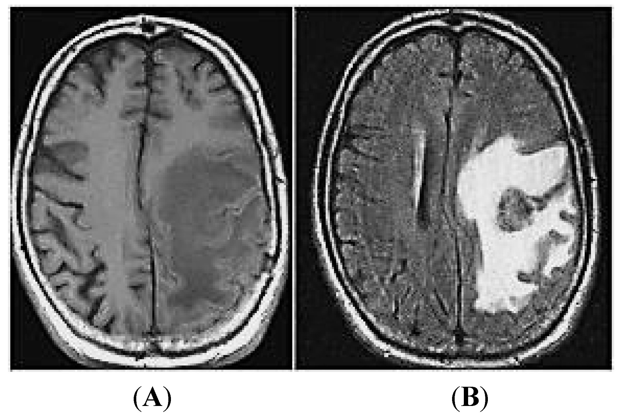
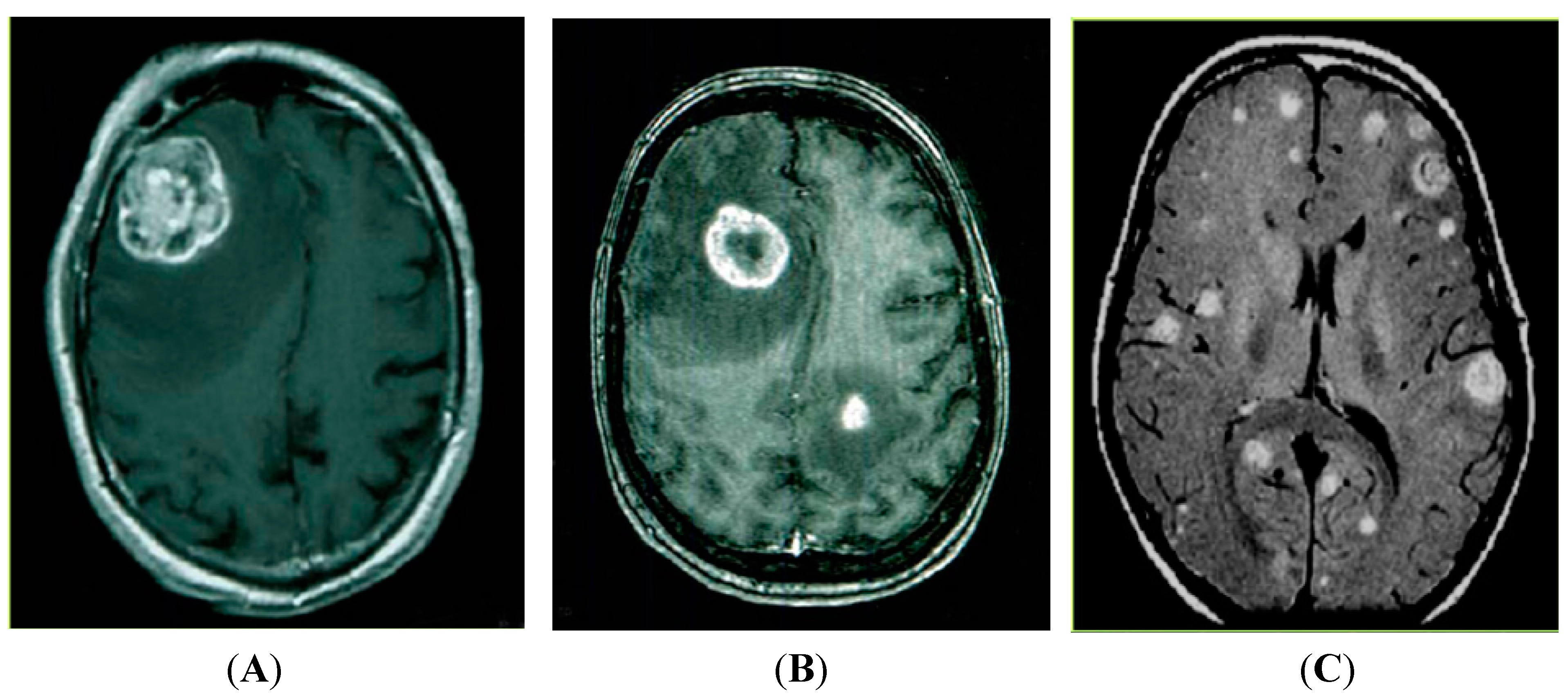
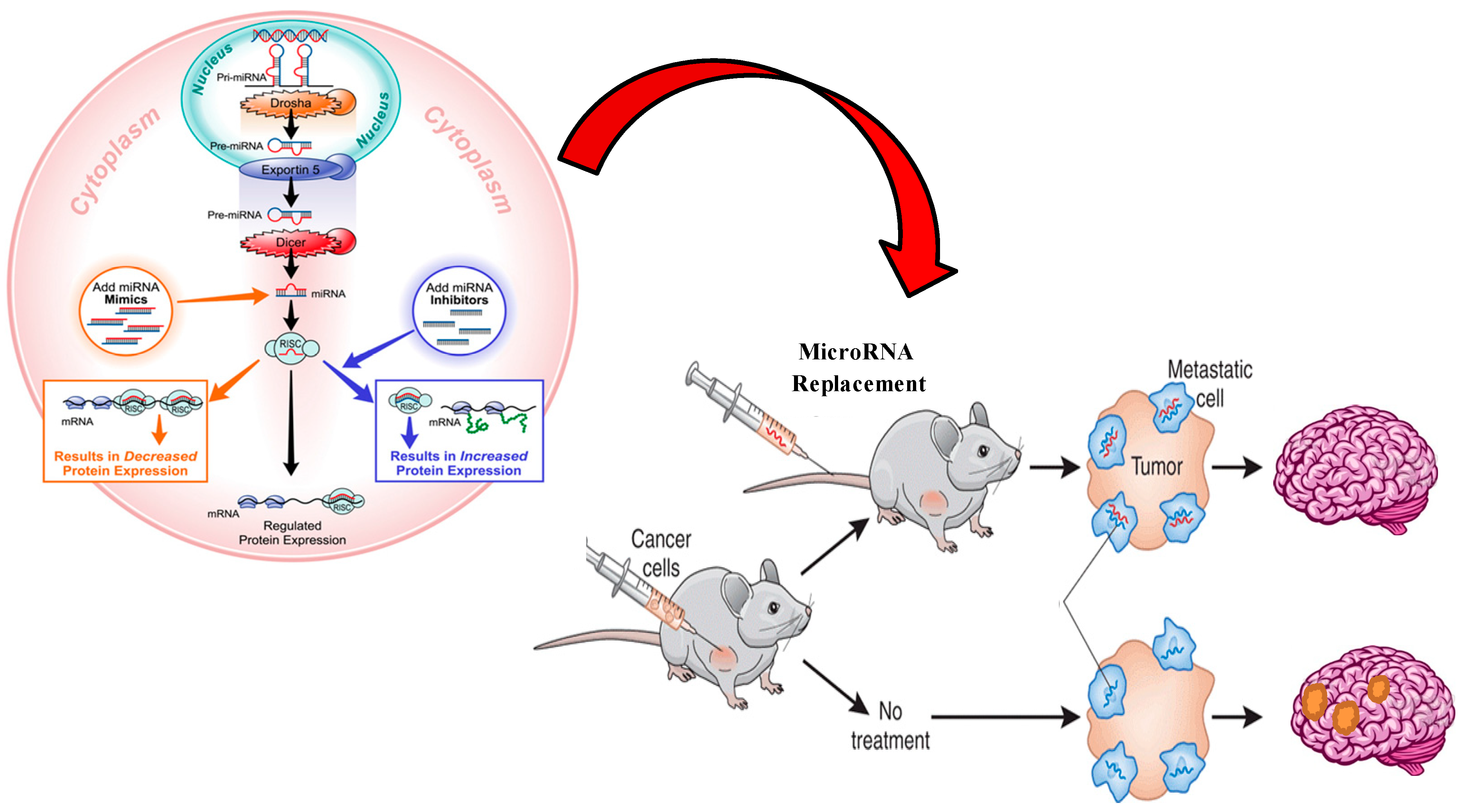
2. MiRNAs and Brain Metastases

Genetic and epigenetic changes allow cancer cells to find the brain microenvironment—“the soil”—a favorable niche for tumorigenic “seeds” to implant, grow and blossom [24]. However, the precise manner in which the brain microenvironment promotes the growth of solid tumor cells remains a critical barrier. Understanding the precise micro-environment-mediated genetic events triggered in metastatic tumor cells to promote growth and drug resistance should substantially improve our knowledge base and identify new “druggable” targets. Non-coding (nc) RNAs are master regulators of the human genome and their aberrant expression contributes to tumorigenesis, metastasis and the acquisition of therapeutic resistance. However, the precise role of ncRNAs in brain metastases and the acquisition of drug resistance remained unknown. MiRNAs are endogenously expressed, small, non-coding RNA molecules that negatively regulate gene expression at the post-transcriptional level by base pairing to the 3' untranslated region (UTR) of target messenger RNA (mRNA). MiRNAs play a key role in cell development, proliferation, differentiation and apoptosis and, accordingly, alterations in miRNA expression are seen in tissues from all organ systems and contribute to cancers, autoimmune and genetic disorders and infectious processes. The loss of a tumor suppressive miRNA activates inherently oncogenic pathways to promote the generation of a cancer phenotype, tumor initiation, progression and metastasis [25]. Epigenetic alteration of miRNA have also been shown to play a role in cancer since compelling evidence demonstrates that miRNA deregulation promotes generation of a cancer phenotype, tumor initiation, metastatic growth and development of drug resistance [26,27]. Nearly 50% of human miRNA genes are located in areas of the genome associated with carcinogenesis [28]. Studying the miRNA profile of different tumors has gained popularity in the last decade as represents a breakthrough method for tumor classification that can impact cancer diagnosis, prognosis and treatment decisions. For example, miRNA profiling of 103 lymph node negative, breast cancer tumors lead to the identification of miRNA-106b in triple negative tumors and now known to carry a worse prognosis [29]. Different stages of breast cancer were noted to correlate with distinct miRNA profiles, including members of the miR-200 family and miR-9, to suggest that miRNAs are directly involved in tumor progression and metastasis. In colorectal cancer, the detection of circulating miRNA-141 correlated with metastatic disease and poor prognosis [30] and up-regulation of miR-9 was involved in metastases as well through facilitated cell motility and down-regulated α-catenin [31]. Certain miRNAs, e.g., miRNA-34 and let-7, were also found to be directly involved with the survival of tumor-initiating (or metastases-initiating) cancer stem cells (CSCs) [32,33]. MiRNA signatures have been identified within individual tumor types and may improve useful as diagnostics or prognostics of therapeutic response.
MiRNA profiling of tumor tissue may facilitate the identification of primary tumors based upon the miRNA profile of the metastatic brain lesion. Even with advanced imaging techniques, a small percentage of metastatic brain tumors remain of unknown origin. A recent study successfully identified the tumor of origin in 84% of brain metastases using a quantitative real-time polymerase chain reaction (qRT-PCR) of 48 different miRNAs [34]. Other studies have shown that miR-92b and miRNA-9/9* are over-expressed in primary brain tumors compared to metastatic brain tumors to aid in the diagnosis of brain lesions [35].
The role of miRNAs in the biology of brain metastases has been established in studies investigating a number of primary tumor types (Table 1) [36,37,38,39,40]. In breast cancer, miR-1258 alterations were directly related to heparanase expression, a known prometastatic enzyme found in brain metastatic breast cancer cells that degrades heparan sulfate chains to affect the cytoskeleton and render cells more capable of crossing the BBB [41,42]. The migratory and invasive capacity of breast CSCs was found to be related to the KLF4 gene expression which is inversely related to miRNA-7 expression [43]. Similarly, in lung cancers, miRNA-145 down-regulation was involved in the growth of lung adenocarcinoma and promoted the formation of brain metastases [44]. MiRNA-328 in non-small cell lung cancer (NSCLC) regulated cell migration and the formation of brain metastases through altered expression of the PRKCA genes [45]. MiRNA-378 promoted brain metastases in NSCLC by increasing expression levels of MMP-7, MMP-9 and VEGF and decreasing levels of Sufu, all key genes involved in angiogenesis and extracellular matrix invasion [46]. MiRNA-200 family members were exclusively elevated in the CSF of patients with metastatic brain lesions from various primary tumor types when compared with glioblastoma and non-cancer patients [47].
Table 1. MiRNAs deregulated in brain metastases compared to the primary tumor. Deregulated miRNAs identified in metastatic brain tumor cells compared to their matched primary tumors. NSCLC, non-small cell lung cancer; MMP, matrix metalloproteinase; VEGF, Vascular endothelial growth factor; PTB1b, protein tyrosine phosphatase-1B; HIF-1α: Hypoxia-inducible factor 1-α.
Deregulated MiRNADirection of Expression in Brain MetastasesPrimary TumorPutative Target
miR-1258 [41]Down-regulatedBreastHeparanase
miR-7 [43]Down-regulatedBreastKLF4 gene
miR-145 [44]Down-regulatedLung adenocarcinoma3'-UTR of the JAM-A and fascin
miR-146-a [48]Down-regulatedBreastB-catenin and hnRNPC
miR-768-3p [49]Down-regulatedLung and breastK-RAS
miR-19a [50]Down-regulatedBreast3'-UTR of tissue factor transcript [36]
miR-29c [50]Down-regulatedBreast and melanomaInduced myeloid leukemia cell differentiation protein MCL1 [37]
miR-31 [51]Down-regulatedColonp53 [38]
miR-328 [45]Up-regulatedNSCLCPRKCA gene
miR-378 [46]Up-regulatedNSCLCMMP-7, MMP-9 and VEGF
miR-200 [47]Up-regulatedBreast and lungE-cadherin transcriptional repressors ZEB1 and ZEB2 [39]
miR-210 [50]Up-regulatedBreast and melanomaPTP1b and HIF-1α [40]
miR-1, miR-145, miR-146a, miR-143, miR-10b, miR-22 [51]Up-regulatedColonMultiple genes related to apoptosis and oncogenesis
The brain micro-environment, represented mainly by the astrocytes, is an active player and key regulator in the increased growth and chemoresistance of metastatic brain tumors (Figure 3). Astrocytes up-regulate a number of survival genes within the neighboring tumor cells and render these cells more aggressive, independent of primary tumor histology or p-glycoprotein activity [15]. MiRNAs are directly involved in the changes that the brain microenvironment implies on the metastatic tumor cells as many studies have shown that the brain microenvironment change the miRNA profile of the tumor cells when compared with the primary tumor. Rhabdoid tumor cells showed different miRNA profiles when originated in the brain compared to the kidney [52]. MiRNA-146a was noted to be suppressed in brain metastases compared to the original tumors in animal models, associated with decreased β-catenin protein levels and increased heterogeneous nuclear ribonucleoprotein C1/C2 (hnRNPC) which may increase migratory and invasive capabilities [48]. MiRNA-768-3p was down-regulated in tumor cells when co-cultured with astrocytes and this was validated in human brain metastatic tissues from lung cancer, breast cancer and melanoma when compared to match-paired primary tumor from the same patient. MiRNA-768-3p down-regulation led to an increase in K-ras expression and translated into increased tumor growth and drug resistance [49]. Different miRNA profiles were found between primary colorectal tumors and matched metastatic brain tumors [51] where over-expression of miRNA-145, 1, 146a, 576-5p, 126*, HS287, 28-5p, 143, 199b-5p, 199a-5p, 10b, 22, 133b, 145*, 199a, 133a, 125b and down-regulation of miRNA-31 and HS170 were observed in brain-metastatic carcinomas. Moreover, miRNAs isolated from exosomes of parental breast cancer and melanoma cells were different from those isolated from their corresponding metastatic brain variants. MiRNA-210 was over-expressed while miRNAs-19a and 29c were down-regulated in brain metastases [50]. These studies demonstrate that the brain microenvironment induces changes in the miRNA signature of the tumor cells to activate pro-growth signaling pathways and leads to more aggressive, drug resistant metastatic lesions. Studies suggest that the microenvironment influence on tumor cells that “seed” in the brain may be a universal effect [47]. This represents such an appealing concept to target key miRNAs involved in metastasis.

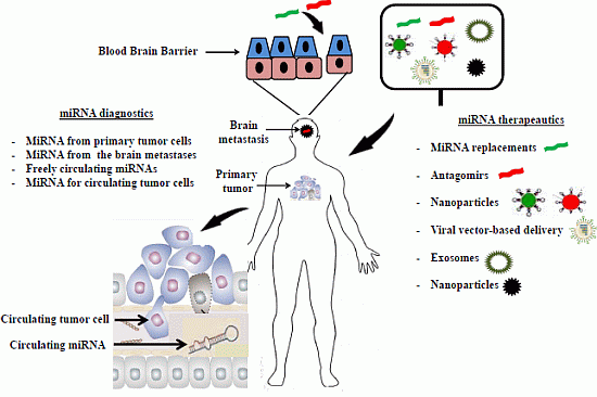
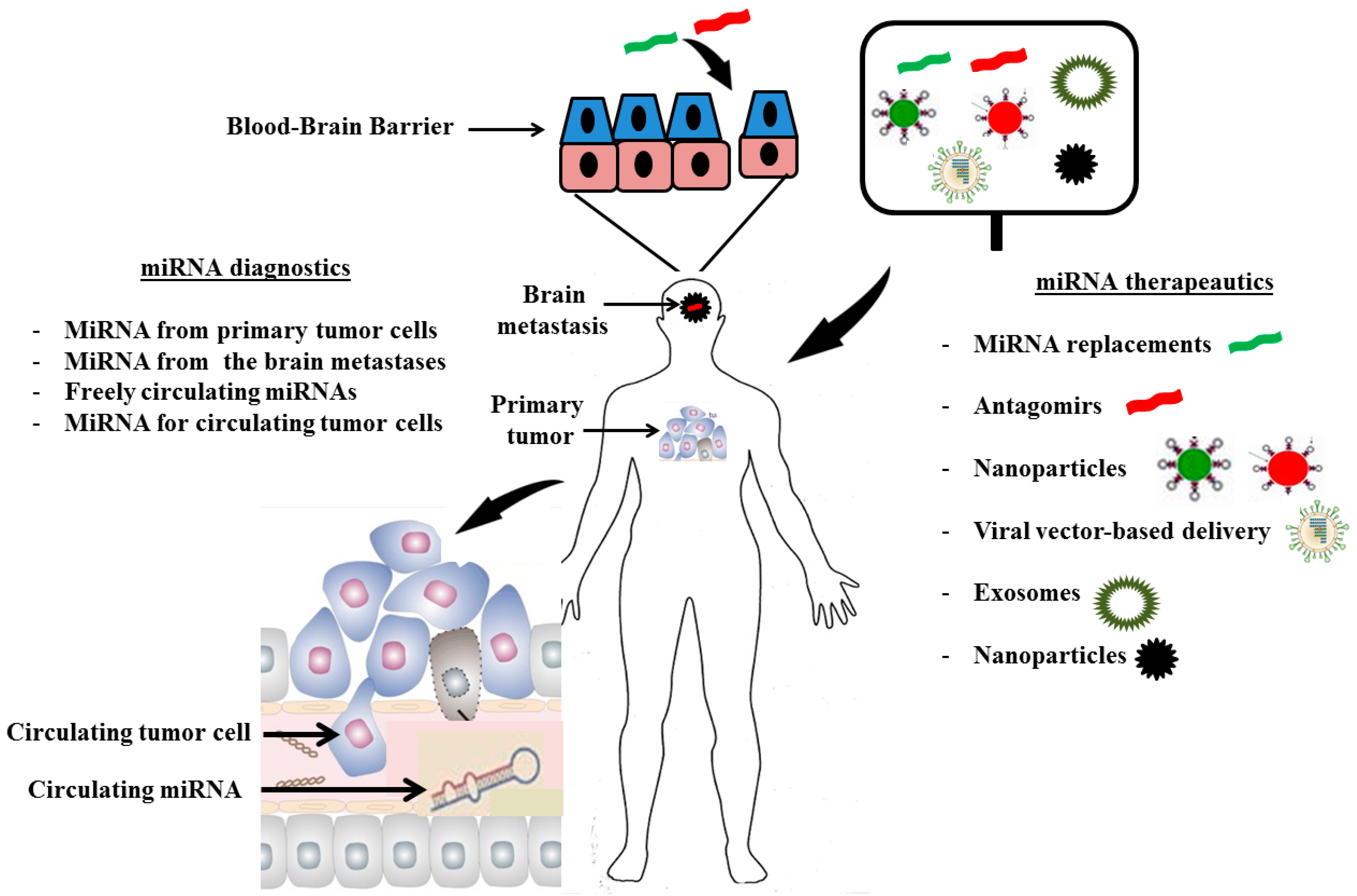
3. MiRNA Diagnostics

Understanding of the role of miRNAs in the biology of brain metastases has generated a greater demand to practically apply this knowledge in clinical practice. MiRNAs hold promise as diagnostics, prognostics and therapeutics to improve cancer patient outcome [53]. For example, miRNAs are being developed to improve detection of the plasma cell dyscrasia multiple myeloma (MM) [54]. Similarly, miRNA-based diagnostics may more readily detect metastatic brain lesions and distinguish primary from metastatic lesions [34]. MiRNA signatures may eventually be incorporated in clinical decision making as prognostic indicators to formulate treatment plans. Multiple miRNA signatures in primary tumors were shown to correlate with more aggressive, invasive, “brain-seeking” behavior. MiRNA-378 in NSCLC is associated with a greater likelihood of tumor seeding within the brain [46]. Clinical trials are needed to determine if miRNA signatures are predictive of worse prognosis. Such signatures could trigger more intensive treatment plans, e.g., prophylactic cranial irradiation or targeted therapy, to prevent the development of metastases or cranial recurrence. Early detection of brain micrometastases may be based upon deregulated miRNAs known to be altered within metastatic brain tumors. Changes in miRNA levels, e.g., loss of miRNA-768-3p signal [49] or increase in miRNA-200 [47], may provide an early signal to prompt aggressive treatment. MiRNAs are also readily detectable and stable within human plasma [30,55,56]. These miRNAs are protected from endogenous RNase activity as free-circulating molecules, within circulating tumor cells (CTCs) or in membrane-derived small membrane vesicles, exosomes, that are released by cells [57]. MiRNAs from plasma, CTCs and exosomes have been successfully detected using RT-PCR techniques and may serve as readily-available diagnsotics [58,59,60]. Deregulated levels of miRNAs have been detected in the plasma of patients with lymphoma (miRNA-155, 210, 21) [61], leukemia (miRNA-92, 150, 342) [62,63], colon cancer (miRNA-29a, 92a) [64], breast cancer (miRNA-195, 21, 92a, let-7a) [65,66], prostate cancer (miRNA-375, 141) [67], ovarian cancer (miRNA-21, 92, 93) [68], pancreatic cancer (miRNA-155, 196a, 642b, 885, 5p, 22, 16) [69,70,71,72], gastric cancer (miRNA-17, 1, 106a, 106b, let-7a and 18a) [73,74,75] and lung cancer (miRNA-486, 30d, 1, 499 and 375) [58,76,77]. MiRNAs were also found to be stable in the cerebrospinal fluid (CSF) of patients with neoplasms as well as neurologic disorders [47,78]. Given the relative invasive nature of CSF sampling, the challenge in miRNA diagnostics in brain metastases is the BBB and whether miRNAs (either as free molecules, in CTCs or other form of transport system such as exosomes) are able to cross the BBB and be readily detectable in the serum of patients. Studies in glioblastomas have shown that miRNA signals can be detected within exosomes in the serum of these patients [59]. These results support disruption of the BBB during metastasis [19]. Studies also detected relevant miRNAs in the plasma of Alzheimer’s or Huntington’s disease patients even though the BBB is thought to remain intact in these conditions [79]. Membrane-derived extracellular vesicles (EVs) containing miRNAs originate from CNS tumors and may function as intercellular communication with the microenvironment and across the BBB [80]. Now that it has been established that the miRNA profile of brain metastases is distinct from primary tumors, it would be of great importance to be able to routinely and inexpensively detect these miRNAs in the blood, serum, CSF or urine of patients.

4. MiRNA Therapeutics

Discoveries in miRNA biology, and their close relationship to oncogenesis in many tumor types, has led to attempts to translate this information into miRNA therapeutics (Figure 3) [81,82]. Currently, however, there is a lack of miRNA-based therapeutics to directly target brain metastases. A well-established feature is that a single miRNA is capable of regulating multiple genes, which makes endogenous miRNAs appealing therapeutic targets. Altering miRNA signatures was also found to sensitize tumor cells to other forms of treatment in, otherwise, chemo-resistant tumors [83]. Two strategies exist for miRNA-based therapeutics: a direct approach which involves either miRNA mimics to replace the loss of a tumor suppressor miRNA or miRNA antagomirs which are antisense oligonucleotides that block oncogenic miRNAs; and an indirect strategy that involves identifying existing agents that modulate the expression and/or processing of miRNAs in traditional compound-library screens.
The development of antagomirs went through multiple phases to increase their stability since naked RNA has a very short half-life in the bloodstream and the use of phosphodiester oligodeoxynucleotides (ODNs), without further modification was unsuccessful [84]. The in vivo stability of antagomirs has been augmented by multiple chemical modifications such as the development of phosphorothioate containing oligonucleotides [85], 2'-O-methyl-(2'-O-Me) or 2'-O-methoxyethyl-oligonucleotides (2'-O-MOE) which improves ribonuclease resistance and increases the binding affinity to the miRNA [86], locked nucleic acid (LNA) oligonucleotides where the ribose ring is “locked” by a methylene bridge which further increases the affinity towards single stranded RNAs [87,88], peptide nucleic acids (PNA) which are artificially synthesized polymers similar to RNAs but are resistant to enzyme degradation [89] and fluorine-derivative nucleic acids (FANA and 2'-F) [90]. Similar to antagomirs, miRNA sponges inhibit miRNA where plasmids containing multiple tandem-binding sites to the miRNA of interest are transfected into the cells and help “fool” the miRNA into binding to the sponge instead of its target mRNA [91]. MiRNA masks are single-stranded 2-O-methyl antisense oligonucleotides that are complementary to the supposed miRNA binding sites in the 3'-UTR of the mRNA [92]. MiRNA replacement therapy aims at restoring a tumor suppressor miRNA that is down-regulated in tumor cells with oligonucleotide mimics similar to the original miRNA. Using longer strands that mimic the pre-miRNA have also been proposes but these require different delivery systems to ensure intranuclear availability [93]. The delivery of miRNA mimics with tumor suppressor effect into tumor cells have shown to be effective in inducing cell death (Figure 4) [49,94,95,96].
Figure 3. MiRNA diagnostics and therapeutics for brain metastases. A synthesized antisense nucleotide (antagomir, red) or miRNA replacement (green) is loaded onto a delivery system. The delivery system can be a viral vector such as adenovirus or a non-viral liposome or nanoparticle. The preparation is then administered intravenously to the patient with a metastatic brain tumor and remains stable in the blood stream. The compound crosses the blood–brain barrier and reaches the tumor cells and undergoes endocytosis to the intracellular space. The antagomir is then released from the delivery system which gets degraded. The antagomir binds to the miRNA of interest in blue and antagonizes its oncogenic effect which eventually leads to apoptosis and tumor regression.
Ijms 15 10508 g003
A current dilemma in miRNA therapeutics is an efficient system to guarantee stability in the blood and adequate delivery to tissues of interest. Viral and non-viral delivery methods have been used with variable success. Adenovirus-associated vectors (AAV) emerged as an appealing method since they have acceptable toxicity profiles [97] and were successfully injected intravenously in mouse models to restore miRNA-26 expression in hepatocellular carcinoma cells [98]. Different AAV serotypes can successfully target distinct tumor types. Non-viral delivery methods may be superior to AAV methods given their stable formulations. For example, liposomes composed of phospholipid bilayers were used to deliver miRNA-133b to lung cancer cells in mice [99]. Liposome use, however, is limited by their toxicity, related to their strong cationic charge [100]. Liposomes have gone through multiple levels of development to improve their stability and minimize the side effects. Hyaluronic acid was added to form polycationic liposome-hyaluronic acid (LPH) which successfully delivered siRNA and miRNA-34a into mouse melanoma models [101]. To overcome the toxicity of liposomes, a neutral lipid emulsion was developed which has a natural predilection to accumulate in the lung compared to the liver predilection of cationic liposomes. The neutral lipid emulsion successfully delivered let-7 and miRNA-34a to lung cancer cells in mice [102]. Liposomes have a short half-life and require continuous infusion or frequent administration which limits their use. Multiple attempts were made to overcome these problems which led to the development of sustained-release polymer formulations [103]. Other forms of non-viral delivery systems include dendrimers which are repetitively-branched perfectly-structured particles that have a high surface to volume ratio and were successfully used in delivering anti-miR-21 and 5-flurouracil to glioblastoma cells in vitro [104]. Other nanoparticles, microspheres and hydrogels have been developed [105] such as the polylactide-co-glycolide (PLGA) particles which are stable particles that allow the delivery of miRNA over time and are highly adaptable and can be used to load multiple cargos. PLGA particles delivered anti-miRNA-155 to malignant pre-B lymphoma cells in mouse models with good results [106].
Figure 4. Model to illustrate the effect of microRNA-based therapeutics for the treatment of brain metastases at the cellular and animal levels. Oncology-directed miRNA replacement therapy. Loss of a tumor suppressor miRNA leads to hyperactivation of inherently oncogenic pathways and tumorigenesis. Administration of a miRNA mimic reinstates the function of the missing tumor suppressor miRNA, suppresses oncogenic pathways and cancer cell growth.
Ijms 15 10508 g004
Throughout the different stages of development of gene and miRNA delivery systems, a major obstacle has been crossing the BBB. Although surgically-implanted wafers and intra-thecal routes are established methods to administer chemotherapy, oral or intravenous routes remain the most convenient. The BBB only allows lipophilic molecules, less than 400 Da, to penetrate the CNS [107]. A few novel techniques have been used to overcome this obstacle. The Trojan Horse Liposome (THL) system encapsulates the genomic material, i.e., miRNA replacements or antagomirs, within the liposome to protect it from nuclease degradation. The compound is constructed using polyethyleneglycol (PEG) to stabilize the liposome [108]. Part of the PEG can be engineered with peptidomimetic monoclonal antibodies (mAbs) that target specific BBB receptors (such as the insulin receptor or the transferrin receptor) and facilitate the transcytosis of the compound. The THL technology has been used to administer compounds that cross the BBB and deliver genetic material to the CNS [109]. Another novel method to bypass the BBB is through polyethylenimine (PEI)-based delivery systems as are widely used in gene therapy [110]. PEI complexes are positively charged that bind negatively charged nucleic acid, i.e., miRNAs. The compound retains an overall positive charge that interacts with negatively charged polysaccharides on the cell surface. This process is followed by endocytosis of the compound to evade the endosome by inducing an influx of protons and water leading to swelling and disruption of the endosome and release of the compound containing the miRNA in the cytoplasm. PEI-based systems have been modified to cross the BBB by adding a short peptide inspired from the rabies virus glycoprotein (RVG) which binds the acetylcholine receptor [111]. Mannitol also is added to increase the permeability of the BBB [112]. The PEI–RVG compound crossed the BBB and delivered the neuron specific miR-124a to brain cells. Even with rapidly emerging understanding of miRNA biology and the development of novel delivery systems, the clinical use of miRNA therapeutics to treat brain metastases remains limited in pre-clinical development and has yet to be exploited. The previous misconception of the brain as a sanctuary organ that systemic or targeted therapies cannot penetrate has contributed to delays in clinical advancement. Future studies are needed to better define miRNA signatures within brain metastases and to correlate these signatures with the miRNA profile of the primary tumor. Advances have been made in pre-clinical and translational studies to identify miRNAs that change after growth in the brain microenvironment but require validation from patient tumor samples [49].

5. Conclusions

Despite advances in developing miRNA diagnostics and therapeutics, significant challenges remain. Since miRNA are upstream regulators of hundreds of genes, the off-target effects of miRNA therapeutics are a potential limitation. Toxicities associated with miRNA therapeutics are not limited to the delivery system since studies have shown that oversaturating small RNA pathways can be lethal [113]. The induction of interferon-α through the toll like receptor (TLR-7) by short interfering RNA (siRNA) leads to systemic immune responses and poor outcomes [111]. The availability of a reliable delivery system that has minimal toxicities, crosses the BBB and successfully unloads the miRNA therapeutic is needed to promote clinical advancement (Figure 4).
In summary, the survival of patients with brain metastases remains poor due to the lack of effective treatments. MiRNAs are key regulators of gene expression and their role in multiple cancer types is well-established. Multiple miRNA signatures are altered in brain metastases relative to the primary tumor and are, in fact, induced through interaction with the brain microenvironment. Identifying miRNA signatures within brain metastases represents a promising approach to target these lesions. However, numerous challenges exist in translating this information into clinical practice. MiRNA therapeutics may eventually provide individualized therapy for patient and this approach is applicable to molecularly heterogeneous diseases with distinct genetic subtypes, such as brain metastases [54].

Acknowledgments

We thank members of the Driscoll lab (http://www.driscolllab.com/index.html#banner) for critical reading of the review.

Author Contributions

The review was conceived, designed, written and edited by the three authors.

Conflicts of Interest

The authors declare no conflict of interest.

References

  1. Jemal, A.; Siegel, R.; Ward, E.; Hao, Y.; Xu, J.; Murray, T.; Thun, M.J. Cancer statistics, 2008. CA Cancer J. Clin. 2008, 58, 71–96. [Google Scholar] [CrossRef]
  2. Nayak, L.; Lee, E.Q.; Wen, P.Y. Epidemiology of brain metastases. Curr. Oncol. Rep. 2012, 14, 48–54. [Google Scholar] [CrossRef]
  3. Disibio, G.; French, S.W. Metastatic patterns of cancers: Results from a large autopsy study. Arch. Pathol. Lab. Med. 2008, 132, 931–939. [Google Scholar]
  4. Barnholtz-Sloan, J.S.; Sloan, A.E.; Davis, F.G.; Vigneau, F.D.; Lai, P.; Sawaya, R.E. Incidence proportions of brain metastases in patients diagnosed (1973 to 2001) in the Metropolitan Detroit Cancer Surveillance System. J. Clin. Oncol. 2004, 22, 2865–2872. [Google Scholar] [CrossRef]
  5. Schouten, L.J.; Rutten, J.; Huveneers, H.A.; Twijnstra, A. Incidence of brain metastases in a cohort of patients with carcinoma of the breast, colon, kidney, and lung and melanoma. Cancer 2002, 94, 2698–2705. [Google Scholar] [CrossRef]
  6. Eichler, A.F.; Loeffler, J.S. Multidisciplinary management of brain metastases. Oncologist 2007, 12, 884–898. [Google Scholar] [CrossRef]
  7. Rizzi, A.; Tondini, M.; Rocco, G.; Rossi, G.; Robustellini, M.; Radaelli, F.; Della Pona, C. Lung cancer with a single brain metastasis: Therapeutic options. Tumori 1990, 76, 579–581. [Google Scholar]
  8. Nugent, J.L.; Bunn, P.A., Jr.; Matthews, M.J.; Ihde, D.C.; Cohen, M.H.; Gazdar, A.; Minna, J.D. CNS metastases in small cell bronchogenic carcinoma: Increasing frequency and changing pattern with lengthening survival. Cancer 1979, 44, 1885–1893. [Google Scholar] [CrossRef]
  9. Knights, E.M., Jr. Metastatic tumors of the brain and their relation to primary and secondary pulmonary cancer. Cancer 1954, 7, 259–265. [Google Scholar] [CrossRef]
  10. Langley, R.R.; Fidler, I.J. The seed and soil hypothesis revisited—The role of tumor–stroma interactions in metastasis to different organs. Int. J. Cancer 2011, 128, 2527–2535. [Google Scholar] [CrossRef]
  11. Chishima, T.; Miyagi, Y.; Wang, X.; Yamaoka, H.; Shimada, H.; Moossa, A.; Hoffman, R.M. Cancer invasion and micrometastasis visualized in live tissue by green fluorescent protein expression. Cancer Res. 1997, 57, 2042–2047. [Google Scholar]
  12. Yang, M.; Baranov, E.; Jiang, P.; Sun, F.-X.; Li, X.-M.; Li, L.; Hasegawa, S.; Bouvet, M.; Al-Tuwaijri, M.; Chishima, T. Whole-body optical imaging of green fluorescent protein-expressing tumors and metastases. Proc. Natl. Acad. Sci. USA 2000, 97, 1206–1211. [Google Scholar] [CrossRef]
  13. Hoffman, R.M. Orthotopic metastatic mouse models for anticancer drug discovery and evaluation: A bridge to the clinic. Investig. New Drugs 1999, 17, 343–360. [Google Scholar] [CrossRef]
  14. Hoffman, R. In vivo imaging of metastatic cancer with fluorescent proteins. Cell Death Differ. 2002, 9, 786–789. [Google Scholar] [CrossRef]
  15. Kim, S.J.; Kim, J.S.; Park, E.S.; Lee, J.S.; Lin, Q.; Langley, R.R.; Maya, M.; He, J.; Kim, S.W.; Weihua, Z.; et al. Astrocytes up-regulate survival genes in tumor cells and induce protection from chemotherapy. Neoplasia 2011, 13, 286–298. [Google Scholar]
  16. Langley, R.R.; Fan, D.; Guo, L.; Zhang, C.; Lin, Q.; Brantley, E.C.; McCarty, J.H.; Fidler, I.J. Generation of an immortalized astrocyte cell line from H-2Kb-tsA58 mice to study the role of astrocytes in brain metastasis. Int. J. Oncol. 2009, 35, 665–672. [Google Scholar]
  17. JuanYin, J.; Tracy, K.; Zhang, L.; Munasinghe, J.; Shapiro, E.; Koretsky, A.; Kelly, K. Noninvasive imaging of the functional effects of anti-VEGF therapy on tumor cell extravasation and regional blood volume in an experimental brain metastasis model. Clin. Exp. Metastasis 2009, 26, 403–414. [Google Scholar] [CrossRef]
  18. Lorger, M.; Felding-Habermann, B. Capturing changes in the brain microenvironment during initial steps of breast cancer brain metastasis. Am. J. Pathol. 2010, 176, 2958–2971. [Google Scholar] [CrossRef]
  19. Deeken, J.F.; Loscher, W. The blood–brain barrier and cancer: Transporters, treatment, and Trojan horses. Clin. Cancer Res. 2007, 13, 1663–1674. [Google Scholar] [CrossRef]
  20. Fukumura, D.; Xu, L.; Chen, Y.; Gohongi, T.; Seed, B.; Jain, R.K. Hypoxia and acidosis independently up-regulate vascular endothelial growth factor transcription in brain tumors in vivo. Cancer Res. 2001, 61, 6020–6024. [Google Scholar]
  21. Hobbs, S.K.; Monsky, W.L.; Yuan, F.; Roberts, W.G.; Griffith, L.; Torchilin, V.P.; Jain, R.K. Regulation of transport pathways in tumor vessels: Role of tumor type and microenvironment. Proc. Natl. Acad. Sci. USA 1998, 95, 4607–4612. [Google Scholar]
  22. Sharom, F.J. Complex interplay between the p-glycoprotein multidrug efflux pump and the membrane: Its role in modulating protein function. Front. Oncol. 2014, 4, 41. [Google Scholar]
  23. Hida, K.; Akiyama, K.; Ohga, N.; Maishi, N.; Hida, Y. Tumour endothelial cells acquire drug resistance in a tumour microenvironment. J. Biochem. 2013, 153, 243–249. [Google Scholar] [CrossRef]
  24. Hanahan, D.; Weinberg, R.A. Hallmarks of cancer: The next generation. Cell 2011, 144, 646–674. [Google Scholar] [CrossRef]
  25. Melo, S.A.; Esteller, M. Dysregulation of microRNAs in cancer: Playing with fire. FEBS Lett. 2011, 585, 2087–2099. [Google Scholar] [CrossRef]
  26. Calin, G.A.; Croce, C.M. MicroRNA signatures in human cancers. Nat. Rev. Cancer 2006, 6, 857–866. [Google Scholar] [CrossRef]
  27. Aigner, A. MicroRNAs (miRNAs) in cancer invasion and metastasis: Therapeutic approaches based on metastasis-related miRNAs. J. Mol. Med. 2011, 89, 445–457. [Google Scholar] [CrossRef]
  28. Calin, G.A.; Sevignani, C.; Dumitru, C.D.; Hyslop, T.; Noch, E.; Yendamuri, S.; Shimizu, M.; Rattan, S.; Bullrich, F.; Negrini, M. Human microRNA genes are frequently located at fragile sites and genomic regions involved in cancers. Proc. Natl. Acad. Sci. USA 2004, 101, 2999–3004. [Google Scholar] [CrossRef]
  29. Janssen, E.A.; Slewa, A.; Gudlaugsson, E.; Jonsdottir, K.; Skaland, I.; Soiland, H.; Baak, J.P. Biologic profiling of lymph node negative breast cancers by means of microRNA expression. Mod. Pathol. 2010, 23, 1567–1576. [Google Scholar] [CrossRef]
  30. Cheng, H.; Zhang, L.; Cogdell, D.E.; Zheng, H.; Schetter, A.J.; Nykter, M.; Harris, C.C.; Chen, K.; Hamilton, S.R.; Zhang, W. Circulating plasma miR-141 is a novel biomarker for metastatic colon cancer and predicts poor prognosis. PLoS One 2011, 6, e17745. [Google Scholar]
  31. Zhu, L.; Chen, H.; Zhou, D.; Li, D.; Bai, R.; Zheng, S.; Ge, W. MicroRNA-9 up-regulation is involved in colorectal cancer metastasis via promoting cell motility. Med. Oncol. 2012, 29, 1037–1043. [Google Scholar] [CrossRef]
  32. Ji, Q.; Hao, X.; Zhang, M.; Tang, W.; Yang, M.; Li, L.; Xiang, D.; Desano, J.T.; Bommer, G.T.; Fan, D.; et al. MicroRNA miR-34 inhibits human pancreatic cancer tumor-initiating cells. PLoS One 2009, 4, e6816. [Google Scholar] [CrossRef]
  33. Zimmerman, A.L.; Wu, S. MicroRNAs, cancer and cancer stem cells. Cancer Lett. 2011, 300, 10–19. [Google Scholar] [CrossRef]
  34. Mueller, W.C.; Spector, Y.; Edmonston, T.B.; Cyr, B.S.; Jaeger, D.; Lass, U.; Aharonov, R.; Rosenwald, S.; Chajut, A. Accurate classification of metastatic brain tumors using a novel microRNA-based test. Oncologist 2011, 16, 165–174. [Google Scholar] [CrossRef]
  35. Nass, D.; Rosenwald, S.; Meiri, E.; Gilad, S.; Tabibian-Keissar, H.; Schlosberg, A.; Kuker, H.; Sion-Vardy, N.; Tobar, A.; Kharenko, O.; et al. MiR-92b and miR-9/9* are specifically expressed in brain primary tumors and can be used to differentiate primary from metastatic brain tumors. Brain Pathol. 2009, 19, 375–383. [Google Scholar] [CrossRef]
  36. Zhang, X.; Yu, H.; Lou, J.R.; Zheng, J.; Zhu, H.; Popescu, N.I.; Lupu, F.; Lind, S.E.; Ding, W.Q. MicroRNA-19 (miR-19) regulates tissue factor expression in breast cancer cells. J. Biol. Chem. 2011, 286, 1429–1435. [Google Scholar]
  37. Mott, J.L.; Kobayashi, S.; Bronk, S.F.; Gores, G.J. Mir-29 regulates Mcl-1 protein expression and apoptosis. Oncogene 2007, 26, 6133–6140. [Google Scholar] [CrossRef]
  38. Creighton, C.J.; Fountain, M.D.; Yu, Z.; Nagaraja, A.K.; Zhu, H.; Khan, M.; Olokpa, E.; Zariff, A.; Gunaratne, P.H.; Matzuk, M.M.; et al. Molecular profiling uncovers a p53-associated role for microRNA-31 in inhibiting the proliferation of serous ovarian carcinomas and other cancers. Cancer Res. 2010, 70, 1906–1915. [Google Scholar] [CrossRef]
  39. Gregory, P.A.; Bert, A.G.; Paterson, E.L.; Barry, S.C.; Tsykin, A.; Farshid, G.; Vadas, M.A.; Khew-Goodall, Y.; Goodall, G.J. The miR-200 family and miR-205 regulate epithelial to mesenchymal transition by targeting ZEB1 and SIP1. Nat. Cell Biol. 2008, 10, 593–601. [Google Scholar] [CrossRef]
  40. Li, L.; Huang, K.; You, Y.; Fu, X.; Hu, L.; Song, L.; Meng, Y. Hypoxia-induced miR-210 in epithelial ovarian cancer enhances cancer cell viability via promoting proliferation and inhibiting apoptosis. Int. J. Oncol. 2014, 44, 2111–2120. [Google Scholar]
  41. Zhang, L.; Sullivan, P.S.; Goodman, J.C.; Gunaratne, P.H.; Marchetti, D. MicroRNA-1258 suppresses breast cancer brain metastasis by targeting heparanase. Cancer Res. 2011, 71, 645–654. [Google Scholar]
  42. Ridgway, L.D.; Wetzel, M.D.; Ngo, J.A.; Erdreich-Epstein, A.; Marchetti, D. Heparanase-induced GEF-H1 signaling regulates the cytoskeletal dynamics of brain metastatic breast cancer cells. Mol. Cancer Res. 2012, 10, 689–702. [Google Scholar] [CrossRef]
  43. Okuda, H.; Xing, F.; Pandey, P.R.; Sharma, S.; Watabe, M.; Pai, S.K.; Mo, Y.Y.; Iiizumi-Gairani, M.; Hirota, S.; Liu, Y.; et al. miR-7 suppresses brain metastasis of breast cancer stem-like cells by modulating KLF4. Cancer Res. 2013, 73, 1434–1444. [Google Scholar] [CrossRef]
  44. Zhao, C.; Xu, Y.; Zhang, Y.; Tan, W.; Xue, J.; Yang, Z.; Zhang, Y.; Lu, Y.; Hu, X. Down-regulationof miR-145 contributes to lung adenocarcinoma cell growth to form brain metastases. Oncol. Rep. 2013, 30, 2027–2034. [Google Scholar]
  45. Arora, S.; Ranade, A.R.; Tran, N.L.; Nasser, S.; Sridhar, S.; Korn, R.L.; Ross, J.T.; Dhruv, H.; Foss, K.M.; Sibenaller, Z.; et al. MicroRNA-328 is associated with (non-small) cell lung cancer (NSCLC) brain metastasis and mediates NSCLC migration. Int. J. Cancer 2011, 129, 2621–2631. [Google Scholar] [CrossRef]
  46. Chen, L.T.; Xu, S.D.; Xu, H.; Zhang, J.F.; Ning, J.F.; Wang, S.F. MicroRNA-378 is associated with non-small cell lung cancer brain metastasis by promoting cell migration, invasion and tumor angiogenesis. Med. Oncol. 2012, 29, 1673–1680. [Google Scholar] [CrossRef]
  47. Teplyuk, N.M.; Mollenhauer, B.; Gabriely, G.; Giese, A.; Kim, E.; Smolsky, M.; Kim, R.Y.; Saria, M.G.; Pastorino, S.; Kesari, S.; et al. MicroRNAs in cerebrospinal fluid identify glioblastoma and metastatic brain cancers and reflect disease activity. Neuro-Oncol. 2012, 14, 689–700. [Google Scholar] [CrossRef]
  48. Hwang, S.J.; Seol, H.J.; Park, Y.M.; Kim, K.H.; Gorospe, M.; Nam, D.H.; Kim, H.H. MicroRNA-146a suppresses metastatic activity in brain metastasis. Mol. Cells 2012, 34, 329–334. [Google Scholar] [CrossRef]
  49. Subramani, A.; Alsidawi, S.; Jagannathan, S.; Sumita, K.; Sasaki, A.T.; Aronow, B.; Warnick, R.E.; Lawler, S.; Driscoll, J.J. The brain microenvironment negatively regulates miRNA-768-3p to promote K-ras expression and lung cancer metastasis. Sci. Rep. 2013, 3, 2392. [Google Scholar]
  50. Camacho, L.; Guerrero, P.; Marchetti, D. MicroRNA and protein profiling of brain metastasis competent cell-derived exosomes. PLoS One 2013, 8, e73790. [Google Scholar] [CrossRef]
  51. Li, Z.; Gu, X.; Fang, Y.; Xiang, J.; Chen, Z. MicroRNA expression profiles in human colorectal cancers with brain metastases. Oncol. Lett. 2012, 3, 346–350. [Google Scholar]
  52. Grupenmacher, A.T.; Halpern, A.L.; Bonaldo Mde, F.; Huang, C.C.; Hamm, C.A.; de Andrade, A.; Tomita, T.; Sredni, S.T. Study of the gene expression and microRNA expression profiles of malignant rhabdoid tumors originated in the brain (AT/RT) and in the kidney (RTK). Childs Nerv. Syst. 2013, 29, 1977–1983. [Google Scholar] [CrossRef]
  53. Lu, Y.; Govindan, R.; Wang, L.; Liu, P.Y.; Goodgame, B.; Wen, W.; Sezhiyan, A.; Pfeifer, J.; Li, Y.F.; Hua, X.; et al. MicroRNA profiling and prediction of recurrence/relapse-free survival in stage I lung cancer. Carcinogenesis 2012, 33, 1046–1054. [Google Scholar] [CrossRef]
  54. Ahmad, N.; Haider, S.; Jagannathan, S.; Anaissie, E.; Driscoll, J. MicroRNA theragnostics for the clinical management of multiple myeloma. Leukemia 2014, 28, 732–738. [Google Scholar] [CrossRef]
  55. Mitchell, P.S.; Parkin, R.K.; Kroh, E.M.; Fritz, B.R.; Wyman, S.K.; Pogosova-Agadjanyan, E.L.; Peterson, A.; Noteboom, J.; O’Briant, K.C.; Allen, A.; et al. Circulating microRNAs as stable blood-based markers for cancer detection. Proc. Natl. Acad. Sci. USA 2008, 105, 10513–10518. [Google Scholar] [CrossRef]
  56. Jones, C.I.; Zabolotskaya, M.V.; King, A.J.; Stewart, H.J.; Horne, G.A.; Chevassut, T.J.; Newbury, S.F. Identification of circulating microRNAs as diagnostic biomarkers for use in multiple myeloma. Br. J. Cancer 2012, 107, 1987–1996. [Google Scholar] [CrossRef]
  57. Vlassov, A.V.; Magdaleno, S.; Setterquist, R.; Conrad, R. Exosomes: Current knowledge of their composition, biological functions, and diagnostic and therapeutic potentials. Biochim. Biophys. Acta 2012, 1820, 940–948. [Google Scholar] [CrossRef]
  58. Cazzoli, R.; Buttitta, F.; di Nicola, M.; Malatesta, S.; Marchetti, A.; Rom, W.N.; Pass, H.I. MicroRNAs derived from circulating exosomes as noninvasive biomarkers for screening and diagnosing lung cancer. J. Thorac. Oncol. 2013, 8, 1156–1162. [Google Scholar] [CrossRef]
  59. Manterola, L.; Guruceaga, E.; Pérez-Larraya, J.G.; González-Huarriz, M.; Jauregui, P.; Tejada, S.; Diez-Valle, R.; Segura, V.; Samprón, N.; Barrena, C. A small noncoding RNA signature found in exosomes of GBM patient serum as a diagnostic tool. Neuro-Oncol. 2014. [Google Scholar] [CrossRef]
  60. Zomer, A.; Vendrig, T.; Hopmans, E.S.; van Eijndhoven, M.; Middeldorp, J.M.; Pegtel, D.M. Exosomes: Fit to deliver small RNA. Commun. Integr. Biol. 2010, 3, 447–450. [Google Scholar] [CrossRef]
  61. Lawrie, C.H.; Gal, S.; Dunlop, H.M.; Pushkaran, B.; Liggins, A.P.; Pulford, K.; Banham, A.H.; Pezzella, F.; Boultwood, J.; Wainscoat, J.S.; et al. Detection of elevated levels of tumour-associated microRNAs in serum of patients with diffuse large B-cell lymphoma. Br. J. Haematol. 2008, 141, 672–675. [Google Scholar]
  62. Tanaka, M.; Oikawa, K.; Takanashi, M.; Kudo, M.; Ohyashiki, J.; Ohyashiki, K.; Kuroda, M. Down-regulation of miR-92 in human plasma is a novel marker for acute leukemia patients. PLoS One 2009, 4, e5532. [Google Scholar]
  63. Fayyad-Kazan, H.; Bitar, N.; Najar, M.; Lewalle, P.; Fayyad-Kazan, M.; Badran, R.; Hamade, E.; Daher, A.; Hussein, N.; ElDirani, R.; et al. Circulating miR-150 and miR-342 in plasma are novel potential biomarkers for acute myeloid leukemia. J. Transl. Med. 2013, 11, 31. [Google Scholar] [CrossRef]
  64. Huang, Z.; Huang, D.; Ni, S.; Peng, Z.; Sheng, W.; Du, X. Plasma microRNAs are promising novel biomarkers for early detection of colorectal cancer. Int. J. Cancer 2010, 127, 118–126. [Google Scholar] [CrossRef]
  65. Heneghan, H.M.; Miller, N.; Lowery, A.J.; Sweeney, K.J.; Newell, J.; Kerin, M.J. Circulating microRNAs as novel minimally invasive biomarkers for breast cancer. Ann. Surg. 2010, 251, 499–505. [Google Scholar] [CrossRef]
  66. Si, H.; Sun, X.; Chen, Y.; Cao, Y.; Chen, S.; Wang, H.; Hu, C. Circulating microRNA-92a and microRNA-21 as novel minimally invasive biomarkers for primary breast cancer. J. Cancer Res. Clin. Oncol. 2013, 139, 223–229. [Google Scholar] [CrossRef]
  67. Brase, J.C.; Johannes, M.; Schlomm, T.; Falth, M.; Haese, A.; Steuber, T.; Beissbarth, T.; Kuner, R.; Sultmann, H. Circulating miRNAs are correlated with tumor progression in prostate cancer. Int. J. Cancer 2011, 128, 608–616. [Google Scholar] [CrossRef]
  68. Resnick, K.E.; Alder, H.; Hagan, J.P.; Richardson, D.L.; Croce, C.M.; Cohn, D.E. The detection of differentially expressed microRNAs from the serum of ovarian cancer patients using a novel real-time PCR platform. Gynecol. Oncol. 2009, 112, 55–59. [Google Scholar]
  69. Wang, J.; Chen, J.; Chang, P.; LeBlanc, A.; Li, D.; Abbruzzesse, J.L.; Frazier, M.L.; Killary, A.M.; Sen, S. MicroRNAs in plasma of pancreatic ductal adenocarcinoma patients as novel blood-based biomarkers of disease. Cancer Prev. Res. 2009, 2, 807–813. [Google Scholar] [CrossRef]
  70. Ganepola, G.A.; Rutledge, J.R.; Suman, P.; Yiengpruksawan, A.; Chang, D.H. Novel blood-basedmicroRNA biomarker panel for early diagnosis of pancreatic cancer. World J. Gastrointest. Oncol. 2014, 6, 22–33. [Google Scholar] [CrossRef]
  71. Gao, L.; He, S.B.; Li, D.C. Effects of miR-16 plus CA19-9 detections on pancreatic cancer diagnostic performance. Clin. Lab. 2014, 60, 73–77. [Google Scholar]
  72. Schultz, N.A.; Dehlendorff, C.; Jensen, B.V.; Bjerregaard, J.K.; Nielsen, K.R.; Bojesen, S.E.; Calatayud, D.; Nielsen, S.E.; Yilmaz, M.; Hollander, N.H.; et al. MicroRNA biomarkers in whole blood for detection of pancreatic cancer. JAMA 2014, 311, 392–404. [Google Scholar] [CrossRef]
  73. Tsujiura, M.; Ichikawa, D.; Komatsu, S.; Shiozaki, A.; Takeshita, H.; Kosuga, T.; Konishi, H.; Morimura, R.; Deguchi, K.; Fujiwara, H.; et al. Circulating microRNAs in plasma of patients with gastric cancers. Br. J. Cancer 2010, 102, 1174–1179. [Google Scholar] [CrossRef]
  74. Ma, G.J.; Gu, R.M.; Zhu, M.; Wen, X.; Li, J.T.; Zhang, Y.Y.; Zhang, X.M.; Chen, S.Q. Plasma post-operative miR-21 expression in the prognosis of gastric cancers. Asian Pac. J. Cancer Prev. 2013, 14, 7551–7554. [Google Scholar] [CrossRef]
  75. Tsujiura, M.; Komatsu, S.; Ichikawa, D.; Shiozaki, A.; Konishi, H.; Takeshita, H.; Moriumura, R.; Nagata, H.; Kawaguchi, T.; Hirajima, S.; et al. Circulating miR-18a in plasma contributes to cancer detection and monitoring in patients with gastric cancer. Gastric Cancer 2014. [Google Scholar] [CrossRef]
  76. Hu, Z.; Chen, X.; Zhao, Y.; Tian, T.; Jin, G.; Shu, Y.; Chen, Y.; Xu, L.; Zen, K.; Zhang, C.; et al. Serum microRNA signatures identified in a genome-wide serum microRNA expression profiling predict survival of non-small-cell lung cancer. J. Clin. Oncol. 2010, 28, 1721–1726. [Google Scholar] [CrossRef]
  77. Yu, H.; Jiang, L.; Sun, C.; Guo, L.; Lin, M.; Huang, J.; Zhu, L. Decreased circulating miR-375: A potential biomarker for patients with non-small-cell lung cancer. Gene 2014, 534, 60–65. [Google Scholar] [CrossRef]
  78. Pacifici, M.; Delbue, S.; Kadri, F.; Peruzzi, F. Cerebrospinal fluid microRNA profiling using quantitative real time PCR. J. Vis. Exp. 2014, 83, e51172. [Google Scholar]
  79. Sheinerman, K.S.; Umansky, S.R. Circulating cell-free microRNA as biomarkers for screening, diagnosis and monitoring of neurodegenerative diseases and other neurologic pathologies. Front. Cell. Neurosci. 2013, 7, 150. [Google Scholar]
  80. D’Asti, E.; Garnier, D.; Lee, T.H.; Montermini, L.; Meehan, B.; Rak, J. Oncogenic extracellular vesicles in brain tumor progression. Front. Physiol. 2012, 3, 294. [Google Scholar]
  81. Zhang, Y.; Wang, Z.; Gemeinhart, R.A. Progress in microRNA delivery. J. Control. Release 2013, 172, 962–974. [Google Scholar] [CrossRef]
  82. Garzon, R.; Marcucci, G.; Croce, C.M. Targeting microRNAs in cancer: Rationale, strategies and challenges. Nat. Rev. Drug Discov. 2010, 9, 775–789. [Google Scholar] [CrossRef]
  83. Corsten, M.F.; Miranda, R.; Kasmieh, R.; Krichevsky, A.M.; Weissleder, R.; Shah, K. MicroRNA-21 knockdown disrupts glioma growth in vivo and displays synergistic cytotoxicity with neural precursor cell delivered S-TRAIL in human gliomas. Cancer Res. 2007, 67, 8994–9000. [Google Scholar] [CrossRef]
  84. Cook, P.D. Medicinal chemistry of antisense oligonucleotides—Future opportunities. Anti-Cancer Drug Des. 1991, 6, 585–607. [Google Scholar]
  85. Crooke, S.T.; Graham, M.J.; Zuckerman, J.E.; Brooks, D.; Conklin, B.S.; Cummins, L.L.; Greig, M.J.; Guinosso, C.J.; Kornbrust, D.; Manoharan, M.; et al. Pharmacokinetic properties of several novel oligonucleotide analogs in mice. J. Pharmacol. Exp. Ther. 1996, 277, 923–937. [Google Scholar]
  86. Yoo, B.H.; Bochkareva, E.; Bochkarev, A.; Mou, T.C.; Gray, D.M. 2'-O-Methyl-modified phosphorothioate antisense oligonucleotides have reduced non-specific effects in vitro. Nucleic Acids Res. 2004, 32, 2008–2016. [Google Scholar] [CrossRef]
  87. Wahlestedt, C.; Salmi, P.; Good, L.; Kela, J.; Johnsson, T.; Hokfelt, T.; Broberger, C.; Porreca, F.; Lai, J.; Ren, K.; et al. Potent and nontoxic antisense oligonucleotides containing locked nucleic acids. Proc. Natl. Acad. Sci. USA 2000, 97, 5633–5638. [Google Scholar] [CrossRef]
  88. Vester, B.; Wengel, J. LNA (locked nucleic acid): High-affinity targeting of complementary RNA and DNA. Biochemistry 2004, 43, 13233–13241. [Google Scholar] [CrossRef]
  89. Zhilina, Z.V.; Ziemba, A.J.; Ebbinghaus, S.W. Peptide nucleic acid conjugates: Synthesis, properties and applications. Curr. Top. Med. Chem. 2005, 5, 1119–1131. [Google Scholar] [CrossRef]
  90. Pallan, P.S.; Greene, E.M.; Jicman, P.A.; Pandey, R.K.; Manoharan, M.; Rozners, E.; Egli, M. Unexpected origins of the enhanced pairing affinity of 2'-fluoro-modified RNA. Nucleic Acids Res. 2011, 39, 3482–3495. [Google Scholar]
  91. Ebert, M.S.; Neilson, J.R.; Sharp, P.A. MicroRNA sponges: Competitive inhibitors of small RNAs in mammalian cells. Nat. Methods 2007, 4, 721–726. [Google Scholar] [CrossRef]
  92. Choi, W.Y.; Giraldez, A.J.; Schier, A.F. Target protectors reveal dampening and balancing of Nodal agonist and antagonist by miR-430. Science 2007, 318, 271–274. [Google Scholar] [CrossRef]
  93. Terasawa, K.; Shimizu, K.; Tsujimoto, G. Synthetic pre-miRNA-based shRNA as potent RNAi triggers. J. Nucleic Acids 2011, 2011, 131579. [Google Scholar]
  94. Bonci, D.; Coppola, V.; Musumeci, M.; Addario, A.; Giuffrida, R.; Memeo, L.; D’Urso, L.; Pagliuca, A.; Biffoni, M.; Labbaye, C. The miR-15a–miR-16-1 cluster controls prostate cancer by targeting multiple oncogenic activities. Nat. Med. 2008, 14, 1271–1277. [Google Scholar] [CrossRef]
  95. Xiong, Y.; Fang, J.H.; Yun, J.P.; Yang, J.; Zhang, Y.; Jia, W.H.; Zhuang, S.M. Effects of microRNA-29 on apoptosis, tumorigenicity, and prognosis of hepatocellular carcinoma. Hepatology 2010, 51, 836–845. [Google Scholar]
  96. Garzon, R.; Heaphy, C.E.; Havelange, V.; Fabbri, M.; Volinia, S.; Tsao, T.; Zanesi, N.; Kornblau, S.M.; Marcucci, G.; Calin, G.A. MicroRNA 29b functions in acute myeloid leukemia. Blood 2009, 114, 5331–5341. [Google Scholar] [CrossRef]
  97. Michelfelder, S.; Trepel, M. Adeno-associated viral vectors and their redirection to cell-type specific receptors. Adv. Genet. 2009, 67, 29–60. [Google Scholar] [CrossRef]
  98. Kota, J.; Chivukula, R.R.; O’Donnell, K.A.; Wentzel, E.A.; Montgomery, C.L.; Hwang, H.-W.; Chang, T.-C.; Vivekanandan, P.; Torbenson, M.; Clark, K.R. Therapeutic microRNA delivery suppresses tumorigenesis in a murine liver cancer model. Cell 2009, 137, 1005–1017. [Google Scholar] [CrossRef]
  99. Wu, Y.; Crawford, M.; Yu, B.; Mao, Y.; Nana-Sinkam, S.P.; Lee, L.J. MicroRNA delivery by cationic lipoplexes for lung cancer therapy. Mol. Pharm. 2011, 8, 1381–1389. [Google Scholar] [CrossRef]
  100. Lv, H.; Zhang, S.; Wang, B.; Cui, S.; Yan, J. Toxicity of cationic lipids and cationic polymers in gene delivery. J. Control. Release 2006, 114, 100–109. [Google Scholar] [CrossRef]
  101. Chen, Y.; Zhu, X.; Zhang, X.; Liu, B.; Huang, L. Nanoparticles modified with tumor-targeting scFv deliver siRNA and miRNA for cancer therapy. Mol. Ther. 2010, 18, 1650–1656. [Google Scholar] [CrossRef]
  102. Trang, P.; Wiggins, J.F.; Daige, C.L.; Cho, C.; Omotola, M.; Brown, D.; Weidhaas, J.B.; Bader, A.G.; Slack, F.J. Systemic delivery of tumor suppressor microRNA mimics using a neutral lipid emulsion inhibits lung tumors in mice. Mol. Ther. 2011, 19, 1116–1122. [Google Scholar] [CrossRef]
  103. Chirila, T.V.; Rakoczy, P.E.; Garrett, K.L.; Lou, X.; Constable, I.J. The use of synthetic polymers for delivery of therapeutic antisense oligodeoxynucleotides. Biomaterials 2002, 23, 321–342. [Google Scholar] [CrossRef]
  104. Ren, Y.; Kang, C.S.; Yuan, X.B.; Zhou, X.; Xu, P.; Han, L.; Wang, G.X.; Jia, Z.; Zhong, Y.; Yu, S.; et al. Co-delivery of as-miR-21 and 5-FU by poly(amidoamine) dendrimer attenuates human glioma cell growth in vitro. J. Biomater. Sci. 2010, 21, 303–314. [Google Scholar] [CrossRef]
  105. Zhao, X.; Pan, F.; Holt, C.M.; Lewis, A.L.; Lu, J.R. Controlled delivery of antisense oligonucleotides: A brief review of current strategies. Expert Opin. Drug Deliv. 2009, 6, 673–686. [Google Scholar] [CrossRef]
  106. Babar, I.A.; Cheng, C.J.; Booth, C.J.; Liang, X.; Weidhaas, J.B.; Saltzman, W.M.; Slack, F.J. Nanoparticle-based therapy in an in vivo microRNA-155 (miR-155)-dependent mouse model of lymphoma. Proc. Natl. Acad. Sci. USA 2012, 109, E1695–E1704. [Google Scholar]
  107. Pardridge, W.M. Drug and gene delivery to the brain: The vascular route. Neuron 2002, 36, 555–558. [Google Scholar] [CrossRef]
  108. Boado, R.J. Blood–brain barrier transport of non-viral gene and RNAi therapeutics. Pharm. Res. 2007, 24, 1772–1787. [Google Scholar] [CrossRef]
  109. Zhang, Y.; Zhang, Y.F.; Bryant, J.; Charles, A.; Boado, R.J.; Pardridge, W.M. Intravenous RNA interference gene therapy targeting the human epidermal growth factor receptor prolongs survival in intracranial brain cancer. Clin. Cancer Res. 2004, 10, 3667–3677. [Google Scholar] [CrossRef]
  110. Boussif, O.; Lezoualc’h, F.; Zanta, M.A.; Mergny, M.D.; Scherman, D.; Demeneix, B.; Behr, J.-P. A versatile vector for gene and oligonucleotide transfer into cells in culture and in vivo: polyethylenimine. Proc. Natl. Acad. Sci. USA 1995, 92, 7297–7301. [Google Scholar]
  111. Hornung, V.; Guenthner-Biller, M.; Bourquin, C.; Ablasser, A.; Schlee, M.; Uematsu, S.; Noronha, A.; Manoharan, M.; Akira, S.; de Fougerolles, A. Sequence-specific potent induction of IFN-α by short interfering RNA in plasmacytoid dendritic cells through TLR7. Nat. Med. 2005, 11, 263–270. [Google Scholar] [CrossRef]
  112. Hwang, D.W.; Son, S.; Jang, J.; Youn, H.; Lee, S.; Lee, D.; Lee, Y.-S.; Jeong, J.M.; Kim, W.J.; Lee, D.S. A brain-targeted rabies virus glycoprotein-disulfide linked PEI nanocarrier for delivery of neurogenic microRNA. Biomaterials 2011, 32, 4968–4975. [Google Scholar] [CrossRef]
  113. Grimm, D.; Streetz, K.L.; Jopling, C.L.; Storm, T.A.; Pandey, K.; Davis, C.R.; Marion, P.; Salazar, F.; Kay, M.A. Fatality in mice due to oversaturation of cellular microRNA/short hairpin RNA pathways. Nature 2006, 441, 537–541. [Google Scholar] [CrossRef]

