Article

Synthesis, Characterization and Photocatalytic Activity of New Photocatalyst ZnBiSbO4 under Visible Light Irradiation

State Key Laboratory of Pollution Control and Resource Reuse, School of the Environment, Nanjing University, Nanjing 210093, China
*
Author to whom correspondence should be addressed.
Int. J. Mol. Sci. 2014, 15(6), 9459-9480; https://doi.org/10.3390/ijms15069459
Submission received: 9 March 2014 / Revised: 9 May 2014 / Accepted: 16 May 2014 / Published: 28 May 2014
(This article belongs to the Section Materials Science)

Abstract

:
In this paper, ZnBiSbO4 was synthesized by a solid-state reaction method for the first time. The structural and photocatalytic properties of ZnBiSbO4 had been characterized by X-ray diffraction, scanning electron microscopy, X-ray photoelectron spectroscopy, Fourier-transform infrared spectroscopy, transmission electron microscope and UV-visible spectrometer. ZnBiSbO4 crystallized with a pyrochlore-type structure and a tetragonal crystal system. The band gap of ZnBiSbO4 was estimated to be 2.49 eV. The photocatalytic degradation of indigo carmine was realized under visible light irradiation with ZnBiSbO4 as a catalyst compared with nitrogen-doped TiO2 (N-TiO2) and CdBiYO4. The results showed that ZnBiSbO4 owned higher photocatalytic activity compared with N-TiO2 or CdBiYO4 for the photocatalytic degradation of indigo carmine under visible light irradiation. The reduction of the total organic carbon, the formation of inorganic products, SO42− and NO3, and the evolution of CO2 revealed the continuous mineralization of indigo carmine during the photocatalytic process. One possible photocatalytic degradation pathway of indigo carmine was obtained. The phytotoxicity of the photocatalytic-treated indigo carmine (IC) wastewater was detected by examining its effect on seed germination and growth.

1. Introduction

Dye effluents from textile industries and photographic industries are becoming a serious environmental problem because of their toxicity, unacceptable color, high chemical oxygen demand content and biological degradation [1]. The presence of dye in water is not only aesthetically displeasing, but also affects water transparency, which results in the reduction of sunlight penetration, gas solubility and the photosynthetic reaction [2]. In order to solve this problem, many scientists look forward to degrading harmful dye effluents from contaminated water with photocatalytic techniques before appropriate disposal, and these scientists have been contributing their various efforts to this career for more than 40 years [3,4,5]. Nowadays, photocatalytic degradation processes have been widely applied as techniques for the destruction of organic pollutants in wastewater and effluents, especially the degradation of dyes [6].
The choice of the wavelength of incident light is important for a photocatalytic degradation system, because light was the source of energy. Previous studies have shown that such semiconductor compounds could degrade most kinds of persistent organic pollutants, such as dyes, pesticide, detergents and volatile organic compounds, under UV-light irradiation [7,8,9,10], which has a higher energy than visible light. As is well known, ultraviolet light occupies only 4% of sunlight; on the other hand, visible light accounts for a share of about 43%. Thus, it seems more practical and favorable to use visible light rather than ultraviolet light for the degradation process. Therefore, it is necessary and urgent to make substantive efforts to develop new photocatalysts, which have a response to visible light and show a relatively high efficiency of degradation. In general, the majority of visible light-driven catalysts in previous research could mainly be classified into two types: one kind is called the TiO2-based catalyst, whose maximum absorption wavelength had been extended to visible light by ion doping [11,12,13,14,15,16,17,18,19,20] and cocatalyst recombination [21,22,23,24,25,26,27,28,29,30,31]; the other one is a multiple complex oxide, such as BiVO4, Bi12TiO20, K6Nb10.8O30, etc. [32,33,34,35,36,37,38]. Recently, a spinel type oxide with a formula of AB2O4 had been found to perform excellent for degrading dyes that existed in wastewater under visible light irradiation. For example, MIn2O4 (M = Ca, Sr, Ba) [11,39,40], NiCo2O4 [11] and ZnFe2O4/MWCNTs [41] were synthesized for methylene blue degradation under visible light irradiation. Additionally, ZnFe2O4 [42] was also reported to perform outstandingly for methyl orange degradation under visible light irradiation.
In this paper, newly synthesized semiconductor catalyst ZnBiSbO4 which belongs to the AB2O4 compound family is presented. We choose indigo carmine (IC) as the model pollutant to evaluate the degradation activity of ZnBiSbO4 under visible light irradiation, because indigo carmine is widely applied and difficult for biodegradation. Additionally, the structural and photocatalytic properties of ZnBiSbO4 have been investigated in detail. For comparison, we select N-TiO2, one kind of traditional photocatalyst, as a catalyst for IC under visible light irradiation. Besides, comparative experiments have been conducted by using CdBiYO4 [43], one representative catalyst of the AB2O4-type compounds, which has been published in our former work, to demonstrate further the superiority of the degradation activity of ZnBiSbO4.

2. Results and Discussion

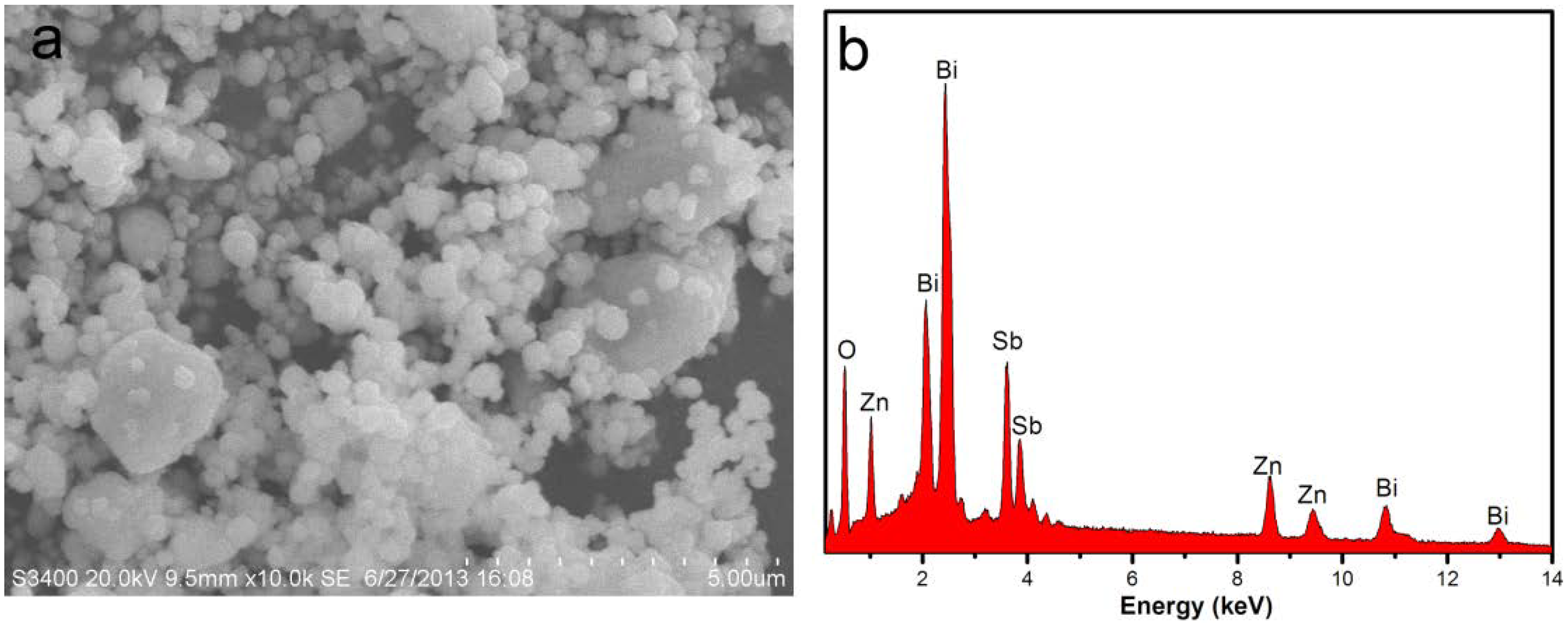
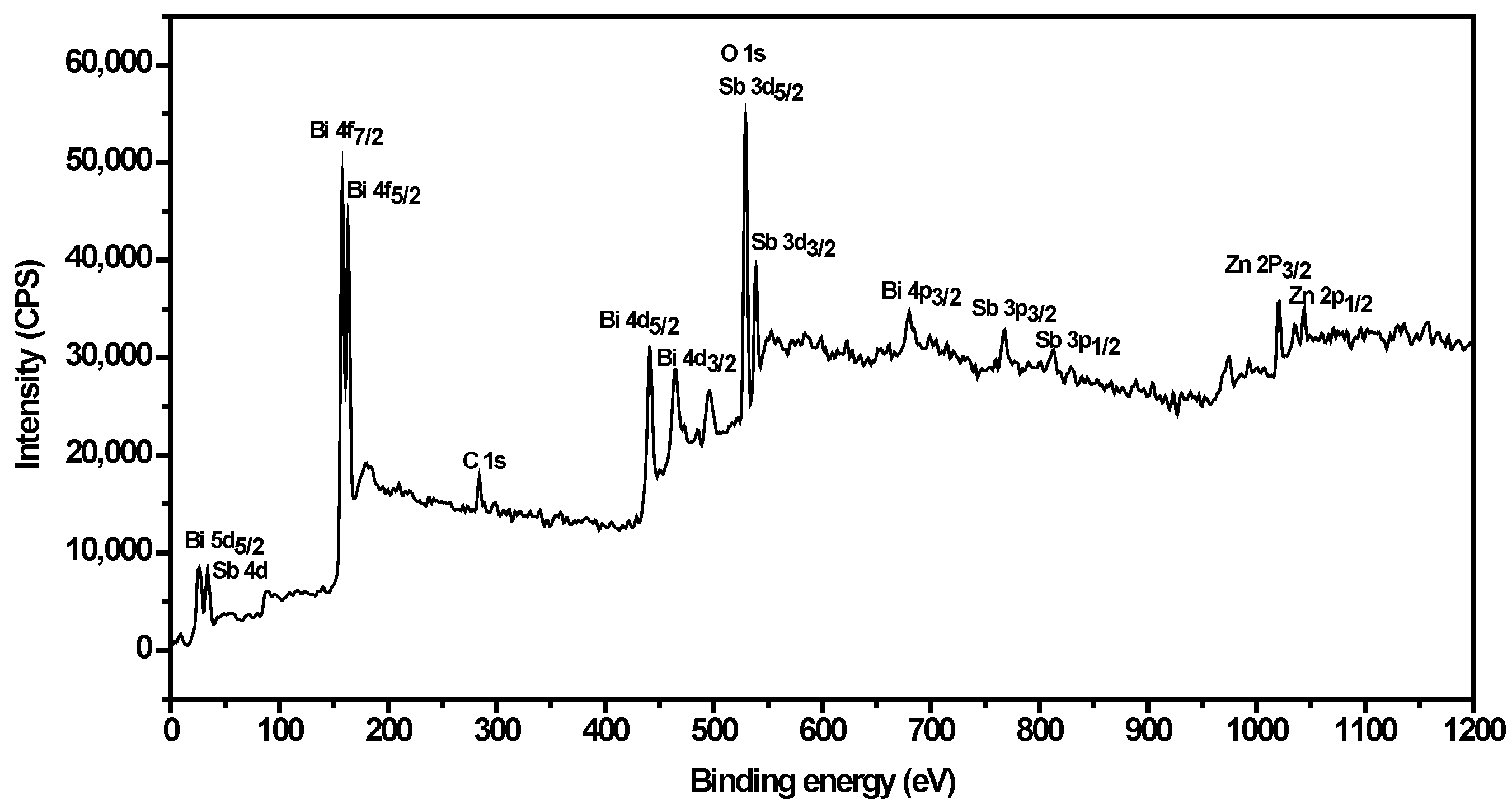
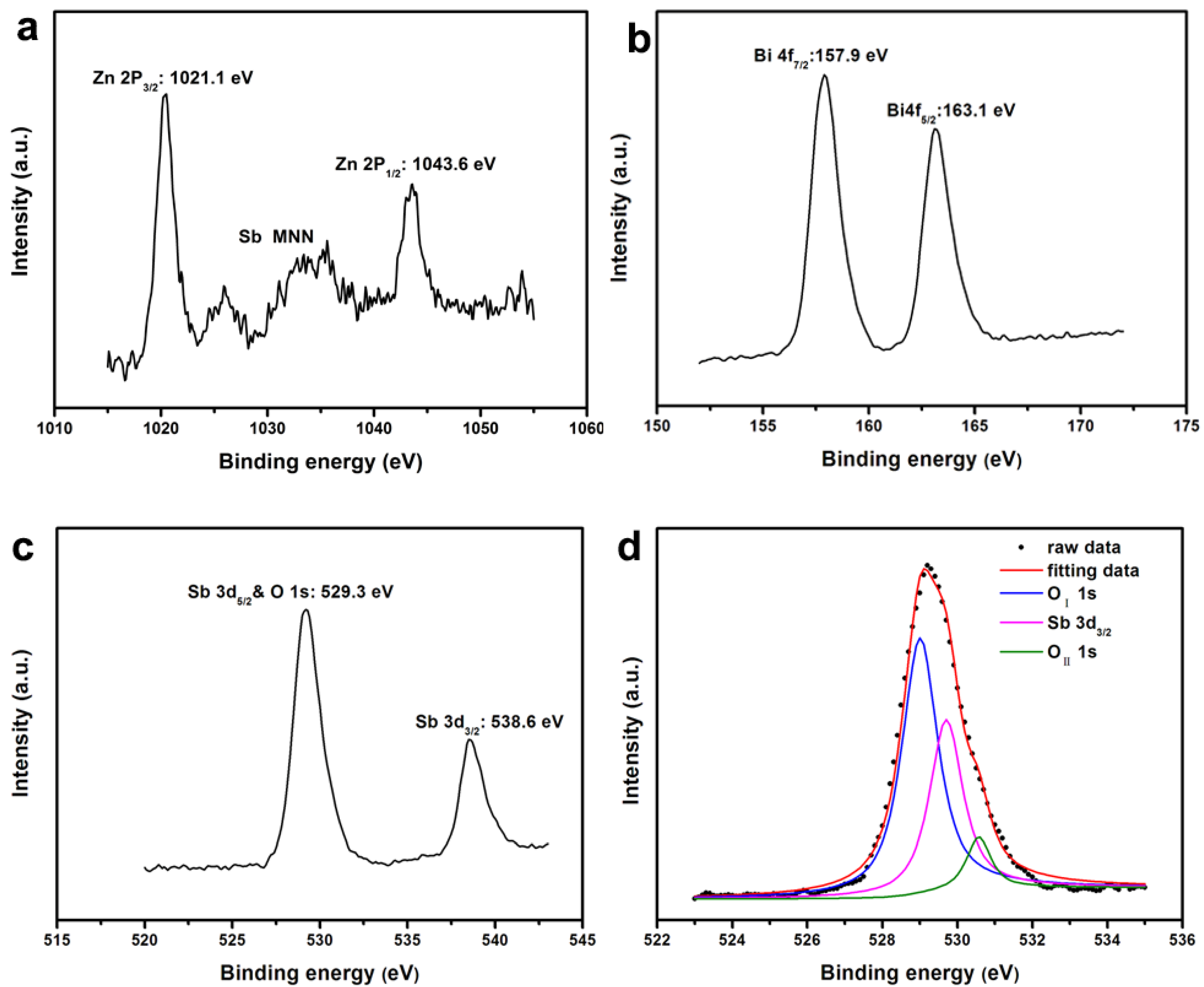
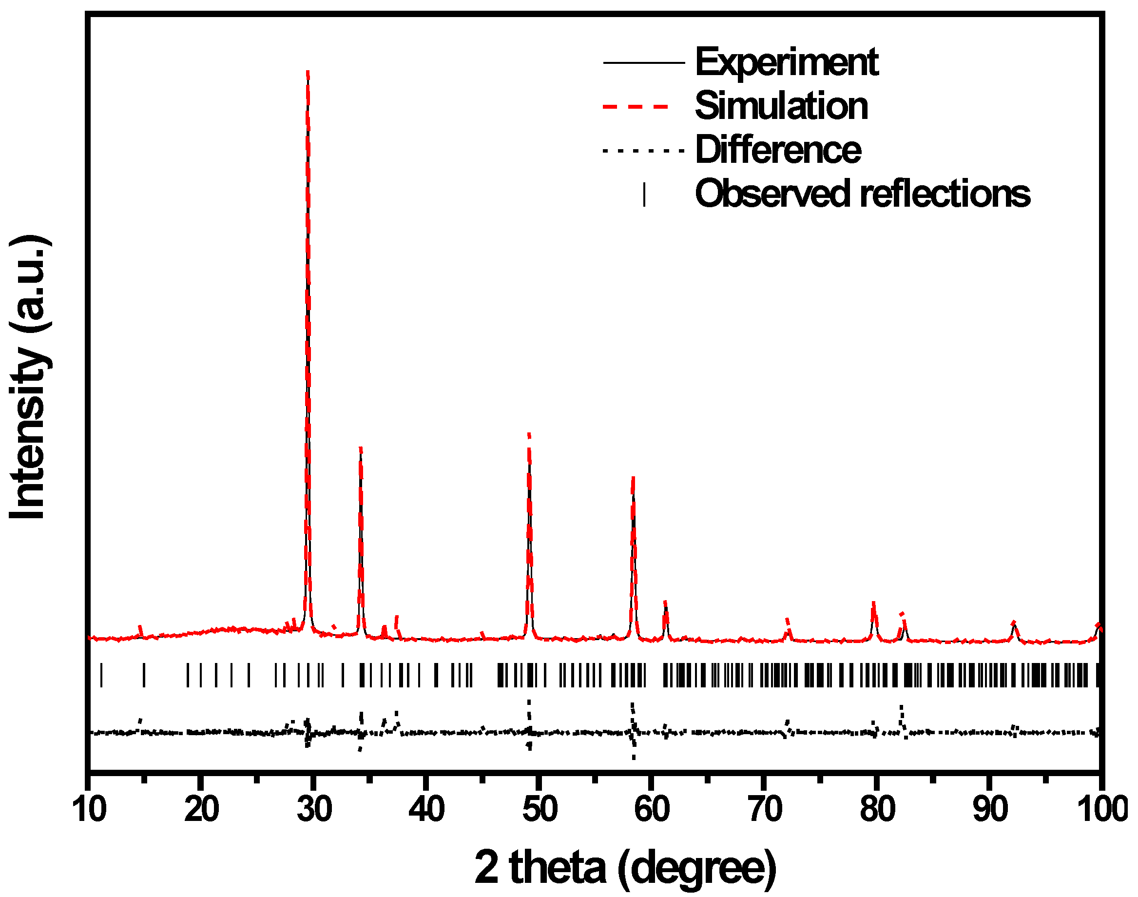
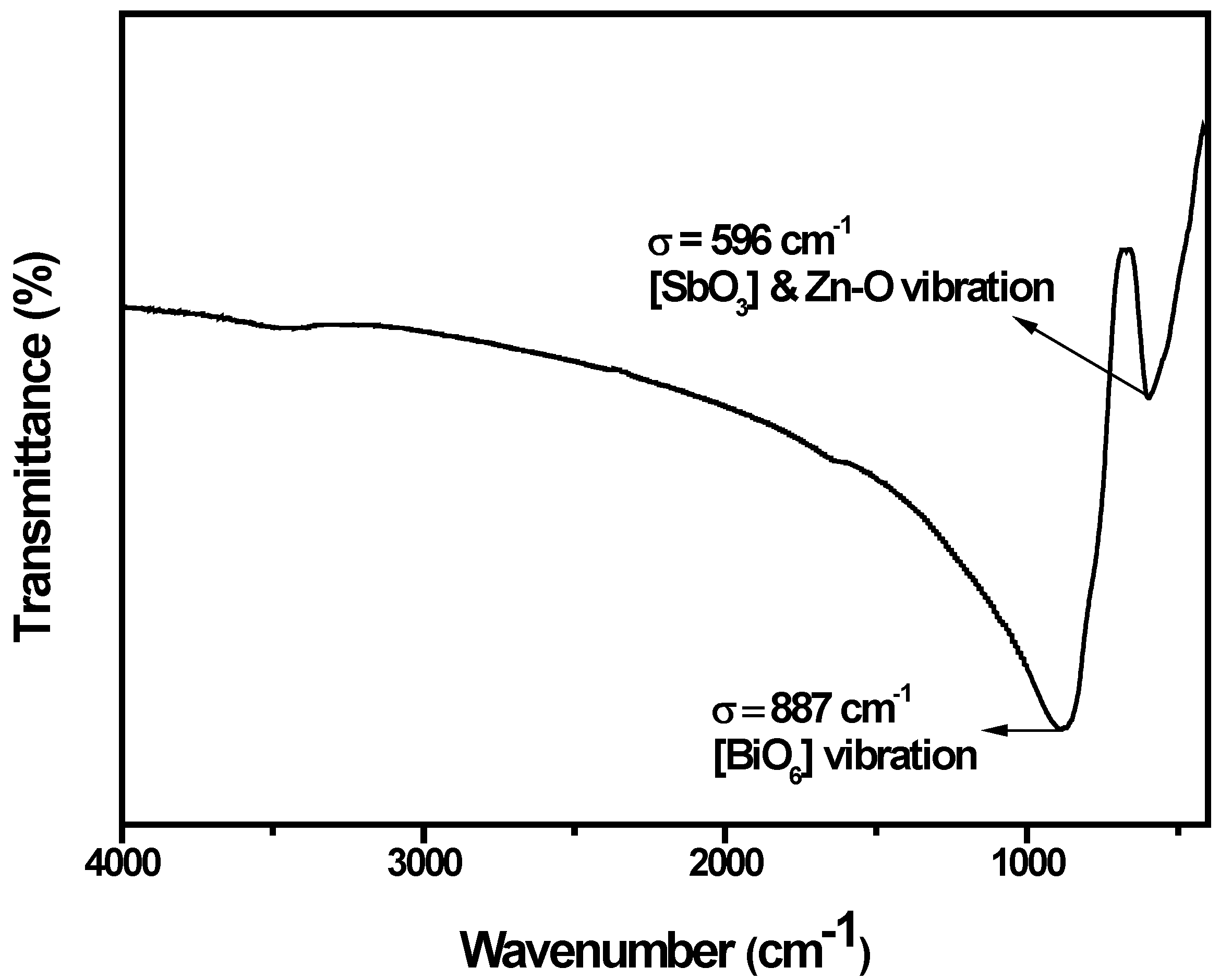
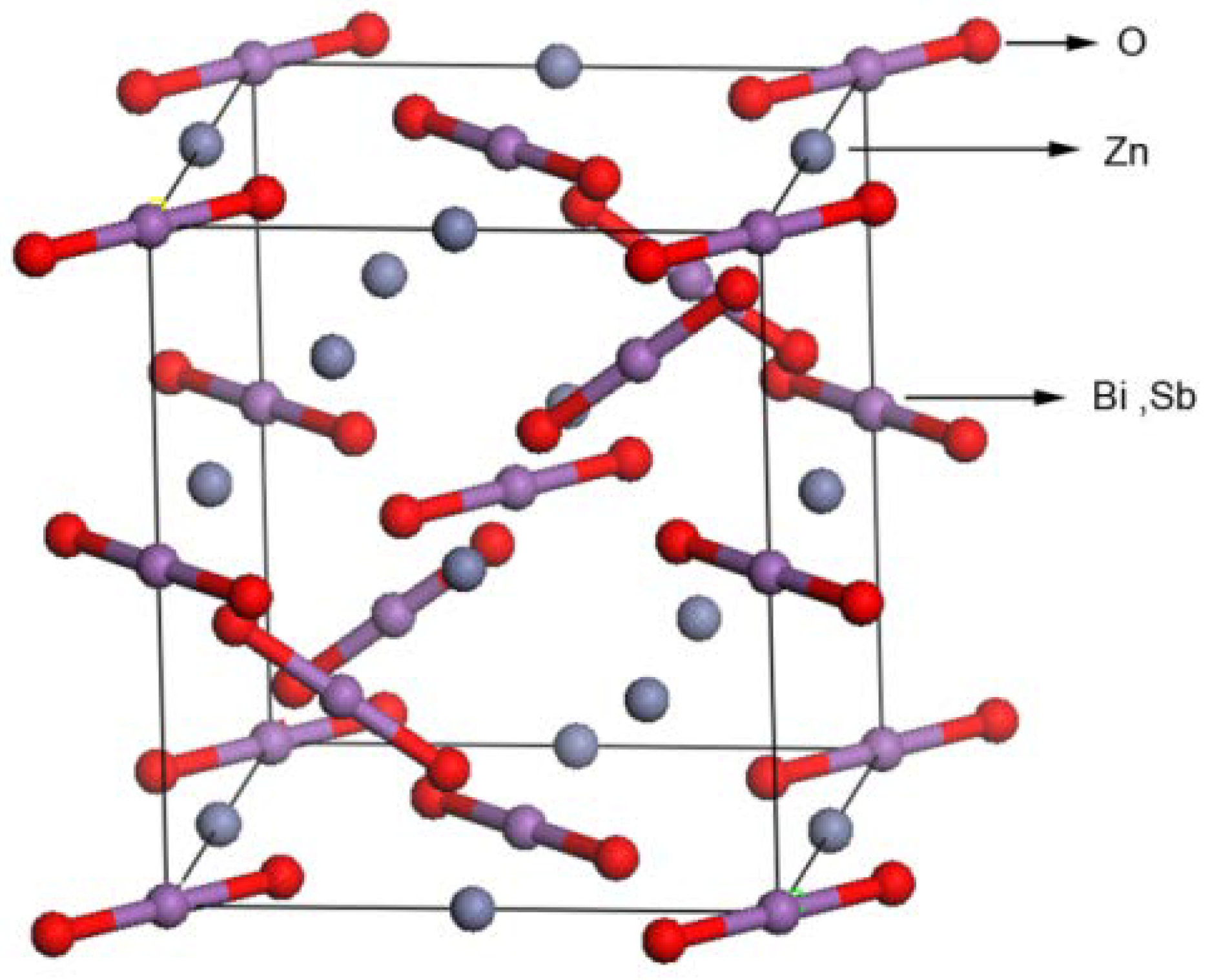
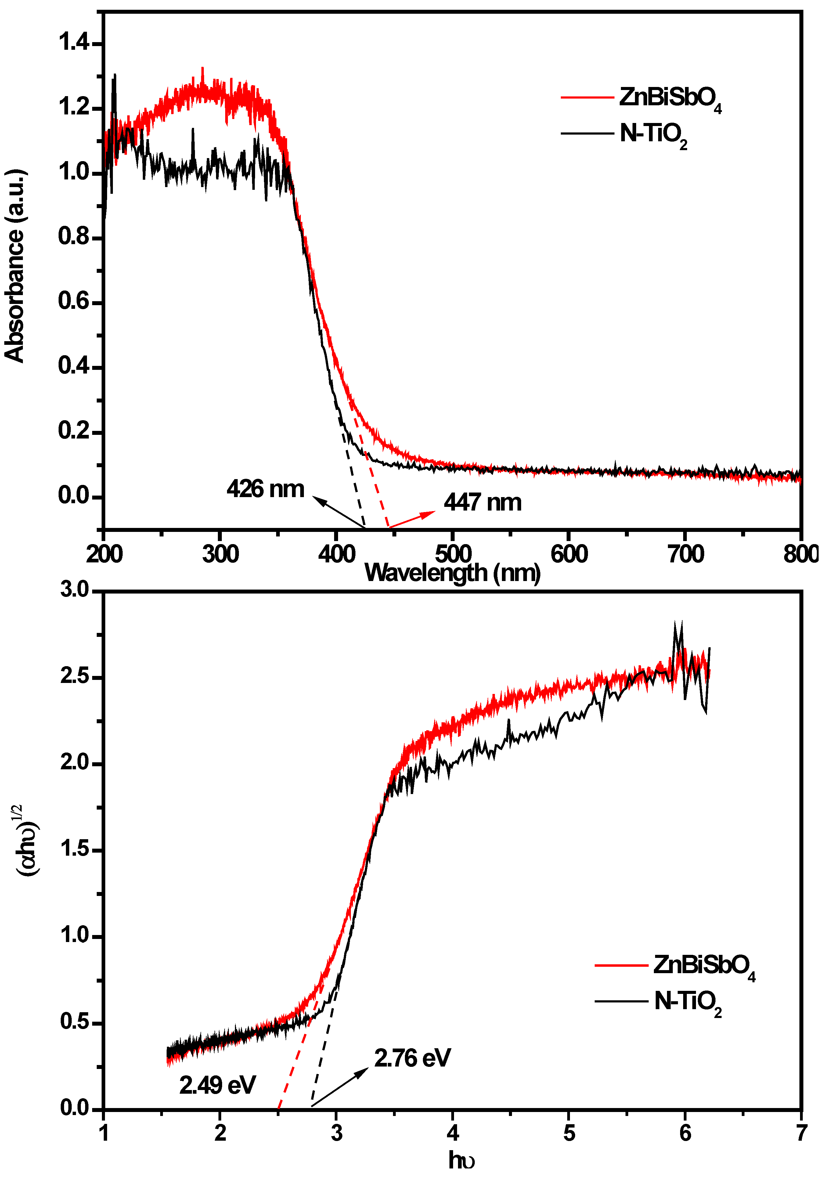
2.1. Crystal Structure and Optical Properties

The transmission electron microscopy (TEM) image of the prepared catalyst, ZnBiSbO4, is shown in Figure 1. It could be observed clearly from Figure 1a,b that the particles of ZnBiSbO4 had a nanostructure and irregular shapes. Additionally, we could also acknowledge that the particles of ZnBiSbO4 crystallized well, and the average particle size was about 220 nm in diameter. Figure 1b also shows the selected area electron diffraction pattern of ZnBiSbO4. It could be seen from Figure 1b that ZnBiSbO4 crystallized with a pyrochlore-type structure, a tetragonal crystal system and a space group I41/A, and the lattice parameters for ZnBiSbO4 were proven to be a = b = 12.040078 Å and c = 10.731416 Å. According to the calculation results from Figure 1b, the (h k l) value for the main peaks of ZnBiSbO4 could be found and indexed. The histogram of the size distribution of ZnBiSbO4 was also demonstrated in Figure 1c, and we could find that the particle size of the majority of ZnBiSbO4 was from 170 to 270 nm. Brunauer–Emmett–Teller (BET) measurements were also detected, and the specific surface area of ZnBiSbO4 was 2.87 m2·g−1.
Figure 1. (a) transmission electron microscopy (TEM) image of ZnBiSbO4; and (b) The selected area electron diffraction pattern of ZnBiSbO4; and (c) histogram of the size distribution of ZnBiSbO4 calcinated at 800 °C for 35 h.
Ijms 15 09459 g001
Figure 2 presents the scanning electron microscopy-energy-dispersive spectrometry (SEM-EDS) spectrum of ZnBiSbO4, indicating the presence of zinc, bismuth, antimony and oxygen element. In order to avoid the influence of inhomogeneity phenomenon on the selected surface, ten different specimen areas selection of ZnBiSbO4 were conducted in an EDS test. The mean of the results of the above EDS spectra taken from prepared ZnBiSbO4 indicated that the stoichiometric ratio of zinc, bismuth, antimony and oxygen was estimated to be 14.83:14.55:13.56:57.06, namely 1.05:1.03:0.96:4.04.
Figure 2. (a) scanning electron microscopy (SEM) spectrum of ZnBiSbO4; (b) energy-dispersive spectrometry (EDS) spectrum of ZnBiSbO4 calcinated at 800 °C for 35 h.
Ijms 15 09459 g002
In order to get a better understanding of the chemical state of all elements on the catalyst surface, the X-ray photoelectron spectroscopy (XPS) full spectrum of ZnBiSbO4 was measured and is displayed in Figure 3. The atom XPS spectra of Zn, Bi and Sb are shown in Figure 4a–c, respectively. As to the O atom XPS spectrum, we could observe that the O 1s peak was asymmetric and should be overlaid by the Sb 3d5/2 peak and the O 1s peak. Besides lattice oxygen (OI) in multiple oxide catalyst, the existence of chemisorbed oxygen (OII) on the surface of the catalyst should also be taken into consideration [44]. Thus, the fitted curves of Sb 3d5/2 and O 1s for XPS spectra are given in Figure 4d, and the fitting results are reported in Table 1. The various elemental peaks, which correspond to the specific binding energies of ZnBiSbO4, are given in Table 2. The area ratio of OI/OT was 87.36% (OT contained OI and OII), meaning that the atom ratio of OI/OT is 87.36% on the catalyst surface. The results further suggested that the oxidation state of Zn, Bi, Sb and O ions from ZnBiSbO4 were +2, +3, +3 and −2, respectively. After calculation with the reported method [45], the average atomic ratio of Zn:Bi:Sb:O for ZnBiSbO4 was 1.00:0.98:1.02:4.12 based on our XPS results. The slight difference between the results that were found in XPS and EDS measurements could be ascribed to the inhomogeneity of different selected areas of ZnBiSbO4 during the testing. Therefore, it could be deduced that the obtained material was of high purity under our preparation conditions.
Figure 3. The X-ray photoelectron spectroscopy (XPS) full spectrum of ZnBiSbO4 calcinated at 800 °C for 35 h.
Ijms 15 09459 g003
Figure 4. (a) The XPS spectrum of Zn 2p; (b) the XPS spectrum of Bi 4f; (c) the XPS spectrum of Sb 3d; and (d) peak curves of Sb 3d5/2 and O 1s for the XPS spectrum of ZnBiSbO4 (OI, lattice oxygen; OII, chemisorbed oxygen).
Ijms 15 09459 g004
Table 1. XPS data corresponding to Figure 4d.
ElementSb 3d5/2O 1s
OIOII
Binding energy (eV)529.7529.0530.6
Table 2. Binding energies for key elements of ZnBiSbO4.
ElementZn 2p3/2Bi 4f7/2Sb 3d5/2O 1s
Binding energy (eV)1021.1157.9529.7529.0
Figure 5 shows the powder X-ray diffraction pattern of ZnBiSbO4 with the full-profile structure refinements of the collected data, which were obtained by the RIETAN™ [46] program based on the Rietveld analysis. It could be seen from Figure 5 that ZnBiSbO4 turned out to be a single phase. Additionally, the results of the final refinements for ZnBiSbO4 indicated a good agreement between the observed intensities and calculated intensities for the pyrochlore-type structure, a tetragonal crystal system and a space group, I41/A (O atoms were included in the model). The lattice parameters for ZnBiSbO4 were a = b = 12.040078 Å and c = 10.731416 Å. All the diffraction peaks for ZnBiSbO4 could be successfully indexed according to the lattice constant and above space group. The atomic coordinates and structural parameters of ZnBiSbO4 are listed in Table 3. According to above results, the structural model of ZnBiSbO4 which is simulated by Materials Studio software is demonstrated in Figure 6.
Figure 5. X-ray diffraction (XRD) pattern and Rietveld refinements of ZnBiSbO4.
Ijms 15 09459 g005
Table 3. Structural parameters of ZnBiSbO4 calcinated at 800 °C for 35 h.
AtomxyzOccupation factor
Zn000.51
Bi0001
Sb0001
O0.767310.140130.081881
Figure 6. The structural model of ZnBiSbO4 simulated by Materials Studio software corresponding to the XRD pattern shown in Figure 5.
Ijms 15 09459 g006
Fourier transform infrared (FTIR) spectrum analysis of ZnBiSbO4 particles is investigated in this study, as shown in Figure 7. From this picture, we could find that the absorption bands of ZnBiSbO4 prepared by a solid-state reaction method at 800 °C are at 596 and 887 cm−1. The strong band near 887 cm−1 should be attributed to the Bi–O vibration in the distorted [BiO6] unit [47]. The band situated at 596 cm−1 was overlaid by the symmetric bending and stretching of the [SbO3] unit [48] and Zn–O stretching [49].
Figure 7. Fourier transform infrared (FTIR) spectrum of ZnBiSbO4 calcinated at 800 °C for 35 h.
Ijms 15 09459 g007
The absorption spectrum of ZnBiSbO4 is presented in Figure 8. For a crystalline semiconductor, the optical absorption near the band edge following Equation (1) [50]:
α = A × (Eg)n
Eg = 1240/λ
Here, A, α, Eg, ν and λ were the proportional constant, absorption coefficient, band gap, light frequency and absorption edge, respectively. In this equation, n determined the character of the transition in a semiconductor. Eg and n could be calculated by the following steps [51]: (i) plotting ln (αhν) vs. ln (hν − Eg) by assuming an approximate value of Eg, which can be calculated by Equation (2); (ii) deducing the value of n; and (iii) refining the value of Eg. From Figure 8, we could find that the absorption edge of ZnBiSbO4 was about 447 nm, meaning that the estimated Eg of ZnBiSbO4 was 2.78 eV. Then, plot ln (αhν) vs. ln (hν − Eg), where we could find the slope of the line part, was about 1.65. Therefore, the n of ZnBiSbO4 was two. After plotting (α)1/2 vs. hν and extrapolating the plot to (αhν)1/2 = 0, the accurate value of Eg of ZnBiSbO4 was calculated as 2.49 eV. Applying the same calculation process to N-TiO2, we found that for N-TiO2: n = 2 and Eg = 2.76 eV. The above results indicated that the optical transition for ZnBiSbO4 or N-TiO2 was indirectly allowed, and ZnBiSbO4 possessed a narrow band gap compared with N-TiO2. The results in our past work showed that the optical transition for CdBiYO4 was directly allowed and that the band gap of CdBiYO4 was 2.41 eV [43].
Figure 8. The absorption spectrum, plot of (αhν)1/2 vs. hν for ZnBiSbO4 calcinated at 800 °C for 35 h and N-TiO2.
Ijms 15 09459 g008

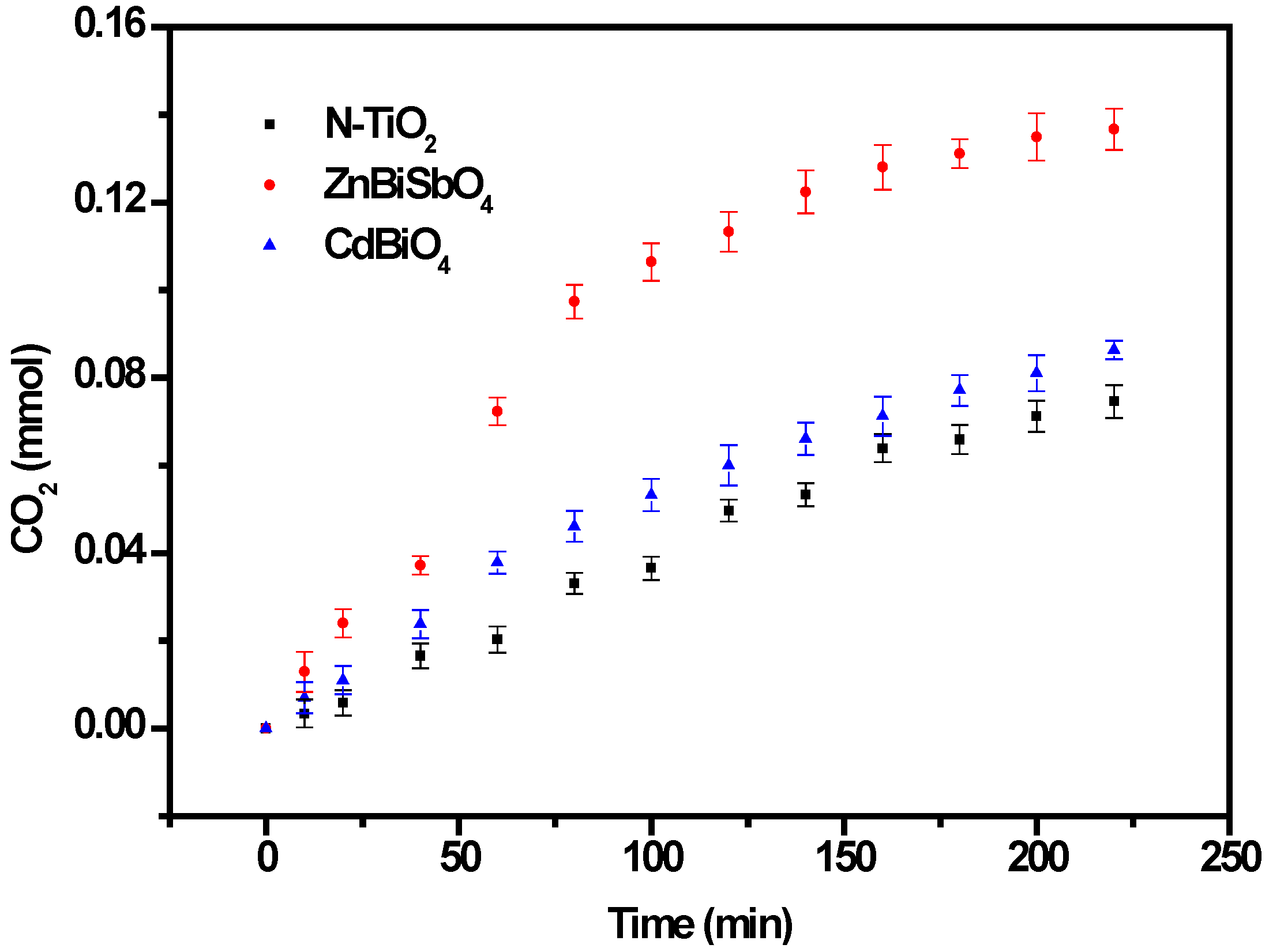
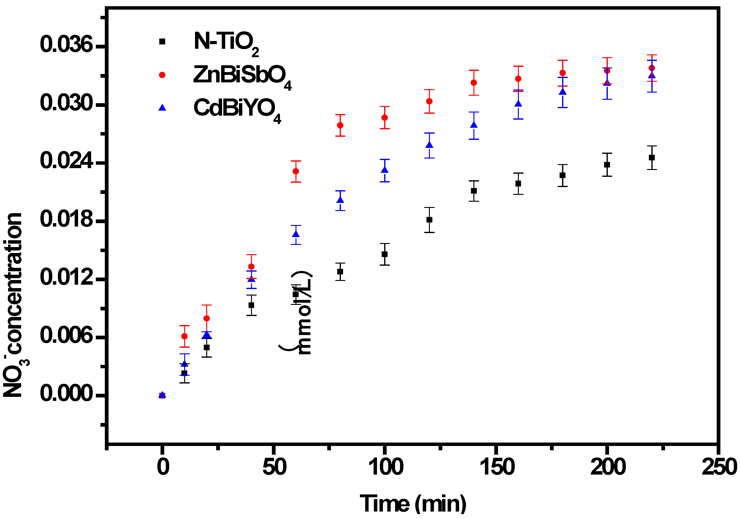
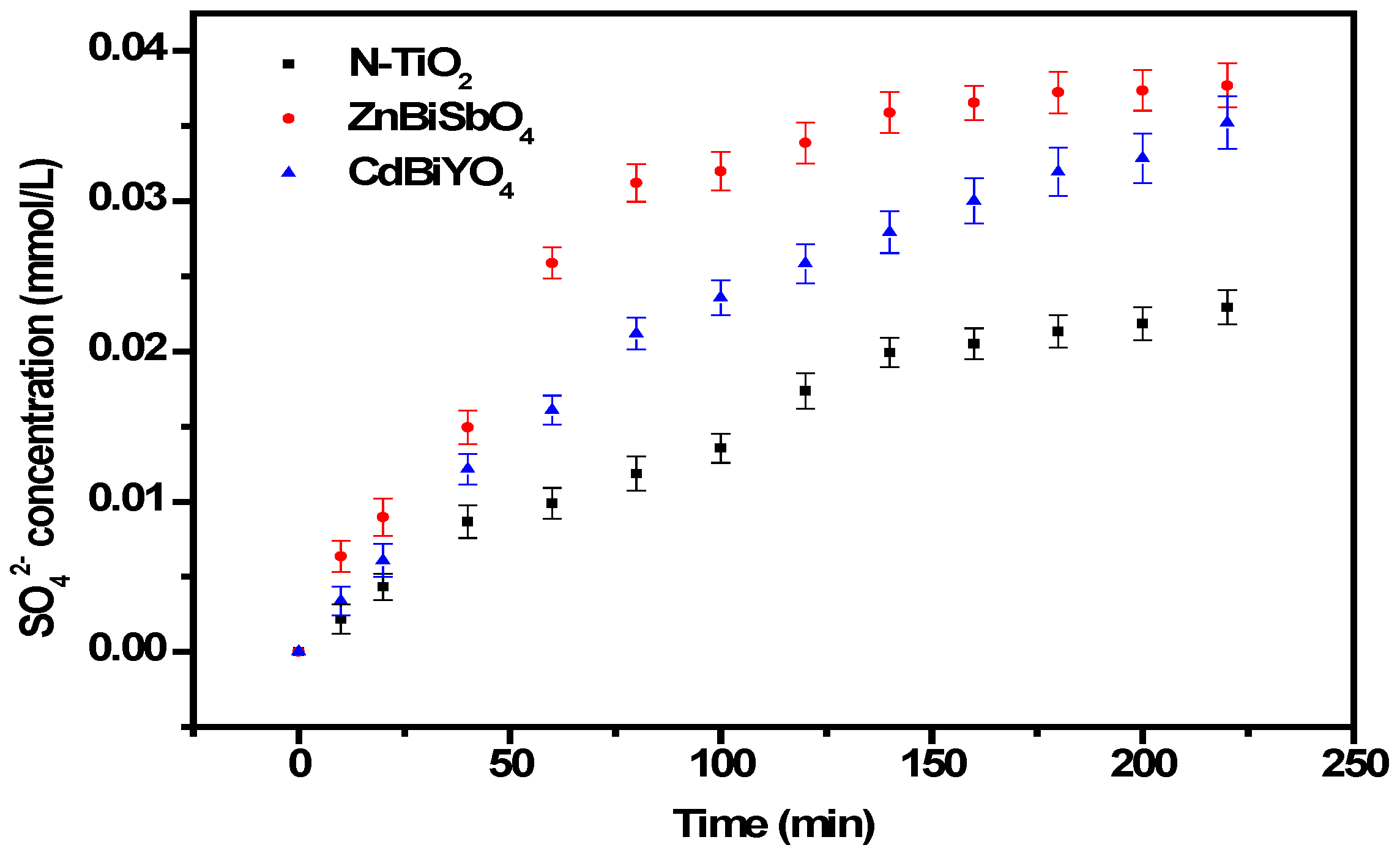
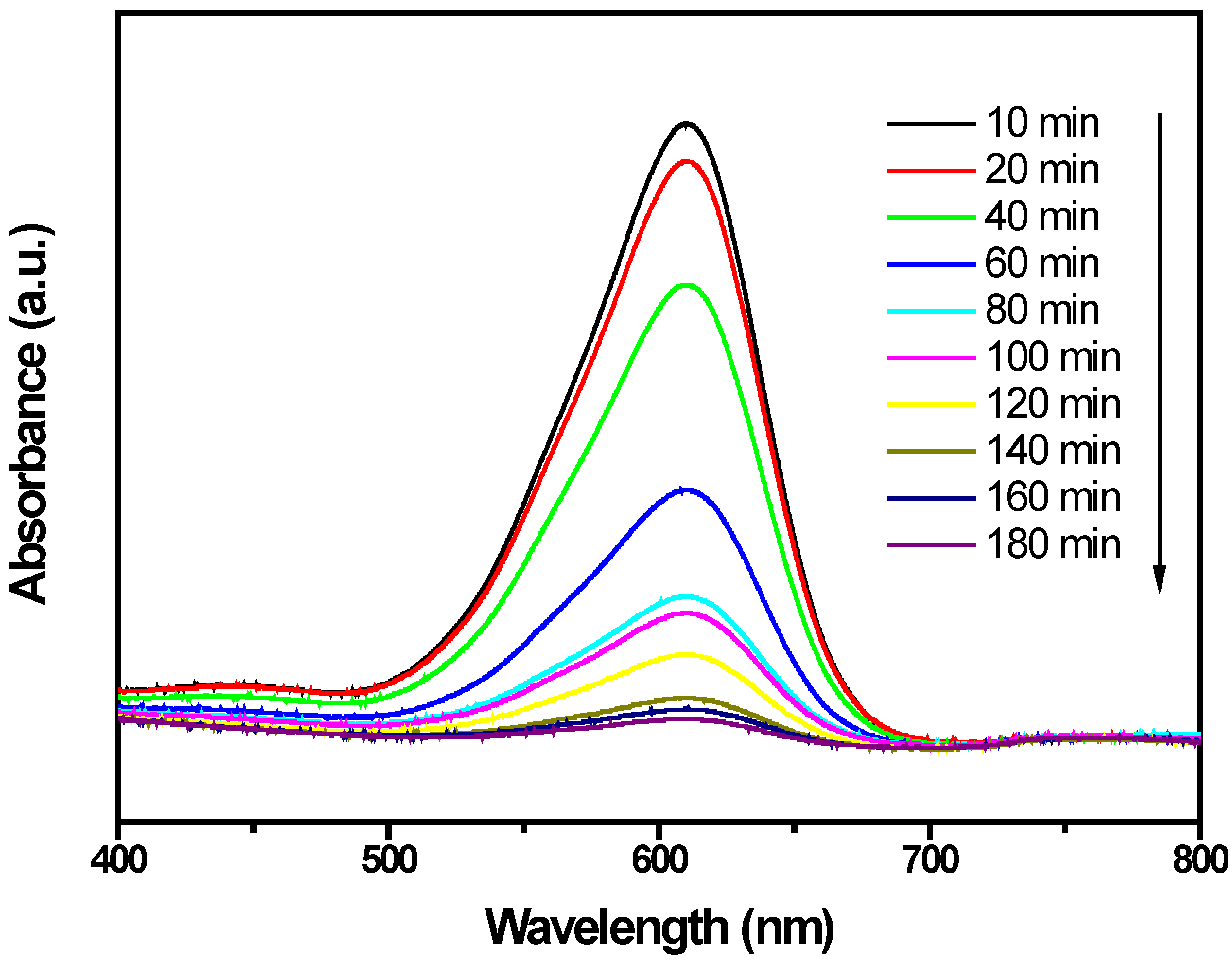
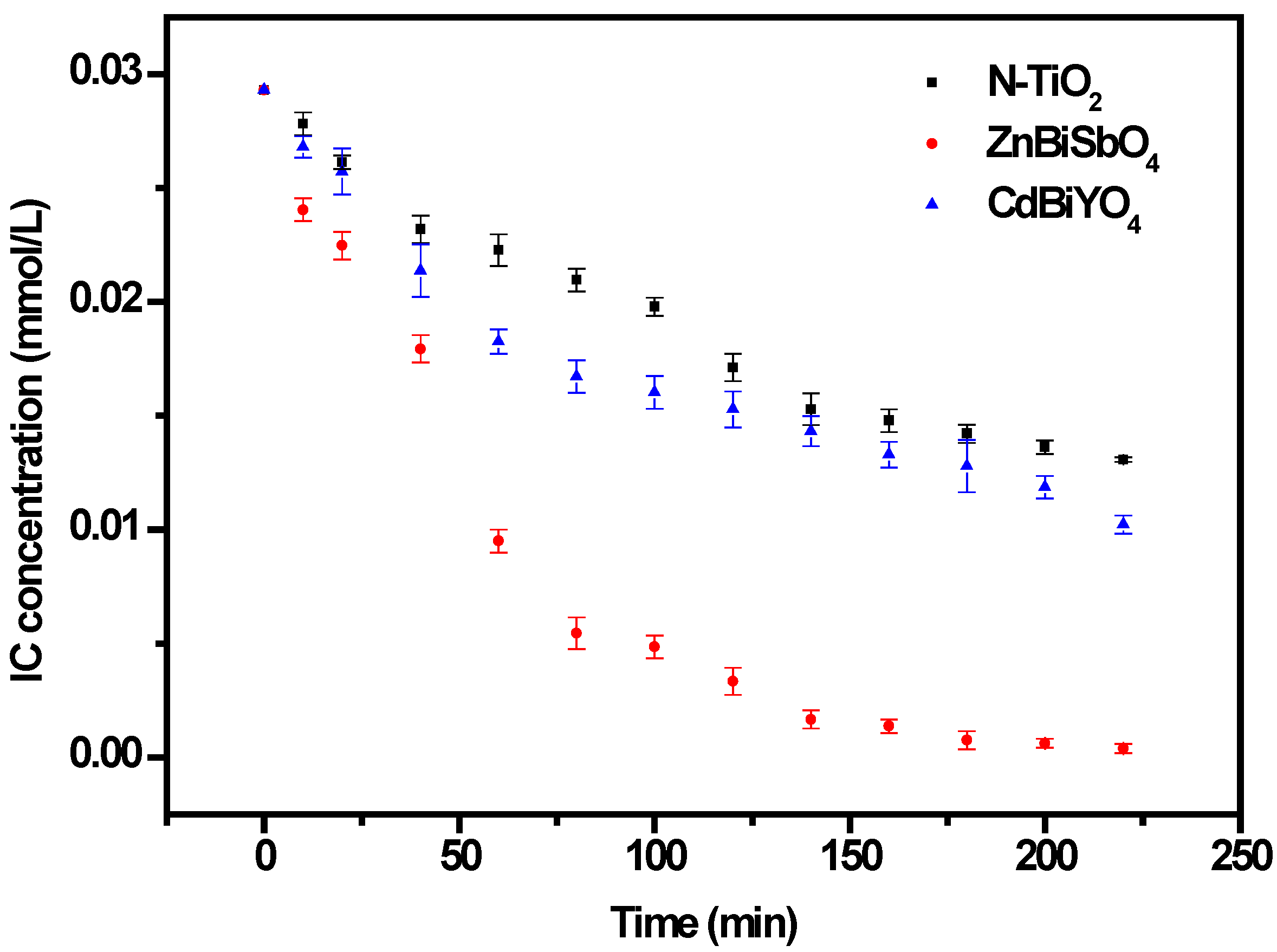
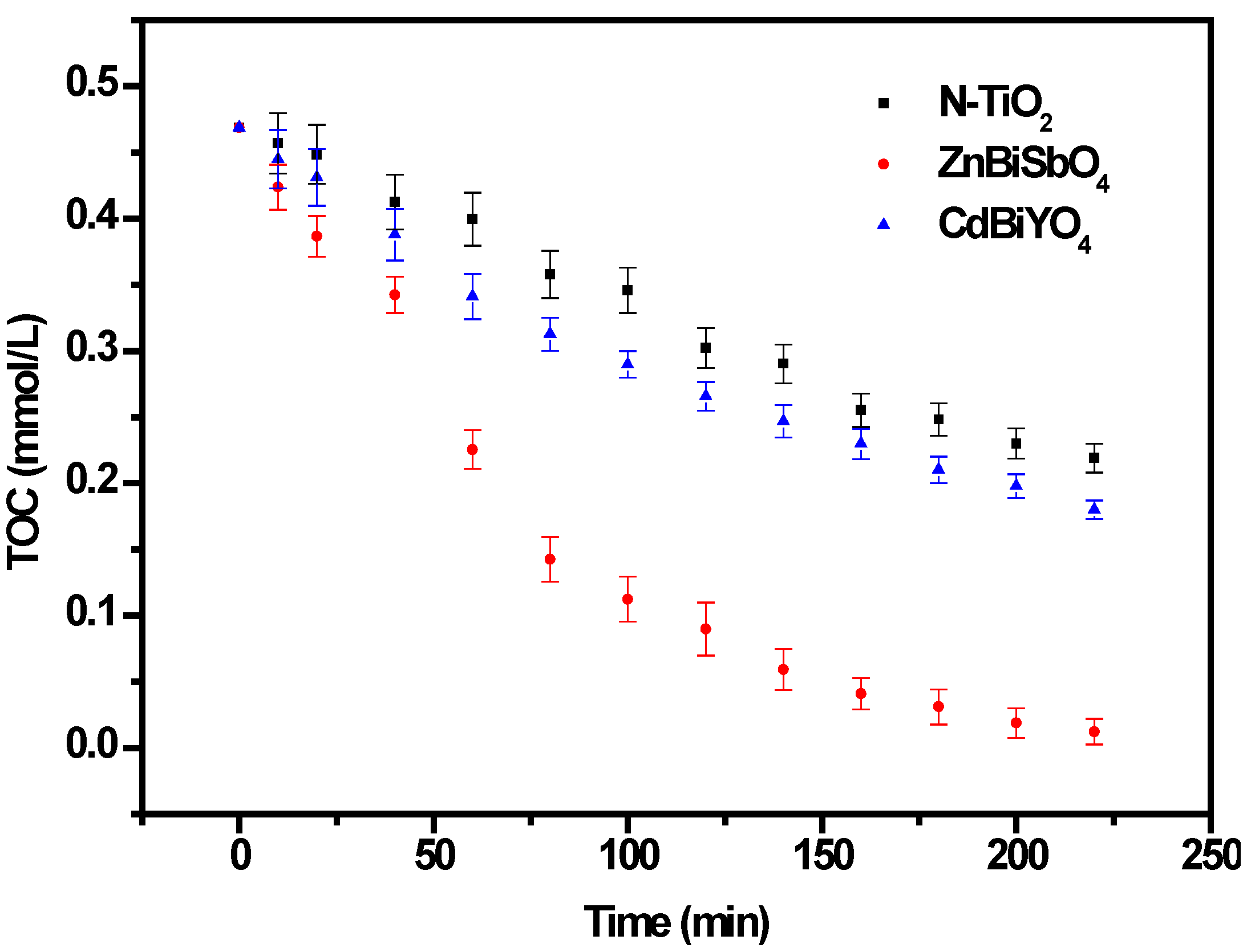
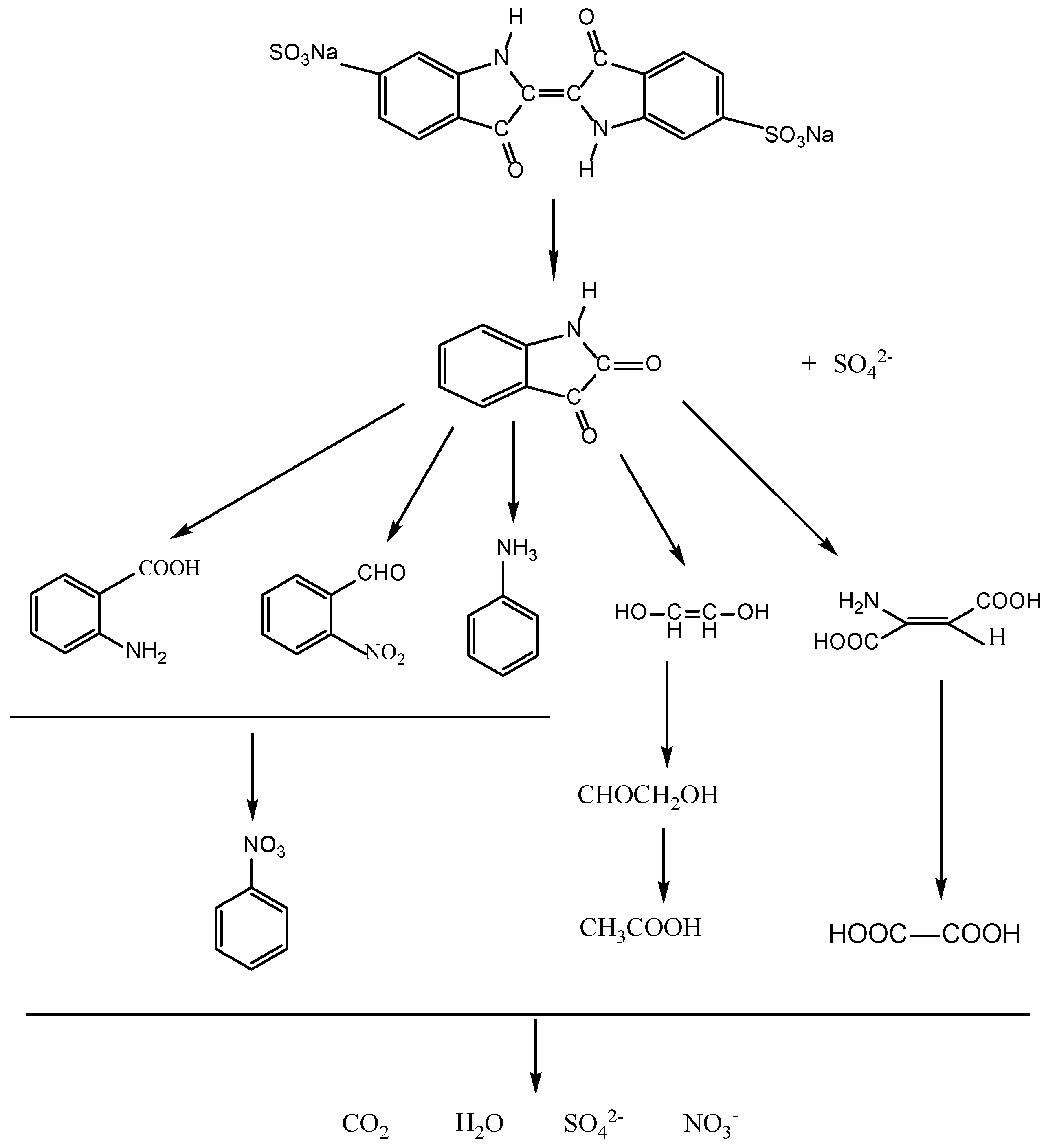
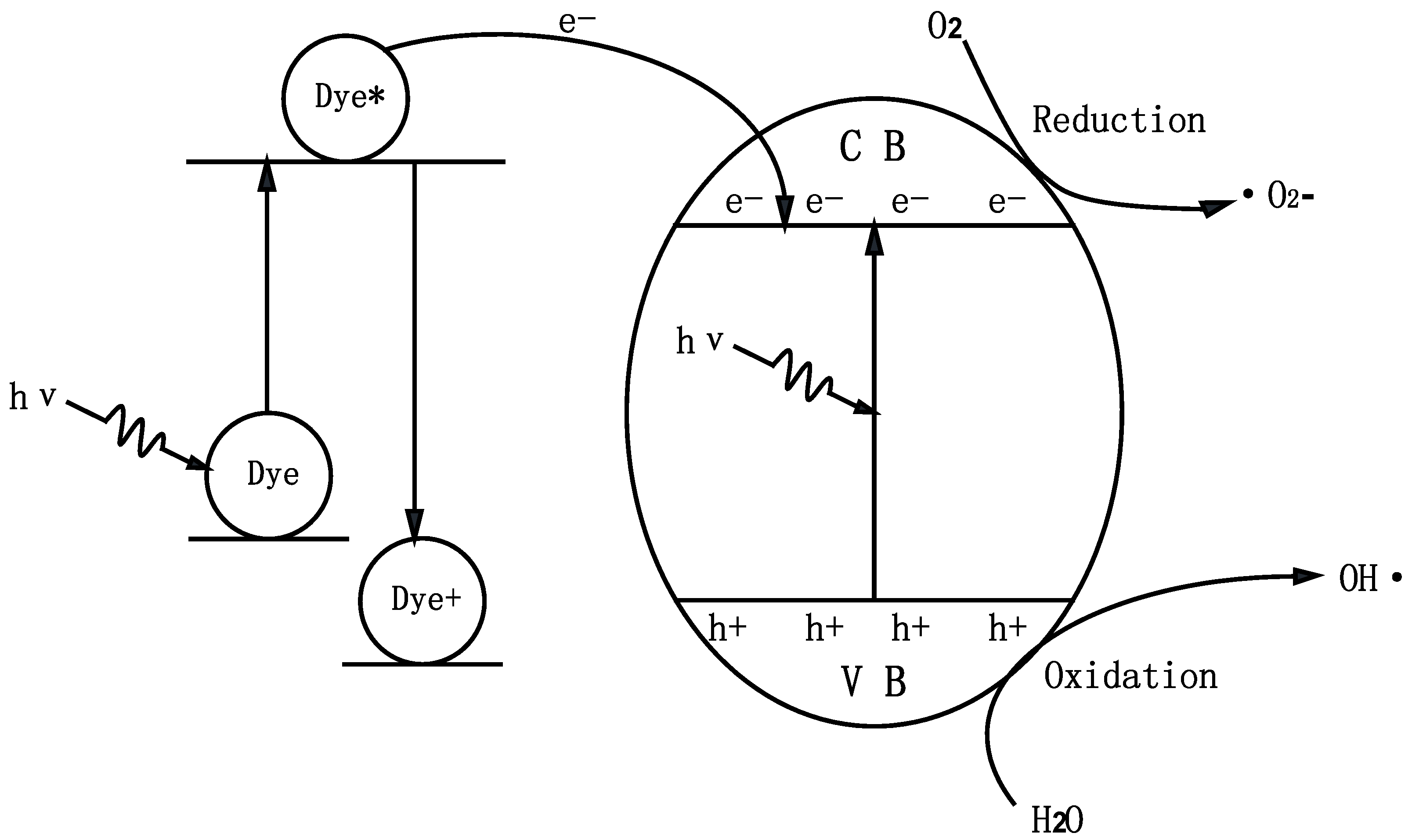
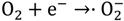
2.2. Photocatalytic Properties

The progress of photocatalysis using the semiconductor compound could be described briefly as follows [52,53]. Firstly, the semiconductor compound absorbed photons, resulting in the generation of electron-hole pairs within the semiconductor compound particles and, subsequently, the diffusion of the charge carriers to the surface of the semiconductor compound particle would be followed; at the same time, the active sites of the surface of the semiconductor compound particles had been adsorbing a lot of pollutants particles; finally, the decomposition of pollutants would be performed by charge carriers.
Figure 9 presents the changes in the UV-Vis spectra of IC under visible light irradiation (λ > 400 nm) with the presence of ZnBiSbO4. The above measurements were performed under oxygen-saturation conditions ([O2]sat = 1.02 × 10⁻³ M). It could be clearly noticed from Figure 9 that the typical IC peaks were at 609.5 nm. An obvious color change from deep red into a colorless solution could be observed within 230 min. For further comparison, Figure 10 depicts the concentration changes of IC with ZnBiSbO4, CdBiYO4 and N-TiO2 as photocatalysts under visible light irradiation, respectively. It could be seen from Figure 10 that the photonic efficiency (λ = 420 nm) was estimated to be 0.0460%, 0.0258% and 0.0304% with ZnBiSbO4, N-TiO2 and CdBiYO4, respectively. When ZnBiSbO4 or CdBiYO4 or N-TiO2 was utilized as a catalyst, the photodegradation conversion rate of IC was 98.66% or 65.12% or 55.39% after visible light irradiation for 220 min. The results showed that the photodegradation rate of IC and the photonic efficiency with ZnBiSbO4 as a catalyst were both higher than those with N-TiO2 or CdBiYO4 as a catalyst. The above results showed that complete removal of indigo carmine was observed after visible light irradiation for 230 min with ZnBiSbO4 as a catalyst. Besides, based on the absorbance changes of IC with irradiation time, the kinetics of IC degradation under visible light irradiation was figured out. The above results demonstrated that the photocatalytic kinetics of IC degradation with ZnBiSbO4, CdBiYO4 and N-TiO2 as photocatalysts followed a first order nature. The first-order rate constants for IC degradation were estimated to be 0.01945, 0.00405 and 0.00502 min−1 with ZnBiSbO4, N-TiO2 and CdBiYO4 as catalysts, respectively. This fact indicated that ZnBiSbO4 was more efficient than N-TiO2 or CdBiYO4 for the photocatalytic degradation of IC under visible light irradiation.
Figure 9. Spectral changes of aqueous solutions of indigo carmine (IC) due to visible light irradiation with the presence of ZnBiSbO4 calcinated at 800 °C for 35 h.
Ijms 15 09459 g009
Figure 10. Photocatalytic degradation of IC under visible light irradiation with the presence of ZnBiSbO4, CdBiYO4 and N-TiO2 as photocatalysts, respectively. The results are means of triplicates, and error bars represent ±1 SD.
Ijms 15 09459 g010
Figure 11 shows the change of total organic carbon (TOC) during the photocatalytic degradation of IC with ZnBiSbO4, CdBiYO4 or N-TiO2 as a catalyst under visible light irradiation. The TOC measurements revealed the disappearance of organic carbon when the IC solution, which contained ZnBiSbO4 or CdBiYO4 or N-TiO2, was exposed under visible light irradiation. The results showed that 97.37% or 61.59% or 53.26% of a TOC decrease was obtained after visible light irradiation for 220 min when ZnBiSbO4 or CdBiYO4 or N-TiO2 was utilized as the photocatalyst. Consequently, it could be seen from Figure 11 that the entire mineralization of indigo carmine was realized after visible light irradiation for 270 min with ZnBiSbO4 as a catalyst, because of the 100% TOC removal. The apparent first order rate constant, k, was estimated to be 0.01536, 0.00449 and 0.00350 min−1 with ZnBiSbO4, CdBiYO4 or N-TiO2 as the photocatalyst, respectively.
During the progress of IC degradation, IC was converted into smaller organic species and was ultimately mineralized to inorganic products, such as carbon dioxide and water. Figure 12 presents the CO2 yield during the photocatalytic degradation of IC with ZnBiSbO4 or CdBiYO4 or N-TiO2 as the photocatalyst under visible light irradiation. The amount of CO2 increased gradually with increasing reaction time when IC was photodegraded with ZnBiSbO4 or CdBiYO4 or N-TiO2 as the photocatalyst. The results showed that the production rate of CO2 from the ZnBiSbO4-IC system was higher than that from the CdBiYO4-IC system or N-TiO2-IC system with increasing reaction time. For example, the production amount of CO2 was 0.08103 or 0.07122 mmol with CdBiYO4 or N-TiO2 as the photocatalyst after a visible light irradiation of 200 min. However, the production amount of CO2 was 0.1349 mmol with ZnBiSbO4 as the photocatalyst after a visible light irradiation of 200 min.
Figure 11. Total organic carbon (TOC) plots during the photocatalytic degradation of IC under visible light irradiation with ZnBiSbO4, CdBiYO4 and N-TiO2 as photocatalysts, respectively. The results are means of triplicates, and error bars represent ±1 SD.
Ijms 15 09459 g011
Figure 12. CO2 production during the photocatalytic degradation of IC with ZnBiSbO4, CdBiYO4 and N-TiO2 as photocatalysts, respectively, under visible light irradiation. The results are means of triplicates, and error bars represent ±1 SD.
Ijms 15 09459 g012
In order to explore the mechanism of the IC degradation with ZnBiSbO4 or CdBiYO4 or N-TiO2 as photocatalyst under visible light irradiation, we also tested the concentration of inorganic ions, NO3 and SO42−, which are shown in Figure 13 and Figure 14, which may be formed as the end products of nitrogen and sulfur atoms that existed in IC. From Figure 13 and Figure 14, we could be sure that both NO3 and SO42− appeared during IC degradation with ZnBiSbO4 or CdBiYO4 or N-TiO2 as the photocatalyst. NO3 and SO42− ions were generated more quickly and effectively with ZnBiSbO4 compared with N-TiO2 or CdBiYO4 as photocatalyst, which was in accord with the above analysis about the degradation progress of IC. According to the NO3 concentration in Figure 13, we could calculate that 57.66% or 68.95% or 41.89% of sulfur from IC was converted into sulfate ions with ZnBiSbO4 or CdBiYO4 or N-TiO2 as the photocatalyst after visible light irradiation for 220 min. Meanwhile, it could be also concluded that 64.32% or 60.09% or 39.11% of sulfur from IC was converted into sulfate ions with ZnBiSbO4 or CdBiYO4 or N-TiO2 as photocatalyst after visible light irradiation for 220 min. It was noteworthy that the amount of SO42− and NO3 that were released into the solution were sharply lower than the stoichiometry value of 100%. One possible reason could be a loss of sulfur-containing volatile compounds or SO2 for the S element and nitrogen-containing volatile compounds or NH3. The second possible reason was a partially irreversible adsorption of some SO42− and NO3 on the surface of the photocatalyst, which had been observed by Lachheb et al. with titanium dioxide [54].
Figure 13. NO3 production during the photocatalytic degradation of IC with ZnBiSbO4, CdBiYO4 and N-TiO2 as photocatalysts, respectively, under visible light irradiation. The results are means of triplicates, and error bars represent ±1 SD.
Ijms 15 09459 g013
In addition, the intermediates generated during the degradation process were detected by high-performance liquid chromatography (HPLC) and gas chromatography-mass spectrometry (GC-MS) and identified by comparison with commercial standard samples. The intermediates in our experiment were identified as follows: indoline-2,3-dione, anthranilic acid, O-nitrobenzyl dehyde, aniline, ethene-1,2-diol, amino-fumaric acid, oxalic acid, glycolal dehyde and acetic acid. The sulfur was first hydrolytically removed and subsequently was oxidized and transformed into SO42−. At the same time, nitrogen atoms in the −3 oxidation state produced NH4+ cations that subsequently were oxidized into NO3 ions. Based on the above results, we have deduced a degradation pathway of IC with ZnBiSbO4, as shown in Figure 15. This pathway was similar, but not identical, to the pathway proposed by Manon Vautier et al. [55]. According to Figure 15, the main degradation end-products of IC were CO2, NO3 and SO42. Thus, the mass balance of the C, N and S elements from IC (reaction time = 0 min) to end-products (reaction time = 220 min) during the degradation process of IC in the reactor had been studied. The original amount of C within IC was 140.64 μmol, which was close to the amount of C within the end-product, CO2 (137.00 μmol). The value of the original amount of N within IC vs. the one within end-product NO3 was 17.58 μmol/10.20 μmol. The value of the original amount of N within IC vs. the one within end-product NO3 was 17.58 μmol/11.40 μmol. The above results agreed with the analysis about Figure 12, Figure 13 and Figure 14.
Figure 14. SO42− production during the photocatalytic degradation of IC with ZnBiSbO4, CdBiYO4 and N-TiO2 as photocatalysts, respectively, under visible light irradiation. The results are the means of triplicates, and error bars represent ±1 SD.
Ijms 15 09459 g014
Figure 15. Suggested photocatalytic degradation pathway scheme for IC under visible light irradiation with the presence of ZnBiSbO4 prepared by a solid-state reaction method at 800 °C for 35 h.
Ijms 15 09459 g015
A mechanism scheme of the charge separation and photocatalytic reaction for ZnBiSbO4 is shown in Figure 16. Firstly, photoinduced holes (h+) and photoinduced electrons (e) came into being in the surface of ZnBiSbO4 particles (Equation (3)). Secondly, organic pollutants (R) could be degraded into inorganic products with the effluence of h+ and e. Many published works [56,57,58] had confirmed that two oxidative agents could be mainly concerned under visible light irradiation: OH·radicals and·O2 radicals. Then, h+ reacted with R directly (Equations (4)–(6)). Besides, the effect of dye sensitization should be taken into consideration (Equations (7) and (8)); because IC could be excited by visible light, and then, the sensitizing dye molecules injected electrons into the semiconductor nanocrystallites, which were collected at a conducting surface to generate the photocurrent (Equation (9)) [59,60].
Figure 16. Suggested photodegradation reaction mechanism of IC with ZnBiSbO4 as the photocatalyst under visible light irradiation. (CB, Conduction Band; VB, Valence Band)
Ijms 15 09459 g016
ZnBiSbO4 + → h+ + e
(H2O ↔ H+ + OH) + h+ → H+ +· OH
Ijms 15 09459 i001
Ijms 15 09459 i002
IC + → IC*
IC* + ZnBiSbO4 → IC+ + ZnBiSbO4(e)
Ijms 15 09459 i003

2.3. Phytotoxicity Properties

The phytotoxicity of the photocatalytic-treated IC wastewater was detected by examining its effect on seed germination and growth as shown in Table 4 and Table 5. We could find that when indigo carmine wastewater was straightly used for agriculture, it was so hazardous to the environment that the seeds could not survive. It could be seen from Table 4 and Table 5 that germinations, the lengths of the plumule and radicle of Triticum aestivum and Sorghum vulgare seeds went up consistently with increasing the photocatalytic reaction time. The above result revealed that the metabolites generated during the photodegradation of IC wastewater were less toxic than the compound in the original IC wastewater. Moreover, the growth conditions of the seeds cultured by IC wastewater after 220 min of photocatalytic reaction were approximate to the control group. The phytotoxicity experiment results demonstrated that the photocatalytic degradation with ZnBiSbO4 as a catalyst under visible light irradiation had a significant positive effect on IC wastewater treatment.
Table 4. The phytotoxicity study for Sorghum vulgare seeds by using treated IC wastewater. The results are the mean of triplicates (mean ± SD).
ParametersSorghum vulgare
ControlSample of different photocatalytic reaction time (min)
04080120160220
Germination (%)10007.2 ± 0.621 ± 4.144 ± 6.568 ± 8.092 ± 3.5
Plumule (cm)9.82 ± 1.2301.15 ± 0.182.02 ± 0.364.16 ± 0.585.33 ± 0.798.54 ± 1.23
Radicle (cm)6.79 ± 9.5200.36 ± 0.121.04 ± 0.222.29 ± 0.293.22 ± 0.545.47 ± 0.89
Table 5. The phytotoxicity study for Triticum aestivum seeds by using treated IC wastewater. The results are the mean of triplicates (mean ± SD).
ParametersTriticum aestivum
ControlSample of different photocatalytic reaction time (min)
04080120160220
Germination (%)10005.7 ± 1.316 ± 2.037 ± 6.463 ± 8.787 ± 2.5
Plumule (cm)8.24 ± 1.1201.02 ± 0.221.79 ± 0.293.84 ± 0.685.11 ± 0.828.32 ± 1.25
Radicle (cm)6.13 ± 1.0700.31 ± 0.050.96 ± 0.142.03 ± 0.573.02 ± 0.445.25 ± 0.78

3. Experimental Section

3.1. Synthesis of Nanocatalyst

The novel photocatalysts, ZnBiSbO4 and CdBiYO4, were prepared by the solid-state reaction method. ZnO, Bi2O3 and Sb2O3 with a purity of 99.99% (Sinopharm Group Chemical Reagent Co., Ltd., Shanghai, China) were used as raw materials. In order to synthesize ZnBiSbO4, the precursors were stoichiometrically mixed in a quartz mortar, then pressed into small columns and put into an alumina crucible (Shenyang Crucible Co., Ltd., Shenyang, China). Finally, calcination was carried out at 800 °C for 35 h in an electric furnace (KSL 1700X, Hefei Kejing Materials Technology Co., Ltd., Hefei, China). The last step was sintering and grinding with a quartz mortar, and then, ZnBiSbO4 powder was fabricated. CdO, Bi2O3 and Y2O3 with a purity of 99.99% were utilized as raw materials and utilized without further purification. All powders were dried at 200 °C for 4 h before synthesis. In order to synthesize CdBiYO4, the precursors were stoichiometrically mixed in a quartz mortar, subsequently pressed into small columns and put into an alumina crucible. Finally, calcination was carried out at 900 °C for 36 h in an electric furnace. Nitrogen-doped titania (N-TiO2) catalyst with tetrabutyl titanate as a titanium precursor was prepared by using the sol-gel method at room temperature. The following procedure was that 17 mL tetrabutyl titanate and 40 mL absolute ethyl alcohol were mixed as Solution A; subsequently, Solution A was added dropwise under vigorous stirring into the Solution B that contained 40 mL absolute ethyl alcohol, 10 mL glacial acetic acid and 5 mL double distilled water to form transparent colloidal Suspension C. Subsequently, aqua ammonia with N/Ti proportion of 8 mol % was added into the resulting transparent colloidal suspension under vigorous stirring condition and stirred for 1 h. Finally, the xerogel was formed after being aged for 2 days. The xerogel was ground into powder, which was calcinated at 500 °C for 2 h; subsequently, the above powder was ground in an agate mortar and screened by a shaker to obtain N-TiO2 powders.

3.2. Characterization of ZnBiSbO4

The particle morphologies of ZnBiSbO4 were measured by transmission electron microscope (TEM, Tecnal F20 S-Twin, FEI Corporation, Hillsboro, OR, USA). The chemical composition of the compound was determined by a scanning electron microscope, which was equipped with X-ray energy dispersion spectrum (SEM-EDS, LEO 1530VP, LEO Corporation, Pegnitz, Germany) and X-ray photoelectron spectroscopy (XPS, ESCALABMK-2, VG Scientific Ltd., East Grinstead, UK). The Zn2+ content, Bi3+ content, Sb3+ content and O2− content of ZnBiSbO4 and the valence state of the elements were also analyzed by X-ray photoelectron spectroscopy. The chemical composition within the depth profile of ZnBiSbO4 was examined by the argon ion denudation method when X-ray photoelectron spectroscopy was utilized. The particle sizes of ZnBiSbO4 were measured by Malvern’s mastersize-2000 particle size analyzer (Malvern Instruments Ltd., Malvern, UK). The crystalline phase of ZnBiSbO4 was analyzed by X-ray diffractometer (D/MAX-RB, Rigaku Corporation, Tokyo, Japan) with Cu-Kα radiation (λ = 1.54056 Å). The patterns were collected at 295 K with a step-scan procedure in the range of 2θ = 10°–100°. The step interval was 0.02°, and the time per step was 1 s. The accelerating voltage and applied current were 40 kV and 40 mA, respectively. Fourier transform infrared spectroscopy (FTIR, Nexus, Nicolet Corporation, Madison, WI, USA) was used to examine the FTIR spectra of ZnBiSbO4. The UV-visible diffuse reflectance spectrum of ZnBiSbO4 was measured with a Shimadzu UV-2550 UV-Visible spectrometer (Shimadzu, Santa Clara, CA, USA), and BaSO4 was utilized as the reference material. The Brunauer–Emmett–Teller (BET) surface area was detected by nitrogen-sorption using a Micromeritics ASAP 2020 analyzer (Micromeritics, Atlanta, GA, USA).

3.3. Photocatalytic Activity Experiments

The photocatalytic activity of ZnBiSbO4 was evaluated with indigo carmine (IC) (C16H8N2Na2O8S2) (Tianjin Bodi Chemical Co., Ltd., Tianjin, China) as the model material. The photoreaction was carried out in a photochemical reaction apparatus (Nanjing Xujiang Machine Plant, Nanjing, China). The internal structure of the reaction apparatus was as following: the lamp was put into a quartz hydrazine, which was a hollow structure, and located in the middle of the reactor. The recycling water through the reactor maintained at a near constant reaction temperature (20 °C), and the solution was continuously stirred and aerated. Twelve holes were utilized to put quartz tubes evenly distributed around the lamp, and the distance between the lamp and each hole was equal. Under the condition of magnetic stirring, the photocatalyst within the IC solution was in a state of suspension. In this paper, the photocatalytic degradation of IC was performed with 0.3 g ZnBiSbO4 in a 300-mL, 29.3-μmol/L IC aqueous solution in quartz tubes with 500 W Xenon lamp (400 nm < λ < 800 nm) as the visible light source. Prior to visible light irradiation, the suspensions, which contained the catalyst and IC dye, were magnetically stirred in darkness for 45 min to ensure the establishment of an adsorption/desorption equilibrium among ZnBiSbO4, the IC dye and atmospheric oxygen. During visible light illumination, the suspension was stirred at 500 rpm, and the initial pH value of the IC solution was 7.0 without pH value adjustment in the reaction process. The above experiments were performed under oxygen-saturation conditions ([O2]sat = 1.02 × 10⁻³ mol/L). One of the quartz tubes was taken out from the photochemical reaction apparatus at various time intervals. The suspension was filtered through 0.22-µm membrane filters. The filtrate was subsequently analyzed by a Shimadzu UV-2450 spectrometer (Shimadzu, Santa Clara, CA, USA) with a detecting wavelength at 610 nm. The experimental error was found to be within ±2.2%.
The photonic efficiency was calculated according to the following Equation (10) [61,62]:
ξ = R/I0
where ξ was the photonic efficiency (%), R was the rate of indigo carmine degradation (mol·L−1·s−1), which indicated the concentration decrement of indigo carmine within every second, and I0 was the incident photon flux (Einstein·L−1·s−1). The incident photon flux, I0, which was measured by a radiometer (Model FZ-A, Photoelectric Instrument Factory, Beijing Normal University, Beijing, China), was determined to be 4.76 × 10⁻⁶ Einstein·L−1·s−1 under visible light irradiation (a wavelength range of 400–700 nm).

3.4. Phytotoxicity Experiments

The phytotoxicity study was carried out by using Triticum aestivum and Sorghum vulgare seeds. Fifteen seeds of Triticum aestivum or Sorghum vulgare were placed over two sheets of filter paper (Whatman No. 42) in 110 × 20 mm glass petri dishes at room temperature. Subsequently, IC effluent samples (15 mL) were added to each petri dish, respectively. Control groups contained 15 mL distilled water, instead of effluents, at the same time. Germination (%) and the length of the plumule and radicle were recorded after 9 days.

4. Conclusions

In summary, newly synthesized photocatalyst ZnBiSbO4 showed higher photocatalytic activity compared with N-TiO2 or CdBiYO4 for the photocatalytic degradation of indigo carmine under visible light irradiation. The photocatalytic degradation of indigo carmine with ZnBiSbO4 as a catalyst followed the first-order reaction kinetics. The possible photocatalytic degradation pathway of indigo carmine was obtained. The phytotoxicity of the photocatalytic-treated IC wastewater was detected by examining its effect on seed germination and growth. The phytotoxicity experiment results demonstrated that the photocatalytic degradation with ZnBiSbO4 as a catalyst under visible light irradiation had a significant positive effect on IC wastewater treatment. The results obtained in our investigations proved that ZnBiSbO4/(visible light) photocatalysis might be regarded as a method for the practical treatment of diluted colored waste water in the environment of room-temperature and ordinary pressure.

Acknowledgments

This work was supported by the National Natural Science Foundation of China (No. 21277067). This work was supported by a grant from the China-Israel Joint Research Program in Water Technology and Renewable Energy (No. 5). This work was supported by a grant from The Fourth Technological Development Scheming (Industry) Program of Suzhou City of China from 2010 (SYG201006). This work was supported by a grant from the Fundamental Research Funds for the Central Universities.

Author Contributions

Jingfei Luan and Mengjing Chen conceived and designed the experiment project. Mengjing Chen, Jingfei Luan and Wenhua Hu performed the experiments. Jingfei Luan and Mengjing Chen wrote the paper. All authors read and approved the manuscript.

Conflicts of Interest

The authors declare no conflict of interest.

References

  1. Siddique, M.; Farooq, R.; Khalid, A.; Farooq, A.; Mahmood, Q.; Farooq, U.; Raja, I.A.; Shaukat, S.F. Thermal-pressure-mediated hydrolysis of reactive blue 19 dye. J. Hazard. Mater. 2009, 172, 1007–1012. [Google Scholar] [CrossRef]
  2. Wang, S.B.; Li, H.T. Dye adsorption on unburned carbon: Kinetics and equilibrium. J. Hazard. Mater. 2005, 126, 71–77. [Google Scholar] [CrossRef]
  3. Zhang, M.Y.; Shao, C.L.; Guo, Z.C.; Zhang, Z.Y.; Mu, J.B.; Zhang, P.; Cao, T.P.; Liu, Y.C. Highly efficient decomposition of organic dye by aqueous-solid phase transfer and in situ photocatalysis using hierarchical copper phthalocyanine hollow spheres. ACS Appl. Mater. Interfaces 2011, 3, 2573–2578. [Google Scholar] [CrossRef]
  4. Hoffmann, M.R.; Martin, S.T.; Choi, W.Y.; Bahnemann, D.W. Environmental applications of semiconductor photocatalysis. Chem. Rev. 1995, 95, 69–96. [Google Scholar] [CrossRef]
  5. Binding, D.; Steinbach, F. Homogeneous photocatalysis by organic dyes in liquid phase. Nature 1970, 227, 832–833. [Google Scholar] [CrossRef]
  6. Luan, J.F.; Pan, B.C.; Paz, Y.; Li, Y.M.; Wu, X.S.; Zou, Z.G. Structural, photophysical and photocatalytic properties of new Bi2SbVO7 under visible light irradiation. Phys. Chem. Chem. Phys. 2009, 11, 6289–6298. [Google Scholar] [CrossRef]
  7. Wang, C.; Zhao, J.C.; Wang, X.M.; Mai, B.X.; Sheng, G.Y.; Peng, P.; Fu, J.M. Preparation, characterization and photocatalytic activity of nano-sized ZnO/SnO2 coupled photocatalysts. Appl. Catal. B 2002, 39, 269–279. [Google Scholar] [CrossRef]
  8. Zhang, L.; Zhang, J.P.; Wu, Z.N. Pesticide removal from water suspension by UV/TiO2 process: A parametric study. Desalin. Water Treat. 2014, 52, 1956–1964. [Google Scholar] [CrossRef]
  9. Linsebigler, A.L.; Lu, G.Q.; Yates, J.T. Photocatalysis on TiO2 surfaces—Principles, mechanisms, and selected results. Chem. Rev. 1995, 95, 735–758. [Google Scholar] [CrossRef]
  10. Torimoto, T.; Ito, S.; Kuwabata, S.; Yoneyama, H. Effects of adsorbents used as supports for titanium dioxide loading on photocatalytic degradation of propyzamide. Environ. Sci. Technol. 1996, 30, 1275–1281. [Google Scholar] [CrossRef]
  11. Pham, T.D.; Lee, B.K. Cu doped TiO2/GF for photocatalytic disinfection of escherichia coli in bioaerosols under visible light irradiation: Application and mechanism. Appl. Surf. Sci. 2014, 296, 15–23. [Google Scholar] [CrossRef]
  12. Shirsath, S.R.; Pinjari, D.V.; Gogate, P.R.; Sonawane, S.H.; Pandit, A.B. Ultrasound assisted synthesis of doped TiO2 nano-particles: Characterization and comparison of effectiveness for photocatalytic oxidation of dyestuff effluent. Ultrason. Sonochem. 2013, 20, 277–286. [Google Scholar]
  13. Charanpahari, A.; Umare, S.S.; Gokhale, S.P.; Sudarsan, V.; Sreedhar, B.; Sasikala, R. Enhanced photocatalytic activity of multi-doped TiO2 for the degradation of methyl orange. Appl. Catal. A 2012, 443, 96–102. [Google Scholar]
  14. Karunakaran, C.; SakthiRaadha, S.; Gomathisankar, P. Hot-injection synthesis of bactericidal Sn-doped TiO2 nanospheres for visible-light photocatalysis. Mater. Express 2012, 2, 319–326. [Google Scholar] [CrossRef]
  15. Sano, T.; Mera, N.; Kanai, Y.; Nishimoto, C.; Tsutsui, S.; Hirakawa, T.; Negishi, N. Origin of visible-light activity of n-doped TiO2 photocatalyst: Behaviors of N and S atoms in a wet n-doping process. Appl. Catal. B 2012, 128, 77–83. [Google Scholar] [CrossRef]
  16. Neville, E.M.; Mattle, M.J.; Loughrey, D.; Rajesh, B.; Rahman, M.; MacElroy, J.M.D.; Sullivan, J.A.; Thampi, K.R. Carbon-doped TiO2 and carbon, tungsten-codoped TiO2 through sol-gel processes in the presence of melamine borate: Reflections through photocatalysis. J. Phys. Chem. C 2012, 116, 16511–16521. [Google Scholar] [CrossRef]
  17. Wang, W.; Lu, C.H.; Ni, Y.R.; Su, M.X.; Xu, Z.Z. A new sight on hydrogenation of F and N–F doped {001} facets dominated anatase TiO2 for efficient visible light photocatalyst. Appl. Catal. B 2012, 127, 28–35. [Google Scholar] [CrossRef]
  18. Ma, X.G.; Wu, Y.; Lu, Y.H.; Xu, J.; Wang, Y.J.; Zhu, Y.F. Effect of compensated codoping on the photoelectrochemical properties of anatase TiO2 photocatalyst. J. Phys. Chem. C 2011, 115, 16963–16969. [Google Scholar] [CrossRef]
  19. Zhang, M.L.; Li, L.F.; Meng, X.D. A simple new way to prepare Cu-doped nano-TiO2 with visible light photocatalytic activity. Adv. Mater. Res. 2011, 197–198, 1028–1031. [Google Scholar] [CrossRef]
  20. Ochiai, T.; Fujishima, A. Photoelectrochemical properties of TiO2 photocatalyst and its applications for environmental purification. J. Photochem. Photobiol. C 2012, 13, 247–262. [Google Scholar] [CrossRef]
  21. Rawal, S.B.; Bera, S.; Lee, W.I. Visible-light photocatalytic properties of W18O19/TiO2 and WO3/TiO2 heterocomposites. Catal. Lett. 2012, 142, 1482–1488. [Google Scholar] [CrossRef]
  22. Su, K.; Ai, Z.H.; Zhang, L.Z. Efficient visible light-driven photocatalytic degradation of pentachlorophenol with Bi2O3/TiO2-xBx. J. Phys. Chem. C 2012, 116, 17118–17123. [Google Scholar]
  23. Wu, D.W.; Li, S.; Zhang, Q.L.; Chen, Y.Q.; Gong, M.C. Composite photocatalyst TiO2/BiNbO4: Preparation and degradation performance for gas phase benzene. Chin. J. Inorg. Chem. 2012, 28, 1383–1388. [Google Scholar]
  24. Fei, X.N.; Jia, G.Z.; Xu, X.J.; Hao, Y.C.; Wang, D.; Guo, J. Study on preparation and sunlight photocatalytic activity of porous coupled ZnO/TiO2 photocatalyst. Optoelectron. Adv. Mater. 2012, 6, 709–712. [Google Scholar]
  25. Lin, B.Z.; Li, X.L.; Xu, B.H.; Chen, Y.L.; Gao, B.F.; Fan, X.R. Improved photocatalytic activity of anatase TiO2-pillared HTaWO6 for degradation of methylene blue. Microporous Mesoporous Mater. 2012, 155, 16–23. [Google Scholar] [CrossRef]
  26. Chai, B.; Peng, T.Y.; Mao, J.; Li, K.; Zan, L. Graphitic carbon nitride (g-C3N4)-Pt-TiO2 nanocomposite as an efficient photocatalyst for hydrogen production under visible light irradiation. Phys. Chem. Chem. Phys. 2012, 14, 16745–16752. [Google Scholar] [CrossRef]
  27. Sun, M.; Chen, G.D.; Zhang, Y.K.; Wei, Q.; Ma, Z.M.; Du, B. Efficient degradation of azo dyes over Sb2S3/TiO2 heterojunction under visible light irradiation. Ind. Eng. Chem. Res. 2012, 51, 2897–2903. [Google Scholar] [CrossRef]
  28. Qian, S.S.; Wang, C.S.; Liu, W.J.; Zhu, Y.H.; Yao, W.J.; Lu, X.H. An enhanced CdS/TiO2 photocatalyst with high stability and activity: Effect of mesoporous substrate and bifunctional linking molecule. J. Mater. Chem. 2011, 21, 4945–4952. [Google Scholar] [CrossRef]
  29. Cropek, D.; Kemme, P.A.; Makarova, O.V.; Chen, L.X.; Rajh, T. Selective photocatalytic decomposition of nitrobenzene using surface modified TiO2 nanoparticles. J. Phys. Chem. C 2008, 112, 8311–8318. [Google Scholar]
  30. Lu, W.S.; Xiao, G.C.; Li, D.Z.; Fu, X.Z.; Wang, X.X. Synthesis and characterization of visible light-driven photocatalyst Pt/InVO4/TiO2. Chin. J. Inorg. Chem. 2005, 21, 1495–1499. [Google Scholar]
  31. Tatsuma, T.; Saitoh, S.; Ngaotrakanwiwat, P.; Ohko, Y.; Fujishima, A. Energy storage of TiO2–WO3 photocatalysis systems in the gas phase. Langmuir 2002, 18, 7777–7779. [Google Scholar] [CrossRef]
  32. Zhang, X.; Ai, Z.H.; Jia, F.L.; Zhang, L.Z.; Fan, X.X.; Zou, Z.G. Selective synthesis and visible-light photocatalytic activities of BiVO4 with different crystalline phases. Mater. Chem. Phys. 2007, 103, 162–167. [Google Scholar] [CrossRef]
  33. Zhou, J.K.; Zou, Z.G.; Ray, A.K.; Zhao, X.S. Preparation and characterization of polycrystalline bismuth titanate Bi12TiO20 and its photocatalytic properties under visible light irradiation. Ind. Eng. Chem. Res. 2007, 46, 745–749. [Google Scholar] [CrossRef]
  34. Zhang, G.K.; Zou, X.; Gong, J.; He, F.S.; Zhang, H.; Zhang, Q.; Liu, Y.; Yang, X.; Hu, B. Preparation and photocatalytic property of potassium niobate K6Nb10.8O30. J. Alloy. Compd. 2006, 425, 76–80. [Google Scholar] [CrossRef]
  35. Peng, F.; Zhou, C.M.; Wang, H.J.; Yu, H.; Liang, J.H.; Yang, J.A. The role of RuO2 in the electrocatalytic oxidation of methanol for direct methanol fuel cell. Catal. Commun. 2009, 10, 533–537. [Google Scholar] [CrossRef]
  36. Li, X.K.; Kako, T.; Ye, J.H. 2-Propanol photodegradation over lead niobates under visible light irradiation. Appl. Catal. A 2007, 326, 1–7. [Google Scholar]
  37. Hou, L.R.; Yuan, C.Z.; Peng, Y. Preparation and photocatalytic property of sunlight-driven photocatalyst Bi38ZnO58. J. Mol. Catal. A 2006, 252, 132–135. [Google Scholar] [CrossRef]
  38. Xie, L.J.; Ma, J.F.; Xu, G.J. Preparation of a novel Bi2MoO6 flake-like nanophotocatalyst by molten salt method and evaluation for photocatalytic decomposition of rhodamine B. Mater. Chem. Phys. 2008, 110, 197–200. [Google Scholar] [CrossRef]
  39. Tang, J.W.; Zou, Z.G.; Yin, J.; Ye, J. Photocatalytic degradation of methylene blue on Caln2O4 under visible light irradiation. Chem. Phys. Lett. 2003, 382, 175–179. [Google Scholar] [CrossRef]
  40. Chu, X.F.; Zheng, C.M. Sulfide-sensing characteristics of MFe2O4 (M = Zn, Cd, Mg and Cu) thick film prepared by co-precipitation method. Sens. Actuators B 2003, 96, 504–508. [Google Scholar] [CrossRef]
  41. Chen, C.H.; Liang, Y.H.; Zhang, W.D. ZnFe2O4/MWCNTs composite with enhanced photocatalytic activity under visible-light irradiation. J. Alloy. Compd. 2010, 501, 168–172. [Google Scholar] [CrossRef]
  42. Qiu, J.X.; Wang, C.Y.; Gu, M.Y. Photocatalytic properties and optical absorption of zinc ferrite nanometer films. Mater. Sci. Eng. B 2004, 112, 1–4. [Google Scholar] [CrossRef]
  43. Du, H.Y.; Luan, J.F. Synthesis, characterization and photocatalytic activity of new photocatalyst CdBiYO4. Solid State Sci. 2012, 14, 1295–1305. [Google Scholar] [CrossRef]
  44. Yang, S.X.; Zhu, W.P.; Jiang, Z.P.; Chen, Z.X.; Wang, J.B. The surface properties and the activities in catalytic wet air oxidation over CeO2-TiO2 catalysts. Appl. Surf. Sci. 2006, 252, 8499–8505. [Google Scholar] [CrossRef]
  45. Matuana, L.M.; Balatinecz, J.J.; Sodhi, R.N.S.; Park, C.B. Surface characterization of esterified cellulosic fibers by XPS and FTIR spectroscopy. Wood Sci. Technol. 2001, 35, 191–201. [Google Scholar] [CrossRef]
  46. Izumi, F. Rietan: A software package for the rietveld analysis and simulation of X-ray and neutron diffraction patterns. J. Crystallogr. 1985, 27, 23. [Google Scholar]
  47. Culea, E.; Pop, L.; Simon, S. Spectroscopic and magnetic behaviour of xGd2O3 (1−x) (Bi2O3 PbO) glasses. Mater. Sci. Eng. B 2004, 112, 59–63. [Google Scholar] [CrossRef]
  48. Soltani, M.; Haddad, S.; Ouennes, K.; Boulegroun, A.; Poulain, M.; Aslan, M.H.; Oral, A.Y. In structural investigations of Sb2O3–M2O (M = Li, Na or K) glasses by the mean of thermal and elastics characterization. AIP Conf. Proc. 2012, 1476, 87–90. [Google Scholar]
  49. Pillai, S.C.; Kelly, J.M.; McCormack, D.E.; O’Brien, P.; Ramesh, R. The effect of processing conditions on varistors prepared from nanocrystalline ZnO. J. Mater. Chem. 2003, 13, 2586–2590. [Google Scholar] [CrossRef]
  50. Tauc, J.; Grigorovici, R.; Vancu, A. Optical properties and electronic structure of amorphous germanium. Phys. Status Solidi 1966, 15, 627–637. [Google Scholar] [CrossRef]
  51. Luan, J.F.; Wang, S.; Ma, K.; Li, Y.M.; Pan, B.C. Structural property and catalytic activity of new In2YbSbO7 and Gd2YbSbO7 nanocatalysts under visible light irradiation. J. Phys. Chem. C 2010, 114, 9398–9407. [Google Scholar]
  52. Yang, Y.J.; Luan, J.F. Synthesis, property characterization and photocatalytic activity of the novel composite polymer polyaniline/Bi2SnTiO7. Molecules 2012, 17, 2752–2772. [Google Scholar] [CrossRef]
  53. Luan, J.F.; Wang, S.; Hu, Z.T.; Zhang, L.Y. Synthesis techniques, properties and applications of polymer nanocomposites. Curr. Org. Synth. 2012, 9, 114–136. [Google Scholar] [CrossRef]
  54. Lachheb, H.; Puzenat, E.; Houas, A.; Ksibi, M.; Elaloui, E.; Guillard, C.; Herrmann, J.M. Photocatalytic degradation of various types of dyes (Alizarin S, Crocein Orange G, Methyl Red, Congo Red, Methylene Blue) in water by UV-irradiated titania. Appl. Catal. B 2002, 39, 75–90. [Google Scholar] [CrossRef]
  55. Vautier, M.; Guillard, C.; Herrmann, J.M. Photocatalytic degradation of dyes in water: Case study of indigo and of indigo carmine. J. Catal. 2001, 201, 46–59. [Google Scholar] [CrossRef]
  56. Shang, X.L.; Li, B.; Li, C.H.; Wang, X.; Zhang, T.Y.; Jiang, S. Preparation and enhanced visible light catalytic activity of TiO2 sensitized with Benzimidazolone Yellow H3G. Dyes Pigment. 2013, 98, 358–366. [Google Scholar] [CrossRef]
  57. Subash, B.; Krishnakumar, B.; Swaminathan, M.; Shanthi, M. Highly efficient, solar active, and reusable photocatalyst: Zr-loaded Ag–ZnO for Reactive Red 120 dye degradation with synergistic effect and dye-sensitized mechanism. Langmuir 2013, 29, 939–949. [Google Scholar] [CrossRef]
  58. Chen, J.; Poon, C.S. Photocatalytic construction and building materials: From fundamentals to applications. Build. Environ. 2009, 44, 1899–1906. [Google Scholar] [CrossRef]
  59. Ding, H.M.; Sun, H.; Shan, Y.K. Preparation and characterization of mesoporous SBA-15 supported dye-sensitized TiO2 photocatalyst. J. Photochem. Photobiol. A 2005, 169, 101–107. [Google Scholar] [CrossRef]
  60. Girotto, E.M.; Gazotti, W.A.; Tormena, C.F.; de Paoli, M.A. Photoelectronic and transport properties of polypyrrole doped with a dianionic dye. Electrochim. Acta 2002, 47, 1351–1357. [Google Scholar]
  61. Marugan, J.; Hufschmidt, D.; Sagawe, G.; Selzer, V.; Bahnemann, D. Optical density and photonic efficiency of silica-supported TiO2 photocatalysts. Water Res. 2006, 40, 833–839. [Google Scholar] [CrossRef]
  62. Sakthivel, S.; Shankar, M.V.; Palanichamy, M.; Arabindoo, B.; Bahnemann, D.W.; Murugesan, V. Enhancement of photocatalytic activity by metal deposition: Characterisation and photonic efficiency of Pt, Au and Pd deposited on TiO2 catalyst. Water Res. 2004, 38, 3001–3008. [Google Scholar] [CrossRef]

