Article

Synthesis and Characterization of New Bivalent Agents as Melatonin- and Histamine H3-Ligands

1
Dipartimento di Farmacia, Università degli Studi di Parma, Parco Area delle Scienze 27/A, I-43124 Parma, Italy
2
Dipartimento di Biotecnologie Mediche e Medicina Traslazionale, Università degli Studi di Milano, Via Vanvitelli 32, I-20129 Milano, Italy
3
Dipartimento di Scienze Biomolecolari, Università degli Studi di Urbino "Carlo Bo", Piazza Rinascimento 6, I-61029 Urbino, Italy
*
Author to whom correspondence should be addressed.
Int. J. Mol. Sci. 2014, 15(9), 16114-16133; https://doi.org/10.3390/ijms150916114
Submission received: 1 August 2014 / Revised: 3 September 2014 / Accepted: 4 September 2014 / Published: 12 September 2014
(This article belongs to the Special Issue Advances in the Research of Melatonin 2014)

Abstract

:
Melatonin is an endogenous molecule involved in many pathophysiological processes. In addition to the control of circadian rhythms, its antioxidant and neuroprotective properties have been widely described. Thus far, different bivalent compounds composed by a melatonin molecule linked to another neuroprotective agent were synthesized and tested for their ability to block neurodegenerative processes in vitro and in vivo. To identify a novel class of potential neuroprotective compounds, we prepared a series of bivalent ligands, in which a prototypic melatonergic ligand is connected to an imidazole-based H3 receptor antagonist through a flexible linker. Four imidazolyl-alkyloxy-anilinoethylamide derivatives, characterized by linkers of different length, were synthesized and their binding affinity for human MT1, MT2 and H3 receptor subtypes was evaluated. Among the tested compounds, 14c and 14d, bearing a pentyl and a hexyl linker, respectively, were able to bind to all receptor subtypes at micromolar concentrations and represent the first bivalent melatonergic/histaminergic ligands reported so far. These preliminary results, based on binding affinity evaluation, pave the way for the future development of new dual-acting compounds targeting both melatonin and histamine receptors, which could represent promising therapeutic agents for the treatment of neurodegenerative pathologies.

Graphical Abstract

1. Introduction

Melatonin (1, Figure 1) is a tryptophan-derived hormone primarily secreted by the pineal gland according to a circadian rhythm, with peak concentrations at night. Most of the regulatory functions exerted by this hormone are mediated by two high-affinity G-protein-coupled receptors, named MT1 and MT2 [1], which are mainly expressed in the central nervous system (CNS) but are also present in different peripheral organs [2,3]. Nonetheless, melatonin has shown to bind also to several other cellular targets, including the MT3 binding site (quinone reductase 2), calmodulin, calreticulin, and tubulin [4], which could have a role in those phenotypic effects of melatonin that are independent of the activation of membrane-bound receptors. Additionally, melatonin effects could be also sustained by its metabolites, since it is rapidly transformed in the peripheral sites [5]. In addition to its well-established function in the regulation of the sleep-wake cycle and in the entrainment of circadian rhythms [6], melatonin is involved in a variety of other pathophysiological processes, including radical scavenging, attenuation of oxidative damage and neuroprotection [7,8,9,10]. Experimental evidence highlights the important role played by the activation of MT1 and MT2 receptors in sustaining its antioxidant and neuroprotective actions [11,12,13,14]. Even if the mechanism at the basis of this neuroprotective effect has not been completely elucidated, it is probably related to a multifactorial action exerted within the cell. For example, in motoneurons melatonin attenuated the production of reactive oxygen species, modulated Ca2+ levels and inhibited proapoptotic signaling [11,12]. In this context, not only melatonin but also synthetic compounds, like 2-iodomelatonin or other non-indole melatonergic ligands, have shown to efficiently produce antioxidant and cytoprotective effects and to block neurodegenerative processes both in vitro and in vivo [15,16]. Recently, different series of dual-acting compounds, constituted by melatonin linked to another known neuroprotective agent, have been reported as novel potential therapeutic agents for the treatment of neurodegenerative disorders. Indeed, hybrid melatonin-tacrine compounds showed potent anticholinesterase and antioxidant activity and melatonin-N,N-dibenzyl(N-methyl)amine hybrids also showed neuroprotective effects and were proposed as new potential therapeutic agents for neurodegenerative pathologies such as Parkinson’s and Alzheimer’s diseases. [17,18] Curcumin-melatonin hybrids showed antioxidant and neuroprotective actions as well, as demonstrated in in vitro studies on a cellular model of Alzheimer’s disease [19].
Histamine H3 receptors are mainly expressed in the CNS, with a predominantly presynaptic localization. They have been characterized as both autoreceptors and heteroreceptors, exerting a negative feedback mechanism on the synthesis and release of histamine and on the release of other neurotransmitters, such as acetylcholine, noradrenaline, dopamine, serotonin, etc. [20]. Histamine H3 receptor antagonists, which increase the release of such neurotransmitters, have been extensively investigated for the treatment of different CNS pathologies, such as narcolepsy, attention deficit hyperactivity disorder, obesity, and Alzheimer’s disease [21,22] and a number of potent and selective compounds are currently undergoing clinical trials. H3-antagonists have been also investigated for their neuroprotective potential and the improvement of cognitive disorders [23]. Several experimental studies evaluated the neuroprotective effects of different classes of H3 receptor antagonists in vitro and in vivo [24,25,26], highlighting their potential usefulness in the treatment of cognitive pathologies. Indeed, pretreatment with the H3 receptor antagonist ABT-239 was able to significantly attenuate kainic acid-mediated behavioral and excitotoxic effects [27]. Recently, a new class of compounds has been reported in which the pharmacophore for H3 receptor antagonists was combined with a 3-indolyl-alkyl portion. These compounds exhibited potent H3 receptor antagonist activity and free radical scavenging properties and were hypothesized to be superior agents for Alzheimer’s disease therapy by acting in a complementary manner [28].
Figure 1. Structures of melatonin and of MT1-selective compounds bearing an aryl-alkyloxy chain.
Ijms 15 16114 g001
Given the promising neuroprotective effects shown by melatonin receptor agonists and H3 receptor antagonists, we combined the pharmacophore elements of these classes into a single molecule, with the aim to retain the ability to bind both receptor subtypes. Structure-activity relationships (SARs) for melatonin receptor ligands showed that the introduction of an aryl-alkyloxy chain of suitable length in a position corresponding to that of the 5-methoxy group of melatonin is tolerated and leads to potent compounds selective for the MT1 subtype (e.g., compounds 24, Figure 1) [29,30,31]. According to the results of docking studies on a homology model of the MT1 receptor, it has been hypothesized that the substituent conferring subtype selectivity can be accommodated within a lipophilic channel, exposed to the solvent at its cytosolic terminus, which is available in the MT1 receptor. In the MT2 receptor this channel is much more crowded, given the presence of bulkier amino acids hampering the accommodation of the aryl-alkyloxy chain in the same manner as in the MT1 receptor [31].
The classical pharmacophore model for H3 receptor antagonists is composed by three main portions, i.e., a basic group, a central lipophilic core usually connected through an alkyl spacer and a terminal group, which displays high chemical diversity as it could be a polar group, a lipophilic group or another basic center [32]. The first H3 receptor antagonists were imidazole derivatives, while the second generation of compounds carried a different basic group, usually a piperidine or a pyrrolidine. Several series of potent imidazole-based H3 antagonists lacking a second basic center have been reported [33]. A polar atom/group is often present in these compounds, such as a thiourea (compound 5, Figure 2), a sulfonamide, or an oxygen atom (compounds 6 and 7). Good antagonists were also obtained with the insertion of a lipophilic chain only (compound 8), indicating that the presence of the imidazole ring is sufficient to preserve binding to the H3 receptor [34]. In addition, SARs for these non-basic imidazole-based H3 antagonists showed that the length of the alkyl linker can be significantly increased, sometimes reaching 9–10 methylene units, while maintaining significant H3 binding affinity [35,36,37,38].
Figure 2. Structures of imidazole-based H3 antagonists.
Ijms 15 16114 g002
The possibility of obtaining high H3 receptor binding affinity for compounds lacking a strongly basic center is an important point in light of the merging of the pharmacophore elements of H3-antagonists with those of melatonergic agonists. In fact, introduction of basic groups on the scaffold of melatonergic ligands invariably brought to a loss of binding affinity [39,40]. On the other hand, the tolerance shown by the H3 receptor for the length of the alkyl spacer supports the possibility of combining H3 receptor and melatonin receptor pharmacophore elements through a suitable alkyl spacer. In fact, good MT1 binding affinities could be obtained with aryl-alkyloxy substituents having alkyl chains of four methylene units or longer.
The melatonergic pharmacophore was provided by an anilinoethylamide fragment which already proved to bioisosterically reproduce the indol-3-ylethylamide portion of melatonin, affording compounds with similar binding affinity and intrinsic activity [41].Indeed, being the anilinoethylamide smaller than the indol-3-ylethylamide of melatonin, it could be better tolerated at the H3 receptor binding site. The anilinoethylamide fragment was decorated with an aryl-alkyloxy substituent containing the elements necessary for molecular recognition at the H3 receptor. The aryl-alkyloxy chain was a 4(5)-imidazolyl-alkyloxy one, seen in ciproxifan (7) and its analogs [42] (Figure 3).
Figure 3. Schematic representation of the strategy followed in the design of dual melatonergic-H3 receptor ligands.
Ijms 15 16114 g003
Docking studies suggested that these compounds could be accommodated within the MT1 receptor binding site. The 4(5)-imidazolyl-alkyl portion could occupy the lipophilic pocket delimited by transmembrane (TM) helices 3, 4, and 5, with the terminal imidazole ring positioned at the rim of the TM portion of the receptor, where it could undertake polar interactions with amino acids in helices or extracellular loops (Figure 4, left).
The H3 receptor should be able to interact with these compounds by binding their imidazole ring through E206 on TM5, as supported by mutagenesis experiments on histamine and other imidazole-based ligands [43]. The anilinoethylamide fragment could be docked within a lipophilic cavity identified in H3 receptor models [44,45], mainly delimited by TM2, 6 and 7, and roughly perpendicular to the region where E206 is located (Figure 4, right). The existence of this lipophilic cavity is supported by the high binding affinity shown by H3 receptor antagonists with two basic centers and a lipophilic substituent that could extend over both binding site cavities (Figure 5) [45,46,47].
Figure 4. (Left) hypothetical binding conformation of compound 14d (green carbons) within an MT1 receptor model (light gray carbons) [31]. Transmembrane helices 3, 5, 6 and 7 are colored red, green, blue and yellow, respectively, while extracellular loop 2 is depicted in orange; (Right) hypothetical binding conformation of 14d within an H3 receptor model (light gray carbons) [45]. Transmembrane helices 3, 5, 6 and 7 are colored red, green, blue and yellow, respectively, while extracellular loop 2 is depicted in orange.
Ijms 15 16114 g004
Figure 5. Dibasic H3 receptor antagonists carrying a lipophilic substituent.
Ijms 15 16114 g005
We report here the synthesis and evaluation of the binding affinity for histamine H3 and melatonin MT1 and MT2 receptors of four 3-(4(5)-imidazolyl-alkyloxy)-anilinoethylacetamides in which alkyl spacers of different length were introduced at position 3 of the aniline core, looking for an optimal linker to combine the H3 receptor- and the melatonin receptor-binding moieties.

2. Results and Discussion

2.1. Chemistry

The synthesis of the dual melatonergic/histaminergic ligands is described in Scheme 1 and Scheme 2.
The key starting alcohols 11a, 11cd were prepared from the suitable aldehydes 8a, 8cd following the previously reported three-step sequence (Wittig reaction, hydrogenation, LiAlH4 ester reduction) [48], whereas the alcohol 11b could be obtained by direct reduction of the aldehyde 8d[48] (Scheme 1).
The N-protected compounds 13ad were obtained by mesylation of the suitable (1-trityl-imidazol-4-yl)alkan-1-ol 11ad, followed by substitution of the intermediate methansulfonates 12ad with N-{2-[(3-hydroxyphenyl)methylamino]ethyl}acetamide [31] in the presence of NaH. Deprotection of the imidazole ring under acidic conditions finally yielded the target compounds 14ad (Scheme 2).
Scheme 1. Synthesis of compounds 11ad.a
Ijms 15 16114 g006
a Reagents and conditions: (a) NaH, triethyl phosphonoacetate, THF, r.t.; (b) H2 (1 atm), 10% Pd/C, MeOH, 4 h, r.t.; (c) LiAlH4, THF, 4 h, reflux.
Scheme 2. Synthesis of compounds 14ad.a
Ijms 15 16114 g007
a Reagents and conditions: (a) mesyl chloride, TEA, CH2Cl2, 1 h, 0 °C; (b) NaH, DMF, 16 h, r.t.; (c) 2N HCl, THF, 3 h, 70 °C.

2.2. Binding Affinities of Compounds 14a–d for Melatonin MT1, MT2 and Histamine H3 Receptors

Binding affinities at human MT1, MT2 and H3 receptors of the newly synthesized N-(4(5)-imidazolyl-alkyloxy-anilinoethyl)acetamides 14ad were assessed as described in the Experimental Section and are reported in Table 1.
Table 1. Binding affinities (pKi) and intrinsic activities (IAR) measured for compounds 14ad at the human MT1, MT2 and H3 receptors. N.A. = Not active up to 100 μM. N.D. = Not determined.
Ijms 15 16114 i001
CompoundnhMT1hMT2hH3
pKiIARpKiIARpKi
1 9.60 ± 0.181.00 ± 0.099.44 ± 0.121.00 ± 0.07N.D.
5 N.D.N.D.N.D.N.D.7.28 ± 0.15
14a0N.A. N.A. 5.91 ± 0.01
14b1N.A. N.A. N.D.
14c26.09 ± 0.12N.D.6.28 ± 0.10N.D.6.28 ± 0.03
14d36.79 ± 0.01−0.26 ± 0.096.76 ± 0.06−0.35 ± 0.196.22 ± 0.09
The bivalent melatonergic/histaminergic ligands differ for the length of the alkyl chain connecting the imidazole ring to the anilinoethylamide portion, spanning from three to six methylene units. Indeed, while for imidazole-based histamine H3 receptor ligands an ethyl or a propyl chain is usually preferred, for melatonergic ligands the optimal chain length is four methylene units or longer. Compound 14a with the shortest spacer was totally inactive towards both MT1 and MT2 melatonin receptors, whereas it showed micromolar affinity for the H3 receptor. Elongation of the alkyl chain with a fourth methylene unit (14b) was not sufficient to achieve binding affinity towards melatonin receptors The first active bivalent ligand was obtained with a pentyl linker connecting the imidazole ring to the melatonergic fragment (14c). Indeed, although compound 14c is significantly less potent than melatonin towards MT1 and MT2 receptors, it is able to bind to both melatonin and H3 receptor subtypes with micromolar affinity. A further elongation of the linker yielded the second active derivative 14d, with improved binding affinity for melatonin receptors compared to 14c. When evaluated in the GTPγS assay, 14c behaved as an antagonist at both MT1 and MT2 receptors.
The low affinities of these compounds for melatonin receptors are likely to be ascribed to the presence of the imidazole ring. Indeed, compound 3 (Figure 1) displayed pKi = 8.93 for MT1 receptors and, more generally, in the series of phenyl-alkyloxy-anilinoethylamides alkyl chains longer than four methylene units provided compounds with good binding affinities [31]. According to our docking hypothesis, the partial recovery of binding affinity observed with longer spacers could be due to the accommodation of the imidazole ring into a more peripheral, solvent-exposed region of the receptor. The limited binding affinity might be related to the presence, in this area, of some positively-charged amino acids belonging to extracellular loop 2 (e.g., R164 and R173 in the MT1 receptor) which could negatively interact with the imidazole ring (Figure 4). The presence of the imidazole ring is likely related to the antagonist behavior shown by 14d. It remains to be evaluated if replacement of this ring with other heterocycles, tolerated by the H3 receptor, could restore the agonist activity.
Alkyl chain length seems not to significantly influence binding affinity at the H3 receptor. The longest derivatives 14cd are those with highest binding affinities, about 10 times lower than the reference imidazole-based H3 antagonist thioperamide (5). We may speculate that the anilinoethylamide portion undertakes some unfavorable interaction with the binding site which could contribute to the limited binding affinity observed for these compounds.

3. Experimental Section

3.1. General Experimental Procedures

1H NMR (200 MHz) and 13C NMR (50 MHz) spectra were recorded on a Bruker (Billerica, MA, USA) AVANCE 200 spectrometer, using CDCl3 as solvent. Chemical shifts (δ scale) are reported in parts per million (ppm) relative to the central peak of the solvent. Coupling constants (J values) are given in hertz (Hz). ESI-MS spectra were taken on a Waters (Milford, MA, USA) Micromass Zq instrument. Elemental analyses for C, H and N were performed on a Carlo Erba (Milan, Italy) analyzer, and the results are within 0.4% of the calculated values. UV-Vis spectra were recorded using a Beckman (Brea, CA, USA) DU640 spectrophotometer. Column chromatography purifications were performed under “flash” conditions using Merck 230–400 mesh silica gel. Analytical thin-layer chromatography (TLC) was carried out on Merck (Darmstadt, Germany) silica gel 60 F254 plates. Reagents were obtained from commercial suppliers and used without further purification. The aldehyde 8a was commercially available; aldheydes 8cd [48] and N-{2-[(3-hydroxyphenyl)methylamino]ethyl}acetamide [31] were prepared as previously reported. The two radioligands 2-[125I]iodomelatonin (specific activity, 2000 Ci/mMol) and [35S]GTPγS ([35S]guanosine-5'-O-(3-thio-triphosphate); specific activity, 1000 Ci/mMol) were purchased from PerkinElmer (Waltham, MA, USA). [3H](R)-α-methylhistamine (specific activity, 47.0 Ci/mMol) was purchased from Amersham Bioscience (Amersham, UK).

3.2. Synthetic Procedures

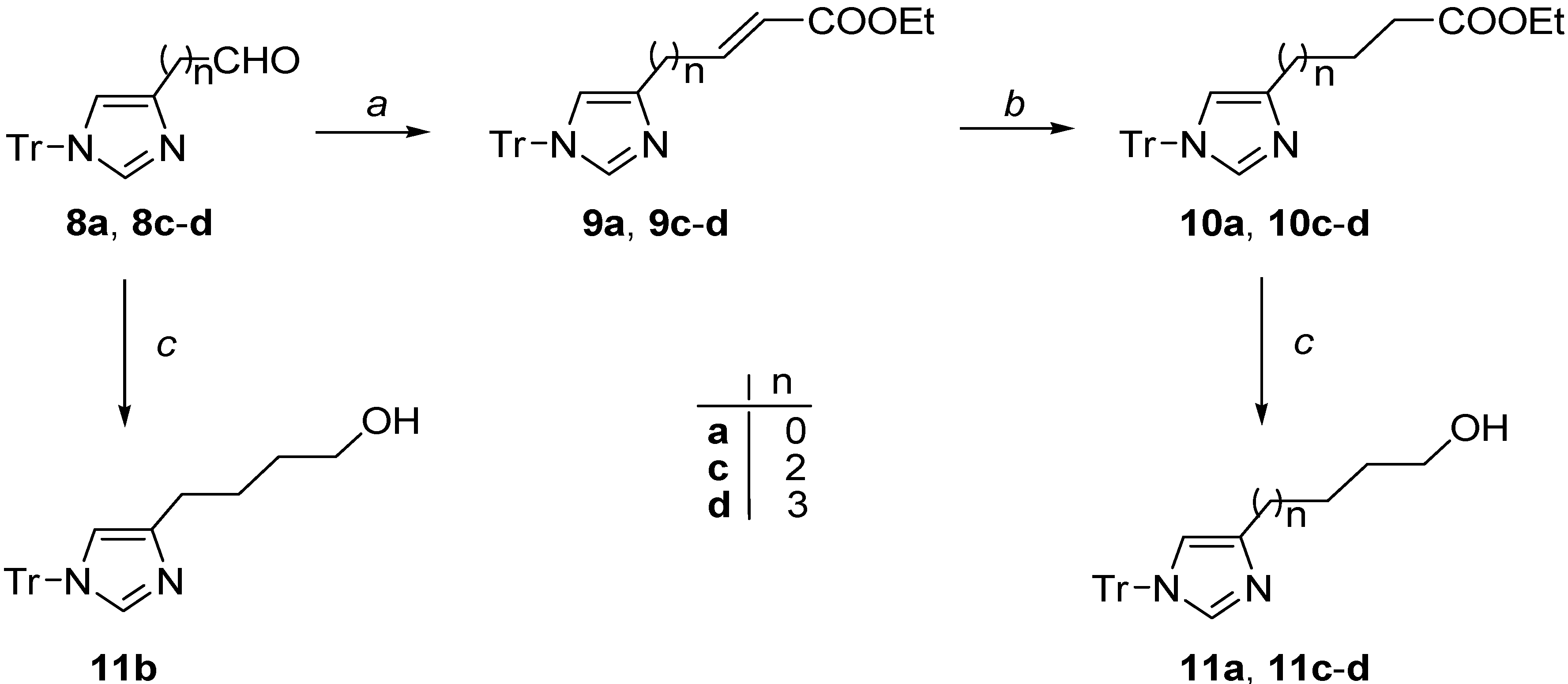
3.2.1. Synthesis of Unsaturated Esters 9a, 9cd

General Procedure: A solution of triethyl phosphonoacetate (0.2 mL, 1 mMol) in THF (6 mL) was added to an ice-cooled suspension of NaH (80% dispersed in mineral oil, 0.030 g, 1 mMol) in THF (6 mL) and the resulting mixture was stirred at room temperature for 30 min. A solution of the opportune aldehyde 8a, 8cd [48] (0.9 mMol) in THF (6 mL) was added dropwise and the mixture was stirred for 16 h at room temperature. The reaction mixture was then poured into iced water and extracted 3× with diethyl ether; the organic phases were combined, dried (Na2SO4) and concentrated to give a crude residue which was purified by flash chromatography (silica gel, EtOAc as eluent).

3.2.2. (E)-Ethyl 3-(1-trityl-1H-imidazol-4-yl)acrylate (9a)

The chemical physical data are identical to those reported in literature [49].

3.2.3. (E)-Ethyl 5-(1-trityl-1H-imidazol-4-yl)pent-2-enoate (9c)

ESI MS (m/z): 437 (M + 1)+; 243 (Ph3C+). The chemical physical data are identical to those reported in literature [48].

3.2.4. (E-Ethyl 6-(1-trityl-1H-imidazol-4-yl)hex-2-enoate (9d)

Oil; 46% yield. ESI MS (m/z): 451 (M + 1)+; 243 (Ph3C+). 1H NMR (CDCl3): δ 1.29 (t, 3H J = 7.0), 1.76–1.88 (m, 2H), 2.17–2.24 (m, 2H), 2.54–2.61 (m, 2H), 4.18 (q, 2H J = 7.0), 5.78 (d, 1H, J = 16.0), 6.53 (s, 1H), 6.89–7.04 (dt, 1H, J = 7.0 and 16.0), 7.12–7.36 (m, 16H).

3.2.5. Synthesis of Ester Derivatives 10a, 10cd

General Procedure: A solution of the suitable ethyl ester 9a, 9cd (1 mMol) in MeOH (5 mL) was hydrogenated under hydrogen atmosphere in the presence of 10% Pd/C (40 mg) for 4 h at room temperature. The catalyst was removed by filtration on Celite, and the filtrate was concentrated under reduced pressure to afford a crude residue which was purified by flash chromatography (silica gel, EtOAc as eluent).

3.2.6. Ethyl 3-(1-trityl-1H-imidazol-4-yl)propanoate (10a)

White solid; 84% yield. ESI MS (m/z): 411 (M + 1)+, 243 (Ph3C+). 1H NMR (CDCl3): δ 1.24 (t, 3H, J = 7.0), 2.12 (t, 2H, J = 7.5), 2.64 (t, 2H, J = 7.5), 4.15 (q, 2H, J = 7.0), 6.63 (s, 1H), 7.14–7.39 (m, 16H).

3.2.7. Ethyl 5-(1-trityl-1H-imidazol-4-yl)pentanoate (10c)

ESI MS (m/z): 439 (M + 1)+; 243 (Ph3C+). The chemical physical data are identical to those reported in literature [48].

3.2.8. Ethyl 6-(1-trityl-1H-imidazol-4-yl)hexanoate (10d)

Oil; 86% yield. ESI MS (m/z): 453 (M + 1)+, 243 (Ph3C+). 1H NMR (CDCl3): δ 1.25 (t, 3H J = 7.0), 1.25–1.42 (m, 2H), 1.57–1.72 (m, 4H), 2.25–2.32 (m, 2H), 2.51–2.59 (m, 2H), 4.12 (q, 2H J = 7.0), 6.53 (s, 1H), 7.12–7.38 (m, 16H).

3.2.9. Synthesis of Alcohol Derivatives 11a, 11cd

General Procedure: A solution of appropriate ester 10a, 10cd (1 mMol) in dry THF (4 mL) was added dropwise to a suspension of LiAlH4 (0.076 g, 2 mMol) in dry THF (4 mL) and the resulting mixture was refluxed for 4 h. After cooling to 0 °C the reaction mixture was quenched by slow addition of a saturated aqueous solution of NaHCO3 and EtOAc. The mixture was filtered on Celite, the filtrate was concentrated under reduced pressure to afford a residue which was taken up in CH2Cl2 and washed with a saturated aqueous solution of NaHCO3. The organic phase was dried (Na2SO4) and concentrated in vacuo to give a crude product which was purified by flash chromatography (silica gel, EtOAc to EtOAc/MeOH 97/3 as eluent).

3.2.10. 3-(1-Trityl-1H-imidazol-4-yl)propan-1-ol (11a)

ESI MS (m/z): 369 (M + 1)+; 243 (Ph3C+). The chemical physical data are identical to those reported in literature [48].

3.2.11. 4-(1-Trityl-1H-imidazol-4-yl)butan-1-ol (11b)

The product was obtained by direct reduction of the aldehyde 8d as previously reported and the chemical physical data are identical to those reported in literature [48]. ESI MS (m/z): 383 (M + 1)+; 243 (Ph3C+).

3.2.12. 5-(1-Trityl-1H-imidazol-4-yl)pentan-1-ol (11c)

ESI MS (m/z): 397 (M + 1)+; 243 (Ph3C+). The chemical physical data are identical to those reported in literature [48].

3.2.13. 6-(1-Trityl-1H-imidazol-4-yl)hexan-1-ol (11d)

Oil; 94% yield. ESI MS (m/z): 411 (M + 1)+, 243 (Ph3C+). 1H NMR (CDCl3): δ 1.23–1.39 (m, 4H), 1.48–1.67 (m, 4H), 2.52–2.60 (m, 2H), 3.59–3.65 (m, 2H), 6.52 (s, 1H), 7.09–7.40 (m, 16H).

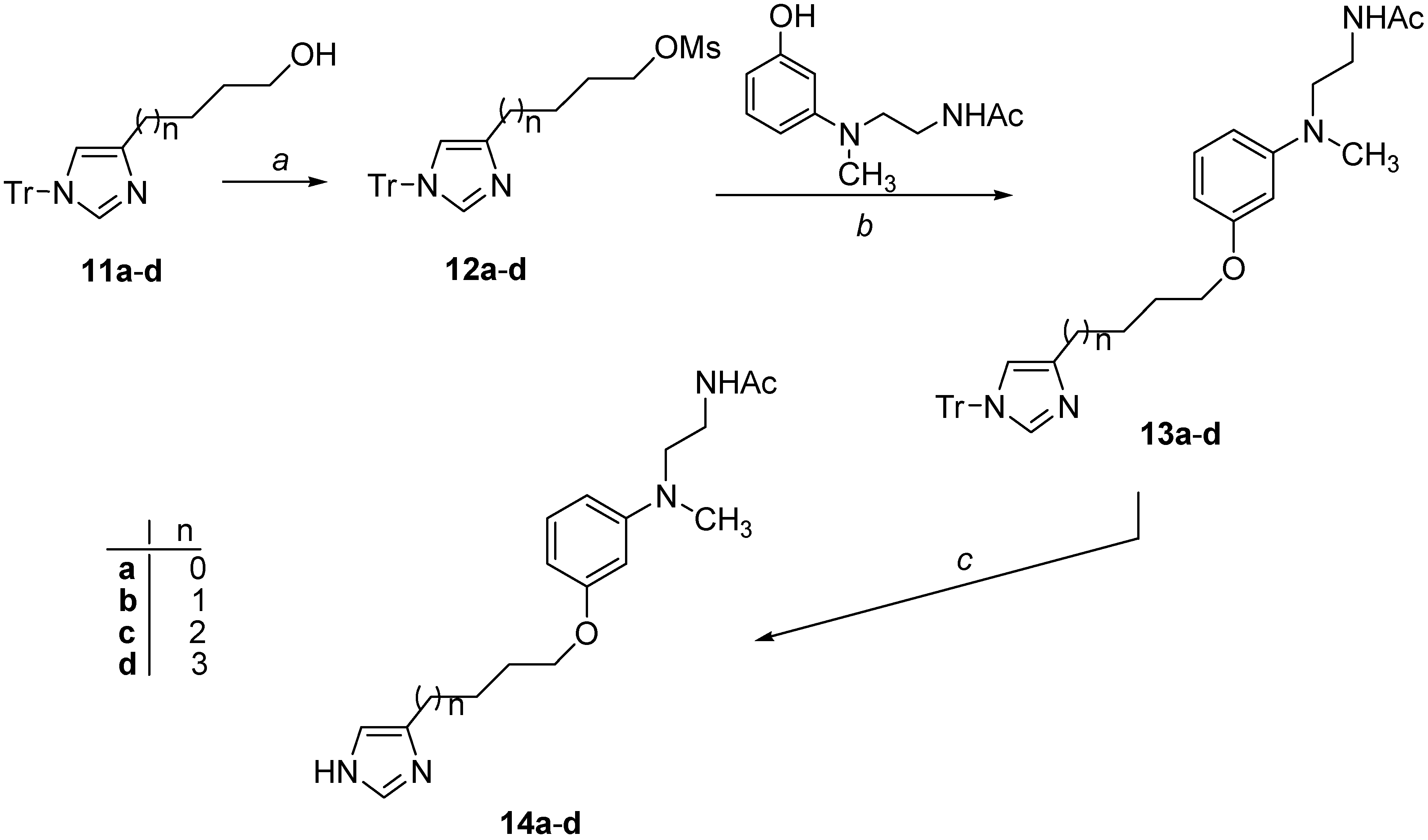
3.2.14. Synthesis of Mesyl Derivatives 12ad

General Procedure: Methanesulfonyl chloride (0.25 g, 2.2 mMol) was added to an ice-cooled solution of the opportune alcohol 11ad (1.8 mMol) in dry CH2Cl2 (8 mL) and TEA (0.24 g, 2.4 mMol). The reaction mixture was stirred under nitrogen atmosphere at 0 °C for 1 h (for 10a further 3 h at room temperature), quenched with water and washed once with an aqueous solution of 5% NaHCO3. The organic phase was dried over Na2SO4 and the solvent removed by distillation to afford the crude mesylate (12ad), which was used in the next step without any further purification.

3.2.15. 3-(1-Trityl-1H-imidazol-4-yl)propyl methansulfonate (12a)

Oil; 92% yield. ESI MS (m/z): 447 (M + 1)+, 243 (Ph3C+). 1H NMR (CDCl3): δ 2.11 (quint, 2H), 2.65–2.77 (m, 2H), 2.97 (s, 3H), 4.26 (t, 2H), 6.59 (s, 1H), 7.12–7.44 (m, 16H).

3.2.16. 4-(1-Trityl-1H-imidazol-4-yl)butyl methansulfonate (12b)

Oil; 95% yield. ESI MS (m/z): 461 (M + 1)+, 243 (Ph3C+).

3.2.17. 5-(1-Trityl-1H-imidazol-4-yl)pentyl methansulfonate (12c)

Oil; 87% yield. ESI MS (m/z): 475 (M + 1)+, 243 (Ph3C+).

3.2.18. 6-(1-Trityl-1H-imidazol-4-yl)hexyl methansulfonate (12d)

Oil; 88% yield. ESI MS (m/z): 489 (M + 1)+, 243 (Ph3C+).

3.2.19. Synthesis of Derivatives 13ad

General Procedure: NaH (80% in mineral oil, 0.036 g, 1.2 mMol) was added to a solution of N-{2-[(3-hydroxyphenyl)methylamino]ethyl}acetamide [31] (0.21 g, 1 mMol) in dry DMF (2.2 mL) under nitrogen atmosphere. After stirring for 30 min at room temperature, a solution of the suitable mesyl derivative 12ad (1 mMol) in dry DMF (1 mL) was added to the reaction mixture and stirring continued for 16 h. The reaction mixture was poured into water and extracted 3× with EtOAc. The organic phases were combined, washed once with brine, dried (Na2SO4) and concentrated to give a crude residue, which was purified by flash chromatography (silica gel, EtOAc as eluent).

3.2.20. N-[2-(Methyl{3-[3-(1-trityl-1H-imidazol-4-yl)propoxy]phenyl}amino)ethyl]acetamide (13a)

Oil; 67% yield. ESI MS (m/z): 559 (M + 1)+, 243 (Ph3C+). 1H NMR (CDCl3): δ 1.92 (s, 3H), 2.06-2.18 (m, 2H), 2.74 (t, 2H, J = 7.5), 2.93 (s, 3H), 3.45 (m, 4H), 3.98 (t, 2H, J = 6.5), 5.86 (brs, 1H), 6.25 (dd, 1H, J = 2.0 and 8.0), 6.27 (m, 1H), 6.36 (dd, 1H, J = 2.0 and 8.0), 6.58 (s, 1H), 7.08–7.38 (m, 17H).

3.2.21. N-[2-(Methyl{3-[4-(1-trityl-1H-imidazol-4-yl)butoxy]phenyl}amino)ethyl]acetamide (13b)

Oil; 40% yield. ESI MS (m/z): 573 (M + 1)+, 243 (Ph3C+). 1H NMR (CDCl3): δ 1.67–1.78 (m, 4H), 1.91 (s, 3H), 2.75 (t, 2H), 2.94 (s, 3H), 3.48 (m, 4H), 4.00 (t, 2H), 5.82 (brs, 1H), 6.26 (dd, 1H, J = 2.0 and 8.0), 6.29 (m, 1H), 6.38 (dd, 1H, J = 2.0 and 8.0), 6.59 (s, 1H), 7.06–7.41 (m, 17H). 13C NMR (CDCl3): δ 170.4, 160.3, 150.8, 142.3, 141.1, 138.0, 129.9, 129.7, 128.0, 117.9, 105.4, 102.4, 99.7, 75.3, 67.6, 51.7, 38.4, 37.2, 29.7, 28.9, 27.9 25.8, 23.2.

3.2.22. N-[2-(Methyl{3-[5-(1-trityl-1H-imidazol-4-yl)pentyloxy]phenyl}amino)ethyl]acetamide (13c)

Oil; 71% yield. ESI MS (m/z): 587 (M + 1)+, 243 (Ph3C+). 1H NMR (CDCl3): δ 1.44–1.55 (m, 2H), 1.62–1.82 (m, 4H), 1.91 (s, 3H), 2.57 (t, 2H J = 7.5), 2.92 (s, 3H), 3.41–3.44 (m, 4H), 3.93 (t, 2H J = 6.5), 5.96 (brs, 1H), 6.25 (dd, 1H, J = 2.0 and 8.0), 6.27 (m, 1H), 6.36 (dd, 1H, J = 2.0 and 8.0), 6.54 (s, 1H), 7.10–7.36 (m, 17H). 13NMR (CDCl3): δ 170.4, 160.4, 150.8, 142.5, 141.7, 138.2, 129.9, 129.8, 128.0, 117.8, 105.4, 102.3, 99.7, 75.1, 67.8, 51.7, 38.4, 37.3, 29.5, 29.2, 29.1, 28.3, 25.8, 23.2.

3.2.23. N-[2-(Methyl{3-[6-(1-trityl-1H-imidazol-4-yl)hexyloxy]phenyl}amino)ethyl]acetamide (13d)

Oil; 85% yield. ESI MS (m/z): 601 (M + 1)+, 243 (Ph3C+). 1H NMR (CDCl3): δ 1.26–1.52 (m, 4H), 1.53–1.75 (m, 4H), 1.95 (s, 3H), 2.60 (t, 2H J = 7.5), 2.94 (s, 3H), 3.45–3.47 (m, 4H), 3.94 (t, 2H J = 6.5), 5.75 (brs, 1H), 6.27 (dd, 1H, J = 2.0 and 8.0), 6.29 (m, 1H), 6.37 (dd, 1H, J = 2.0 and 8.0), 6.57 (s, 1H), 7.13–7.35 (m, 17H).

3.2.24. Synthesis of Target Derivatives 14ad

General Procedure: A solution of the suitable trityl derivative 13ad (0.5 mMol) and 2 N HCl (2 mL) in THF (1 mL) was heated at 70 °C for 3 h. The reaction mixture was washed with diethyl ether, the aqueous phase basified with 2 N Na2CO3 and extracted 3× with diethyl ether. The organic phases were combined, dried (Na2SO4) and concentrated to give a residue that was purified by filtration through a silica gel plug (EtOAc-MeOH 85:15 as eluent).

3.2.25. N-[2-({3-[3-(1H-Imidazol-4-yl)propoxy]phenyl}methylamino)ethyl]acetamide (14a)

Oil; 57% yield. ESI MS (m/z): 317 (M + 1)+. 1H NMR (CDCl3): δ 1.96 (s, 3H), 2.06–2.15 (m, 2H), 2.83 (t, 1H, J = 7.0), 2.94 (s, 3H), 3.45 (m, 4H), 4.03 (t, 1H, J = 6.5), 6.10 (brs, 1H), 6.26–6.37 (m, 3H), 6.83 (s, 1H), 7.13 (dd, 1H, J1 = J2 = 8.0), 7.60 (s, 1H). 13C NMR (CDCl3): δ 170.9, 160.2, 150.7, 135.6, 134.5, 130.0, 118.3, 105.3, 102.6, 99.3, 67.0, 51.6, 38.4, 37.1, 29.7, 29.3, 23.2. UV-Vis (MeOH) λ = 253 (12,600), 293 (4300) nm (ε). Anal. calcd for C17H24N4O2: C 64.53, H 7.65, N 17.71, found: C 64.29, H 7.70, N 17.39.

3.2.26. N-[2-({3-[4-(1H-Imidazol-4-yl)butoxy]phenyl}methylamino)ethyl]acetamide (14b)

Oil; 66% yield. ESI MS (m/z): 331 (M + 1)+. 1H NMR (CDCl3): δ 1.86–1.90 (m, 2H), 1.99 (s, 3H), 2.75–2.82 (m, 4H), 2.95 (s, 3H), 3.46 (m, 4H), 4.02 (t, 2H J = 6.5), 6.24 (brs, 1H), 6.26–6.41 (m, 3H), 6.97 (s, 1H), 7.13 (dd, 1H, J1 = J2 = 8.0), 7.93 (s, 1H). 13C NMR (CDCl3): δ 170.9, 160.2, 150.7, 136.3, 133.6, 130.0, 117.5, 105.4, 102.4, 99.7, 67.5, 51.7, 38.4, 37.1, 28.5, 25.9, 25.7, 23.2. Anal. calcd for C18H26N4O2: C 65.43, H 7.93, N 16.96, found: C 65.18, H 8.01, N 16.60.

3.2.27. N-[2-({3-[5-(1H-Imidazol-4-yl)pentyloxy]phenyl}methylamino)ethyl]acetamide (14c)

Oil; 92% yield. ESI MS (m/z): 345 (M + 1)+. 1H NMR (CDCl3): δ 1.43–1.56 (m, 2H), 1.60–1.84 (m, 4H), 1.95 (s, 3H), 2.67 (t, 2H J = 7.0), 2.94 (s, 3H), 3.45–3.47 (m, 4H), 3.96 (t, 2H J = 6.5), 6.05 (brs, 1H), 6.25–6.37 (m, 3H), 6.81 (s, 1H), 7.13 (dd, 1H, J1 = J2 = 8.0), 7.61 (s, 1H). 13C NMR (CDCl3): δ 170.9, 160.2, 150.7, 136.1, 134.1, 129.9, 118.0, 105.3, 102.2, 99.5, 67.5, 51.6, 38.4, 37.2, 28.9, 28.9, 26.2, 25.6, 23.2. UV-Vis (MeOH) λ = 253 (13,000), 293 (4100) nm (ε). Anal. calcd for C19H28N4O2: C 66.25, H 8.19, N 16.27, found: C 66.03, H 8.16, N 15.88.

3.2.28. N-[2-({3-[6-(1H-Imidazol-4-yl)hexyloxy]phenyl}methylamino)ethyl]acetamide (14d)

Oil; 84% yield. ESI MS (m/z): 359 (M + 1)+. 1H NMR (CDCl3): δ 1.41–1.47 (m, 4H), 1.61–1.80 (m, 4H), 1.94 (s, 3H), 2.64 (t, 2H J = 7.0), 2.94 (s, 3H), 3.45–3.47 (m, 4H), 3.95 (t, 2H J = 6.5), 5.88 (brs, 1H), 6.27–6.39 (m, 3H), 6.78 (s, 1H), 7.13 (dd, 1H, J1 = J2 = 8.0), 7.58 (s, 1H). 13C NMR (CDCl3): 170.7, 160.3, 150.8, 136.3, 134.1, 129.9, 117.8, 105.3, 102.2, 99.6, 67.6, 51.7, 38.4, 37.3, 29.1, 29.1, 28.7, 26.2, 25.7, 23.2. UV-Vis (MeOH) λ = 252 (13,200), 294 (4100) nm (ε). Anal. calcd for C20H30N4O2: C 67.01, H 8.44, N 15.63, found: C 66.70, H 8.42, N 15.33.

3.3. Human Histamine H3 Receptor Binding Assay

Homogenates of SK–N–MC cells expressing human histamine H3 receptors (hH3Rs) were used to determine affinity values of the new compounds in radioligand displacement studies according to Lovenberg et al.’s method [50]. SK–N–MC cells in confluent culture plates were harvested using trypsin digestion and then centrifuged for 5 min at 1100 rpm. Pellets, either fresh or stored at −80 °C until the moment of use, were mechanically homogenized with Potter-Elvhejem in 20 mM Tris-HCl/0.5 mM EDTA. Supernatants from a 2000 rpm spin (10 min) were collected and re-centrifuged at 10,000 rpm for 30 min. Pellets were re-homogenized in 50 mM Tris-HCl/5 mM EDTA (pH 7.4). Membranes were incubated for 60 min at room temperature with 0.5 nM [3H](R)-α-methylhistamine in 50 mM Tris-HCl/5 mM EDTA (pH 7.4) with or without competing ligands. Each concentration was tested in triplicate. Incubation was terminated by rapid filtration over Millipore (Darmstadt, Germany) AAWPO2500 filters followed by two washes with ice-cold buffer (50 mM Tris-HCl/5 mM EDTA). Filters retained radioactivity was determined by liquid scintillation counting (Ultima Gold XR Perkin Elmer (Waltham, MA, USA) scintillation liquid, Tri-Carb 2810 TR Perkin Elmer (Waltham, MA, USA) Liquid Scintillation Analyzer). Non-specific binding was defined with 10 μM histamine as competing ligand. pIC50 and Hill coefficient values were estimated from the displacement curves of the tested compounds (10 nM–100 μM) versus [3H](R)-α-methylhistamine, using Prism (San Diego, CA, USA) GraphPad 2005, and converted into pKi values according to the Cheng–Prusoff equation [51]. Data are expressed as mean ± SEM of three independent experiments.

3.4. Human Melatonin Receptors Binding Assay

Binding affinities were determined using 2-[125I]iodomelatonin as the labeled ligand in competition experiments with cloned human MT1 and MT2 receptors expressed in NIH3T3 rat fibroblast cells. The characterization of NIH3T3 MT1 and MT2 cells was already described in detail [52,53]. Membranes were incubated for 90 min at 37 °C in binding buffer (50 mM Tris/HCl, pH 7.4). The final membrane concentration was 5–10 μg of protein per tube. The membrane protein level was determined in accordance with a previously reported method [54]. 2-[125I]Iodomelatonin (100 pM) and different concentrations of the tested compounds were incubated with the receptor preparation for 90 min at 37 °C. Nonspecific binding was assessed with 10 μM melatonin; IC50 values were determined by nonlinear fitting strategies with Prism (GraphPad SoftWare Inc., San Diego, CA, USA). The pKi values were calculated from the IC50 values according to the Cheng–Prusoff equation [51]. The pKi values are the mean of at least three independent determinations performed in duplicate.
To determine the functional activity of compound 14d at MT1 and MT2 receptor subtypes, [35S]GTPγS binding assays in NIH3T3 cells expressing human-cloned MT1 or MT2 receptors were performed. The amount of bound [35S]GTPγS is proportional to the level of the analog-induced G protein activation and is related to the intrinsic activity of the compound under study. The detailed description and validation of this method were reported elsewhere [52,55]. Membranes (15–25 μg of protein, final incubation volume 100 μL) were incubated at 30 °C for 30 min in the presence and absence 14d, in assay buffer consisting of [35S]GTPγS (0.3–0.5 nM), GDP (50 μM), NaCl (100 mM), and MgCl2 (3 mM). Nonspecific binding was defined using GTPγS (10 μM). In cell lines expressing human MT1 or MT2 receptors, melatonin produced concentration-dependent stimulation of basal [35S]GTPγS binding with a maximal stimulation above basal levels of 370% and 250% in MT1 and MT2 receptors, respectively. Basal stimulation is the amount of [35S]GTPγS specifically bound in the absence of compounds and was taken as 100%. The maximal G protein activation was measured in each experiment using melatonin (100 nM). Compounds were added at three different concentrations (one concentration equivalent to 100 nM melatonin, a second one, 10-fold smaller, and a third one, 10-fold larger), and the percent stimulation above basal was determined. The equivalent concentration was estimated on the basis of the ratio of the affinity of the test compound to that of melatonin. It was assumed that, at the equivalent concentration, the test compound occupies the same number of receptors as 100 nM melatonin. All of the measurements were performed in triplicate. The relative intrinsic activity (IAR) values were obtained by dividing the maximum ligand induced stimulation of [35S]GTPγS binding by that of melatonin, as measured in the same experiment.

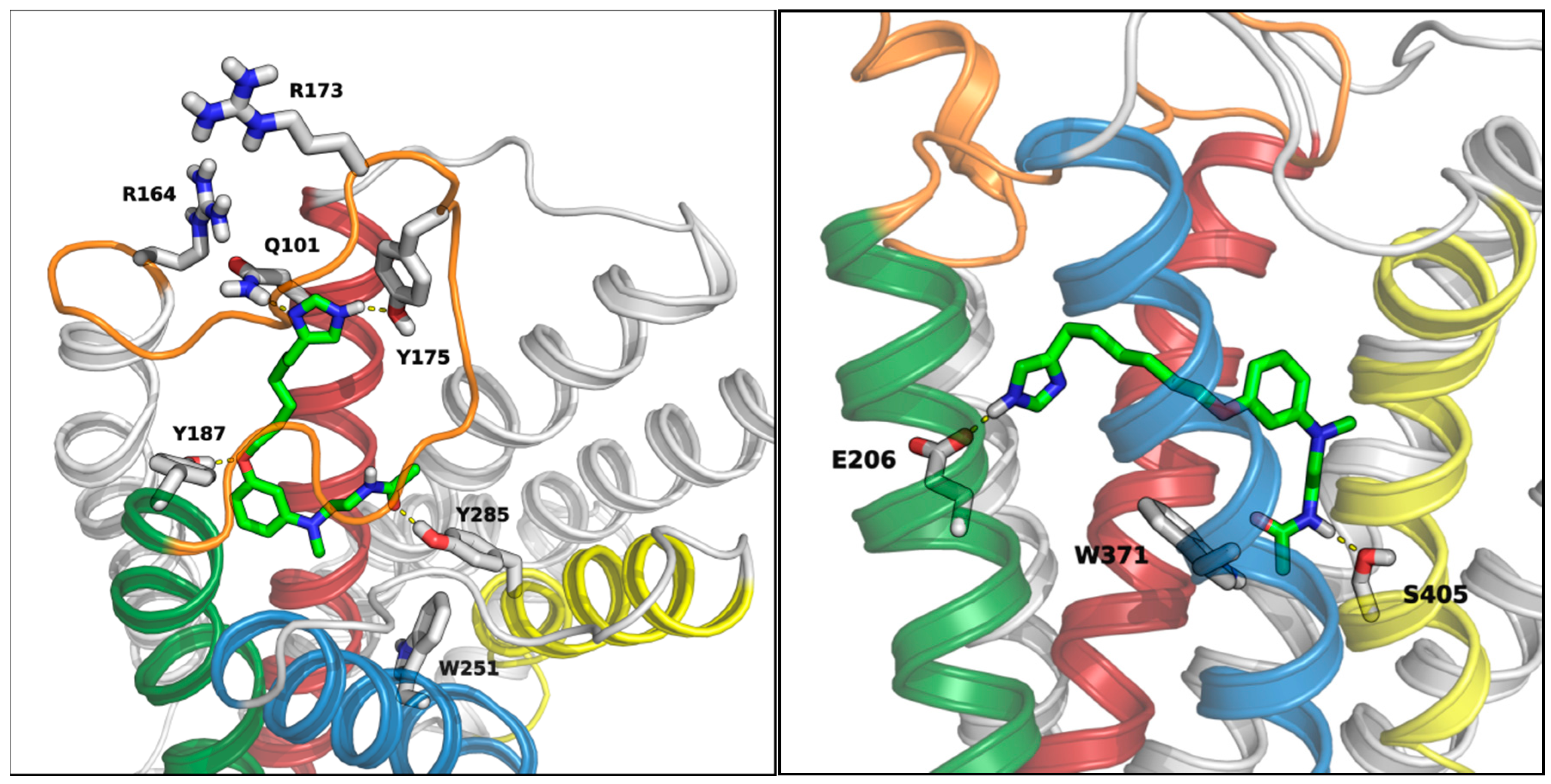
3.5. Docking Studies

All docking simulations were performed with Glide 5.7 [56]; Maestro 9.2 [57] was applied to prepare ligand structures and to refine protein-ligand complexes.

3.5.1. MT1 Receptor

A previously-reported MT1 receptor model [31] was taken as starting point for induced-fit docking (IFD) of compounds 14ad. An initial softened-potential docking run was performed applying van der Waals radii scaling of 0.7 and 0.5 on protein and ligand non-polar atoms, respectively. Amino acids hampering the accommodation of the imidazole ring of 14ad, i.e., Q101 and Q169, were temporarily mutated to alanines. Energy grids generated for the initial softened-potential docking were centered in the putative binding site of the receptor, setting enclosing and bounding boxes to default dimensions. During flexible docking runs, two hydrogen bond constraints were applied between Y187 hydroxyl group and the phenolic oxygen of the ligand, and between Y285 hydroxyl group and the amide oxygen of the ligand, to reproduce the main polar interactions previously proposed for melatonergic ligands. Ligand docking was performed in standard precision (SP) mode, collecting fifty poses. The resulting ligand–receptor complexes were then submitted to a protein structure refinement stage; once amino acid side chains that had previously been removed were re-introduced, residues within a shell of 5 Å around any ligand pose were refined by a side chain conformational search, followed by energy minimization of the residues and the ligand molecule. In the final docking stage, each ligand structure obtained at the end of the protein structure refinement was energetically optimized (refined) in the field of the receptor and subsequently scored using default Glide settings. The final ligand-protein complexes were ranked according to their IFD score, a composite score that accounts for ligand-receptor interaction energy, receptor strain and solvation terms. The best-ranked MT1-14ad complexes were minimized applying the OPLS2005 force field [58] to a convergence threshold of 0.05 kJ·mol−1·Å−1. During this minimization procedure, the ligand and residues within 8 Å from the ligand were free to move, while all other atoms were retained fixed.

3.5.2. H3 Receptor

A previously-reported model of the histamine H3 receptor [45] was used as reference structure for docking studies of compounds 14ad. Glide grids were centered in the putative binding site of the receptor, located between D114 and E206, setting the dimension of enclosing and bounding boxes to default values. Compounds 14ad were flexibly docked within the H3 receptor binding cavity in SP mode, applying a van der Waals radii scaling of 0.7 on ligand non-polar atoms. During docking runs, a hydrogen bond constraint was applied between E206 and the imidazole ring of the ligands. Fifty poses were collected for each ligand and ranked according to their Emodel value. The best-scored ligand conformations were merged into the H3 receptor model and the resulting complexes were minimized applying the OPLS2005 force field to an energy gradient of 0.05 kJ·mol−1 Å−1. During this minimization phase, the docked compound and all residues within 8 Å from the ligand were free to move, whereas all other atoms were retained fixed.

4. Conclusions

We reported the synthesis and binding affinity evaluation of a series of bivalent melatonergic/histaminergic ligands characterized by an imidazolyl-alkyloxy-anilinoethylamide structure. In this series, four derivatives with alkyl linkers of different length were tested for their ability to bind human MT1, MT2 and H3 receptor subtypes. Among the tested compounds, 14c and 14d, bearing a pentyl and a hexyl linker between the imidazole ring and the aniline core, respectively, were able to bind to all receptor subtypes at micromolar concentrations. Although these two compounds might possess sub-optimal binding affinities, they are the first bivalent melatonergic/histaminergic ligands reported so far and might represent promising starting points for the development of potent dual-acting agents, potentially useful for the treatment of cognitive disorders.

Author Contributions

Daniele Pala: molecular modeling and writing; Laura Scalvini and Alessio Lodola: molecular modeling; Marco Mor: drug design; Lisa Flammini and Elisabetta Barocelli: H3 receptor assays; Valeria Lucini and Francesco Scaglione: melatonin receptor assays; Silvia Bartolucci and Annalida Bedini: synthesis; Silvia Rivara: drug design and writing; Gilberto Spadoni: drug design and synthesis.

Conflicts of Interest

The authors declare no conflict of interest.

References

  1. Dubocovich, M.L.; Delagrange, P.; Krause, D.N.; Sugden, D.; Cardinali, D.P.; Olcese, J. International Union of Basic and Clinical Pharmacology. LXXV. Nomenclature, classification, and pharmacology of G protein-coupled melatonin receptors. Pharmacol. Rev. 2010, 62, 343–380. [Google Scholar] [CrossRef]
  2. Slominski, R.M.; Reiter, R.J.; Schlabritz-Loutsevitch, N.; Ostrom, R.S.; Slominski, A.T. Melatonin membrane receptors in peripheral tissues: Distribution and functions. Mol. Cell Endocrinol. 2012, 35, 152–166. [Google Scholar]
  3. Zlotos, D.P.; Jockers, R.; Cecon, E.; Rivara, S.; Witt-Enderby, P.A. MT1 and MT2 melatonin receptors: Ligands, models, oligomers, and therapeutic potential. J. Med. Chem. 2014, 57, 3161–3185. [Google Scholar] [CrossRef]
  4. Pandi-Perumal, S.R.; Trakht, I.; Srinivasan, V.; Spence, D.W.; Maestroni, G.J.M.; Zisapel, N.; Cardinali, D.P. Physiological effects of melatonin: Role of melatonin receptors and signal transduction pathways. Prog. Neurobiol. 2008, 85, 335–353. [Google Scholar] [CrossRef]
  5. Kim, T.K.; Kleszczynski, K.; Janjetovic, Z.; Sweatman, T.; Lin, Z.; Li, W.; Reiter, R.J.; Fischer, T.W.; Slominski, A.T. Metabolism of melatonin and biological activity of intermediates of melatoninergic pathway in human skin cells. FASEB J. 2013, 27, 2742–2755. [Google Scholar] [CrossRef]
  6. Cajochen, C.; Kräuchi, K.; Wirz-Justice, A. Role of melatonin in the regulation of human circadian rhythms and sleep. J. Neuroendocrinol. 2003, 15, 432–437. [Google Scholar] [CrossRef]
  7. Reiter, R.J.; Tan, D.-X.; Mayo, J.C.; Sainz, R.M.; Leon, J.; Czarnocki, Z. Melatonin as an antioxidant: Biochemical mechanisms and pathophysiological implications in humans. Acta Biochim. Pol. 2003, 50, 1129–1146. [Google Scholar]
  8. Srinivasan, V.; Pandi-Perumal, S.R.; Cardinali, D.P.; Poeggeler, B.; Hardeland, R. Melatonin in Alzheimer’s disease and other neurodegenerative disorders. Behav. Brain Funct. BBF 2006, 2, 15. [Google Scholar] [CrossRef]
  9. Wang, J.; Wang, Z. Role of melatonin in Alzheimer-like neurodegeneration. Acta Pharmacol. Sin. 2006, 27, 41–49. [Google Scholar] [CrossRef]
  10. Wang, X. The antiapoptotic activity of melatonin in neurodegenerative diseases. CNS Neurosci. Ther. 2009, 15, 345–357. [Google Scholar] [CrossRef]
  11. Das, A.; McDowell, M.; Pava, M.J.; Smith, J.A.; Reiter, R.J.; Woodward, J.J.; Varma, A.K.; Ray, S.K.; Banik, N.L. The inhibition of apoptosis by melatonin in VSC4.1 motoneurons exposed to oxidative stress, glutamate excitotoxicity, or TNF-alpha toxicity involves membrane melatonin receptors. J. Pineal Res. 2010, 48, 157–169. [Google Scholar] [CrossRef]
  12. Das, A.; Wallace, G.; Reiter, R.J.; Varma, A.K.; Ray, S.K.; Banik, N.L. Overexpression of melatonin membrane receptors increases calcium-binding proteins and protects VSC4.1 motoneurons from glutamate toxicity through multiple mechanisms. J. Pineal Res. 2013, 54, 58–68. [Google Scholar]
  13. Chern, C.-M.; Liao, J.-F.; Wang, Y.-H.; Shen, Y.-C. Melatonin ameliorates neural function by promoting endogenous neurogenesis through the MT2 melatonin receptor in ischemic-stroke mice. Free Radic. Biol. Med. 2012, 52, 1634–1647. [Google Scholar] [CrossRef]
  14. Kaneko, Y.; Hayashi, T.; Yu, S.; Tajiri, N.; Bae, E.C.; Solomita, M.A.; Chheda, S.H.; Weinbren, N.L.; Parolini, O.; Borlongan, C.V. Human amniotic epithelial cells express melatonin receptor MT1, but not melatonin receptor MT2: A new perspective to neuroprotection. J. Pineal Res. 2011, 50, 272–280. [Google Scholar] [CrossRef]
  15. Carocci, A.; Catalano, A.; Bruno, C.; Lovece, A.; Roselli, M.G.; Cavalluzzi, M.M.; de Santis, F.; de Palma, A.; Rusciano, M.R.; Illario, M.; et al. N-(phenoxyalkyl)amides as M1 and MT2 ligands: Antioxidant properties and inhibition of Ca2+/CaM-dependent kinase II. Bioorg. Med. Chem. 2013, 21, 847–851. [Google Scholar] [CrossRef] [Green Version]
  16. Wang, X.; Sirianni, A.; Pei, Z.; Cormier, K.; Smith, K.; Jiang, J.; Zhou, S.; Wang, H.; Zhao, R.; Yano, H.; et al. The melatonin MT1 receptor axis modulates mutant Huntingtin-mediated toxicity. J. Neurosci. 2011, 31, 14496–14507. [Google Scholar] [CrossRef]
  17. Rodríguez-Franco, M.I.; Fernández-Bachiller, M.I.; Pérez, C.; Hernández-Ledesma, B.; Bartolomé, B. Novel tacrine-melatonin hybrids as dual-acting drugs for Alzheimer disease, with improved acetylcholinesterase inhibitory and antioxidant properties. J. Med. Chem. 2006, 49, 459–462. [Google Scholar] [CrossRef]
  18. López-Iglesias, B.; Pérez, C.; Morales-García, J.A.; Alonso-Gil, S.; Pérez-Castillo, A.; Romero, A.; López, M.G.; Villarroya, M.; Conde, S.; Rodríguez-Franco, M.I. New melatonin-N,N-dibenzyl(N-methyl)amine hybrids: Potent neurogenic agents with antioxidant, cholinergic, and neuroprotective properties as innovative drugs for Alzheimer’s disease. J. Med. Chem. 2014, 57, 3773–3785. [Google Scholar] [CrossRef]
  19. Chojnacki, J.E.; Liu, K.; Yan, X.; Toldo, S.; Selden, T.; Estrada, M.; Rodríguez-Franco, M.I.; Halquist, M.S.; Ye, D.; Zhang, S. Discovery of 5-(4-Hydroxyphenyl)-3-oxo-pentanoic acid [2-(5-methoxy-1H-indol-3-yl)-ethyl]-amide as a neuroprotectant for Alzheimer’s disease by hybridization of curcumin and melatonin. ACS Chem. Neurosci. 2014, 5, 690–699. [Google Scholar] [CrossRef]
  20. Leurs, R.; Bakker, R.A.; Timmerman, H.; de Esch, I.J. The histamine H3 receptor: From gene cloning to H3 receptor drugs. Nat. Rev. Drug Discov. 2005, 4, 107–120. [Google Scholar] [CrossRef]
  21. Singh, M.; Jadhav, H.R. Histamine H3 receptor function and ligands: Recent developments. Mini-Rev. Med. Chem. 2013, 12, 47–57. [Google Scholar] [CrossRef]
  22. Gemkow, M.J.; Davenport, A.J.; Harich, S.; Ellenbroek, B.A.; Cesura, A.; Hallett, D. The histamine H3 receptor as a therapeutic drug target for CNS disorders. Drug Discov. Today 2009, 14, 509–515. [Google Scholar] [CrossRef]
  23. Brioni, J.D.; Esbenshade, T.A.; Garrison, T.R.; Bitner, S.R.; Cowart, M.D. Discovery of histamine H3 antagonists for the treatment of cognitive disorders and Alzheimer’s disease. J. Pharmacol. Exp. Ther. 2011, 336, 38–46. [Google Scholar] [CrossRef]
  24. Bhowmik, M.; Khanam, R.; Vohora, D. Histamine H3 receptor antagonists in relation to epilepsy and neurodegeneration: A systemic consideration of recent progress and perspectives. Br. J. Pharmacol. 2012, 167, 1398–1414. [Google Scholar] [CrossRef]
  25. Bitner, R.S.; Markosyan, S.; Nikkel, A.L.; Brioni, J.D. In-vivo histamine H3 receptor antagonism activates cellular signaling suggestive of symptomatic and disease modifying efficacy in Alzheimer’s disease. Neuropharmacology 2011, 60, 460–466. [Google Scholar] [CrossRef]
  26. Yan, H.; Zhang, X.; Hu, W.; Ma, J.; Hou, W.; Zhang, X.; Wang, X.; Gao, J.; Shen, Y.; Lv, J.; et al. Histamine H3 receptors aggravate cerebral ischaemic injury by histamine-independent mechanisms. Nat. Commun. 2014, 5, 3334. [Google Scholar]
  27. Bhowmik, M.; Saini, N.; Vohora, D. Histamine H3 receptor antagonism by ABT-239 attenuates kainic acid induced excitotoxicity in mice. Brain Res. 2014. [Google Scholar] [CrossRef]
  28. Tang, L.; Zhao, L.; Hong, L.; Yang, F.; Sheng, R.; Chen, J.; Shi, Y.; Zhou, N.; Hu, Y. Design and synthesis of novel 3-substituted-indole derivatives as selective H3 receptor antagonists and potent free radical scavengers. Bioorg. Med. Chem. 2013, 21, 5936–5944. [Google Scholar] [CrossRef]
  29. Mésangeau, C.; Pérès, B.; Descamps-François, C.; Chavatte, P.; Audinot, V.; Coumailleau, S.; Boutin, J.A.; Delagrange, P.; Bennejean, C.; Renard, P.; et al. Design, synthesis and pharmacological evaluation of novel naphthalenic derivatives as selective MT1 melatoninergic ligands. Bioorg. Med. Chem. 2010, 18, 3426–3436. [Google Scholar] [CrossRef]
  30. Markl, C.; Clafshenkel, W.P.; Attia, M.I.; Sethi, S.; Witt-Enderby, P.A.; Zlotos, D.P. N-acetyl-5-arylalkyloxytryptamine analogs: Probing the melatonin receptors for MT1-selectivity. Arch. Pharm. (Weinheim) 2011, 344, 666–674. [Google Scholar] [CrossRef]
  31. Rivara, S.; Pala, D.; Lodola, A.; Mor, M.; Lucini, V.; Dugnani, S.; Scaglione, F.; Bedini, A.; Lucarini, S.; Tarzia, G.; et al. MT1-selective melatonin receptor ligands: Synthesis, pharmacological evaluation, and molecular dynamics investigation of N-{[(3-O-substituted)anilino]alkyl}amides. ChemMedChem 2012, 7, 1954–1964. [Google Scholar] [CrossRef]
  32. Celanire, S.; Wijtmans, M.; Talaga, P.; Leurs, R.; de Esch, I.J.P. Histamine H3 receptor antagonists reach out for the clinic. Drug Discov. Today 2005, 10, 1613–1627. [Google Scholar] [CrossRef]
  33. Wingen, K.; Stark, H. Scaffold variations in amine warhead of histamine H3 receptor antagonists. Drug Discov. Today Technol. 2013, 10, e483–e489. [Google Scholar] [CrossRef]
  34. Tedford, C.E.; Hoffmann, M.; Seyedi, N.; Maruyama, R.; Levi, R.; Yates, S.L.; Ali, S.M.; Phillips, J.G. High antagonist potency of GT-2227 and GT-2331, new histamine H3 receptor antagonists, in two functional models. Eur. J. Pharmacol. 1998, 351, 307–311. [Google Scholar] [CrossRef]
  35. De Esch, I.J.; Gaffar, A.; Menge, W.M.; Timmerman, H. Synthesis and histamine H3 receptor activity of 4-(n-alkyl)-1H-imidazoles and 4-(ω-phenylalkyl)-1H-imidazoles. Bioorg. Med. Chem. 1999, 7, 3003–3009. [Google Scholar] [CrossRef]
  36. Vollinga, R.C.; Menge, W.M.; Leurs, R.; Timmerman, H. New analogs of burimamide as potent and selective histamine H3 receptor antagonists: The effect of chain length variation of the alkyl spacer and modifications of the N-thiourea substituent. J. Med. Chem. 1995, 38, 2244–2250. [Google Scholar] [CrossRef]
  37. Tozer, M.J.; Harper, E.A.; Kalindjian, S.B.; Pether, M.J.; Shankley, N.P.; Watt, G.F. From histamine to imidazolylalkyl-sulfonamides: The design of a novel series of histamine H3-receptor antagonists. Bioorg. Med. Chem. Lett. 1999, 9, 1825–1830. [Google Scholar] [CrossRef]
  38. Grassmann, S.; Apelt, J.; Sippl, W.; Ligneau, X.; Pertz, H.H.; Zhao, Y.H.; Arrang, J.M.; Ganellin, C.R.; Schwartz, J.C.; Schunack, W.; et al. Imidazole derivatives as a novel class of hybrid compounds with inhibitory histamine N-methyltransferase potencies and histamine hH3 receptor affinities. Bioorg. Med. Chem. 2003, 11, 2163–2174. [Google Scholar] [CrossRef]
  39. Ettaoussi, M.; Sabaouni, A.; Rami, M.; Boutin, J.A.; Delagrange, P.; Renard, P.; Spedding, M.; Caignard, D.H.; Berthelot, P.; Yous, S. Design, synthesis and pharmacological evaluation of new series of naphthalenic analogues as melatoninergic (MT1/MT2) and serotoninergic 5-HT2C dual ligands (I). Eur. J. Med. Chem. 2012, 49, 310–323. [Google Scholar] [CrossRef]
  40. Zlotos, D.P.; Attia, M.I.; Julius, J.; Sethi, S.; Witt-Enderby, P.A. 2-[(2,3-dihydro-1H-indol-1-yl)methyl]melatonin analogues: A novel class of MT2-selective melatonin receptor antagonists. J. Med. Chem. 2009, 52, 826–833. [Google Scholar] [CrossRef]
  41. Rivara, S.; Lodola, A.; Mor, M.; Bedini, A.; Spadoni, G.; Lucini, V.; Pannacci, M.; Fraschini, F.; Scaglione, F.; Sanchez, R.O.; et al. N-(Substituted-anilinoethyl)amides: Design, synthesis, and pharmacological characterization of a new class of melatonin receptor ligands. J. Med. Chem. 2007, 50, 6618–6626. [Google Scholar] [CrossRef]
  42. Stark, H.; Ligneau, X.; Sadek, B.; Ganellin, C.R.; Arrang, J.M.; Schwartz, J.C.; Schunack, W. Analogues and derivatives of ciproxifan, a novel prototype for generating potent histamine H3-receptor antagonists. Bioorg. Med. Chem. Lett. 2000, 10, 2379–2382. [Google Scholar] [CrossRef]
  43. Uveges, A.J.; Kowal, D.; Zhang, Y.; Spangler, T.B.; Dunlop, J.; Semus, S.; Jones, P.G. The role of transmembrane helix 5 in agonist binding to the human H3 receptor. J. Pharmacol. Exp. Ther. 2002, 301, 451–458. [Google Scholar] [CrossRef]
  44. Yao, B.B.; Hutchins, C.W.; Carr, T.L.; Cassar, S.; Masters, J.N.; Bennani, J.L.; Esbenshade, T.A.; Hancock, A.A. Molecular modeling and pharmacological analysis of spacies-related histamine H3 receptor heterogeneity. Neuropharmacology 2003, 44, 773–786. [Google Scholar] [CrossRef]
  45. Bordi, F.; Rivara, S.; Dallaturca, E.; Carmi, C.; Pala, D.; Lodola, A.; Vacondio, F.; Flammini, L.; Bertoni, S.; Ballabeni, V.; et al. Dibasic biphenyl H3 receptor antagonists: Steric tolerance for a lipophilic side chain. Eur. J. Med. Chem. 2012, 48, 214–230. [Google Scholar] [CrossRef]
  46. Ly, K.S.; Letavic, M.A.; Keith, J.M.; Miller, J.M.; Stocking, E.M.; Barbier, A.J.; Bonaventure, P.; Lord, B.; Jiang, X.; Boggs, J.D.; et al. Synthesis and biological activity of piperazine and diazepane amides that are histamine H3 antagonists and serotonin reuptake inhibitors. Bioorg. Med. Chem. Lett. 2008, 18, 39–43. [Google Scholar] [CrossRef]
  47. Roche, O.; Rodriguez Sarmiento, R.M. A new class of histamine H3 receptor antagonists derived from ligand based design. Bioorg. Med. Chem. Lett. 2007, 17, 3670–3675. [Google Scholar] [CrossRef]
  48. Kitbunnadaj, R.; Hoffmann, M.; Fratantoni, S.A.; Bongers, G.; Bakker, R.A.; Wieland, K.; el Jilali, A.; de Esch, I.J.P.; Menge, W.M.P.B.; Timmerman, H.; et al. New high affinity H3 receptor agonists without a basic side chain. Bioorg. Med. Chem. 2005, 13, 6309–6323. [Google Scholar] [CrossRef]
  49. Griffith, R.K.; DiPietro, R.A. Improved syntheses of vinylimidazoles. Synth. Commun. 1986, 16, 1761–1770. [Google Scholar] [CrossRef]
  50. Lovenberg, T.W.; Roland, B.L.; Wilson, S.J.; Jiang, X.; Pyati, J.; Huvar, A.; Jackson, M.R.; Erlander, M.G. Cloning and functional expression of the human histamine H3 receptor. Mol. Pharmacol. 1999, 55, 1101–1107. [Google Scholar]
  51. Cheng, Y.; Prusoff, W.H. Relationship between the inhibition constant (K1) and the concentration of inhibitor which causes 50 per cent inhibition (I50) of an enzymatic reaction. Biochem. Pharmacol. 1973, 22, 3099–3108. [Google Scholar] [CrossRef]
  52. Nonno, R.; Lucini, V.; Pannacci, M.; Mazzucchelli, C.; Angeloni, D.; Fraschini, F.; Stankov, B.M. Pharmacological characterization of the human melatonin Mel8a receptor following stable transfection into NIH3T3 cells. Br. J. Pharmacol. 1998, 124, 485–492. [Google Scholar] [CrossRef]
  53. Nonno, R.; Pannacci, M.; Lucini, V.; Angeloni, D.; Fraschini, F.; Stankov, B.M. Ligand efficacy and potency at recombinant human MT2 melatonin receptors: Evidence for agonist activity of some mt1-antagonists. Br. J. Pharmacol. 1999, 127, 1288–1294. [Google Scholar] [CrossRef]
  54. Bradford, M.M. A rapid and sensitive method for the quantitation of microgram quantities of protein utilizing the principle of protein-dye binding. Anal. Biochem. 1976, 72, 248–254. [Google Scholar] [CrossRef]
  55. Spadoni, G.; Balsamini, C.; Bedini, A.; Diamantini, G.; di Giacomo, B.; Tontini, A.; Tarzia, G.; Mor, M.; Plazzi, P.V.; Rivara, S.; et al. 2-[N-Acylamino(C1-C3)alkyl]indoles as MT1 melatonin receptor partial agonists, antagonists, and putative inverse agonists. J. Med. Chem. 1998, 41, 3624–3634. [Google Scholar] [CrossRef]
  56. Glide, version 5.7; Schrödinger, LLC:: New York, NY, USA, 2011.
  57. Maestro, version 9.2; Schrödinger, LLC: New York, NY, USA, 2011.
  58. Banks, J.L.; Beard, H.S.; Cao, Y.; Cho, A.E.; Damm, W.; Farid, R.; Felts, A.K.; Halgren, T.A.; Mainz, D.T.; Maple, J.R.; et al. Integrated modeling program, applied chemical theory (IMPACT). J. Comput. Chem. 2005, 26, 1752–1780. [Google Scholar] [CrossRef]

