Article

Golgi-Related Proteins GOLPH2 (GP73/GOLM1) and GOLPH3 (GOPP1/MIDAS) in Cutaneous Melanoma: Patterns of Expression and Prognostic Significance

1
Department of Pathomorphology and Oncological Cytology, Wroclaw Medical University, Borowska 213, 50-556 Wroclaw, Poland
2
Faculty of Mathematics and Information Science, Warsaw University of Technology, Koszykowa 75, 00-662 Warsaw, Poland
3
Department of Oncology, Wroclaw Medical University; pl. Hirszfelda 12, 53-413 Wroclaw, Poland
4
Lower Silesian Cancer Center, Hirszfelda 12, 53-413 Wroclaw, Poland
*
Author to whom correspondence should be addressed.
Int. J. Mol. Sci. 2016, 17(10), 1619; https://doi.org/10.3390/ijms17101619
Submission received: 31 July 2016 / Revised: 5 September 2016 / Accepted: 12 September 2016 / Published: 1 October 2016
(This article belongs to the Special Issue Advances in Molecular Oncology)

Abstract

:
GOLPH2 and GOLPH3 are Golgi-related proteins associated with aggressiveness and progression of a number of cancers. Their prognostic significance in melanoma has not yet been analyzed. We performed immunohistochemical analysis for GOLPH2 and GOLPH3 in 20 normal skin, 30 benign nevi and 100 primary melanoma tissue samples and evaluated their expression in three compartments: cancer cells, tumor-associated macrophages (TAMs) and cancer-associated fibroblasts (CAFs). High levels of both proteins in melanoma cells were associated with characteristics of aggressive disease, and shorter disease-free survival (DFS) and cancer-specific overall survival (CSOS). On the contrary, increased numbers of GOLPH2-positive and GOLPH3-positive TAMs were observed in thinner, non-ulcerated tumors, with brisk lymphocytic reaction and absent lymphangioinvasion. Distant metastases were not observed among patients with high numbers of GOLPH2-positive TAMs. Increased expression of either protein in TAMs was related to prolonged CSOS and DFS. Similarly, GOLPH3-expressing CAFs were more frequent in thin melanomas with low mitotic rate, without ulceration and lymphangioinvasion. Moreover, increased GOLPH3-positive CAFs correlated with the absence of regional or distant metastases, and with longer CSOS and DFS. GOLPH2 expression was not observed in CAFs. Our results suggest that GOLPH2 and GOLPH3 play a role in melanoma progression and are potential targets for molecular-based therapies.

Graphical Abstract

1. Introduction

Golgi apparatus is a multifunctional organelle described for the first time over 100 years ago. Its best characterized functions involve modification of proteins and lipids, their sorting and dispatching in transport vesicles to various intracellular destinations or for exocytosis. Given the central role of Golgi for various cell processes, its dysregulation is a likely feature in carcinogenesis. However, alterations of Golgi function in cancer are still insufficiently studied.
GOLPH2 (Golgi phosphoprotein 2, also named GP73 and GOLM1) is a resident 73 kDa Golgi membrane glycoprotein expressed chiefly in epithelial lineage of cells [1]. While the physiological role of GOLPH2 remains vague, its upregulated expression was observed in a number of diseases. Initially, GOLPH2 upregulation was reported in neoplastic and non-neoplastic liver pathologies, and a number of studies postulated its serum level as a novel marker of hepatocellular carcinoma [2,3,4,5,6,7,8]. Subsequently, enhanced GOLPH2 expression was reported in other cancer settings [9,10,11,12,13,14,15]. To the best of our knowledge, GOLPH2 expression in melanoma patients has not been previously analyzed.
GOLPH3 (Golgi phosphoprotein 3, also named GOPP1/MIDAS), a highly conserved 34 kDa protein localized mostly in the trans-Golgi network, provides numerous cellular functions [16]. Binding of PI4P and MYO18A by GOLPH3 links Golgi membrane to F-actin cytoskeleton and is essential for vesicular trafficking [17,18]. As a side effect of these interactions, Golgi apparatus adopts a characteristic extended ribbon structure [18]. GOLPH3 also plays a role in mitochondrial biogenesis [19,20], cell adhesion and migration [21,22,23], cytokinesis [24], and promotes Golgi dispersal and survival response after DNA damage [25]. Its regulatory contribution to protein glycosylation in the Golgi may affect a wide range of cell functions [16]. A recent study established that GOLPH3 has oncogenic properties mediated via mTOR signaling, and that GOLPH3 gene at 5p13 region is frequently amplified in a number of cancers [26]. Close association between GOLPH3 expression and patient survival in a range of neoplasms (a meta-analysis in [27]) make the protein a potential target of new therapies and warrant further investigations. We believe that no clinicopathological analysis of GOLPH3 expression in tissue specimens from patients with cutaneous melanoma has been published so far.
Tumor stroma has long been known to be a relevant and active player in carcinogenesis. Accumulating evidence shows that cancer-associated fibroblasts (CAFs) and tumor-associated macrophages (TAMs) representing key non-malignant constituents of melanoma microenvironment, strongly influence cancer growth and progression [28,29]. Secretion of growth factors, modulation of local immune response, regulation of angiogenesis, remodeling of extracellular matrix by secreted metalloproteinases are some of the mechanisms involved [28,29]. Moreover, recent studies demonstrate that fibroblasts and macrophages play a critical role in treatment resistance in melanoma [30,31]. Considering the distinct position of melanoma-associated stromal cells during carcinogenesis and a complexity of interactions between them and malignant melanocytes, we decided to individually examine neoplastic and stromal components. There is no published analysis of GOLPH2 and 3 expression in melanoma’s stroma and available studies in other malignancies lack a systematical assessment of these proteins in a non-neoplastic tumor microenvironment.
We performed immunohistochemical analysis for GOLPH2 and GOLPH3 and evaluated their expression in three compartments: cancer cells, TAMs and CAFs. This is the first immunohistochemical analysis of GOLPH2 and 3 expression in a clinical melanoma cohort, and the first one addressing the significance of GOLPH2 and GOLPH3 expression in the stromal components. The goal of our study was to examine the relationship of GOLPH2 and GOLPH3 expression levels in different compartments of primary melanoma tissue samples with other histopathological and clinicopathological parameters, especially with patient survival.

2. Results

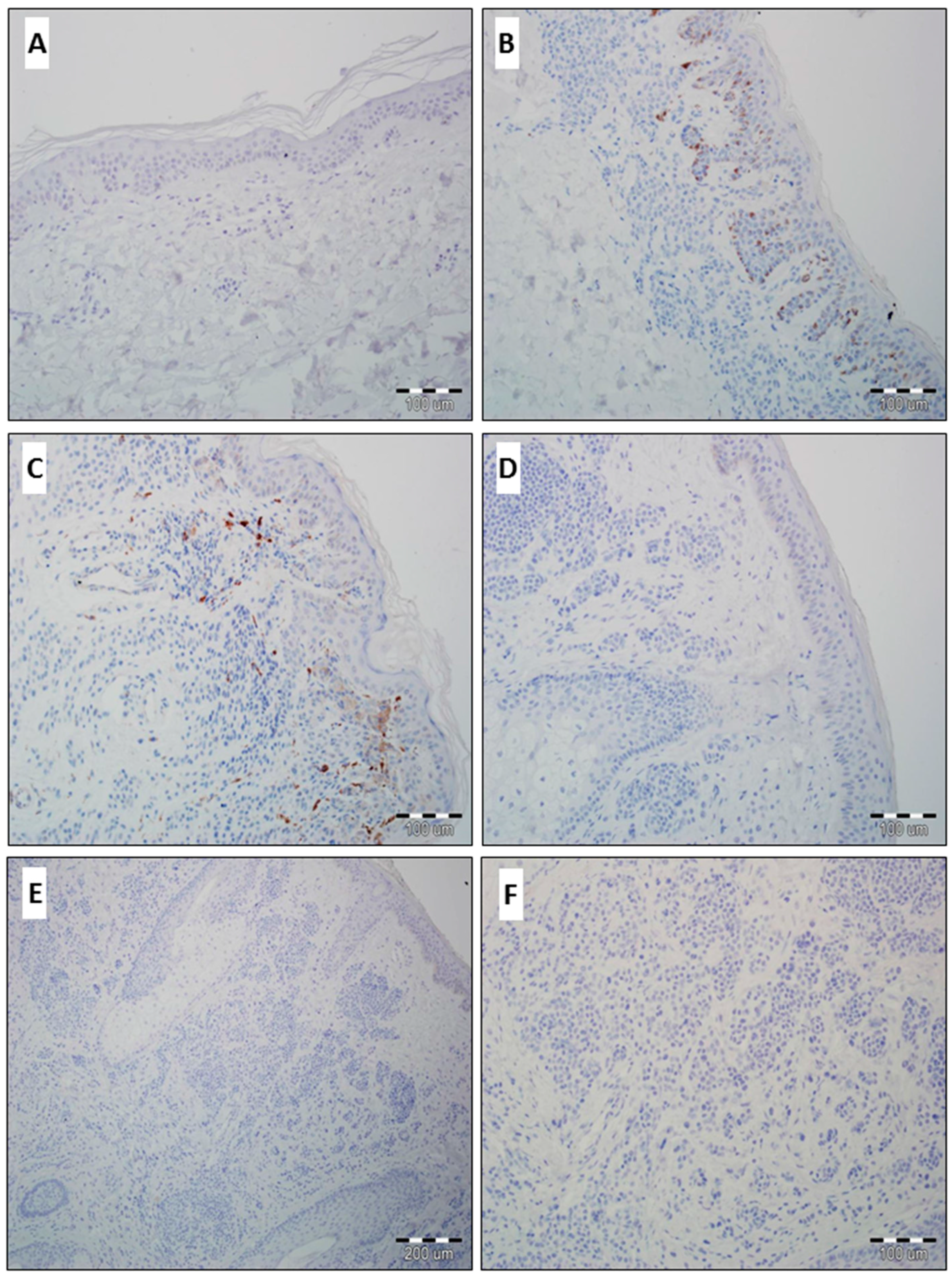
2.1. Expression of GOLPH2 and GOLPH3 in Normal Skin and Benign Nevi

In all cases of normal skin, the epidermis was negative for GOLPH2 and GOLPH3 (Figure 1A). We observed predominantly similar results in benign skin nevi. Only in four cases (all were diagnosed as compound benign skin nevus) scanty immunoexpression of GOLPH2 and GOLPH3 in benign melanocytes and stromal compartment of the nevi was observed (Figure 1B,C). Other benign nevi were negative (Figure 1D–F).

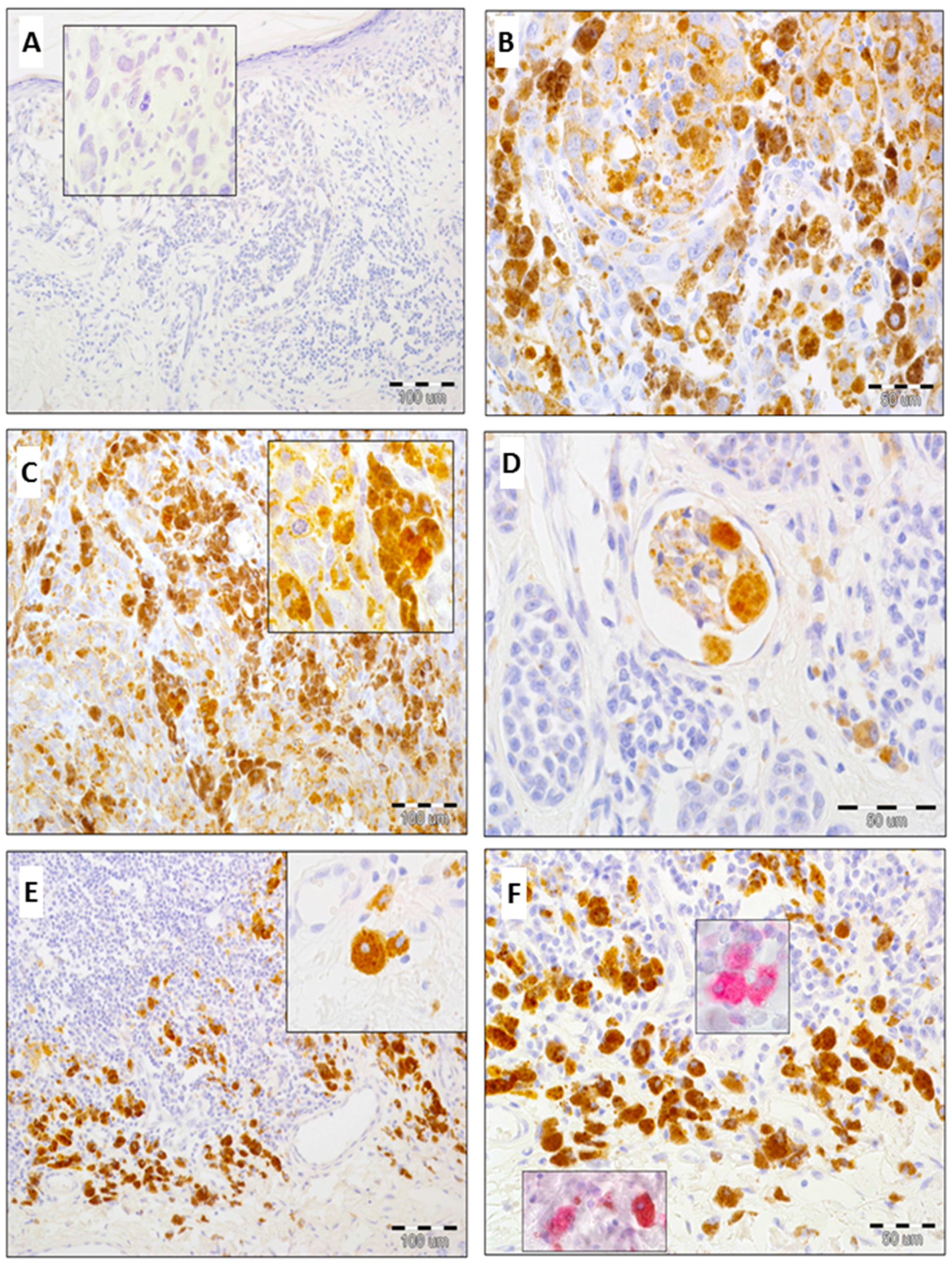
2.2. Expression of GOLPH2 in Melanoma Cells and Tumor-Associated Macrophages (TAMs)

The expression of analyzed proteins in evaluated compartments was divided into two levels based on IRS score for melanoma cells (low: <6 vs. high: ≥6), the number of GOLPH2- and GOLPH3-positive macrophages (<20 vs. ≥20) and the number of GOLPH3-positive fibroblasts (<10 vs. ≥10). The IRS scale for melanoma cells assigns a score for the percentage of cells demonstrating reaction (0–4 points) and for reaction intensity (0–3 points). The IRS score is the product of the scores for these two parameters (0–12 points). A detailed description of pathological assessment of protein expression is included in the Material and Methods section.
High expression of GOLPH2 in cancer cells was detected in 31% of melanomas (31/100), whereas low/absent expression was found in 69% of cases (69/100). No GOLPH2 expression in melanoma cells was observed in 29 primary tumors (Figure 2A–D). Mean IRS score for malignant melanocytes was 3.27 ± 3.1 (median: 2).
Upregulated expression of GOLPH2 in TAMs was observed in 63% of cases (63/100), low/absent expression in 37% of melanomas (37/100). 23 primary tumors presented no GOLPH2 immunoreactivity in TAMs (Figure 2E,F).

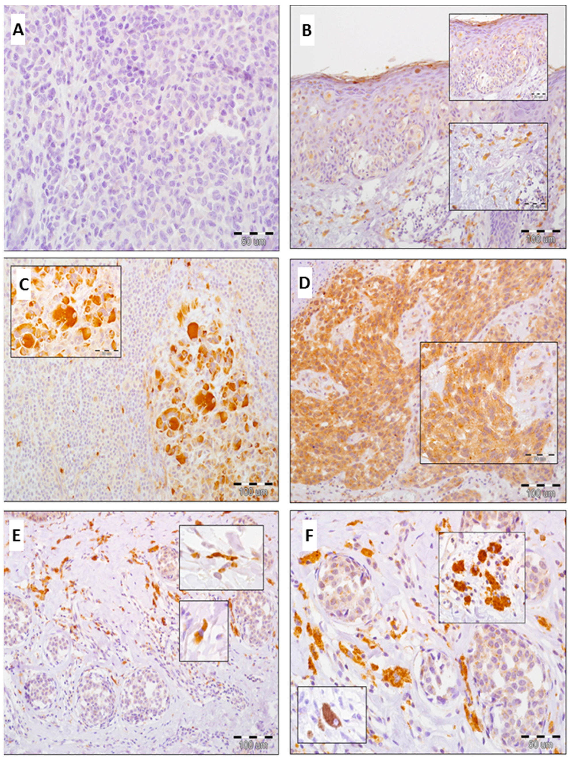
2.3. Expression of GOLPH3 in Melanoma Cells, Tumor-Associated Macrophages (TAMs) and Cancer-Associated Fibroblasts (CAFs)

Elevated level of GOLPH3 expression in melanoma cells was detected in 51% of cases (51/100), whereas low/absent expression in 49% of cases (49/100) (Figure 3A–D). Mean IRS score for melanoma cells was 5.16 ± 3.0 (median: 6).
GOLPH3 expression in TAMs and CAFs was upregulated in 68% and 78% primary tumors, respectively. Low/absent expression of GOLPH3 in TAMs and CAFs was demonstrated in 32% and 22% primary tumors, respectively (Figure 3E,F).

2.4. Analysis of Correlations between GOLPH2 and GOLPH3 Immunoreactivity in Melanoma Cells and Clinicopathological Parameters

Increased immunoreactivity of GOLPH2 and GOLPH3 in melanoma cells was statistically linked with thicker tumors, regional lymph node metastases (only SLN metastases for GOLPH3), locoregional recurrence, lymphangioinvasion, presence of ulceration, higher mitotic rate, and lower TIL (tumor-infiltrating lymphocytes) grade (Table 1, Table 2, Table 3 and Table 4). Overexpression of GOLPH3 was more frequently observed in nodular and acral-lentiginous melanomas (Table 4).

2.5. Analysis of Correlations Between GOLPH2 and GOLPH3 Immunoreactivity in Stromal Compartment and Clinicopathological Parameters

Increased numbers of GOLPH2-positive and GOLPH3-positive macrophages correlated with thinner tumors, absence of ulceration, brisk TILs, lack of lymphangioinvasion and lower locoregional recurrence rate (Table 1, Table 2, Table 3 and Table 4). Moreover, increased GOLPH3-positive macrophages were related to low proliferative activity of cancer cells and characterized tumors localized on the trunk and extremities. (Table 3 and Table 4). Distant metastases were not observed among patients with high numbers of GOLPH2-positive TAMs (Table 1).
Increased GOLPH3-positive fibroblasts were observed in thinner, non-ulcerated melanomas, with low mitotic rate and without lymphangioinvasion. Moreover, high numbers of CAFs correlated with absence of regional and distant metastases (Table 3 and Table 4).

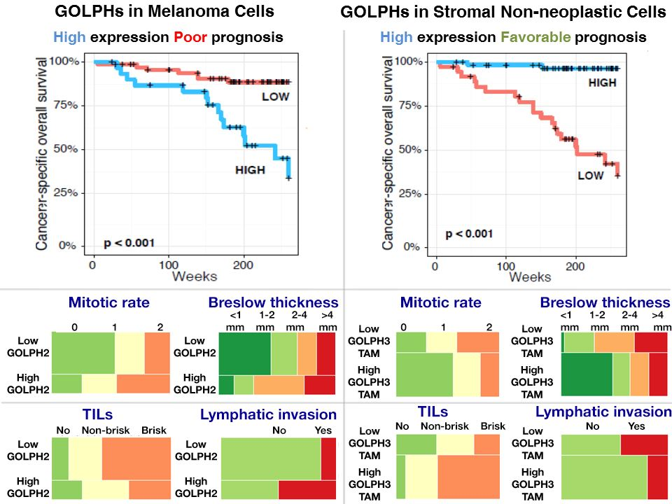
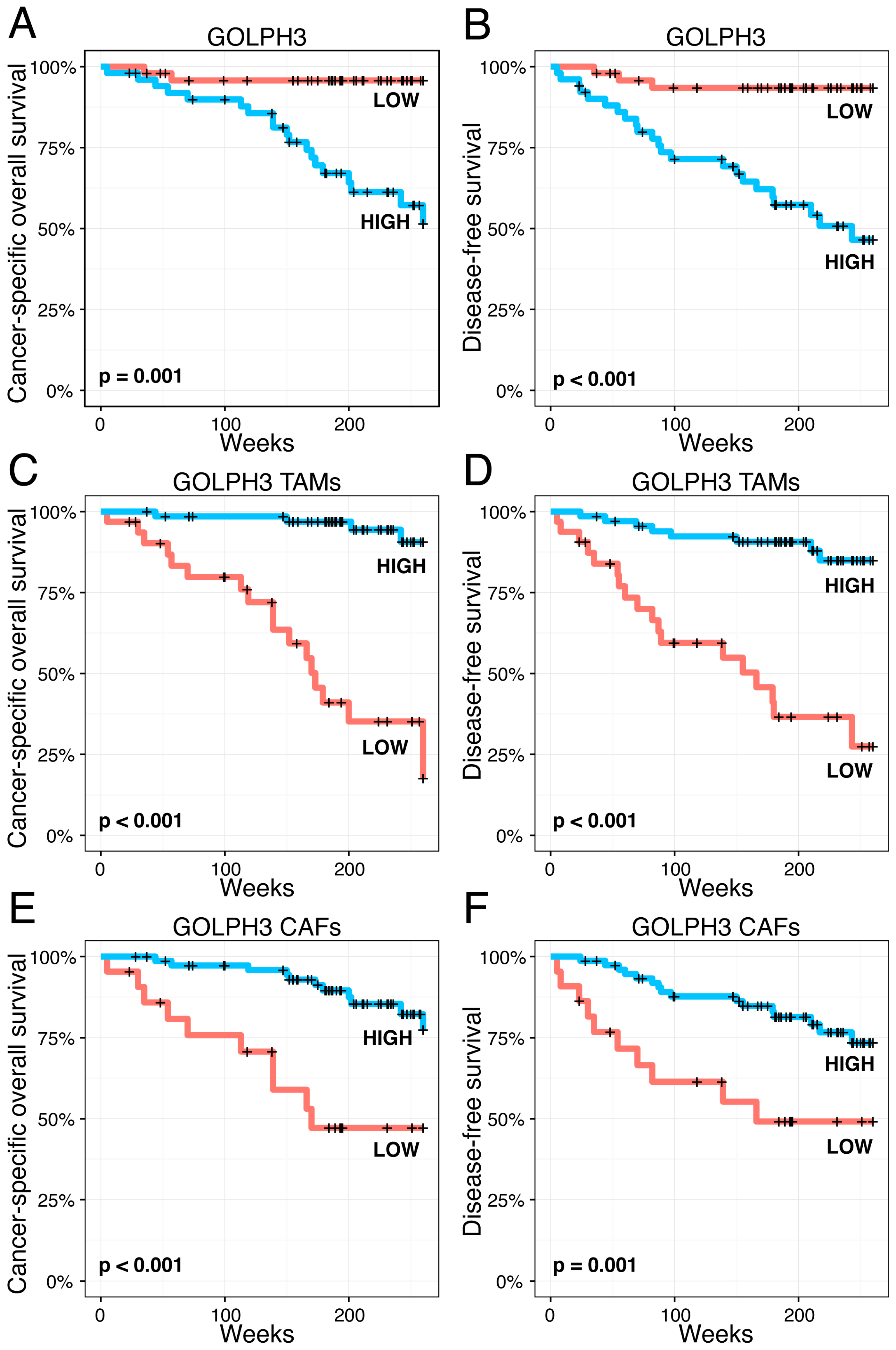
2.6. Impact of Golgi-Related Protein Expression in Neoplastic Cells and Stromal Compartment on Melanoma Patient Survival—Kaplan-Meier Analysis

We revealed that enhanced GOLPH2 and GOLPH3 immunoreactivity in melanoma cells is associated with shorter cancer-specific overall survival (CSOS) and disease-free survival (DFS) in the whole group of patients (Figure 4 and Figure 5). An analogous relation was observed in patients without nodal metastases (data not shown).
Stromal expression of GOLPH2 in TAMs also demonstrated prognostic significance-upregulation of GOLPH2 in TAMs strongly correlated to longer CSOS and DFS both in the whole group of patients (Figure 4) and in a subgroup without nodal metastases (data not shown).
Moreover, Kaplan-Meier analysis showed prognostic significance of GOLPH3 expression in TAMs and CAFs—enhanced immunoreactivity in the stromal compartment of melanoma was associated with prolonged CSOS and DFS, both in the whole study population (Figure 5) and in patients with no nodal metastases (data not shown). GOLPH3 expression in CAFs was an exception not being associated with DFS in patients without nodal metastases (data not shown).

3. Discussion

In this study we aimed to examine the expression of GOLPH2 and GOLPH3 in malignant melanocytes and stromal CAFs and TAMs, and to reveal the relationship between these expression levels and other histopathological and clinicopathological parameters. High levels of both proteins in melanoma cells were associated with features of aggressive disease, i.e., greater thickness, ulceration, visible mitotic figures, weak lymphocytic infiltrate (relation with GOLPH3), lymphangioinvasion, nodal metastases and recurrence. Moreover, upregulation of GOLPH2 and GOLPH3 in cancer cells was associated with shorter DFS and CSOS. To the best of our knowledge, this is the first analysis of GOLPH2 and GOLPH3 expression in melanoma in the clinical setting.
Little is known about GOLPH2 functions, but available studies provide some insight into the mechanisms connecting GOLPH2 and carcinogenesis. Jin et al. reported that GOLPH2 promotes HCC invasion by activation of matrix metalloproteinase-13 (MMP-13) [32]. MMP-13 has a documented role in melanoma. Apart from exacerbating melanoma invasiveness [33], it seems to mediate proliferation of cancer cells [34]. In line with in vitro studies, Corte et al. reported a positive correlation between MMP-13 expression and MR in clinical melanoma tissue samples [35]. In their study on gastric cancer, Tang et al. showed that GOLPH2 overexpression attenuated anti-tumor Th1 lymphocyte response [36]. This effect is likely to apply to melanoma, in which Th1-type immune response is a key defensive mechanism.
Recent study by Scott et al. identified GOLPH3 as a potent oncogene amplified in several cancers, including melanoma, which enhances mTOR signaling [26]. mTOR-related pathways control cell growth, proliferation and survival, and are often dysregulated in malignancies [37]. Following studies revealed a number of mechanisms relating GOLPH3 to cancer progression. It mediates formation of metastases by upregulation of MMPs [38,39] and acts as a promotor of angiogenesis [40,41]. Dai et al. showed that GOLPH3 activates NF-κB pathway in HCC [42], while Zeng et al. proved that overexpression of GOLPH3 inhibits transcriptional activity of FOXO1 and, as a consequence, facilitates G1–S-phase transition in breast cancer cells [43]. Both these pathways also play a role in cutaneous melanoma [44,45].
Our results suggest that expression of GOLPH2 and GOLPH3 in TAMs is also clinically relevant for patients with melanoma. We observed increased numbers of GOLPH2-positive and GOLPH3-positive macrophages in thinner, non-ulcerated tumors, with brisk TILs and absent lymphangioinvasion. Distant metastases were not observed among patients with high numbers of GOLPH2-positive TAMs. Increased GOLPH3- and GOLPH2-expressing TAMs were related to prolonged CSOS and DFS. This is the first study that addresses the prognostic significance of GOLPH2 and GOLPH3 expression in TAMs.
TAMs seem to promote melanomagenesis and favor immune escape by their metabolic activity in the local immune milieu [28,46,47]. However, macrophages form a heterogeneous population of cells which dynamically change phenotype during carcinogenesis. The initial response to tumor antigens is mediated by M1 type macrophages, but they often rapidly switch to M2 phenotype under the influence of cytokines secreted by tumor cells [48,49]. Hypoxic conditions emerging within the tumor also stimulate the M1-M2 switch [48]. M1 cells play a great role in melanoma as they promote inflammatory response mediated by TNF-a. Evolution from M1 to M2 phenotype promotes escape of the tumor from immune surveillance [47].
Having no factual insight into the role of GOLPH2 and GOLPH3 in TAMs, we only speculate that the proteins may be associated with prolonged stabilization of M1 phenotype of macrophages, which could result in observed favorable clinical outcome. An indirect indication to an association between activation of local immunity and macrophages expressing GOLPH2 and GOLPH3 is a positive correlation between their number and TIL intensity observed in our study. Careful functional investigations are needed to explore the roles of tumor-associated macrophages in melanoma.
Beneficial influence on survival of high GOLPH3 expression in CAFs is another interesting finding in our results. CAFs are thought to act as promotors of melanoma, but their precise functions and mechanisms of action are not fully understood [50,51,52,53,54]. In our experiment, GOLPH3-expressing fibroblasts were more frequent in thinner, non-ulcerated melanomas, with low mitotic rate and without lymphangioinvasion. Moreover, increased GOLPH3-positive CAFs correlated with absence of regional and distant metastases.
It seems that most of CAFs in melanoma are residual skin fibroblasts which, stimulated by humoral mediators and direct contact with malignant melanocytes, undergo a metabolic transformation to CAFs. This hypothetical process of paraoncogenic transformation of normal fibroblasts to CAFs gradually changes cytobiochemical characteristics of fibroblasts, affecting also their fundamental function, namely the synthesis of extracellular matrix (ECM) components [51,54].
Overexpression of GOLPH3, observed only in some CAFs, might be a feature of initial steps of postulated paraoncogenic transformation in a selected subpopulation of fibroblasts. Stimulation of their metabolism and otherwise quiescent pathway (a manifestation of which would be upregulated GOLPH3) might be an attempt to restrict the expansion of melanoma by increase of ECM production. In such a case, enhanced GOLPH3 immunoreactivity in CAFs might be a potential marker for normal fibroblasts or early stage CAFs, which do not exhibit genetic aberrations or metabolic changes (e.g., increased production of cancer-promoting MMPs) typical for fully developed CAFs [51,54]. The presented interpretation of our results is largely hypothetical due to lack of other studies addressing the role of GOLPH3 in CAFs and no clear criteria discriminating normal fibroblasts and CAFs. The topic warrants further, minute investigations with the use of molecular biology techniques.
To conclude, consistent with findings in other cancer settings, our results suggest a role for GOLPH2 and GOLPH3 in melanoma pathogenesis, but functional studies are needed to evaluate the mechanisms involved.

4. Materials and Methods

4.1. Patients

100 patients diagnosed with cutaneous melanoma between 2005 and 2010 and receiving treatment in the Lower Silesian Oncology Center in Wroclaw, Poland were enrolled in the study. Enrollment criteria were the availability of tissue material (paraffin blocks and histopathology slides) and medical documentation. Archival medical records were the source of comprehensive clinical data. Information on the diagnostic and therapeutic procedures were retrieved from the medical records at the Lower Silesian Oncology Center‘s oncology outpatient clinic and from the Lower Silesian Cancer Registry and Civil Register Office. The study was approved by the ethical committee of the Wroclaw Medical University, Wrocław, Poland.
Based on the medical records, it was determined that the patients under study were treated by then-available methods and the primary procedure was as follows: the patients underwent excisional biopsy of the primary lesion. When histopathological examination showed cutaneous melanoma, the scar was excised with a margin of 5, 10 or 20 mm of unaffected skin depending on Breslow thickness and primary tumor location, if any. Sentinel lymph node biopsy (SNLB) was performed in patients with no clinically manifest nodal metastases (cN0) and Breslow thickness above 1 mm (>pT1a). Lymphadenectomy was performed when metastases were found (either clinically or by SLNB) in the regional lymph nodes.
The clinical and pathological data of the patients investigated in this study included age and gender, primary tumor location, tumor stratification according to AJCC (pT), presence or absence of nodal (pN) and distant (pM) metastases, information on disease recurrence and SLNB procedures (Table 1 and Table 3).
For tumor samples and histopathological evaluation, archival formalin-fixed and paraffin-embedded tumor specimens were studied. Two pathologists examined all hematoxylin and eosin-stained sections. Breslow thickness, Clark level, growth phase, histologic type, mitotic rate (number of mitotic figures per 1 mm2), presence of ulceration, lymphangioinvasion, microsatellitosis, intensity of TILs and microscopic evidence of regression (Table 2 and Table 4) were recorded in pathology reports for the primary tumor.
The semi-quantitative method described below was used to assess TILs. Absence of TILs: there are no lymphocytes present or lymphocytes are present but they do not infiltrate tumor at all. Non-brisk TILs: lymphocytes infiltrate melanoma only focally or not along the entire base of the vertical growth phase. Brisk TILs: lymphocytes diffusely infiltrate the entire base of the vertical growth phase or the entire invasive component of the melanoma.

4.2. Immunohistochemistry

GOLPH2 and GOLPH3 immunoreactivity was analyzed in 20 normal skin specimens (sampled during autopsy or non-oncological procedures), 30 benign nevi and 100 cutaneous melanoma patients. Immunohistochemical evaluation of GOLPH2 expression (mouse monoclonal antibody, clone F-2, sc-365817; dilution: 1:100; Santa Cruz Biotechnology, Dallas, TX, USA) and GOLPH3 (rabbit polyclonal antibody, LS-B5044; dilution 1:1000; LifeSpan Biosciences, Seattle, WA, USA) was performed on 4 µm-thick paraffin sections mounted on sialinized slides (code number S 3003; DAKO, Glostrup, Denmark), which were then subjected to deparaffinization, rehydration and heat-induced epitope unmasking, performed using PT Link, with EnVisionTM Target Retrieval Solution used for 20–40 min incubation at 97 °C. Autostainer Link was used to perform immunological test using detection reagents DakoEnVisionTMFLEX/HRP (SM802).
To visualize TAMs and CAFs, slides were stained immunohistochemically with anti-CD68 (monoclonal mouse antibody, clone KP1, nr IS 609 RTU*FLEX, dilution by the producer, DAKO) and anti-SMA (monoclonal mouse antibody, clone 1A4, nr IS 611 RTU*FLEX, dilution by the producer, DAKO) antibodies and anti-Melan-A antibody (mouse monoclonal antibody, clone A103, nr IS 633 RTU*FLEX, dilution by the producer, DAKO) as a control reaction.
Cells positively stained for CD68 and negative for SMA and Melan-A were regarded as macrophages, whereas cells positive for SMA and negative for CD68 and Melan-A were regarded as fibroblasts. Besides immunohistochemical profile, visual assessment of cell cytomorphology was the basis of the final interpretation of microscopic image.
Only an overlap between immunohistochemical (IHC) profile and adequate, non-malignant morphology of cells (small, normochromatic nuclei, no aberrations of nuclear shape, absent nucleoli, proper nuclear/cytoplasmic ratio) qualified them as macrophages or fibroblasts.
In interpretation of IHC reactions, to avoid errors related to the use of brown chromogen to visualize expression of GOLPH2 and GOLPH3 in TAMs (which may contain melanin, imitating a positive reaction), control IHC reactions were performed in Autostainer Link using detection reagents EnVisionTM G/2 System/AP Rabbit/Mouse/Permanent Red (nr K 5355, DAKO) with red chromogen.

4.3. Evaluation of Immunohistochemistry

Two independent pathologists evaluated the IHC reaction intensity. In case of doubt, the samples were re-evaluated under a double-headed microscope and the result was discussed until final agreement.
The expression of GOLPH2 was observed in two cell populations: melanoma cells and TAMs. GOLPH3 immunoreactivity was observed in three compartments: melanoma cells, TAMs and CAFs. For every cell type, an individual system of IHC reaction assessment was used.
A semi-quantitative method was used to evaluate GOLPH2 and GOLPH3 expression in melanoma cells. The two immunohistochemical reaction parameters used were the percentage of cells with a positive cytoplasmic reaction (the percentage of reactive tissue) and the intensity of reaction. The semi-quantitative IRS (ImmunoReactive Score) scale of Remmele and Stenger [55] was used to calculate the final immunohistochemical reaction results. In this approach, points are given depending on the percentage of reactive cells (0–4 points) and intensity of reaction (0–3 points). The scores for these two parameters are multiplied to give the final result referred to as IRS factor or score (0–12 points).
GOLPH2 and GOLPH3 immunoreactivity in TAMs and CAFs was evaluated in five hot-spots characterized by the highest intensity of a corresponding IHC reaction at 400× magnification. A mean number of GOLPH2- and GOLPH3-positive cells was calculated in each case. Final interpretation of IHC reaction with anti-GOLPH2 and anti-GOLPH3 antibodies included three other microscopic parameters, i.e., cyto- and immunopathological features of macrophage or fibroblast phenotype: (1) non-malignant morphology of cells (small, normochromatic nuclei, no aberrations of nuclear shape, absent nucleoli, proper nuclear/cytoplasmic ratio); (2) positive anti-CD68 or anti-SMA stain; and (3) lack of Melan-A positivity.

4.4. Statistical Analysis

Statistical analysis was performed using the R language (Available online: https://www.r-project.org/). Continuous variables like the age or proportions of lymphocytes were summarized with the use of the mean, median, min and max values. The expression of analyzed proteins in evaluated compartments was divided into two levels based on IRS score for melanoma cells (<6 vs. ≥6), the number of GOLPH2- and GOLPH3-positive macrophages (<20 vs. ≥20) and the number of GOLPH3-positive fibroblasts (<10 vs. ≥10). For CSOS DFS, we performed log-tests and Kaplan-Meier curves; all such analyses were conducted with the survival package for R. To assess the relation between dichotomized GOLPH2 and GOLPH3 expression in melanoma cells, TAMs and CAFs, and continuous variables, the Wilcoxon two-sample test was used. The relation of their immunoreactivity with binary variables was assessed by Fisher’s exact test while the relation with other categorical variables was assessed by chi-square test. All relations were summarized by a suitable p-value, and all p-values smaller than 0.05 were considered as significant.

5. Conclusions

GOLPH2 and GOLPH3 are Golgi-related proteins associated with aggressiveness and progression of a number of cancers, but their prognostic significance in melanoma has not been investigated. Using immunohistochemical stainings, we analyzed GOLPH2 and GOLPH3 expression in cancer cells, TAMs and CAFs in 100 primary melanoma tissue samples. To the best of our knowledge, this is the first analysis of GOLPH2 and GOLPH3 expression in clinical melanoma specimens, and the first one addressing the significance of their expression in the stromal components. We revealed that high levels of both proteins in melanoma cells were associated with characteristics of aggressive disease, and shorter DFS and CSOS. On the contrary, increased numbers of GOLPH3-positive CAFs and TAMs, and GOLPH2-positive TAMs were more frequently observed in less aggressive tumors, and were related to longer DFS and CSOS. GOLPH2 expression was not observed in CAFs. Our results suggest that GOLPH2 and GOLPH3 play a role in melanoma progression and are potential targets for molecular-based therapies. We report for the first time the significance, contrary to that in cancer cells, of GOLPH2 and GOLPH3 expression in stromal TAMs and CAFs.

Acknowledgments

This study was supported by Wroclaw Medical University research grant ST.B130.16.049.

Author Contributions

Piotr Donizy, Rafal Matkowski: study concepts and design; Piotr Donizy, Agnieszka Halon, Maciej Kaczorowski, Rafal Matkowski: data acquisition; Piotr Donizy, Przemyslaw Biecek, Rafal Matkowski, Maciej Kaczorowski: data analysis and interpretation; Przemyslaw Biecek: statistical analysis; Teresa Szkudlarek, Piotr Donizy: immunohistochemical analysis; Piotr Donizy, Maciej Kaczorowski, Rafal Matkowski: manuscript preparation and editing. All authors read and approved the final manuscript.

Conflicts of Interest

The authors declare no conflict of interest.

References

  1. Kladney, R.D.; Bulla, G.A.; Guo, L.; Mason, A.L.; Tollefson, A.E.; Simon, D.J.; Koutoubi, Z.; Fimmel, C.J. GP73, a novel Golgi-localized protein upregulated by viral infection. Gene 2000, 249, 53–65. [Google Scholar] [CrossRef]
  2. Iftikhar, R.; Kladney, R.D.; Havlioglu, N.; Schmitt-Graff, A.; Gusmirovic, I.; Solomon, H.; Luxon, B.A.; Bacon, B.R.; Fimmel, C.J. Disease- and cell-specific expression of GP73 in human liver disease. Am. J. Gastroenterol. 2004, 99, 1087–1095. [Google Scholar] [CrossRef] [PubMed]
  3. Kladney, R.D.; Cui, X.; Bulla, G.A.; Brunt, E.M.; Fimmel, C.J. Expression of GP73, a resident Golgi membrane protein, in viral and nonviral liver disease. Hepatology 2002, 35, 1431–1440. [Google Scholar] [CrossRef] [PubMed]
  4. Mao, Y.; Yang, H.; Xu, H.; Lu, X.; Sang, X.; Du, S.; Zhao, H.; Chen, W.; Xu, Y.; Chi, T.; et al. Golgi protein 73 (GOLPH2) is a valuable serum marker for hepatocellular carcinoma. Gut 2010, 59, 1687–1693. [Google Scholar] [CrossRef] [PubMed]
  5. Marrero, J.A.; Romano, P.R.; Nikolaeva, O.; Steel, L.; Mehta, A.; Fimmel, C.J.; Comunale, M.A.; D'Amelio, A.; Lok, A.S.; Block, T.M. GP73, a resident Golgi glycoprotein, is a novel serum marker for hepatocellular carcinoma. J. Hepatol. 2005, 43, 1007–1012. [Google Scholar] [CrossRef] [PubMed]
  6. Sun, Y.; Yang, H.; Mao, Y.; Xu, H.; Zhang, J.; Li, G.; Lu, X.; Sang, X.; Zhao, H.; Zhong, S.; et al. Increased Golgi protein 73 expression in hepatocellular carcinoma tissue correlates with tumor aggression but not survival. J. Gastroenterol. Hepatol. 2011, 26, 1207–1212. [Google Scholar] [CrossRef] [PubMed]
  7. Wang, Y.; Yang, H.; Xu, H.; Lu, X.; Sang, X.; Zhong, S.; Huang, J.; Mao, Y. Golgi protein 73, not Glypican-3, may be a tumor marker complementary to alpha-Fetoprotein for hepatocellular carcinoma diagnosis. J. Gastroenterol. Hepatol. 2014, 29, 597–602. [Google Scholar] [CrossRef] [PubMed]
  8. Wright, L.M.; Huster, D.; Lutsenko, S.; Wrba, F.; Ferenci, P.; Fimmel, C.J. Hepatocyte GP73 expression in Wilson disease. J. Hepatol. 2009, 51, 557–564. [Google Scholar] [CrossRef] [PubMed]
  9. Fritzsche, F.R.; Kristiansen, G.; Riener, M.O.; Dietel, M.; Oelrich, B. GOLPH2 expression may serve as diagnostic marker in seminomas. BMC Urol. 2010, 10. [Google Scholar] [CrossRef] [PubMed] [Green Version]
  10. Kristiansen, G.; Fritzsche, F.R.; Wassermann, K.; Jager, C.; Tolls, A.; Lein, M.; Stephan, C.; Jung, K.; Pilarsky, C.; Dietel, M.; et al. GOLPH2 protein expression as a novel tissue biomarker for prostate cancer: Implications for tissue-based diagnostics. Br. J. Cancer 2008, 99, 939–948. [Google Scholar] [CrossRef] [PubMed] [Green Version]
  11. Li, W.; Wang, X.; Li, B.; Lu, J.; Chen, G. Diagnostic significance of overexpression of Golgi membrane protein 1 in prostate cancer. Urology 2012, 80, 952.e1–952.e7. [Google Scholar] [CrossRef] [PubMed]
  12. Liu, G.; Zhang, Y.; He, F.; Li, J.; Wei, X.; Li, Y.; Liao, X.; Sun, J.; Yi, W.; Niu, D. Expression of GOLPH2 is associated with the progression of and poor prognosis in gastric cancer. Oncol. Rep. 2014, 32, 2077–2085. [Google Scholar] [CrossRef] [PubMed]
  13. Varambally, S.; Laxman, B.; Mehra, R.; Cao, Q.; Dhanasekaran, S.M.; Tomlins, S.A.; Granger, J.; Vellaichamy, A.; Sreekumar, A.; Yu, J.; et al. Golgi protein GOLM1 is a tissue and urine biomarker of prostate cancer. Neoplasia 2008, 10, 1285–1294. [Google Scholar] [CrossRef] [PubMed]
  14. Wei, S.; Dunn, T.A.; Isaacs, W.B.; de Marzo, A.M.; Luo, J. GOLPH2 and MYO6: Putative prostate cancer markers localized to the Golgi apparatus. Prostate 2008, 68, 1387–1395. [Google Scholar] [CrossRef] [PubMed]
  15. Zhang, F.; Gu, Y.; Li, X.; Wang, W.; He, J.; Peng, T. Up-regulated Golgi phosphoprotein 2 (GOLPH2) expression in lung adenocarcinoma tissue. Clin. Biochem. 2010, 43, 983–991. [Google Scholar] [CrossRef] [PubMed]
  16. Sechi, S.; Frappaolo, A.; Belloni, G.; Colotti, G.; Giansanti, M.G. The multiple cellular functions of the oncoprotein Golgi phosphoprotein 3. Oncotarget 2015, 6, 3493–3506. [Google Scholar] [CrossRef] [PubMed]
  17. Bishe, B.; Syed, G.H.; Field, S.J.; Siddiqui, A. Role of phosphatidylinositol 4-phosphate (PI4P) and its binding protein GOLPH3 in hepatitis C virus secretion. J. Biol. Chem. 2012, 287, 27637–27647. [Google Scholar] [CrossRef] [PubMed]
  18. Dippold, H.C.; Ng, M.M.; Farber-Katz, S.E.; Lee, S.K.; Kerr, M.L.; Peterman, M.C.; Sim, R.; Wiharto, P.A.; Galbraith, K.A.; Madhavarapu, S.; et al. GOLPH3 bridges phosphatidylinositol-4-phosphate and actomyosin to stretch and shape the Golgi to promote budding. Cell 2009, 139, 337–351. [Google Scholar] [CrossRef] [PubMed]
  19. Nakashima-Kamimura, N.; Asoh, S.; Ishibashi, Y.; Mukai, Y.; Shidara, Y.; Oda, H.; Munakata, K.; Goto, Y.; Ohta, S. MIDAS/GPP34, a nuclear gene product, regulates total mitochondrial mass in response to mitochondrial dysfunction. J. Cell Sci. 2005, 118, 5357–5367. [Google Scholar] [CrossRef] [PubMed]
  20. Salem, A.F.; Whitaker-Menezes, D.; Lin, Z.; Martinez-Outschoorn, U.E.; Tanowitz, H.B.; Al-Zoubi, M.S.; Howell, A.; Pestell, R.G.; Sotgia, F.; Lisanti, M.P. Two-compartment tumor metabolism: Autophagy in the tumor microenvironment and oxidative mitochondrial metabolism (OXPHOS) in cancer cells. Cell Cycle 2012, 11, 2545–2556. [Google Scholar] [CrossRef] [PubMed]
  21. Isaji, T.; Im, S.; Gu, W.; Wang, Y.; Hang, Q.; Lu, J.; Fukuda, T.; Hashii, N.; Takakura, D.; Kawasaki, N.; et al. An oncogenic protein Golgi phosphoprotein 3 up-regulates cell migration via sialylation. J. Biol. Chem. 2014, 289, 20694–20705. [Google Scholar] [CrossRef] [PubMed]
  22. Tokuda, E.; Itoh, T.; Hasegawa, J.; Ijuin, T.; Takeuchi, Y.; Irino, Y.; Fukumoto, M.; Takenawa, T. Phosphatidylinositol 4-phosphate in the Golgi apparatus regulates cell-cell adhesion and invasive cell migration in human breast cancer. Cancer Res. 2014, 74, 3054–3066. [Google Scholar] [CrossRef] [PubMed]
  23. Zhang, X.; Ding, Z.; Mo, J.; Sang, B.; Shi, Q.; Hu, J.; Xie, S.; Zhan, W.; Lu, D.; Yang, M.; et al. GOLPH3 promotes glioblastoma cell migration and invasion via the mTOR-YB1 pathway in vitro. Mol. Carcinog. 2015, 54, 1252–1263. [Google Scholar] [CrossRef] [PubMed]
  24. Sechi, S.; Colotti, G.; Belloni, G.; Mattei, V.; Frappaolo, A.; Raffa, G.D.; Fuller, M.T.; Giansanti, M.G. GOLPH3 is essential for contractile ring formation and Rab11 localization to the cleavage site during cytokinesis in Drosophila melanogaster. PLoS Genet. 2014, 10. [Google Scholar] [CrossRef] [PubMed]
  25. Farber-Katz, S.E.; Dippold, H.C.; Buschman, M.D.; Peterman, M.C.; Xing, M.; Noakes, C.J.; Tat, J.; Ng, M.M.; Rahajeng, J.; Cowan, D.M.; et al. DNA damage triggers Golgi dispersal via DNA-PK and GOLPH3. Cell 2014, 156, 413–427. [Google Scholar] [CrossRef] [PubMed]
  26. Scott, K.L.; Kabbarah, O.; Liang, M.C.; Ivanova, E.; Anagnostou, V.; Wu, J.; Dhakal, S.; Wu, M.; Chen, S.; Feinberg, T.; et al. GOLPH3 modulates mTOR signalling and rapamycin sensitivity in cancer. Nature 2009, 459, 1085–1090. [Google Scholar] [CrossRef] [PubMed]
  27. Jiang, Y.; Su, Y.; Zhao, Y.; Pan, C.; Chen, L. Golgi phosphoprotein3 overexpression is associated with poor survival in patients with solid tumors: A meta-analysis. Int. J. Clin. Exp. Pathol. 2015, 8, 10615–10624. [Google Scholar] [PubMed]
  28. Hussein, M.R. Tumour-associated macrophages and melanoma tumourigenesis: Integrating the complexity. Int. J. Exp. Pathol. 2006, 87, 163–176. [Google Scholar] [CrossRef] [PubMed]
  29. Li, G.; Satyamoorthy, K.; Meier, F.; Berking, C.; Bogenrieder, T.; Herlyn, M. Function and regulation of melanoma-stromal fibroblast interactions: When seeds meet soil. Oncogene 2003, 22, 3162–3171. [Google Scholar] [CrossRef] [PubMed]
  30. Flach, E.H.; Rebecca, V.W.; Herlyn, M.; Smalley, K.S.; Anderson, A.R. Fibroblasts contribute to melanoma tumor growth and drug resistance. Mol. Pharm. 2011, 8, 2039–2049. [Google Scholar] [CrossRef] [PubMed]
  31. Wang, T.; Xiao, M.; Ge, Y.; Krepler, C.; Belser, E.; Lopez-Coral, A.; Xu, X.; Zhang, G.; Azuma, R.; Liu, Q.; et al. BRAF Inhibition Stimulates Melanoma-Associated Macrophages to Drive Tumor Growth. Clin. Cancer Res. 2015, 21, 1652–1664. [Google Scholar] [CrossRef] [PubMed]
  32. Jin, D.; Tao, J.; Li, D.; Wang, Y.; Li, L.; Hu, Z.; Zhou, Z.; Chang, X.; Qu, C.; Zhang, H. Golgi protein 73 activation of MMP-13 promotes hepatocellular carcinoma cell invasion. Oncotarget 2015, 6, 33523–33533. [Google Scholar] [PubMed]
  33. Zigrino, P.; Kuhn, I.; Bauerle, T.; Zamek, J.; Fox, J.W.; Neumann, S.; Licht, A.; Schorpp-Kistner, M.; Angel, P.; Mauch, C. Stromal expression of MMP-13 is required for melanoma invasion and metastasis. J. Investig. Dermatol. 2009, 129, 2686–2693. [Google Scholar] [CrossRef] [PubMed]
  34. Meierjohann, S.; Hufnagel, A.; Wende, E.; Kleinschmidt, M.A.; Wolf, K.; Friedl, P.; Gaubatz, S.; Schartl, M. MMP13 mediates cell cycle progression in melanocytes and melanoma cells: In vitro studies of migration and proliferation. Mol. Cancer 2010, 9. [Google Scholar] [CrossRef] [PubMed]
  35. Corte, M.D.; Gonzalez, L.O.; Corte, M.G.; Quintela, I.; Pidal, I.; Bongera, M.; Vizoso, F. Collagenase-3 (MMP-13) expression in cutaneous malignant melanoma. Int. J. Biol. Markers 2005, 20, 242–248. [Google Scholar] [PubMed]
  36. Tang, Q.F.; Ji, Q.; Tang, Y.; Hu, S.J.; Bao, Y.J.; Peng, W.; Yin, P.H. Golgi phosphoprotein 2 down-regulates the Th1 response in human gastric cancer cells by suppressing IL-12A. Asian Pac. J. Cancer Prev. 2013, 14, 5747–5751. [Google Scholar] [CrossRef] [PubMed]
  37. Guertin, D.A.; Sabatini, D.M. Defining the role of mTOR in cancer. Cancer Cell 2007, 12, 9–22. [Google Scholar] [CrossRef] [PubMed]
  38. Li, W.; Qi, K.; Wang, Z.; Gu, M.; Chen, G.; Guo, F. Golgi phosphoprotein 3 regulates metastasis of prostate cancer via matrix metalloproteinase 9. Int. J. Clin. Exp. Pathol. 2015, 8, 3691–3700. [Google Scholar] [PubMed]
  39. Wang, R.; Ke, Z.F.; Wang, F.; Zhang, W.H.; Wang, Y.F.; Li, S.H.; Wang, L.T. GOLPH3 overexpression is closely correlated with poor prognosis in human non-small cell lung cancer and mediates its metastasis through upregulating MMP-2 and MMP-9. Cell. Physiol. Biochem. 2015, 35, 969–982. [Google Scholar] [CrossRef] [PubMed]
  40. Guo, Y.T.; Qiu, C.Z.; Huang, Z.X.; Yu, W.S.; Yang, X.F.; Wang, M.Z. Correlational research of Golgi phosphorylation protein 3 expression in colorectal cancer. World J. Gastroenterol. 2015, 21, 13473–13479. [Google Scholar] [CrossRef] [PubMed]
  41. Lu, M.; Tian, Y.; Yue, W.M.; Li, L.; Li, S.H.; Qi, L.; Hu, W.S.; Gao, C.; Si, L.B.; Tian, H. GOLPH3, a good prognostic indicator in early-stage NSCLC related to tumor angiogenesis. Asian Pac. J. Cancer Prev. 2014, 15, 5793–5798. [Google Scholar] [CrossRef] [PubMed]
  42. Dai, T.; Zhang, D.; Cai, M.; Wang, C.; Wu, Z.; Ying, Z.; Wu, J.; Li, M.; Xie, D.; Li, J.; et al. Golgi phosphoprotein 3 (GOLPH3) promotes hepatocellular carcinoma cell aggressiveness by activating the NF-κB pathway. J. Pathol. 2015, 235, 490–501. [Google Scholar] [CrossRef] [PubMed]
  43. Zeng, Z.; Lin, H.; Zhao, X.; Liu, G.; Wang, X.; Xu, R.; Chen, K.; Li, J.; Song, L. Overexpression of GOLPH3 promotes proliferation and tumorigenicity in breast cancer via suppression of the FOXO1 transcription factor. Clin. Cancer Res. 2012, 18, 4059–4069. [Google Scholar] [CrossRef] [PubMed]
  44. Ren, J.W.; Li, Z.J.; Tu, C. MiR-135 post-transcriptionally regulates FOXO1 expression and promotes cell proliferation in human malignant melanoma cells. Int. J. Clin. Exp. Pathol. 2015, 8, 6356–6366. [Google Scholar] [PubMed]
  45. Ueda, Y.; Richmond, A. NF-kappaB activation in melanoma. Pigment Cell Res. 2006, 19, 112–124. [Google Scholar] [CrossRef] [PubMed]
  46. Mantovani, A.; Sica, A. Macrophages, innate immunity and cancer: Balance, tolerance, and diversity. Curr. Opin. Immunol. 2010, 22, 231–237. [Google Scholar] [CrossRef] [PubMed]
  47. Nazimek, K.; Bryniarski, K. The biological activity of macrophages in health and disease. Postepy Hig. Med. Dosw. 2012, 66, 507–520. [Google Scholar] [CrossRef]
  48. Fairweather, D.; Cihakova, D. Alternatively activated macrophages in infection and autoimmunity. J. Autoimm. 2009, 33, 222–230. [Google Scholar] [CrossRef] [PubMed]
  49. Zamarron, B.F.; Chen, W. Dual roles of immune cells and their factors in cancer development and progression. Int. J. Biol. Sci. 2011, 7, 651–658. [Google Scholar] [CrossRef] [PubMed]
  50. Hanahan, D.; Weinberg, R.A. The hallmarks of cancer. Cell 2000, 100, 57–70. [Google Scholar] [CrossRef]
  51. Labrousse, A.L.; Ntayi, C.; Hornebeck, W.; Bernard, P. Stromal reaction in cutaneous melanoma. Crit. Rev. Oncol. Hematol. 2004, 49, 269–275. [Google Scholar] [CrossRef] [PubMed]
  52. Li, H.; Fan, X.; Houghton, J. Tumor microenvironment: The role of the tumor stroma in cancer. J. Cell. Biochem. 2007, 101, 805–815. [Google Scholar] [CrossRef] [PubMed]
  53. Liotta, L.A.; Kohn, E.C. The microenvironment of the tumour-host interface. Nature 2001, 411, 375–379. [Google Scholar] [CrossRef] [PubMed]
  54. Remmele, W.; Stegner, H.E. Recommendation for uniform definition of an immunoreactive score (IRS) for immunohistochemical estrogen receptor detection (ER-ICA) in breast cancer tissue. Pathologe 1987, 8, 138–140. [Google Scholar] [PubMed]
  55. Xing, F.; Saidou, J.; Watabe, K. Cancer associated fibroblasts (CAFs) in tumor microenvironment. Front. Biosci. 2010, 15, 166–179. [Google Scholar] [CrossRef]
Figure 1. Normal skin without GOLPH3 immunoreactivity ((A) 200×, hematoxylin); Scanty expression of GOLPH2 in a compound skin nevus ((B) 200×, hematoxylin); Scanty expression of GOLPH3 in a compound skin nevus ((C) focal and low GOLPH3 reactivity in benign melanocytes and a small percentage of GOLPH3-positive stromal cells; 200×, hematoxylin); Lack of GOLPH2 expression in an intradermal nevus ((D) 200×, hematoxylin); Lack of GOLPH3 expression in an intradermal nevus ((E), 100×; (F), 200×, hematoxylin).
Ijms 17 01619 g001
Figure 2. Immunohistochemical analysis of GOLPH2 expression in skin melanoma. Lack of GOLPH2 immunoreactivity in melanoma cells, higher magnification: pathologic mitosis with no GOLPH2 expression ((A) 100×, 600×, hematoxylin); High cytoplasmic expression of GOLPH2 in neoplastic melanocytes ((B) 200×, hematoxylin, (C) 100×, 600×, hematoxylin); Lymphangioinvasion with a nest of GOLPH2-positive melanoma cells ((D) 400×, hematoxylin); Strong GOLPH2 expression in tumor-associated macrophages, higher magnification: round cell with a macrophage phenotype, low nuclear/cytoplasmic ratio and strong cytoplasmic GOLPH2 reactivity ((E) 100×, 600×, hematoxylin); Strong TIL (tumor-infiltrating lymphocytes) infiltration (negative for GOLPH2) at the base of tumor mass and enhanced expression of GOLPH2 in macrophages, higher magnification: immunohistochemical reaction (red chromogen) with anti-CD68 antibody ((F) 200×, 600×, hematoxylin).
Ijms 17 01619 g002
Figure 3. GOLPH3 expression in skin melanoma patients. Lack of GOLPH3 reactivity in invasive melanoma ((A) 400×, hematoxylin); In situ component of melanoma with scanty expression of GOLPH3 in neoplastic cells and with moderate reaction in macrophages and fibroblasts ((B) 100×, 200×, 400×, hematoxylin); High cytoplasmic GOLPH3 expression in neoplastic melanocytes and lack of GOLPH3 reactivity in TILs, higher magnification: bizarre neoplastic melanocytes with typical malignant cytomorphology (hyperchromasia and aberrant nuclear shape, high nuclear/cytoplasmic ratio, prominent nucleoli) ((C) 100×, 600×, hematoxylin); Diffuse, strong GOLPH3 reactivity in melanoma cells, higher magnification: GOLPH3-positive melanoma cells with atypical mitotic figures ((D) 100×, 400×, hematoxylin); High percentage of GOLPH3-positive fibroblasts in stromal compartment of melanoma, higher magnification: typical morphology of GOLPH3-positive fibroblasts (elongated shape, adequate nuclear/cytoplasmic ratio, inconspicuous nucleoli) ((E) 100×, 600×, hematoxylin); Tumor-associated macrophages with strong expression of GOLPH3, higher magnification: typical morphology of macrophages (predominantly oval shape without morphological features of malignancy, positive anti-CD68 reaction) ((F) 200×, 600×, hematoxylin).
Ijms 17 01619 g003
Figure 4. Kaplan-Meier analysis of the prognostic impact of GOLPH2 expression in cutaneous melanoma patients. Enhanced GOLPH2 immunoreactivity in melanoma cells was significantly associated with shorter cancer-specific overall survival (CSOS) and disease-free survival (DFS) (A and B, respectively); Upregulation of GOLPH2 in tumor-associated macrophages demonstrated contrary prognostic significance and was strongly correlated with longer CSOS and DFS (C and D, respectively).
Ijms 17 01619 g004
Figure 5. Kaplan-Meier analysis of the prognostic impact of GOLPH3 expression in cutaneous melanoma patients. Upregulation of GOLPH3 in melanoma cells was significantly associated with shorter cancer-specific overall survival (CSOS) and disease-free survival (DFS) (A and B, respectively). Interestingly, elevated numbers of GOLPH3-positive tumor-associated macrophages and fibroblasts demonstrated contrary prognostic significance and were correlated with significantly longer CSOS and DFS (respectively, C and E for CSOS, D and F for DFS).
Ijms 17 01619 g005
Table 1. Correlations between GOLPH2 immunoreactivity in melanoma cells and tumor-associated macrophages, and clinical parameters.
Clinical ParametersGOLPH2 Immunoreactivity
Melanoma CellsTumor-Associated Macrophages (TAMs)
LowHighp ValueLowHighp Value
Age in years (21–79) a
mean, 56 ± 15.4; median, 58 1.000 0.555
Gender b
Female44150.22026330.122
Male25161130
Primary tumor location c
Head/neck1040.911950.145
Extremities31121330
Hand/foot2112
Trunk26141426
Primary tumor (pT) a
pT13040.0025290.005
pT21651011
pT31013914
pT4139139
Regional lymph nodes status (pN) b
No metastases (pN−)64210.00329560.258
Metastases present (pN+)51087
Distant metastases (pM) b
No metastases (pM−)68270.05332630.012
Metastases present (pM+)1450
Sentinel lymph node biopsy status (SNLB) b (57 patients)
No metastases (SNLB−)37100.00710370.398
Metastases present (SNLB+)3746
Recurrence b
No63210.00727570.043
Yes610106
a p value of Wilcoxon two-sample test; b p value of Fisher’s exact test; c p value of chi2 test; statistically significant results (p < 0.05) are in bold text.
Table 2. Correlations between GOLPH2 immunoreactivity in melanoma cells and tumor-associated macrophages, and pathological parameters.
Histopathological ParametersGOLPH2 Immunoreactivity
Melanoma CellsTumor-Associated Macrophages (TAMs)
LowHighp ValueLowHighp Value
Breslow thickness a
≤1 mm3040.0045290.004
1.01–2.00 mm1551010
2.01–4.00 mm1113915
>4 mm139139
Clark level a
I--0.004--0.015
II171315
III35131632
IV14111114
V3672
Histologic type b
Superficial spreading melanoma (SSM)47200.93923440.691
Nodular malignant melanoma (NMM)20101317
Acral-lentiginous melanoma (ALM)2112
Mitotic rate a
03780.01912330.138
≥132232530
Ulceration c
No45100.00515400.043
Yes24212223
TILs c
No1080.1051170.038
Non-brisk19121219
Brisk40111437
Microsatellitosis c
No65300.96034610.537
Yes4132
Lymphatic invasion c
No5815<0.0011954<0.001
Yes1116189
Tumor regression c
No64280.98735570.725
Yes5326
a p value of Wilcoxon two-sample test; b p value of chi2 test; c p value of Fisher’s exact test; statistically significant results (p < 0.05) are in bold text.
Table 3. Correlations between GOLPH3 immunoreactivity in melanoma cells, tumor-associated macrophages and cancer-associated fibroblasts, and clinical parameters.
Clinical ParametersGOLPH3 Immunoreactivity
Melanoma CellsTumor-Associated Macrophages (TAMs)Cancer-Associated Fibroblasts (CAFs)
LowHighp ValueLowHighp ValueLowHighp Value
Age in years (21–79) a
mean, 56 ± 15.4; median, 58 1.000 0.910 0.984
Gender b
Female31280.51818410.86816430.216
Male18231427635
Primary tumor location c
Head/neck860.338950.017590.160
Extremities22211033538
Hand/foot032112
Trunk192111291129
Primary tumor (pT) a
pT12590.001133<0.0011330.005
pT211101011516
pT38151112716
pT45171012913
Regional lymph nodes status (pN) b
No metastases (pN−)45400.11025600.30715700.031
Metastases present (pN+)4117878
Distant metastases (pM) b
No metastases (pM−)48470.38328670.06218770.008
Metastases present (pM+)144141
Sentinel lymph node biopsy status (SNLB) b (57 patients)
No metastases (SNLB−)27200.01810370.3984430.177
Metastases present (SNLB+)194637
Recurrence b
No47370.00422620.01019650.989
Yes214106313
a p value of Wilcoxon two-sample test; b p value of Fisher’s exact test; c p value of chi2 test; statistically significant results (p < 0.05) are in bold text.
Table 4. Correlations between GOLPH3 immunoreactivity in melanoma cells, tumor-associated macrophages and cancer-associated fibroblasts, and pathological parameters.
Histopathological ParametersGOLPH3 Immunoreactivity
Melanoma CellsTumor-Associated Macrophages (TAMs)Cancer-Associated Fibroblasts (CAFs)
LowHighp ValueLowHighp ValueLowHighp Value
Breslow thickness a
≤1 mm259<0.001133<0.0011330.006
1.01−2.00 mm119911515
2.01−4.00 mm8161212717
>4 mm5171012913
Clark level a
I--0.001--0.021--0.012
II162117117
III21271533840
IV10151114817
V275454
Histologic type b 0.042 0.036
Superficial spreading melanoma (SSM)3829165112550.369
Nodular malignant melanoma (NMM)11191416921
Acral-lentiginous melanoma (ALM)032112
Mitotic rate a
031140.0029360.0374410.010
≥1183723321837
Ulceration c
No35200.00210450.0026490.007
Yes143122231629
TILs c
No4140.017126<0.0017110.071
Non-brisk14171120823
Brisk3120942744
Microsatellitosis c
No47481.00029660.37620750.658
Yes233223
Lymphatic invasion c
No4429<0.00118550.01910630.003
Yes52214131215
Tumor regression c
No46460.75728640.45820721.000
Yes354426
a p value of Wilcoxon two-sample test; b p value of chi2 test; c p value of Fisher’s exact test; statistically significant results (p < 0.05) are in bold text.

