Article

Bidirectional Estrogen-Like Effects of Genistein on Murine Experimental Autoimmune Ovarian Disease

1
State Key Laboratory of Food Science and Technology, Nanchang University, Nanchang 330047, China
2
Spice and Beverage Research Institute, Chinese Academy of Tropical Agricultural Sciences, Wanning 571533, China
3
Canadian Food and Wine Institute, Niagara College, 135 Taylor Road, Niagara-on-the-Lake, ON L0S 1J0, Canada
4
School of Chemical Sciences, University of Auckland, Private Bag 92019, New Zealand
*
Author to whom correspondence should be addressed.
These authors contributed equally to this work.
Int. J. Mol. Sci. 2016, 17(11), 1855; https://doi.org/10.3390/ijms17111855
Submission received: 11 September 2016 / Revised: 23 October 2016 / Accepted: 1 November 2016 / Published: 8 November 2016
(This article belongs to the Special Issue Lipidomics and Glycomics: New Advances in Food Science and Nutrition)

Abstract

:
This study was to investigate the bidirectional estrogen-like effects of genistein on murine experimental autoimmune ovarian disease (AOD). Female BALB/c mice were induced by immunization with a peptide from murine zona pellucida. The changes of estrous cycle, ovarian histomorphology were measured, and the levels of serum sex hormone were analyzed using radioimmunoassay. Proliferative responses of the ovary were also determined by immunohistochemistry. Administration of 25 or 45 mg/kg body weight genistein enhanced ovary development with changes in serum sex hormone levels and proliferative responses. Meanwhile, the proportions of growing and mature follicles increased and the incidence of autoimmune oophoritis decreased, which exhibited normal ovarian morphology in administration of 25 or 45 mg/kg body weight genistein, while a lower dose (5 mg/kg body weight genistein) produced the opposite effect. These findings suggest that genistein exerts bidirectional estrogen-like effects on murine experimental AOD, while a high dose (45 mg/kg body weight) of genistein may suppress AOD.

Graphical Abstract

1. Introduction

Premature ovarian failure (POF), also named premature ovarian insufficiency, results in the dysfunction of the ovaries and consequent insufficiency of estrogen in women under the age of 40 [1]. Patients with POF suffer from anovulation, infertility and menopausal symptoms. POF has been reported to play a causative role in many diseases, including osteoporosis, hypothyroidism, Addison’s disease, and other auto-immune disorders. Statistically, the risk of POF for women under the age of 40 is 1% and under the age of 30 is 0.1% [2,3,4]. Multicausal pathogenesis had been suggested in the development of POF, such as genetic abnormalities, previous ovarian surgery, systemic chemotherapy and radiotherapy, infections, enzymatic factors and autoimmune disease [5].
Autoimmune ovarian disease (AOD) is a chronic inflammatory disease. AOD was closely related with lymphocytic infiltration of ovarian follicles in females with POF, which can be induced by susceptible strains of animals via immunizing with zone pellucid (ZP) antigens and adjuvant [6,7,8]. The mammalian ZP is extracellular glycoprotein surrounding the oocytes and plays an important role in spermatozoa–oocyte interaction and fertilization. Furthermore, ZP has been certified to affect the development of follicles. Many researches showed that the presence of antibodies in ZP antigens could alter ovarian function and histology by interfering with cellular immune response [9,10,11].
There are many investigations that have evaluated the functions of phyto-oestrogens, including isoflavones, coumestans and lignans. Soy isoflavones, particularly daidzein and genistein, are beneficial for breast cancer, neuronal injury, prostate cancer, sexual dysfunction, osteoporosis and menopausal symptoms [12,13]. The potential therapeutic benefits are derived from the estrogenic effect of soy isoflavones [14,15,16]. Therefore, soy isoflavones could be used as safe, natural alternatives to traditional hormonal therapies [17,18]. However, experimental evidences are controversial when the safety and efficacy of dietary supplements containing soy isoflavones was regarded [19,20,21,22]. Recently, a review summarized that the beneficial and harmful effects of phyto-estrogens may be related to the exposure time, dosage and form in animals [13]. Genistein could stimulate the proliferation of ERα-positive cells including MCF-7 and T47D cells, but did not stimulate the proliferation of ERα-negative cells including MDA-MB-435 cells. While, another report gave the opposite result, that genistein had an antiproliferative effect in MCF-7 breast cancer cells [23]. Seo et al. concluded that the utilization of phytoestrogens in postmenopausal women might be detrimental [24].Thus, it appears that more researches should be done to clarify the role of soy-based products in the reduction of menopausal symptoms.
Genistein has been reported to bind to and signal through estrogen receptors, thereby making the major site of estrogen receptors-ovary as the target tissue for genistein [25]. Therefore, in order to investigate the role of genistein in experimental AOD, genistein was orally administered to mouse during the acute stage of autoimmune oophoritis. Diverse techniques were employed to examine the changes of estrogen-dependent target tissue and determine ovary and estrogenic levels in the mouse serum.

2. Results

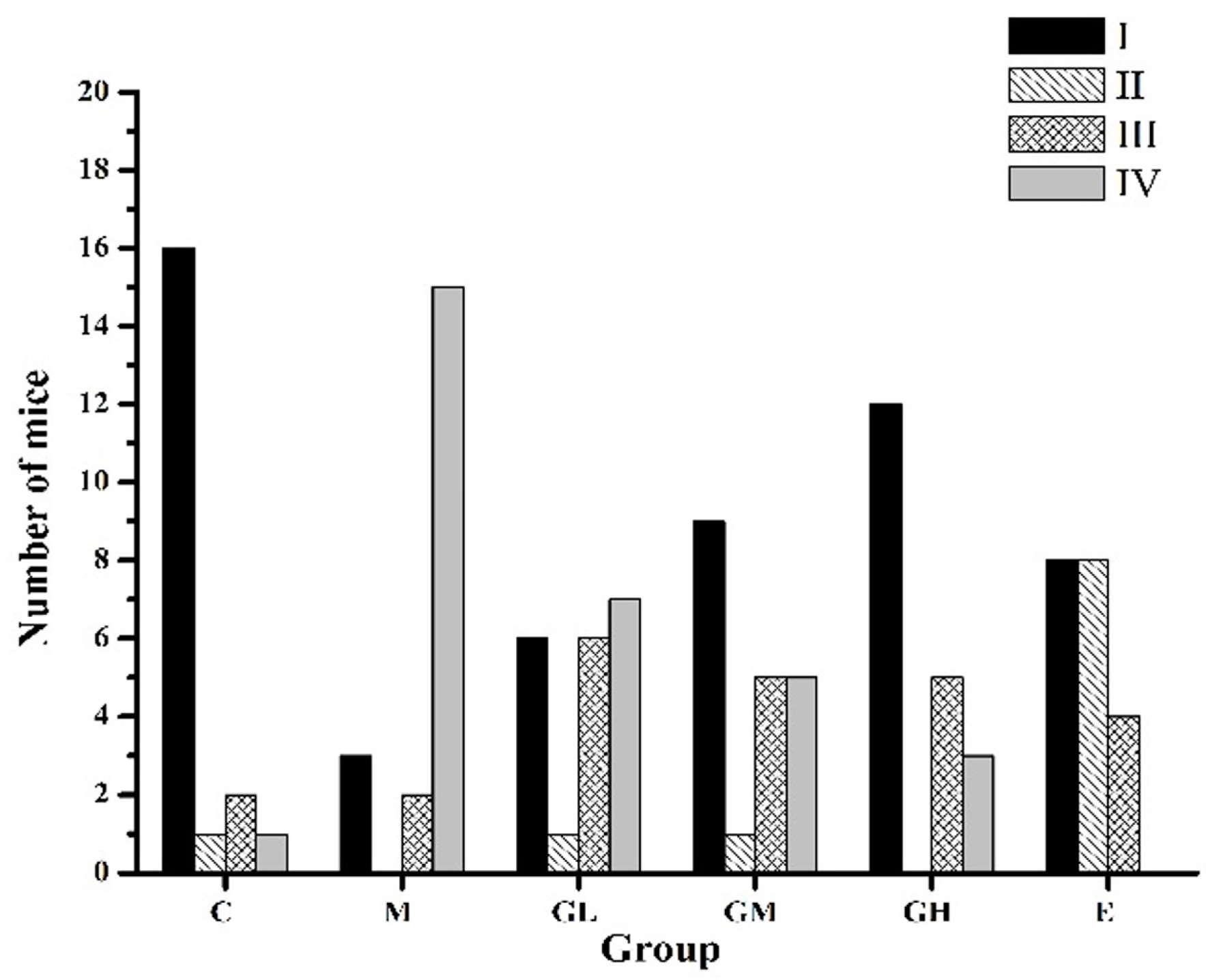
2.1. Regularization of the Estrous Cycle by Oral Administration of Genistein

The estrous cycles of normal female mice were regular and lasted 4–6 days: proestrus and metestrus for 1 day, estrus and diestrus for 1–2 days. Irregular cycles occurred before amenorrhea happened which is the character of POF. The patterns of estrous cycles were categorized based on an increasing degree of abnormality from I to IV, as per the standard in the report given by Bagavant et al. [26].
As shown in Figure 1, estrous cycle patterns changed during genistein administration. Normal estrous cycles (category I) were 85% of the mice in control group (C); however, only 15% of mice in the model group (M) were in normal cycles. The numbers of mice in normal cycles in C and M groups were significantly different (p < 0.01). In contrast, the estrous cycles of mice in genistein and estradiol groups were more regular. It was observed that 30% (low-dose of genistein (5 mg/kg body weight) therapeutic group (GL group)), 45% (moderate-dose of genistein (25 mg/kg body weight) therapeutic group (GM group)), 60% (high-dose of genistein (45 mg/kg body weight) therapeutic group (GH group)), 40% (estradiol (1 mg/kg body weight) therapeutic group (E group)) of mice cycled normally, respectively. The numbers of mice in normal estrous cycles in the GH group were significantly higher than those in the M group (p < 0.05). Furthermore, fewer mice in the GH groups showed no cycles (category IV) (15%) than those in the M group (75%, p < 0.01). The E group tended also to have more mice with regular cycles (40%) and shortened estrus (40%) compared to the C group (5%).

2.2. Estradiol Increased While Those that Are Follicle-Stimulating Hormone and Luteinizing Hormone Decreased after Oral Administration of Genistein

For women with amenorrhea, it would be reasonable to measure basal follicle-stimulating hormone (FSH) and estradiol (E2) concentrations on at least two occasions if the value of FSH is at all elevated [27]. In addition, luteinizing hormone (LH) and FSH value >30 IU/L, and levels of E2 < 50 pg/mL are typical for women with absent or nonfunctioning follicles.
As shown in Table 1, the levels of FSH and LH increased significantly, but E2 decreased significantly in the M group compared to the C group (p < 0.05) in the 1st, 20th, 30th, and 50th day. After administration of genistein, the levels of FSH and LH decreased and E2 enhanced to different extents on the 1st, 20th, 30th, and 50th day. For instance, the levels of FSH reduced by 3%, 23% and 28% in GL, GM and GH groups compared to the M group on the 50th day, respectively. The decrease in GM and GH groups was significantly different (p < 0.05), while in GL group there was no significant difference. Also, the changes of LH were similar to the FSH. However, the levels of E2 increased by 37%, 37% and 46% in GL, GM and GH groups compared to the M group on the 30th day, respectively. The enhancement of E2 in the GH group was significantly different (p < 0.05). In addition, the levels of prolactin (PRL) increased and the levels of testosterone (T) decreased in administration of genistein groups compared to M group with no significant difference (p > 0.05). Meanwhile, FSH and LH reduced and E2 enhanced in the E group during administration of estradiol.

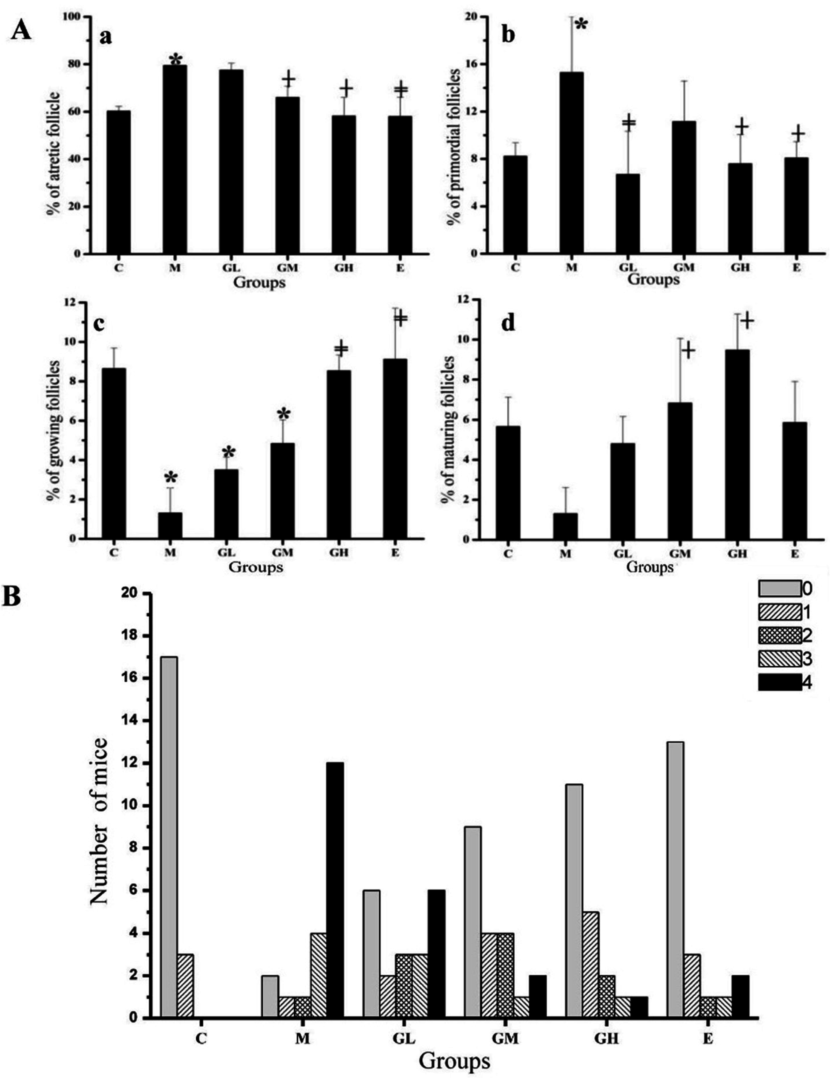
2.3. Decreased Morbidity of Oophoritis after Oral Administration of Genistein

When mice were immunized with ZP3, inflammation developed in the ovarian and in the growing and mature follicles. Moreover, some ovaries exhibited a significant loss of growing and mature follicles. As shown in Figure 2A, the ratio of growing follicles reduced significantly in the M group on day 78 compared to the C group (Figure 2c). The numbers of atretic and primordial follicles in the M group were increased (p < 0.05) relative to mice in the C group on day 78 (Figure 2a,b). The loss of follicles was concomitant with a decrease in corpora lutea [28], indicating that ovarian function was disrupted. Following exposure to genistein and estradiol, the ratio of atretic and primordial follicles decreased, and growing and mature follicles increased compared to the M group, respectively. Moreover, the changes in follicles ratio of every stage in the GM and GH groups were significantly different compared with the M group (p < 0.05). While there was no difference in the ratios of atretic, growing and mature follicles between the M group and GL group (p > 0.05). However, the changes of follicles showed no difference between mice administration of genistein and estradiol (p > 0.05).
The oophoritis on an increasing severity from 1 to 4 was shown in Figure 2B. The oophoritis morbidity was 15% in the C group; however, 90% of the mice in the M group had oophoritis. The oophoritis morbidity in the GL, GM and GH groups was 70%, 55% and 45%, respectively. The oophoritis morbidity decreased significantly in the GL, GM and GH groups compared to the M group (p < 0.01). While the oophoritis morbidity in the E group was 35%, which declined significantly compared to the M group (p < 0.01). However, the mice with oophoritis in the GM and GH groups were focused on the level 1 and 2, while for mice in the GL group, they were mainly on level 4, which is similar to the M group.

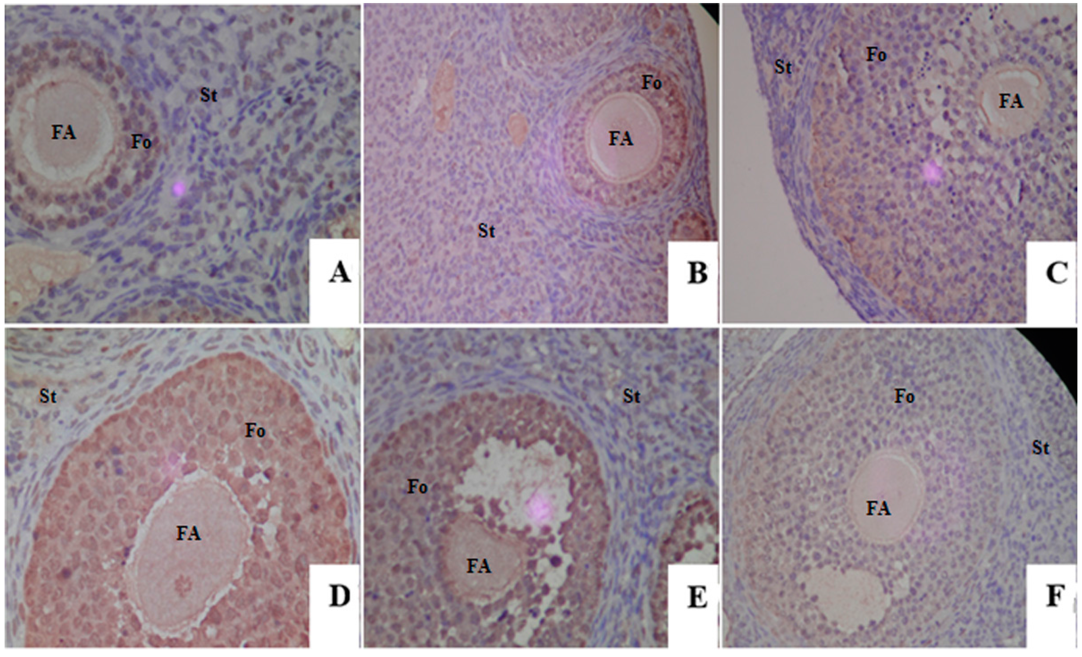
2.4. Proliferative Responses of the Ovary

The proliferation rate of the ovary follicles was determined by using an immunohistochemical staining of proliferating cell nuclear antigen (PCNA) protein (Figure 3), which was an accessory protein to δ polymerase, and was closely related to DNA replication, DNA repair and cell cycle progression [29]. Thus, PCNA was considered as a marker for the ovarian follicle growth. For all groups, the expression of PCNA-positive (brown in color) was visible not only in the follicles, but also in the stroma. Comparing to the C group, the strength and density of PCNA expression were much higher and mainly on the follicles in the M group. For mice exposure to genistein and estradiol, the expression of PCNA was focused on the follicles and changed differently in ovaries of mice in different groups. As shown in Figure 3, the strength and density of PCNA-positive expression in the E2 and GM groups were similar to the M group, while for the GL group the strength and density of PCNA-positive were significantly higher than that in the M group. However, the expression of PCNA in the GH group was similar to the C group and significantly lower compared to the M group.

3. Discussion

Autoimmune ovarian disease is a known cause of human premature ovarian failure. AOD induction by immunizing with ZP3, an ovary-specific glycoprotein, is simple and rapid. Also, the experimental murine model was developed in 1991 [30] and had been successfully exploited in investigating some fundamental issues of self-tolerance and autoimmune disease mechanisms [31]. The irregular estrous cycles, changes in endocrine hormone levels and high oophoritis morbidity in the M group indicated that the autoimmune ovarian disease model was successful. The influence of ZP3 on the ovarian lifespan might be a continuous process through the steady enhancement of FSH and LH and decline of E2. Therefore, the AOD model was widely used to investigate the therapeutic efficacy of materials for POF.
In this study, the mice received the diet supplemented with genistein at different doses (5, 25, 45 mg/kg body weight). The estrous cycle was convenient and useful for monitoring the health state of female mice. The results showed that the estrous cycles were more regular after administration of genistein and eatradiol relative to the M group. Genistein could improve the occurrence of irregular cycles, which was consistent with Zhuang et al. [32]. The levels of endocrine hormone were reported to influence the development of follicles. FSH and LH had a synergistic effect on regulating follicular development and differentiation, E2 was related to secretion of follicles. Our results found that consumption of genistein enhanced the E2 level and could attenuate the preovulatory surge of LH and FSH in GM and GH groups compared to the M group. However, there was no significant difference in the levels of FSH, LH and E2 in GL group compared to the M group. Furthermore, the level of PRL showed a decreasing tendency, while for level of T was an opposite tendency with no significant difference when compared to the M group. These results were proved by multiple studies that had documented the estrogenic activity of genistein which has an indirect effect on the hypothalamic-pituitary-gonadal axis [33,34,35]. Zin et al. [36] found that FSH increased and LH decreased in rats with 10 mg/kg body weight of genistein, FSH and LH both reduced in rats with 100 mg/kg body weight of genistein. They concluded that post-weaning exposure to genistein could influence development of the reproductive system through regulating hormones. Otherwise, the increase of growing and mature follicles, decrease of antral and primordial follicles, and the decline of oophoritis morbidity after administration of moderate and high dose of genistein indicated that genistein could improve the inflammation response in ovary. Xiao et al. found that amniotic fluid stem cells could prevent follicle atresia and sustain the healthy follicles in the ovary of mice which were induced POF by chemotherapeutic drugs [37].
Genistein could have a proliferative or antiproliferative effect depending on the levels of endogenous estrogen, life stage and tumor types. The physiologies such as development and differentiation of ovary could be modified after dietary phytoestrogen exposure [38]. When genistein effects relate to proliferation, it was meaningful to determine the expression profiles of proliferation markers like PCNA [39]. In the ovary, the expression of PCNA may affect follicular growth by regulating PCNA-dependent granulose cell proliferation in follicles [40]. In the present study, PCNA was mainly expressed in the ovarian follicles. The expression of PCNA in the GH group was similar to the C group and significantly lower compared to the M group. However, the expression in the GL group was higher than the M group. Interestingly, the expressions of PCNA after exposure to genistein were consistent with the histomorphological results regarding the follicular development.

4. Materials and Methods

4.1. Animals and Chemicals

One hundred and twenty female mice BALB/c (7–9 weeks) were obtained from the HFK BIOSCIENCE Co., Ltd. (Beijing, China). Before starting the experiments, all the animals were housed at an ambient temperature of 25 ± 2 °C, 12/12 h of light-dark cycle with ad libitum food and water for 1 week. After acclimation, a total of 120 mice were randomly divided into six groups (n = 20 per group): C group, M group, GL group, GM group, GH group, and E group. All groups except for C group received experimental AOD via immunization. In addition, there were not any estrogenic compounds exposure in diet, caging and bedding during the period. Care and treatment of the animals were based on approved protocols in accordance with the NIH guidelines (NIH Publication 85-23, 1996).
The murine ZP3 peptide was synthesized by an automatic peptide synthesizer, and with a purity of more than 90% as determined by HPLC analysis. The amino acid composition was verified by amino acid analysis and the amino acid sequence of the murine ZP3330–342 peptides was NSSSSQFQIHGPR.
Genistein was purchased from Ci Yuan Biotechnology Co., Ltd. (Shanxi, China) with a purity of more than 98% as determined by HPLC analysis. Estradiol and rabbit anti-mouse PCNA, a primary antibody, were obtained from Sigma (St. Louis, MO, USA). The goat anti-rabbit IgG secondary antibody was obtained from Beijing Zhongshan Jinqiao Biotechnology Co., Ltd. (Beijing, China). Other chemicals were of analytical grade and purchased from Zhongshan Jinqiao Biotechnology Co., Ltd. (Beijing, China).

4.2. Estrous Cycle Staging

The estrous cycles were determined by examining vaginal smears. Then, the estrous cycles were staged by vaginal cytological appearance, mainly on the proportions of leukocytes, nucleated epithelial cells and cornified squamous epithelial cells. The vaginal smears were taken according to the published method by Zhuang et al. [32].

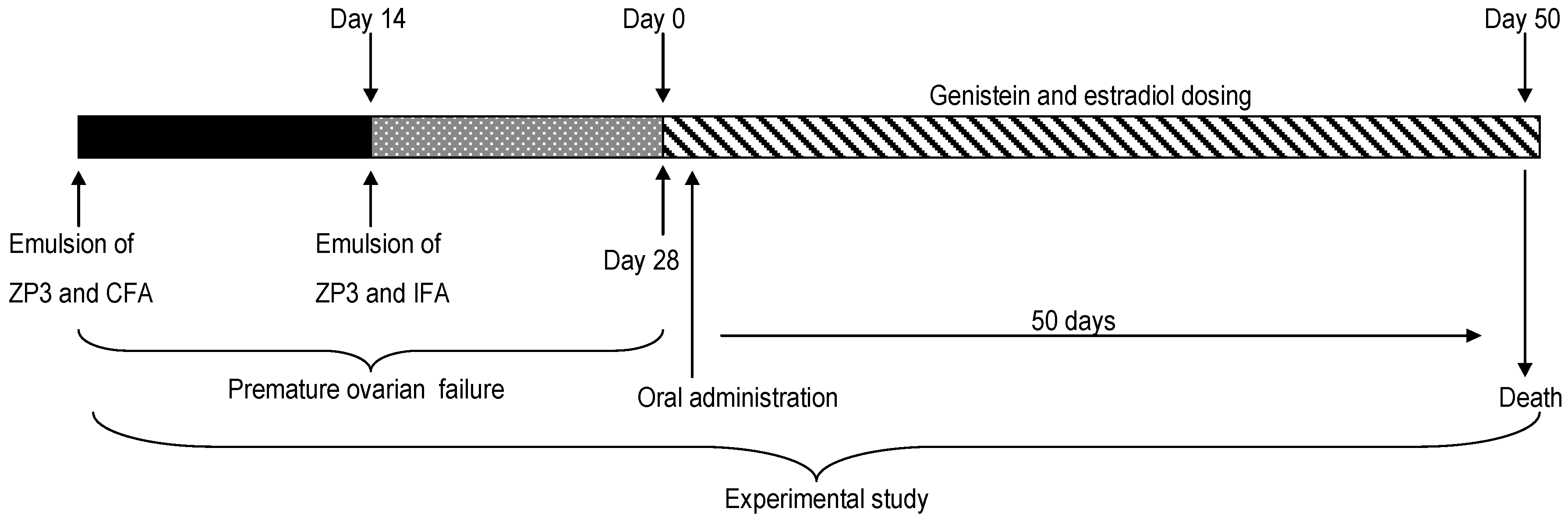
4.3. Immunization and Induction of Genistein

Ultrafiltered ZP3 peptides solution (1 mmol) was emulsified in an equal volume of complete Freund’s adjuvant (Mycobacterium tuberculosis, H37Ra strain; 0.16 mg/mouse). Mice were anesthetized by intraperitoneal injection (i.p.) of tribromoethanol. Each mouse except for C group was immunized subcutaneously in both hind footpads and received 0.15 mL of the mixture that contained 50 nmol of the peptides. After 14 days, 0.15 mL of the emulsion (1 mmol incomplete Freund’s adjuvant and ZP3) was injected into each mouse in the same position. C group received 0.15 mL of double-distilled water each time. Genistein and estradiol were administrated by daily gavage after the immunization (Figure 4: experimental study). Genistein was solubilized in DMSO to yield final concentrations of 0.75, 3.75 and 6.75 mg/mL. The doses of genistein in GL, GM, GH groups were 5, 25, 45 mg/kg body weight, respectively. Estradiol also was solubilized in DMSO solution (0.15 mg/mL (1 mg/kg body weight)) and was used as a positive control. Mice were given the same volume of DMSO in C group and M group. Mice were sacrificed on day 50 after treatment.

4.4. Detection of Serum Sex Hormone by Radio Immunity

Blood samples were collected from mice on day 1, 20, 30 and 50 after immunization and clotted at room temperature. Then, serum samples were obtained by centrifuging at 3287× g for 20 min, and frozen at −80 °C for analysis of sex hormones including E2, FSH, LH, PRL and T, using a solid-phase RIA kit (Diagnostic Products, Beijing Sino-uk institute of Biological Technology, Beijing, China). Aliquots of serum (300 μL or less) were assayed in sextuplicate.

4.5. Ovarian Histomorphology

At the end of the experimental period, animals were sacrificed. Ovaries were fixed in 10% formalin for 24 h and embedded in paraffin. Serial sections (5 μm) were stained with hematoxylin and eosin. Specimens were coded and examined by an independent observer who was blind to experimental details. Oophoritis was classified in an increasing severity from 1 to 4 based on a previous report [41].

4.6. Immunohistochemistry Analysis

For an immunohistochemical analysis of PCNA expression in the ovary, tissues were fixed in 10% formaldehyde solution and embedded in paraffin, blocked and cut into 5 μm sections. After rehydration, protein epitope retrieved with Tris-EDTA (10 mmol; pH 9) for 12–17 h at 60 °C. NOVA Detect Mouse Tissue Detection System protocol with specific primary antibodies against PCNA was used to detect the ovarian PCNA expression.

4.7. Statistical Analysis

Results were presented as mean ± S.D. Student’s t-test, rank-sum test, and one-way ANOVA were used for data analysis with SPASS 17.0 software (Version 17.0, Chicago, IL, USA). p < 0.05 was considered significant.

5. Conclusions

In conclusion, we demonstrated that orally administering moderate and high doses of genistein successfully suppressed experimental AOD in mice in this study, as evidenced by normalization of estrous cycle, decreasing levels of FSH and LH, increasing levels of E2 and alleviating abnormal ovarian histomorphology, especially at high doses. However, a significant improvement was not attained from low doses of genistein when compared to the M group. Comparing the serious side effects of traditional hormonal therapies for POF, there is an urgent need for natural alternatives. Our results proved that phytoestrogens such as genistein may be a promising, safe therapeutic agent for the treatment of premature ovarian failure. Therefore, it is reasonable to carry out further clinical research on genistein, its pharmacological dosage and weak effects for the treatment of POF.

Acknowledgments

The financial support for this study by the National Basic Research Program of China (973 program) (No.: 2012CB720805), International Science & Technology Cooperation Program of China (No.: 2010DFA31780), the Joint Sino-German Research Project of National Natural Science Foundation of China (No.: GZ 731), and the Program for New Century Excellent Talents in University (NCET-12-0749), is gratefully acknowledged.

Author Contributions

Shaoping Nie conceived and designed the experiments; Qiao Ding, Yuxiao Wang, Na Li performed the experiments; Qiao Ding, Yuxiao Wang, Na Li, Jielun Hu and Shaoping Nie analyzed the data; Qiao Ding, Yuxiao Wang, Na Li, Jielun Hu, Kexue Zhu, Sunan Wang, Fan Zhu and Shaoping Nie prepared the paper.

Conflicts of Interest

The authors declare no conflict of interest.

Abbreviations

AODautoimmune ovarian disease
POFpremature ovarian failure
ZPzona pellucid
C groupcontrol group
M groupmodel group
GL grouplow-dose of genistein (5 mg/kg body weight) therapeutic group
GM groupmoderate-dose of genistein (25 mg/kg body weight) therapeutic group
GH grouphigh-dose of genistein (45 mg/kg body weight) therapeutic group
E groupestradiol group
E2estradiol
FSHfollicle-stimulating hormone
LHluteinizing hormone
Ttestosterone
PRLprolactin
PCNAproliferating cell nuclear antigen

References

  1. Cordts, E.B.; Santos, M.C.; Peluso, C.; Kayaki, E.A.; Bianco, B.; Barbosa, C.P.; Christofolini, D.M. COMT polymorphism influences decrease of ovarian follicles and emerges as a predictive factor for premature ovarian insufficiency. J. Ovarian Res. 2014, 7, 47–51. [Google Scholar] [CrossRef] [PubMed]
  2. Meskhi, A.; Seif, M.W. Premature ovarian failure. Curr. Opin. Obstet. Gyn. 2006, 18, 418–426. [Google Scholar] [CrossRef] [PubMed]
  3. Liu, L.; Tan, R.; Cui, Y.; Liu, J.; Wu, J. Estrogen receptor α gene (ESR1) polymorphisms associated with idiopathic premature ovarian failure in chinese women. Gynecol. Endocrinol. 2013, 29, 182–185. [Google Scholar] [CrossRef] [PubMed]
  4. Cordts, E.B.; Christofolini, D.M.; Dos Santos, A.A.; Bianco, B.; Barbosa, C.P. Genetic aspects of premature ovarian failure: A literature review. Arch. Gynecol. Obstet. 2011, 283, 635–643. [Google Scholar] [CrossRef] [PubMed]
  5. Sassarini, J.; Lumsden, M.A.; Critchley, H.O.D. Sex hormone replacement in ovarian failure—New treatment concepts. Best Pract. Res. Clin. Endocrinol. Metab. 2015, 29, 105–114. [Google Scholar] [CrossRef] [PubMed]
  6. Tung, K.S.; Agersborg, S.S.; Alard, P.; Garza, K.M.; Lou, Y.H. Regulatory T-cell, endogenous antigen and neonatal environment in the prevention and induction of autoimmune disease. Immunol. Rev. 2001, 182, 135–148. [Google Scholar] [CrossRef] [PubMed]
  7. Bagavant, H.; Sharp, C.; Kurth, B.; Tung, K.S. Induction and immunohistology of autoimmune ovarian disease in cynomolgus macaques (macaca fascicularis). Am. J. Pathol. 2002, 160, 141–149. [Google Scholar] [CrossRef]
  8. Lahoti, A.; Prasannan, L.; Speiser, P.W. Premature ovarian failure. In Abnormal Female Puberty; Springer: New York, NY, USA, 2016; pp. 67–85. [Google Scholar]
  9. Kelkar, R.L.; Meherji, P.K.; Kadam, S.S.; Gupta, S.K.; Nandedkar, T.D. Circulating auto-antibodies against the zona pellucida and thyroid microsomal antigen in women with premature ovarian failure. J. Reprod. Immunol. 2005, 66, 53–67. [Google Scholar] [CrossRef] [PubMed]
  10. Hoek, A.; Schoemaker, J.; Drexhage, H. Premature ovarian failure and ovarian autoimmunity 1. Endocr. Rev. 1997, 18, 107–134. [Google Scholar] [CrossRef] [PubMed]
  11. Fu, L.; Feng, W.; Li, S.R.; Huang, B.Y. ZP3 peptides administered orally suppress murine experimental autoimmune ovarian disease. J. Reprod. Immunol. 2007, 75, 40–47. [Google Scholar] [CrossRef] [PubMed]
  12. Mahmoud, A.M.; Yang, W.; Bosland, M.C. Soy isoflavones and prostate cancer: A review of molecular mechanisms. J. Steroid Biochem. 2014, 140, 116–132. [Google Scholar] [CrossRef] [PubMed]
  13. Nie, Q.X.; Xing, M.M.; Hu, J.L.; Hu, X.J.; Nie, S.P.; Xie, M.Y. Metabolism and health effects of phyto-estrogens. Crit. Rev. Food Sci. 2015, 11. [Google Scholar] [CrossRef] [PubMed]
  14. Kostelac, D.; Rechkemmer, G.; Briviba, K. Phytoestrogens modulate binding response of estrogen receptors α and β to the estrogen response element. J. Agric. Food Chem. 2003, 51, 7632–7635. [Google Scholar] [CrossRef] [PubMed]
  15. Suetsugi, M.; Su, L.; Karlsberg, K.; Yuan, Y.C.; Chen, S. Flavone and isoflavone phytoestrogens are agonists of estrogen-related receptors 1 national institutes of health grants ES08258 and CA44735. Mol. Cancer Res. 2003, 1, 981–991. [Google Scholar] [PubMed]
  16. Mueller, S.O.; Simon, S.; Chae, K.; Metzler, M.; Korach, K.S. Phytoestrogens and their human metabolites show distinct agonistic and antagonistic properties on estrogen receptor α (ERα) and ERβ in human cells. Toxicol. Sci. 2004, 80, 14–25. [Google Scholar] [CrossRef] [PubMed]
  17. McKee, J.; Warber, S.L. Integrative therapies for menopause. South. Med. J. 2005, 98, 319–326. [Google Scholar] [CrossRef] [PubMed]
  18. Shifren, J.L.; Schiff, I. Role of hormone therapy in the management of menopause. Obstet. Gynecol. 2010, 115, 839–855. [Google Scholar] [CrossRef] [PubMed]
  19. Ferrari, C.K. Functional foods, herbs and nutraceuticals: Towards biochemical mechanisms of healthy aging. Biogerontology 2004, 5, 275–289. [Google Scholar] [CrossRef] [PubMed]
  20. Lethaby, A.; Brown, J.; Marjoribanks, J.; Kronenberg, F.; Roberts, H.; Eden, J. Phytoestrogens for vasomotor menopausal symptoms. Cochrane Database Syst. Rev. 2007, 4. [Google Scholar] [CrossRef]
  21. Xiao, C.W. Health effects of soy protein and isoflavones in humans. J. Nutr. 2008, 138, 1244–1249. [Google Scholar]
  22. Prasain, J.; Carlson, S.; Wyss, J. Flavonoids and age-related disease: Risk, benefits and critical windows. Maturitas 2010, 66, 163–171. [Google Scholar] [CrossRef] [PubMed]
  23. Prietsch, R.; Monte, L.D.; Da Silva, F.; Beira, F.; del Pino, F.; Campos, V.; Collares, T.; Pinto, L.; Spanevello, R.; Gamaro, G. Genistein induces apoptosis and autophagy in human breast MCF-7 cells by modulating the expression of proapoptotic factors and oxidative stress enzymes. Mol. Cell. Biochem. 2014, 390, 235–242. [Google Scholar] [CrossRef] [PubMed]
  24. Seo, H.S.; DeNardo, D.G.; Jacquot, Y.; Laïos, I.; Vidal, D.S.; Zambrana, C.R.; Leclercq, G.; Brown, P.H. Stimulatory effect of genistein and apigenin on the growth of breast cancer cells correlates with their ability to activate ERα. Breast Cancer Res. Treat. 2006, 99, 121–134. [Google Scholar] [CrossRef] [PubMed]
  25. Patel, S.; Zhou, C.Q.; Rattan, S.; Flaws, J.A. Effects of endocrine-disrupting chemicals on the ovary. Biol. Reprod. 2015, 93, 1–9. [Google Scholar] [CrossRef] [PubMed]
  26. Bagavant, H.; Adams, S.; Terranova, P.; Chang, A.; Kraemer, F.W.; Lou, Y.; Kasai, K.; Luo, A.M.; Tung, K.S. Autoimmune ovarian inflammation triggered by proinflammatory (Th1) T cells is compatible with normal ovarian function in mice. Biol. Reprod. 1999, 61, 635–642. [Google Scholar] [CrossRef] [PubMed]
  27. Rebar, R.W. Premature ovarian failure. Obstet. Gynecol. 2009, 113, 1355–1363. [Google Scholar] [CrossRef] [PubMed]
  28. Thatcher, W.W.; Macmillan, K.L.; Hansen, P.J.; Drost, M. Concepts for regulation of corpus luteum function by the conceptus and ovarian follicles to improve fertility. Theriogenology 1989, 31, 149–164. [Google Scholar] [CrossRef]
  29. Thomas, H.; Nasim, M.; Sarraf, C.; Alison, M.; Love, S.; Lambert, H.; Price, P. Proliferating cell nuclear antigen (PNCA) immunostaining—A prognostic factor in ovarian cancer? Br. J. Cancer 1995, 71, 357–362. [Google Scholar] [CrossRef] [PubMed]
  30. Rhim, S.H.; Millar, S.; Robey, F.; Luo, A.; Lou, Y.; Yule, T.; Allen, P.; Dean, J.; Tung, K. Autoimmune disease of the ovary induced by a ZP3 peptide from the mouse zona pellucida. J. Clin. Investig. 1992, 89, 28–35. [Google Scholar] [CrossRef] [PubMed]
  31. Tung, K.; Lou, Y.; Garza, K.; Teuscher, C. Autoimmune ovarian disease: Mechanism of disease induction and prevention. Curr. Opin. Immunol. 1997, 9, 839–845. [Google Scholar] [CrossRef]
  32. Zhuang, X.L.; Fu, Y.C.; Xu, J.J.; Kong, X.X.; Chen, Z.G.; Luo, L.L. Effects of genistein on ovarian follicular development and ovarian life span in rats. Fitoterapia 2010, 81, 998–1002. [Google Scholar] [CrossRef] [PubMed]
  33. Nagata, C.; Takatsuka, N.; Inaba, S.; Kawakami, N.; Shimizu, H. Effect of soymilk consumption on serum estrogen concentrations in premenopausal japanese women. J. Natl. Cancer Inst. 1998, 90, 1830–1835. [Google Scholar] [CrossRef] [PubMed]
  34. Duncan, A.M.; Merz, B.E.; Xu, X.; Nagel, T.C.; Phipps, W.R.; Kurzer, M.S. Soy isoflavones exert modest hormonal effects in premenopausal women 1. J. Clin. Endocr. Metab. 1999, 84, 192–197. [Google Scholar] [CrossRef] [PubMed]
  35. Wu, A.; Stanczyk, F.; Hendrich, S.; Murphy, P.; Zhang, C.; Wan, P.; Pike, M. Effects of soy foods on ovarian function in premenopausal women. Br. J. Cancer. 2000, 82, 1879–1886. [Google Scholar] [CrossRef] [PubMed]
  36. Zin, S.R.M.; Omar, S.Z.; Khan, N.L.A.; Musameh, N.I.; Das, S.; Kassim, N.M. Effects of the phytoestrogen genistein on the development of the reproductive system of Sprague Dawley rats. Clinics 2013, 68, 253–262. [Google Scholar] [CrossRef]
  37. Xiao, G.Y.; Liu, I.H.; Cheng, C.C.; Chang, C.C.; Lee, Y.H.; Cheng, W.T.K.; Wu, S.C. Amniotic fluid stem cells prevent follicle atresia and rescue fertility of mice with premature ovarian failure induced by chemotherapy. PLoS ONE 2014, 9, e106538. [Google Scholar] [CrossRef] [PubMed]
  38. Patisaul, H.B.; Jefferson, W. The pros and cons of phytoestrogens. Front. Neuroendocrinol. 2010, 31, 400–419. [Google Scholar] [CrossRef] [PubMed]
  39. Cline, J.M.; Wood, C.E. The mammary glands of macaques. Toxicol. Pathol. 2008, 36, 130–141. [Google Scholar] [CrossRef] [PubMed]
  40. Zhang, Z.H.; Chen, L.Y.; Wang, F.; Wu, Y.Q.; Su, J.Q.; Huang, X.H.; Cheng, Y.; Wang, Z.C. Expression of hypoxia-inducible factor-1α during ovarian follicular growth and development in Sprague-Dawley rats. Genet. Mol. Res. 2015, 14, 5896–5909. [Google Scholar] [CrossRef] [PubMed]
  41. Garza, K.M.; Agersborg, S.S.; Baker, E.; Tung, K.S. Persistence of physiological self antigen is required for the regulation of self tolerance. J. Immunol. 2000, 164, 3982–3989. [Google Scholar] [CrossRef] [PubMed]
Figure 1. Effects of genistein on estrous cycle patterns of BALB/c female mice. Four patterns of abnormal estrous cycles were graded in an increasing order of abnormality (I–IV) as follows: I: normal; II: regular cycles with a shortened estrus; III: irregular cycles with a prolonged diestrus and normal or prolonged estrus; IV: no cyclicity. C: control group; M: model group; GL: low-dose of genistein (5 mg/kg body weight) therapeutic group; GM: moderate-dose of genistein (25 mg/kg body weight) therapeutic group; GH: high-dose of genistein (45 mg/kg body weight) therapeutic group; E: estradiol (1 mg/kg body weight) therapeutic group.
Ijms 17 01855 g001
Figure 2. The follicles ratio of every stage and oophoritis morbidity in the ovary. (A) The follicles ratio of every stage (a: atretic follicles; b: primordial follicles; c: growing follicles; d: mature follicles); (B) Autoimmune oophoritis was classified according to increasing severity from 1 to 4, 0: normal with no oophoritis. * p < 0.05 compared to control group; p < 0.05 compared to the model group; p < 0.01 compared to the model group.
Ijms 17 01855 g002
Figure 3. Photomicrograph of PCNA from growing follicles in each group. (A) Control group; (B) model group; (C) estradiol group; (D) low-dose genistein group; (E) moderate-dose genistein group; (F) high-dose genistein group. The expression of PCNA-positive was brown in color, while blue in color for PCNA-negative expression. Original magnification 400×. Fo: follicle; St: stroma; FA: follicle antrum.
Ijms 17 01855 g003
Figure 4. Schematic illustration of experimental design.
Ijms 17 01855 g004
Table 1. Levels of sex hormone in mouse serum.
Time/DayGroupE2/pg·mL−1LH/mIU·mL−1FSH/mIU·mL−1PRL/ng·mL−1T/ng·mL−1
1stC9.41 ± 0.4530.15 ± 2.349.33 ± 1.019.28 ± 0.650.531 ± 0.062
M5.24 ± 0.90 a36.51 ± 0.51 aa9.90 ± 0.547.09 ± 0.650.544 ± 0.072
GL7.66 ± 0.3631.41 ± 2.43 b8.68 ± 0.717.07 ± 1.020.448 ± 0.045
GM8.39 ± 0.2231.65 ± 2.07 b7.15 ± 0.2310.77 ± 0.20 b0.514 ± 0.055
GH9.17 ± 3.15 b34.23 ± 0.875.69 ± 1.71 aa,bb8.48 ± 0.360.492 ± 0.021
E6.45 ± 0.7932.02 ± 1.03 b8.24 ± 0.528.74 ± 0.420.421 ± 0.018
20thC13.10 ± 8.5731.50 ± 2.08 b7.60 ± 1.86 b9.01 ± 1.360.468 ± 0.006
M4.66 ± 0.56 aa36.33 ± 1.4110.05 ± 1.117.45 ± 0.970.484 ± 0.020
GL9.09 ± 3.2729.80 ± 1.94 bb,c7.57 ± 0.929.36 ± 1.390.463 ± 0.046
GM5.21 ± 0.85 aa36.39 ± 1.83 a8.96 ± 0.668.43 ± 1.080.432 ± 0.042
GH4.67 ± 0.45 aa32.56 ± 2.19 b6.84 ± 0.26 bb8.25 ± 0.670.479 ± 0.026
E4.79 ± 0.14 aa34.72 ± 3.988.71 ± 0.309.39 ± 0.740.457 ± 0.045
30thC4.32 ± 0.3730.61 ± 0.378.36 ± 0.928.56 ± 1.230.397 ± 0.192
M3.01 ± 0.45 a36.87 ± 0.56 aa10.32 ± 0.12 a8.44 ± 0.750.437 ± 0.016
GL4.13 ± 0.2232.35 ± 1.31 b8.89 ± 0.238.56 ± 0.520.414 ± 0.013
GM4.13 ± 0.8136.73 ± 2.57 aa8.65 ± 0.427.23 ± 0.02 c0.449 ± 0.028
GH4.38 ± 0.57 b31.93 ± 0.90 b7.50 ± 0.19 b8.59 ± 0.390.691 ± 0.234 a,c
E4.29 ± 0.16 b34.05 ± 0.618.48 ± 0.919.87 ± 1.200.399 ± 0.035
50thC4.42 ± 0.3230.23 ± 1.629.02 ± 0.3610.21 ± 1.310.470 ± 0.043
M3.03 ± 0.75 a34.65 ± 0.91 a11.35 ± 0.44 a7.52 ± 0.34 a,c0.486 ± 0.018
GL4.93 ± 0.78 b,c31.50 ± 1.96 b10.97 ± 0.938.76 ± 0.380.471 ± 0.010
GM4.54 ± 0.35 b30.15 ± 1.28 b,c8.78 ± 0.29 b7.78 ± 0.32 a,c0.461 ± 0.032
GH3.84 ± 0.2434.68 ± 1.43 a8.14 ± 0.77 bb8.86 ± 0.560.469 ± 0.025
E3.51 ± 0.1333.65 ± 1.169.34 ± 0.41 b9.98 ± 0.910.448 ± 0.019
All data are presented as mean ± S.D. a: p < 0.05 compared to the control group; aa: p < 0.01 compared to the control group; b: p < 0.05 compared to the model group; bb: p < 0.01 compared to the model group; c: p < 0.05 compared to the estradiol group. C: control group; M: model group; GL: low-dose of genistein (5 mg/kg body weight) therapeutic group; GM: moderate-dose of genistein (25 mg/kg body weight) therapeutic group; GH: high-dose of genistein (45 mg/kg body weight) therapeutic group; E: estradiol (1 mg/kg body weight) therapeutic group.

