Article

Mutations in the Mitochondrial ND1 Gene Are Associated with Postoperative Prognosis of Localized Renal Cell Carcinoma

1
Department of Urology, Tokai University School of Medicine, Kanagawa, Isehara 259-1193, Japan
2
Department of Clinical Pharmacology, Tokai University School of Medicine, Kanagawa, Isehara 259-1193, Japan
3
Department of Pathology, Tokai University School of Medicine, Kanagawa, Isehara 259-1193, Japan
4
Support Center for Medical Research and Education, Tokai University, Kanagawa, Isehara 259-1193, Japan
*
Author to whom correspondence should be addressed.
Int. J. Mol. Sci. 2016, 17(12), 2049; https://doi.org/10.3390/ijms17122049
Submission received: 26 October 2016 / Revised: 28 November 2016 / Accepted: 29 November 2016 / Published: 7 December 2016
(This article belongs to the Section Molecular Pathology, Diagnostics, and Therapeutics)

Abstract

:
We analyzed mutations in the mitochondrial ND1 gene to determine their association with clinicopathological parameters and postoperative recurrence of renal cell carcinoma (RCC) in Japanese patients. Among 62 RCC cases for which tumor pathology was confirmed by histopathology, ND1 sequencing revealed the presence of 30 mutation sites in 19 cases. Most mutations were heteroplasmic, with 16 of 19 cases harboring one or more heteroplasmic sites. Additionally, 12 sites had amino acid mutations, which were frequent in 10 of the cases. The 5-year recurrence-free survival (RFS) rate was significantly worse in patients with tumors >40 mm in diameter (p = 0.0091), pathological T (pT) stage ≥3 (p = 0.0122), Fuhrman nuclear atypia grade ≥III (p = 0.0070), and ND1 mutations (p = 0.0006). Multivariate analysis using these factors revealed that mutations in ND1 were significantly associated with the 5-year RFS rate (p = 0.0044). These results suggest a strong correlation between the presence of ND1 mutations in cancer tissue and postoperative recurrence of localized RCC in Japanese patients.

1. Introduction

Kidney cancer is globally the 9th and 14th most common malignant tumor among males and females, respectively [1]. In developed countries, renal cell carcinoma (RCC) constitutes 85%–90% of all cancers of the kidney [2,3]. There is a 1.5:1 predominance of new RCC cases in males over females, and the peak age of onset is between 60 and 70 years [4]. The advent and widespread use of abdominal imaging techniques, such as abdominal ultrasonography and computed tomography, has led to the increased frequency of the diagnosis of small asymptomatic RCCs, which account for about half of all newly diagnosed cases [5]. Despite the increase in the number of new cases, the mortality rate due to RCC has, in fact, decreased [6,7]. However, even in patients with localized RCC that has been resected with curative surgery, such as total or partial nephrectomy, the relapse rate is about 20%–40%, with progression to metastatic RCC (mRCC) [8].
These observations suggest that even small RCCs can exhibit aggressive metastatic ability to distant organs [9]. In addition, given the lack of an effective adjuvant therapy to reduce the risk of recurrence after curative surgery, early detection and intervention in patients with recurrent RCC is critical to improving prognosis in mRCC. However, there are no established prognostic models to accurately predict the metastatic potential of RCCs. Recent studies suggested that several prognostic models that were developed based on the TNM system may not effectively predict the postoperative prognosis of distinct RCC types, and the development of improved models with increased accuracy for recurrence patterns is sorely needed [10,11,12]. Additionally, diagnostic and prognostic molecular markers for RCC have not yet been fully incorporated into clinical practice [13,14].
Human mitochondrial DNA (mtDNA) is a closed, circular molecule comprising 16,569 nucleotide pairs and encoding 13 essential genes with roles in oxidative phosphorylation as well as the structural rRNA and tRNAs necessary for the expression of these genes [15]. mtDNA is encoded separately from nuclear DNA and is in close proximity to reactive oxygen species (ROS) due to functional necessity. For this reason, and as histone proteins that protect nuclear DNA are missing in mitochondria, mtDNA accumulates mutations at a higher rate than nuclear DNA, which subsequently affect the oxidative phosphorylation capacity of mitochondria [16].
Recent studies proposed that mutations in mtDNA might be utilized as molecular markers for early detection, assessment of the degree of malignancy, and prediction of recurrence and prognosis after radical surgery in several cancer types. [17,18]. mtDNA mutations that resulted in ROS accumulation were shown to be correlated with the acquisition of metastatic potential and poor prognosis [19]. For example, Bai et al. demonstrated the association of a nine-base pair deletion at nucleotide position 8272 (region V) of the mtDNA with postoperative survival rate in RCC [20]. In addition, mutations in NADH dehydrogenase subunit 1 (ND1), a subunit of oxidative phosphorylation complex, were detected in breast and nasopharyngeal carcinomas [21,22]. Furthermore, Yusnita et al. [23] identified 11 missense mutations in ND1 from colorectal tumor tissue specimens. Studies also showed that mutations in ND1 may lead to changes in the secondary structure of the protein and subsequent inactivation of enzymes [23]. The studies described above indicate that there was a correlation between various types of cancer and the ND1 mutations, which may lead to subsequent abrogation of its enzymatic activity [21,22,23]. These studies provide strong evidence for the utility of mtDNA mutations as potential prognostic factors for recurrence and progression in various types of cancer.
In this study, we aimed to determine the correlation between mtDNA mutations and postoperative recurrence in RCC. We performed comparative sequence analysis of mtDNA extracted from formalin-fixed paraffin-embedded (FFPE) RCC and the paired non-cancerous tissue specimens to evaluate postoperative gene markers as prognostic factors.

2. Results

2.1. Clinical Characteristics of Study Subjects

Characteristics of patients and tumor specimens are summarized in Table 1. Median age was 62 (39–80) years, and male-to-female ratio was 3.4:1. Median follow-up period was 66.5 (29–76) months. From 62 subjects in this study that were followed up with for a minimum of two years, 11 had recurrent RCC by June 2016. Among pathological characteristics, median tumor diameter in the surgical specimen was 32 (12–105) mm. Histologically, 55 patients had clear cell RCC (ccRCC), whereas tumors were non-clear cell RCC (nccRCC) in the remaining seven cases. By histopathological diagnosis, tumors were locally confined (stage pT1–2) in 57 patients, whereas tumors invaded the vascular or surrounding tissue (pathological T (pT) stage 3–4) in five patients. By Fuhrman grading of nuclear atypia, 54 and eight cases were grade I/II and III/IV, respectively. Microvessel invasion was observed in 12 patients.

2.2. Nucleotide Differentiation Index Analysis (NST)

In this study comprising 62 Japanese patients with RCC, we investigated the ND1 gene using the nucleotide differentiation index (NST) [24]. As controls, ND1 sequences of 62 healthy Japanese subjects were obtained from GenBank. We determined that the NST was 0.005, indicating that RCC was significantly associated with mutations in the mitochondrial ND1 gene.

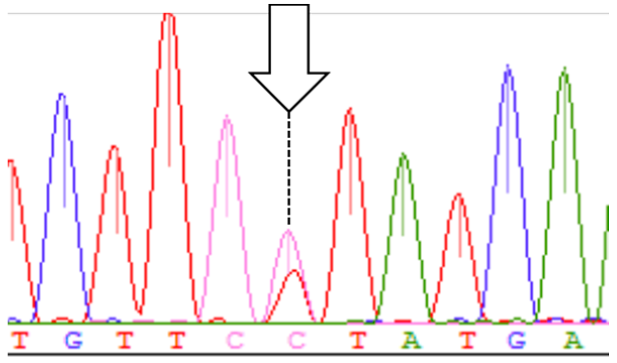
2.3. Analysis of ND1 Mutations

We analyzed the ND1 sequences (956 base pairs) using the FFPE tissue specimens of 62 patients with RCC. We found somatic mutations, including heteroplasmy, at 30 sites in 19 cases (30.6%; 19/62 patients) (Figure 1). Only one site (3970) was confirmed to have both homoplasmic and heteroplasmic mutations. In addition, C3328Y, C4197Y, and C4141Y with heteroplasmic mutations were novel mutations that were not previously reported in Japanese patients. Moreover, mutations leading to amino acid changes were observed at 12 sites in 10 cases (52.6%, 10/19 patients). The ND1 region of patient hk383 harbored a mutation, insertion of a C at position 3572, which caused a frameshift. Heteroplasmic mutations were observed at 24 sites in 16 cases (84.2%, 16/19 patients), and 84% of the cases with mutations harbored heteroplasmic mutations (Table 2). Since mtDNA is generally homoplasmic, heteroplasmic mutations observed in this cohort were considered to have occurred as part of changes in conditions related to RCC.

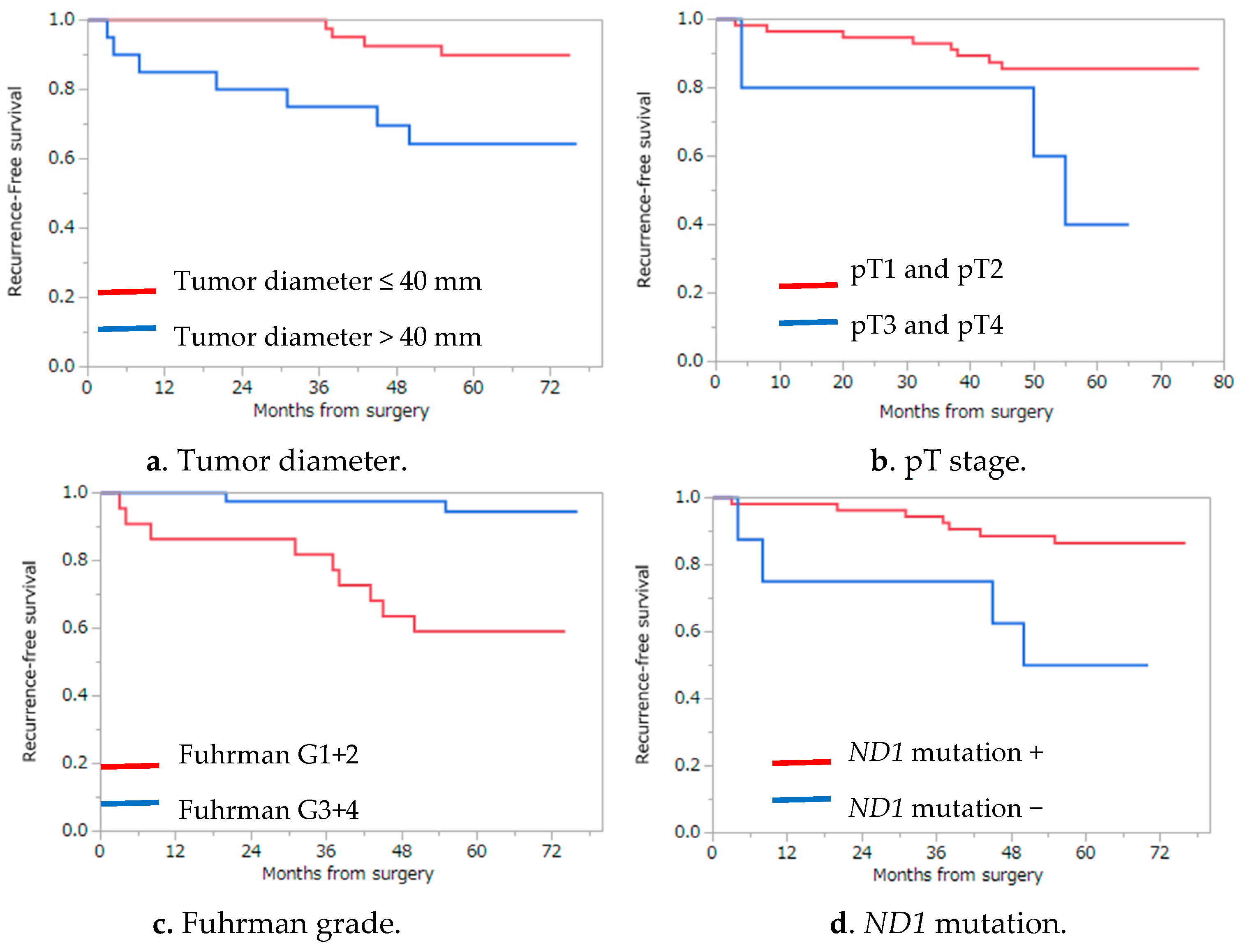
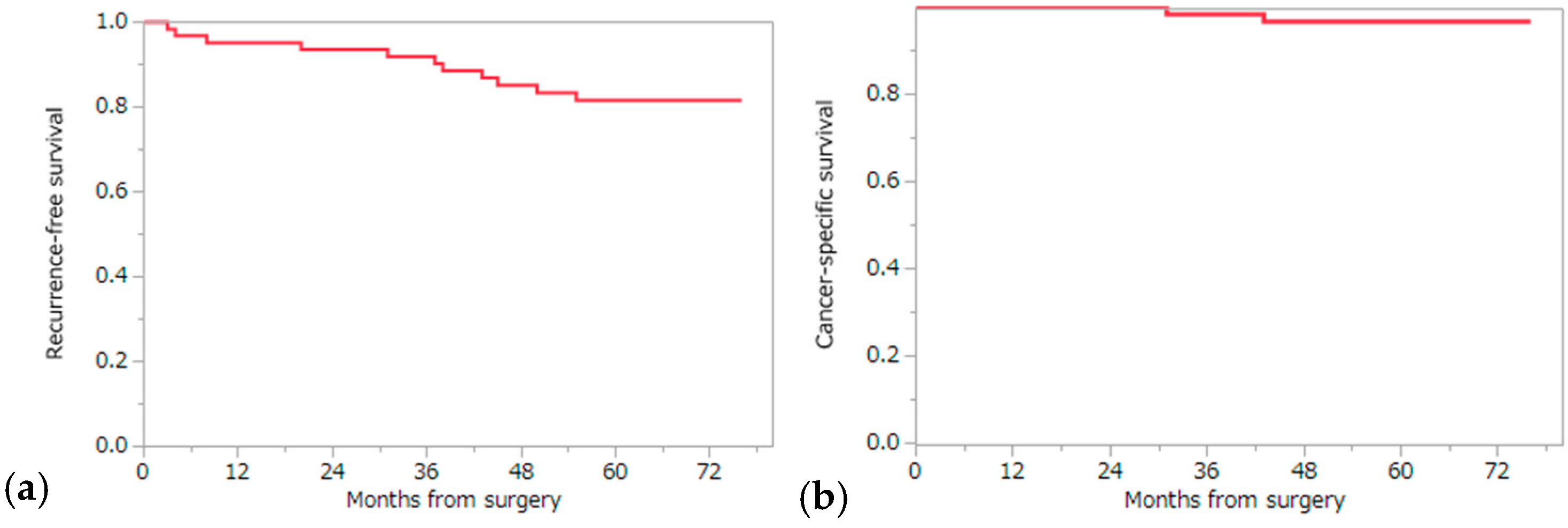
2.4. Analysis of Recurrence Risk Factors and Cancer-Specific and Overall Survival

The 5-year recurrence-free survival (RFS), cancer specific survival (CSS), and overall survival (OS) were 81.6%, 96.7%, and 93.4% by the Kaplan–Meier analysis, respectively (Figure 2). Among the ten factors that were assessed by the log-rank test, four factors (maximum tumor diameter, pT stage, Fuhrman grade, and ND1 mutation) were significantly associated with recurrence, suggesting that these factors might impact RFS: tumor diameter (5-year RFS, 89.9% and 64.3% for ≤40 and >40 mm, respectively; p = 0.0091), pT stage (5-year RFS, 85.6% and 40.0% for ≤pT2 and ≥pT3, respectively; p = 0.012), Fuhrman grade (5-year RFS, 86.6% and 50.0% for I/II and III/IV, respectively; p = 0.0070), and somatic mutation in ND1 (5-year RFS, 59.1% and 94.6% for the presence and absence of somatic mutation/s in ND1, respectively; p = 0.0006) (Table 3) (Figure 3). Multivariate analysis using the Cox proportional hazards model using these four factors showed that only the presence of somatic mutations in the ND1 gene was a significant factor associated with RFS (p = 0.0044) (Table 4). We also examined the association of these ten factors with CSS using the log-rank test. As a result, three factors (maximum tumor diameter, pT stage, and ND1 mutation) were found to be significantly associated with CSS: tumor diameter (5-year CSS, 100% and 90% for ≤40 mm and >40 mm, respectively; p = 0.0415), pT stage (5-year CSS, 98.2% and 80.0% for ≤pT2 and ≥pT3, respectively; p = 0.0330), and somatic mutation in ND1 (5-year CSS, 89.5% and 100% for the presence and absence of somatic mutation/s in ND1, respectively; p = 0.0341) (Table 5). Multivariate analysis using the Cox proportional hazards model for these three factors showed that there was no factor significantly associated with CSS; however, somatic mutation in ND1 showed a tendency toward deterioration of CSS in the presence of ND1 mutation (p = 0.0827) (Table 6). These findings suggest that mutations in ND1 might also be considered as a prognostic factor for recurrence and a possible predictor of CSS. Among the ccRCC subgroup (55 cases), we examined the association of RFS with the same nine factors using the log-rank test and Cox proportional hazards model. As a result of the log-rank test, three factors (maximum tumor diameter (p = 0.0161), Fuhrman grade (p = 0.0051), and ND1 mutation (p = 0.0042)) were candidates of becoming independent predictors of RFS. Finally, ND1 mutation was the only statistically significant predictive factor of RFS in the ccRCC sub group according to the Cox proportional hazards model (p = 0.0208) (Table 7).

3. Discussion

In this study, we conducted a comparative sequence analysis of mtDNA extracted from FFPE RCC specimens and the paired non-cancerous tissue specimens to determine postoperative genetic markers as potential prognostic factors for the recurrence of RCC. As a result, 30 mutations were found in the ND1 sequence of 19 patients. These mutations were not found in healthy Japanese subjects based on the GenBank database and were confirmed to be somatic by comparing with the paired non-cancerous tissue specimens. Majority of the mutations were heteroplasmic; 16 of 19 cases had one or more heteroplasmic sites. Heteroplasmy suggests the presence of a pathogenic mutation as a cause or result of cancer and was previously shown to have a strong association with mutations [25,26,27]. In six cases, there were three new mutation sites (C3328Y, C4141Y, and C4197Y) that were not previously reported and were not found in the database. Of these new mutation sites, C3328Y and C4141Y were missense mutations, whereas L8F and R279W were amino acid changes resulting from the mutations. With respect to amino acid changes, we found mutations that led to changes in amino acids at 12 sites in 10 cases (52.6%; 10/19 patients). Mutations that lead to amino acid changes may alter protein structures and subsequently affect their function and activity.
We identified an insertion of C at position 3572 (C3572ins) in patient hk383, which caused a frameshift and an amino acid change. With this insertion, the protein structure of ND1 and complex I function may be significantly altered [28]. This specific mutation in mitochondrial ND1 has been reported in oncocytic thyroid carcinoma [29], oncocytic pituitary adenoma [30], renal oncocytoma, and chromophobe RCC (eosinophilic variant) [28], suggesting that C3572ins mutation in ND1 is associated with oncocytic tumors or variants. Oncocytic is derived from the granular eosinophilic appearance of cytoplasm due to the abnormal accumulation of mitochondria [28]. The patient with the C3572ins mutation (case hk383) also had a granular eosinophilic cytoplasm and was diagnosed with an eosinophilic variant of ccRCC. While a correlation between oncocytosis and tumorigenesis has been suggested [31], the relationship between C3572ins and tumor progression is not clear. In this study, our patient with the C3572ins mutation had metastatic recurrence in the lymph nodes and the adrenal glands eight months after surgery. However, further studies are needed to assess the utility of this mutation as a predictor for disease recurrence.
Our log-rank test revealed that tumor diameter >40 mm, pT stage≥ 3, Fuhrman grade ≥III, and ND1 mutation were associated with worse RFS rates in patients who underwent surgery for localized RCC. Multivariate analysis using these factors revealed that mutations in ND1 were significantly associated with RFS rates. Although larger tumor size, pT stage, and Fuhrman grade were previously reported as poor prognostic factors [32,33], this is the first study to show that mutations in ND1 were predictors of recurrence in localized RCC.
The mechanism of carcinogenesis in RCC has not yet been elucidated; however, genetic factors were shown to be crucial to its development. For example, mutations of the von Hippel-Lindau (VHL) gene on the short arm of chromosome 3 and another locus on the long arm of chromosome 5 were shown to be associated with the carcinogenesis of RCC and are considered as candidate tumor suppressor genes [34,35]. However, factors that determine the speed and progression of RCC or its metastasis are not yet known. In this study, we focused on mitochondria, in particular, since its relationship with oxidative stress and consequent mtDNA mutations might affect the prognosis of patients with RCC. Mitochondria has a leading role in critical cellular functions such as glucose and lipid metabolism and apoptosis [36]. Furthermore, mitochondria has endogenous angiogenic factors, such as VEGF and TGF-β1, and VHL protein for protein degradation by ubiquitination [37,38]. The loss of VHL protein observed in most ccRCC cases suggest that it has contributions to the downregulation of biosynthesis of the oxidative phosphorylation complex. Solid tumors endure a severely hypoxic environment, and adaptation to such a hypoxic environment is a critical step in tumor progression. Increases in markers of oxidative stress, such as ROS and nitric oxide, were found in some patients with RCC [39]. In addition, patients with increased parameters of oxidative stress and decreased antioxidant levels were associated with higher grade, stage and metastatic disease [39].
This is the first study that demonstrates the relationship between mitochondrial ND1 mutations and postoperative recurrence of RCC in a Japanese patient cohort. The Japanese are a single ethnic group and thus tend to show similar responses to treatments such as that observed with interferon therapy for RCC [40] and hepatocellular carcinoma [41]. Additionally, Japanese patients were recently shown to exhibit side effects that were distinct from those observed in Western populations in response to targeted therapy for RCC [42]. Furthermore, mtDNA sequences are stored in databases based on race and continent, and comparison of mtDNA sequences across different ethnic groups and races based on the findings of this study will be useful in determining the prognosis of RCC in Japanese patients in future studies. Finally, our findings showing that over 80% of the mutations found were heteroplasmic provide very strong evidence that mutations in ND1 can be used as indicators of cancer prognosis in the future [43]. Studies with increased numbers of patients are necessary to confirm and improve the reliability of our findings in the present study.

4. Materials and Methods

4.1. Patients and Tumor Specimens

In this study, FFPE tissue specimens with accompanying clinical data were collected from a total of 62 patients who consecutively underwent radical surgery for localized RCC (clinical T1-4N0M0) at Tokai University School of Medicine Hospital between January and December 2010 and were retrospectively followed for a minimum of two years. This study was approved by the clinical research review committee of the Tokai University School of Medicine.

4.2. Assessment of Formalin-Fixed Paraffin-Embedded Tissue Specimens

Two pathologists microscopically confirmed the location of cancerous tissue and the paired non-cancerous tissue within the FFPE specimens, and tissue punch samples that were obtained with an 18 G needle were in separate 1.5-mL test tubes.

4.3. Amplification of mtDNA with Polymerase Chain Reaction

FFPE tissue punch samples were washed three times with 500 μL lemosol and three times with 99.5% ethanol to remove lemosol. Tissues were next incubated with 200 μg proteinase K in HMW Buffer (10 mM Tris-Cl pH 8.0, 150 mM NaCl, 10 mM EDTA (ethylenediaminetetraacetic acid), 0.1% SDS (sodium dodecyl sulfate)) at 60 °C for overnight, followed by extraction with phenol-chloroform (Phenol/Chloroform/Isoamyl alcohol, 25:24:1), each twice, at 11,000 rpm at room temperature for 10 min. DNA was precipitated by the addition of 0.1 vol 3 M Na-AcOH and 2.5 vol ice-cold ethanol. After centrifugation at 15,000 rpm at 4 °C for 20 min, DNA pellets were rinsed with 70% cold ethanol, dried, and dissolved in TE buffer (10 mM Tris-HCl, pH 8.0 and 1 mM EDTA). Total mtDNA was amplified by polymerase chain reaction (PCR) with the following primer sets that were designed to cover the entire 957-bp mtDNA ND1 region:
  • F3274 (5′-ACAGTCAGAGGTTCAATTCCTCTTCT-3′),
  • R3590 (5′-ATAGGAGGCCTAGGTTGAGGTTGACCA-3′),
  • F3590 (5′-TGGTCAACCTCAACCTAGGCCTCCTAT-3′),
  • R3725 (5′-GATGGCTAGGGTGACTTCATATGAGA-3′),
  • F3731 (5′-ATGAAGTCACCCTAGCCATCATTCTACTA-3′),
  • R4021 (5′-TCATATGTTGTTCCTAGGAAGATTGTAGT-3′),
  • ND1-F (5′-TCCGAACTAGTCTCAGGCTTCA-3′),
  • ND1-R (5′-CACGGAGAATTTTGGATTCTCAG-3′).
PCR was performed by Ex Taq® (Takara Bio, Tokyo, Japan) under the following conditions: melting at 95 °C for 10 s, annealing at 55 °C for 20 s, and extension at 72 °C for 30 s, for a total of 35 cycles. PCR products were treated by EXOSAP-IT® (Affimetrix, Santa Clara, CA, USA) and directly sequenced using the Big Dye® Terminator v3.1 Reaction Kit (Applied Biosystems, Torrance, CA, USA) mix and ABI 3500xL DNA sequencer (Life Technologies, Carlsbad, CA, USA). We also confirmed the sequences in the ND1 region three times to confirm the mutations. The ND1 DNA sequence data were assembled using the ATCG software (Genetyx, Tokyo, Japan). Accession numbers for the nucleotide sequences of ND1 obtained from 62 patients included in the present study are LC178840–LC178901.

4.4. Nucleotide Differentiation Index Analysis

For NST analysis, nucleotide sequences of the ND1 gene of 62 subjects in this study were aligned by ClustalW using the Molecular Evolutionary Genetics Analysis (MEGA, version 6, Tempe, AZ, USA, Pennsylvania, USA, Tokyo, Japan) software [24,44]. Common sites in all sequences with gaps were excluded from analysis. mtDNA sequences were compared with the reference mtDNA from 62 healthy long-living Japanese individuals from the Genbank database (Table S1). Mutation sites were used in homology searches across a range of databases (NCBI-dbSNP, MitoMap, Ensemble, JSNP), and NC_012920.1 (GI: 251831106) was chosen as the reference sequence for ND1. Next, 27 segregation sites from ND1 gene sequences were selected. NSTij for the i-site and the jth subpopulation was defined for the NST analysis (Table S2). The NST formula was as follows [24]:
N S T i j = H T i H S i j H T i

4.5. Detection of Somatic Mutations in ND1 Gene

The mtDNA sequences of 62 healthy long-living Japanese individuals were randomly extracted from the Genbank database (Table S1) and were compared with the mtDNA sequences of RCC tissues. Subsequently, mutation sites in the mtDNA sequences of RCC tissues were extracted and determined. By extracting the sequence data of the non-cancerous renal tissue of the same patient, the mutations in the RCC tissue were confirmed to be unique and somatic. In addition, after examining the frequency of these mutations using the MtMap and Genbank databases, they were regarded as mutations in carcinoma tissues.

4.6. Statistical Analysis

JMP® version 12.0.1 (SAS Institute, Cary, NC, USA) was used for all statistical analyses. RFS, CSS, and OS were calculated by the Kaplan–Meier method. Nine clinical and pathological factors were selected to determine their association with RFS and CSS, categorical variables were compared using the chi-square test, and the log-rank test was performed to examine the differences in their effect on RFS and CSS. The nine factors considered were as follows: age (≤60 years vs. >60 years), sex (male vs. female), nephrectomy (full vs. partial), tumor diameter (≤40 mm vs. >40 mm), histology type (ccRCC vs. nccRCC), pT stage (≤pT2 vs. ≥pT3), Fuhrman grade (I/II vs. III/IV), microvessel invasion (absence or presence), and ND1 mutations (presence or absence). The median age was used for separating the younger patients from the older ones. As the prognosis of ccRCC, which comprises 70% of all RCC cases, is unfavorable compared with other RCC types, other histological RCC types were included in the nccRCC group for comparison with ccRCC (Table 1). We also analyzed the predictive factors of RFS among the ccRCC subgroup. Multivariate analysis using the Cox proportional hazards model was performed for candidate risk factors, which were statistically significant by the log-rank test. Furthermore, the independence of factors that influenced RFS and CSS was examined. p values less than 0.05 were considered as statistically significant.

5. Conclusions

This is the first study to demonstrate a relationship between mutations in ND1 and postoperative recurrence of RCC in Japanese patients. Using microscopic evaluation of FFPE specimens, pathologists confirmed the status of tumors from which DNA was extracted. The NST value of 0.005 indicated that RCC was significantly associated with mutations in the mitochondrial ND1 gene. Moreover, we identified 30 mutation sites in 19 patients. Most of the mutations were heteroplasmy, and 16 out of the 19 patients harbored one or more heteroplasmic sites. Additionally, 12 sites had mutations that led to amino acid change mutations and were frequent in 10 out of the 19 patients. Furthermore, the RFS was significantly worse in patients with larger tumors with a diameter of over >40 mm, those with ≥pT stage 3, those with Fuhrman grade ≥III, and those with ND1 mutations in RCC specimens. Moreover, multivariate analysis revealed that the presence of ND1 mutations was significantly associated with worse RFS.

Supplementary Materials

Supplementary materials can be found at www.mdpi.com/1422-0067/17/12/2049/s1.

Acknowledgments

This work was partially supported by the Support Center for Medical Research and Education, Tokai University.

Author Contributions

Hakushi Kim, Toshiro Terachi, Naoya Nakamura, and Tomoyoshi Komiyama conceived this study and designed the experiments. Tomoyoshi Komiyama analyzed the sequencing date. Hiroshi Kamiguchi examined the PCR and DNA sequencing. Chie Inomoto and Hiroshi Kajiwara examined the RCC tissue samples and confirmed the pathological diagnosis. Hakushi Kim, Tomoyoshi Komiyama, and Hiroyuki Kobayashi verified the results and wrote the paper. All authors read and approved the final draft of the manuscript.

Conflicts of Interest

The authors declare no conflict of interest.

References

  1. Ferlay, J.; Soerjomataram, I.; Dikshit, R.; Eser, S.; Mathers, C.; Rebelo, M.; Parkin, D.M.; Forman, D.; Bray, F. Cancer incidence and mortality worldwide: Sources, methods and major patterns in GLOBOCAN 2012. Int. J. Cancer 2015, 136, E359–E386. [Google Scholar] [CrossRef] [PubMed]
  2. Lipworth, L.; Tarone, R.E.; McLaughlin, J.K. The epidemiology of renal cell carcinoma. J. Urol. 2006, 176, 2353–2358. [Google Scholar] [CrossRef] [PubMed]
  3. Znaor, A.; Lortet-Tieulent, J.; Laversanne, M.; Jemal, A.; Bray, F. International variations and trends in renal cell carcinoma incidence and mortality. Eur. Urol. 2015, 67, 519–530. [Google Scholar] [CrossRef] [PubMed]
  4. Ljungberg, B.; Bensalah, K.; Canfield, S.; Dabestani, S.; Hofmann, F.; Hora, M.; Kuczyk, M.A.; Lam, T.; Marconi, L.; Merseburger, A.S.; et al. EAU guidelines on renal cell carcinoma: 2014 update. Eur. Urol. 2015, 67, 913–924. [Google Scholar] [CrossRef] [PubMed]
  5. Nguyen, M.M.; Gill, I.S.; Ellison, L.M. The evolving presentation of renal carcinoma in the United States: Trends from the Surveillance, Epidemiology, and End Results program. J. Urol. 2006, 176, 2397–2400. [Google Scholar] [CrossRef] [PubMed]
  6. Jemal, A.; Tiwari, R.C.; Murray, T.; Ghafoor, A.; Samuels, A.; Ward, E.; Feuer, E.J.; Thun, M.J.; American Cancer Society. Cancer statistics, 2004. CA Cancer J. Clin. 2004, 54, 8–29. [Google Scholar] [CrossRef] [PubMed]
  7. Siegel, R.; Ma, J.; Zou, Z.; Jemal, A. Cancer statistics, 2014. CA Cancer J. Clin. 2014, 64, 9–29. [Google Scholar] [CrossRef] [PubMed]
  8. Janzen, N.K.; Kim, H.L.; Figlin, R.A.; Belldegrun, A.S. Surveillance after radical or partial nephrectomy for localized renal cell carcinoma and management of recurrent disease. Urol. Clin. N. Am. 2003, 30, 843–852. [Google Scholar] [CrossRef]
  9. Hollingsworth, J.M.; Miller, D.C.; Daignault, S.; Hollenbeck, B.K. Rising incidence of small renal masses: A need to reassess treatment effect. J. Natl. Cancer Inst. 2006, 98, 1331–1334. [Google Scholar] [CrossRef] [PubMed]
  10. Sorbellini, M.; Kattan, M.W.; Snyder, M.E.; Reuter, V.; Motzer, R.; Goetzl, M.; McKiernan, J.; Russo, P. A postoperative prognostic nomogram predicting recurrence for patients with conventional clear cell renal cell carcinoma. J. Urol. 2005, 173, 48–51. [Google Scholar] [CrossRef] [PubMed]
  11. Frank, I.; Blute, M.L.; Cheville, J.C.; Lohse, C.M.; Weaver, A.L.; Zincke, H. An outcome prediction model for patients with clear cell renal cell carcinoma treated with radical nephrectomy based on tumor stage, size, grade and necrosis: The Ssign score. J. Urol. 2002, 168, 2395–2400. [Google Scholar] [CrossRef]
  12. Antonelli, A.; Cozzoli, A.; Zani, D.; Zanotelli, T.; Nicolai, M.; Cunico, S.C.; Simeone, C. The follow-up management of non-metastatic renal cell carcinoma: Definition of a surveillance protocol. BJU Int. 2007, 99, 296–300. [Google Scholar] [CrossRef] [PubMed]
  13. Eichelberg, C.; Junker, K.; Ljungberg, B.; Moch, H. Diagnostic and prognostic molecular markers for renal cell carcinoma: A critical appraisal of the current state of research and clinical applicability. Eur. Urol. 2009, 55, 851–863. [Google Scholar] [CrossRef] [PubMed]
  14. Pastore, A.L.; Palleschi, G.; Silvestri, L.; Moschese, D.; Ricci, S.; Petrozza, V.; Carbone, A.; di Carlo, A. Serum and urine biomarkers for human renal cell carcinoma. Dis. Markers 2015, 2015. [Google Scholar] [CrossRef] [PubMed]
  15. Schon, E.A.; DiMauro, S.; Hirano, M. Human mitochondrial DNA: Roles of inherited and somatic mutations. Nat. Rev. Genet. 2012, 13, 878–890. [Google Scholar] [CrossRef] [PubMed]
  16. Croteau, D.L.; Bohr, V.A. Repair of oxidative damage to nuclear and mitochondrial DNA in mammalian cells. J. Biol. Chem. 1997, 272, 25409–25412. [Google Scholar] [CrossRef] [PubMed]
  17. Lee, H.C.; Yin, P.H.; Lin, J.C.; Wu, C.C.; Chen, C.Y.; Wu, C.W.; Chi, C.W.; Tam, T.N.; Wei, Y.H. Mitochondrial genome instability and mtDNA depletion in human cancers. Ann. N. Y. Acad. Sci. 2005, 1042, 109–122. [Google Scholar] [CrossRef] [PubMed]
  18. Chatterjee, A.; Mambo, E.; Sidransky, D. Mitochondrial DNA mutations in human cancer. Oncogene 2006, 25, 4663–4674. [Google Scholar] [CrossRef] [PubMed]
  19. Ishikawa, K.; Takenaga, K.; Akimoto, M.; Koshikawa, N.; Yamaguchi, A.; Imanishi, H.; Nakada, K.; Honma, Y.; Hayashi, J. ROS-generating mitochondrial DNA mutations can regulate tumor cell metastasis. Science 2008, 320, 661–664. [Google Scholar] [CrossRef] [PubMed] [Green Version]
  20. Bai, Y.; Guo, Z.; Xu, J.; Zhang, J.; Cui, L.; Zhang, H.; Zhang, S. The 9-bp deletion at position 8272 in region V of mitochondrial DNA is associated with renal cell carcinoma outcome. Mitochondrial DNA A 2016, 27, 1973–1975. [Google Scholar] [CrossRef] [PubMed]
  21. Grzybowska-Szatkowska, L.; Slaska, B. Mitochondrial NADH dehydrogenase polymorphisms are associated with breast cancer in Poland. J. Appl. Genet. 2014, 55, 173–181. [Google Scholar] [CrossRef] [PubMed]
  22. Gasparre, G.; Iommarini, L.; Porcelli, A.M.; Lang, M.; Ferri, G.G.; Kurelac, I.; Zuntini, R.; Mariani, E.; Pennisi, L.F.; Pasquini, E.; et al. An inherited mitochondrial DNA disruptive mutation shifts to homoplasmy in oncocytic tumor cells. Hum. Mutat. 2009, 30, 391–396. [Google Scholar] [CrossRef] [PubMed]
  23. Yusnita, Y.; Norsiah, M.D.; Rahman, A.J. Mutations in mitochondrial NADH dehydrogenase subunit 1 (mtND1) gene in colorectal carcinoma. Malays. J. Pathol. 2010, 32, 103–110. [Google Scholar] [PubMed]
  24. Komiyama, T.; Iwama, H.; Osada, N.; Nakamura, Y.; Kobayashi, H.; Tateno, Y.; Gojobori, T. Dopamine receptor genes and evolutionary differentiation in the domestication of fighting cocks and long-crowing chickens. PLoS ONE 2014, 9, e101778. [Google Scholar] [CrossRef] [PubMed]
  25. Naviaux, R.K. Mitochondrial DNA disorders. Eur. J. Pediatr. 2000, 159, S219–S226. [Google Scholar] [CrossRef] [PubMed]
  26. He, Y.; Wu, J.; Dressman, D.C.; Iacobuzio-Donahue, C.; Markowitz, S.D.; Velculescu, V.E.; Diaz, L.A., Jr.; Kinzler, K.W.; Vogelstein, B.; Papadopoulos, N. Heteroplasmic mitochondrial DNA mutations in normal and tumour cells. Nature 2010, 464, 610–614. [Google Scholar] [CrossRef] [PubMed]
  27. Wallace, D.C.; Chalkia, D. Mitochondrial DNA genetics and the heteroplasmy conundrum in evolution and disease. Cold Spring Harb. Perspect. Biol. 2013, 5. [Google Scholar] [CrossRef] [PubMed]
  28. Lang, M.; Vocke, C.D.; Merino, M.J.; Schmidt, L.S.; Linehan, W.M. Mitochondrial DNA mutations distinguish bilateral multifocal renal oncocytomas from familial Birt-Hogg-Dube tumors. Mod. Pathol. 2015, 28, 1458–1469. [Google Scholar] [CrossRef] [PubMed]
  29. Gasparre, G.; Porcelli, A.M.; Bonora, E.; Pennisi, L.F.; Toller, M.; Iommarini, L.; Ghelli, A.; Moretti, M.; Betts, C.M.; Martinelli, G.N.; et al. Disruptive mitochondrial DNA mutations in complex I subunits are markers of oncocytic phenotype in thyroid tumors. Proc. Natl. Acad. Sci. USA 2007, 104, 9001–9006. [Google Scholar] [CrossRef] [PubMed]
  30. Porcelli, A.M.; Ghelli, A.; Ceccarelli, C.; Lang, M.; Cenacchi, G.; Capristo, M.; Pennisi, L.F.; Morra, I.; Ciccarelli, E.; Melcarne, A.; et al. The genetic and metabolic signature of oncocytic transformation implicates HIF1α destabilization. Hum. Mol. Genet. 2010, 19, 1019–1032. [Google Scholar] [CrossRef] [PubMed]
  31. Yeh, J.J.; Lunetta, K.L.; van Orsouw, N.J.; Moore, F.D., Jr.; Mutter, G.L.; Vijg, J.; Dahia, P.L.; Eng, C. Somatic mitochondrial DNA (mtDNA) mutations in papillary thyroid carcinomas and differential mtDNA sequence variants in cases with thyroid tumours. Oncogene 2000, 19, 2060–2066. [Google Scholar] [CrossRef] [PubMed]
  32. Guinan, P.D.; Vogelzang, N.J.; Fremgen, A.M.; Chmiel, J.S.; Sylvester, J.L.; Sener, S.F.; Imperato, J.P. Renal cell carcinoma: Tumor size, stage and survival. Members of the Cancer Incidence and End Results Committee. J. Urol. 1995, 153, 901–903. [Google Scholar] [CrossRef]
  33. Teng, J.; Gao, Y.; Chen, M.; Wang, K.; Cui, X.; Liu, Y.; Xu, D. Prognostic value of clinical and pathological factors for surgically treated localized clear cell renal cell carcinoma. Chin. Med. J. 2014, 127, 1640–1644. [Google Scholar] [PubMed]
  34. Kovacs, G. Molecular cytogenetics of renal cell tumors. Adv. Cancer Res. 1993, 62, 89–124. [Google Scholar] [PubMed]
  35. Long, J.P.; Anglard, P.; Gnarra, J.R.; Walther, M.M.; Merino, M.J.; Liu, S.; Lerman, M.I.; Zbar, B.; Linehan, W.M. The use of molecular genetic analysis in the diagnosis of renal cell carcinoma. World J. Urol. 1994, 12, 69–73. [Google Scholar] [CrossRef] [PubMed]
  36. Green, D.R.; Reed, J.C. Mitochondria and apoptosis. Science 1998, 281, 1309–1312. [Google Scholar] [CrossRef] [PubMed]
  37. Lisztwan, J.; Imbert, G.; Wirbelauer, C.; Gstaiger, M.; Krek, W. The von Hippel-Lindau tumor suppressor protein is a component of an E3 ubiquitin-protein ligase activity. Genes Dev. 1999, 13, 1822–1833. [Google Scholar] [CrossRef] [PubMed]
  38. Shiao, Y.H.; Resau, J.H.; Nagashima, K.; Anderson, L.M.; Ramakrishna, G. The von Hippel-Lindau tumor suppressor targets to mitochondria. Cancer Res. 2000, 60, 2816–2819. [Google Scholar] [PubMed]
  39. Ganesamoni, R.; Bhattacharyya, S.; Kumar, S.; Chauhan, A.; Mete, U.K.; Agarwal, M.M.; Mavuduru, R.; Kaushik, G.; Mandal, A.K.; Singh, S.K. Status of oxidative stress in patients with renal cell carcinoma. J. Urol. 2012, 187, 1172–1176. [Google Scholar] [CrossRef] [PubMed]
  40. Sazuka, T.; Nihei, N.; Nakamura, K.; Sakamoto, S.; Fukasawa, S.; Komaru, A.; Ueda, T.; Igarashi, T.; Ichikawa, T. Interferon treatment for Japanese patients with favorable-risk metastatic renal cell carcinoma in the era of targeted therapy. Korean J. Urol. 2015, 56, 205–211. [Google Scholar] [CrossRef] [PubMed]
  41. Takada, N.; Takase, S.; Takada, A.; Date, T. Differences in the hepatitis C virus genotypes in different countries. J. Hepatol. 1993, 17, 277–283. [Google Scholar] [CrossRef]
  42. Tomita, Y.; Shinohara, N.; Yuasa, T.; Fujimoto, H.; Niwakawa, M.; Mugiya, S.; Miki, T.; Uemura, H.; Nonomura, N.; Takahashi, M.; et al. Overall survival and updated results from a phase II study of sunitinib in Japanese patients with metastatic renal cell carcinoma. Jpn. J. Clin. Oncol. 2010, 40, 1166–1172. [Google Scholar] [CrossRef] [PubMed]
  43. Park, J.S.; Sharma, L.K.; Li, H.; Xiang, R.; Holstein, D.; Wu, J.; Lechleiter, J.; Naylor, S.L.; Deng, J.J.; Lu, J.; et al. A heteroplasmic, not homoplasmic, mitochondrial DNA mutation promotes tumorigenesis via alteration in reactive oxygen species generation and apoptosis. Hum. Mol. Genet. 2009, 18, 1578–1589. [Google Scholar] [CrossRef] [PubMed]
  44. Tamura, K.; Stecher, G.; Peterson, D.; Filipski, A.; Kumar, S. MEGA6: Molecular Evolutionary Genetics Analysis version 6.0. Mol. Biol. Evol. 2013, 30, 2725–2729. [Google Scholar] [CrossRef] [PubMed]
Figure 1. The signal of the heteroplasmic mutation site.
Ijms 17 02049 g001
Figure 2. Kaplan–Meier curve for the 5-year RFS (a) and CSS (b) of a total of 62 patients included in this study.
Ijms 17 02049 g002
Figure 3. Results of Kaplan–Meier analysis for RFS, which showed statistically significant differences by the log-rank test. (a) tumor diameter (p = 0.0091); (b) pT stage (p = 0.0122); (c) Fuhrman grade (p = 0.0070); and (d) ND1 mutation (p = 0.0006) were candidates as postoperative prognostic factors for localized renal cell carcinoma.
Ijms 17 02049 g003
Table 1. Patient and tumor characteristics of 62 cases with localized renal cell carcinoma.
Age (years), Median (range)62 (39–80)
Sex (male/female)48/14
Nephrectomy (total/partial)42/20
Observation (months), median (range)66.5 (29–76)
Tumor diameter (mm), median, range32 (12–105)
Histology type
clear55
papillary2
chromophobe3
multilocular1
acquired cystic disease (ACD)-associated1
Pathological T stage
T1a40
T1b14
T2a3
T2b0
T3a1
T3b3
T41
Fuhrman
G11
G253
G36
G42
Microvessel invasion
V050
V112
Table 2. Nucleotide (single nucleotide mutation or heteroplasmic change) and amino acid changes in the ND1 gene.
PatientBase ChangeAA ChangeHeteroplasmyHistology TypeOutcome
hk347C4197Y * +Clear cellNo recurrence
T4248Y +
hk355C3497TA64V Clear cellNo recurrence
hk357C4197Y * +Clear cellNo recurrence
hk362C3970Y +Clear cellNo recurrence
hk363T4248Y +Clear cellNo recurrence
hk364C3497TA64V Clear cellNo recurrence
G3635AS110N
hk368C4197Y * +Clear cellNo recurrence
T4248Y +
hk372A4200W +Clear cellRecurrence
T4216YY304H+
hk382A4200W +Clear cellNo recurrence
T4216YY304H+
hk383C3572insL89P (Frameshift) Clear cellRecurrence
hk385G3496TA64S Clear cellRecurrence/Cancer Death
C4141Y *R279W+
T4248Y +
hk387C4197Y * +Clear cellRecurrence
T4248Y +
hk392G4048RD248N+ACD-associated RCCRecurrence
C4071Y +
hk393T3368YM21T+Clear cellNo recurrence
hk394A4200W +Clear cellRecurrence
T4216YY304H+
hk399C3970Y +Clear cellRecurrence
hk401T4117Y +Clear cellNo recurrence (Figure 1)
hk403C3328Y *L8F+Clear cellRecurrence/Cancer Death
C3970T
hk405G4113R +Clear cellNo recurrence
* Novel mutations not previously reported in the Japanese population.
Table 3. Univariate analysis of factors predicting recurrence after surgery.
VariablesNo. of Cases5-Year RFS Rate (%)p Value
Age (years) 0.1736
≤602888.7
>603475.7
Sex 0.2690
Female1491.7
Male4878.7
Tumor localization 0.7795
Left2483.1
Right3880.4
Nephrectomy 0.2951
Total4278.1
Partial2089.2
Tumor diameter (mm) 0.0091 **
≤404289.9
>402064.3
Histological type 0.9142
clear5681.6
non-clear680.0
pT stage 0.0122 **
≤pT25785.6
≥pT3540.0
Fuhrman grade 0.0070 **
I/II5486.6
III/IV850.0
Vessel invasion 0.1389
Presence5066.7
Absence1285.5
ND1 somatic mutation 0.0006 **
Presence1957.9
Absence4392.4
** Statistical significance was set to p < 0.05.
Table 4. Multivariate Cox proportional hazards regression analysis for factors associated with postoperative recurrence of localized RCC.
VariablesLower 0.95Upper 0.95Likelihood Ratio of Chi-Squarep Value
Tumor diameter−1.4257070.10538292.902511540.0884
Pathological T stage−1.4208650.27054811.904656330.1676
Fuhrman grade−0.7040151.13318170.165945240.6837
Somatic mutation in ND0.30423891.80777738.114455920.0044 **
** Statistical significance was set to p < 0.05.
Table 5. Univariate analysis of factors predicting CSS after surgery.
VariablesNo. of Cases5-Year CSS Rate (%)p Value
Age (years) 0.2085
≤6028100
>603494.1
Sex 0.4682
Female14100
Male4895.8
Tumor localization 0.2481
Left24100
Right3894.5
Nephrectomy 0.3356
Total4295.2
Partial20100
Tumor diameter (mm) 0.0415 **
≤4042100
>402090.0
Histological type 0.6373
clear5696.3
non-clear6100
pT stage 0.0330 **
≤pT25798.2
≥pT3580.0
Fuhrman grade 0.1272
I/II5498.1
III/IV887.5
Vessel invasion 0.2890
Presence5091.7
Absence1298.0
ND1 somatic mutation 0.0341 **
Presence1989.5
Absence43100
** Statistical significance was set to p < 0.05.
Table 6. Multivariate Cox proportional hazards regression analysis for factors associated with CSS after surgery of localized RCC.
VariablesLower 0.95Upper 0.95Likelihood Ratio of Chi-Squarep Value
Tumor diameter−43,798.7943,776.6742.200910380.1379
Pathological T stage−2.1193791.11192710.486469220.4855
Somatic mutation in ND1−24,003.1124,024.2393.01184060.0827
Table 7. Multivariate Cox proportional hazards regression analysis for factors associated with RFS after surgery of localized ccRCC.
VariablesLower 0.95Upper 0.95Likelihood Ratio of Chi-Squarep Value
Tumor diameter−1.4745450.00661343.416164750.0646
Fuhrman grade−0.874710.78159220.012122360.9123
Somatic mutation in ND10.09457031.52214945.339706210.0208 **
** Statistical significance was set to p < 0.05.

