Article

Hepatoprotective Effect of Cuscuta campestris Yunck. Whole Plant on Carbon Tetrachloride Induced Chronic Liver Injury in Mice

1
Department of Chinese Pharmaceutical Sciences and Chinese Medicine Resources, College of Biopharmaceutical and Food Sciences, China Medical University, 91 Hsueh-Shih Road, Taichung 40402, Taiwan
2
Department of Health and Nutrition Biotechnology, College of Health Science, Asia University, 500 Liufeng Rd., Wufeng, Taichung 41354, Taiwan
3
Department of Optometry, Jen-Teh Junior College of Medicine, Nursing and Management, 79-9 Sha-Luen Hu Xi-Zhou Li Hou-Loung Town, Miaoli 356, Taiwan
4
Graduate Institute of Biotechnology, National Chung Hsing University, 145 Xingda Rd., South Dist., Taichung 402, Taiwan
*
Author to whom correspondence should be addressed.
These authors contributed equally to this work.
Int. J. Mol. Sci. 2016, 17(12), 2056; https://doi.org/10.3390/ijms17122056
Submission received: 11 October 2016 / Revised: 24 November 2016 / Accepted: 1 December 2016 / Published: 7 December 2016
(This article belongs to the Special Issue Biological Activity of Natural Secondary Metabolite Products)

Abstract

:
Cuscuta seeds and whole plant have been used to nourish the liver and kidney. This study was aimed to investigate the hepatoprotective activity of the ethanol extract of Cuscuta campestris Yunck. whole plant (CCEtOH). The hepatoprotective effect of CCEtOH (20, 100 and 500 mg/kg) was evaluated on carbon tetrachloride (CCl4)-induced chronic liver injury. Serum alanine aminotransferase, aspartate aminotransferase, triglyceride and cholesterol were measured and the fibrosis was histologically examined. CCEtOH exhibited a significant inhibition of the increase of serum alanine aminotransferase, aspartate aminotransferase, triglyceride and cholesterol. Histological analyses showed that fibrosis of liver induced by CCl4 were significantly reduced by CCEtOH. In addition, 20, 100 and 500 mg/kg of the extract decreased the level of malondialdehyde (MDA) and enhanced the activities of anti-oxidative enzymes including superoxide dismutase (SOD), glutathione peroxidase (GPx) and glutathione reductase (GRd) in the liver. We demonstrate that the hepatoprotective mechanisms of CCEtOH were likely to be associated to the decrease in MDA level by increasing the activities of antioxidant enzymes such as SOD, GPx and GRd. In addition, our findings provide evidence that C. campestris Yunck. whole plant possesses a hepatoprotective activity to ameliorate chronic liver injury.

Graphical Abstract

1. Introduction

Cuscuta seed, Cuscutae Semen or Tu-Si-Zi, which has been widely used to nourish the liver and kidney, mainly refer to the seeds of Cuscuta chinensis Lam. Some phytochemical and pharmacological studies have reported the beneficial activities of Cuscuta seeds [1]. For example, Cuscuta seeds have activities to improve defective kidneys [2], prevent liver against damage [3] and alleviate inflammation/pain [4]. Crude polysaccharides from Cuscuta seeds have an immunostimulating activity [5]. Alongside with the use of the seeds, the Cuscuta whole plant has also been recorded in the famous book “Shen Nong’s Herbal” and some ancient medicinal books to treat dermatosis. In addition, it has been used as a folk medicine to treat adiposity or as a substitute for the Cuscuta seeds. However, no pharmaceutical study has been reported yet. In this study, C. campestris Yunck. whole plant was locally collected and used for the first time to examine its pharmaceutical activity.
Liver tissue injury can be caused by the ingestion of chemicals or drugs or by infection through virus infiltration [6]. Among them, carbon tetrachloride (CCl4) is commonly used to study hepatotoxicity in animal models [7,8]. CCl4 can be metabolized into the highly reactive trichloromethyl radical [9] and then trigger lipid peroxidation [10]. Therefore, blocking the lipid peroxidation can protect liver against CCl4-induced injury [11,12]. In this study, the hepatoprotective activity of the ethanol extract of C. campestris whole plant (CCEtOH) was investigated on CCl4-induced chronic liver injury in mice. Once liver damage has occurred, liver marker enzymes (alanine aminotransferase (ALT), and aspartate aminotransferase (AST)) and lipid profile (total triglyceride and cholesterol) will be increased [13]. Therefore, the levels of serum ALT, AST, cholesterol and triglyceride were measured in this study. In addition, liver biopsies were performed for examining the pathological changes. To elucidate the underlying mechanism of the hepatoprotective activity, the levels of malondialdehyde (MDA) and the activities of anti-oxidative enzymes (superoxide dismutase (SOD), glutathione peroxidase (GPx), and glutathione reductase (GRd)) in liver were also measured. Silymarin was examined as the positive control as it is a promising agent for liver protection. Our findings in this study provide evidence that C. campestris Yunck. whole plant possesses a hepatoprotective activity and the underlying mechanism is likely to be associated to the increase of the anti-oxidation by increasing the activities of antioxidant enzymes such as SOD, GPx and GRd.

2. Results and Discussion

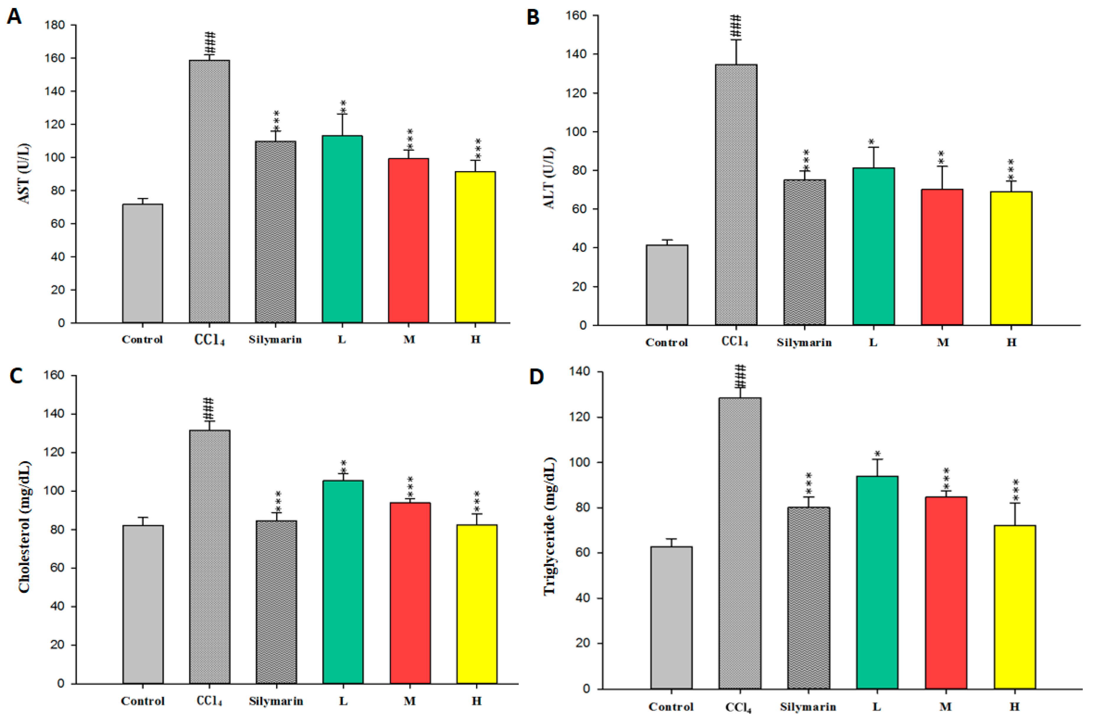
2.1. Effect of CCEtOH on CCl4-Induced Hepatotoxicity

First of all, the hepatotoxicity effect of CCl4 and the protective effect of CCEtOH were examined using the serum of CCl4-induced mice. As shown in Figure 1, the CCl4 group exhibited significant increases of serum ALT, AST, triglyceride and cholesterol. However, these increases were obviously inhibited by treatment with CCEtOH (20, 100 and 500 mg/kg) and silymarin (200 mg/kg). In addition, the inhibitions were in a dose-dependent manner. These results clearly suggested that CCEtOH possess protective properties against CCl4-induced liver injury.

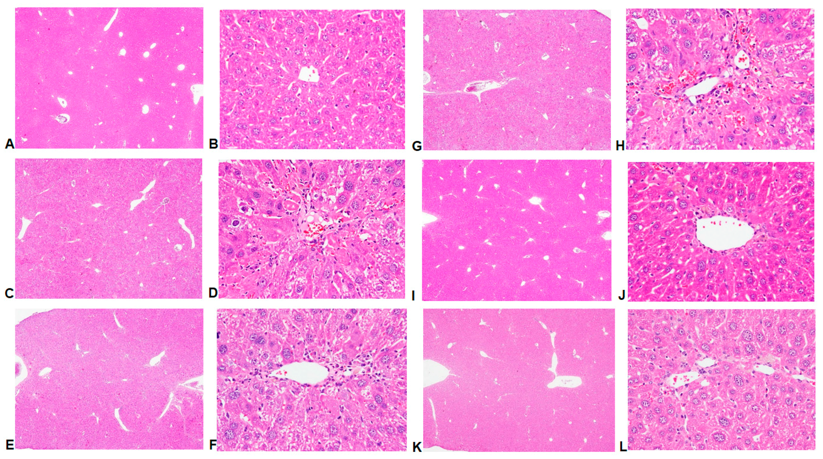
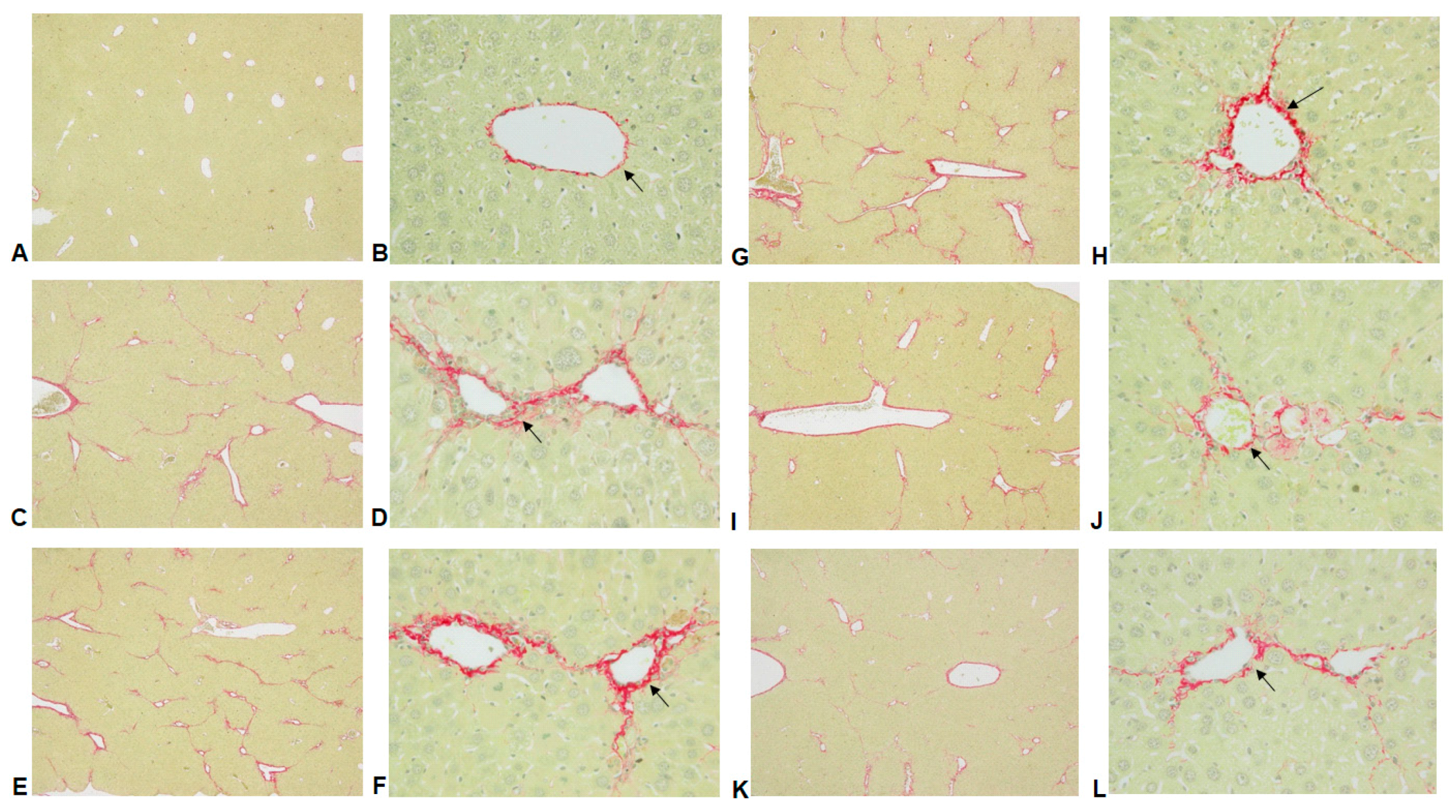
2.2. Histological Analyses

The results of hematoxylin and eosin histological analyses showed that CCl4 induced histological changes including increased hepatic cells cloudy swelling, cytoplasmic vacuolization, lymphocytes infiltration, hepatocellular and necrosis (Figure 2C,D) when compared to the control group (Figure 2A,B). The liver damages were reduced by treatment with CCEtOH (20, 100 and 500 mg/kg) (Figure 2G–L). Since CCl4 induced fibrosis, Sirius Red staining was conducted and the score of liver fibrosis were examined. The results showed that the levels of inflammation and fibrosis are significantly decreased by treatment with CCEtOH (100 and 500 mg/kg) (Figure 2 and Figure 3 and Table 1). Histological examinations showed that treatment with CCEtOH significantly prevents CCl4-induced liver injury.

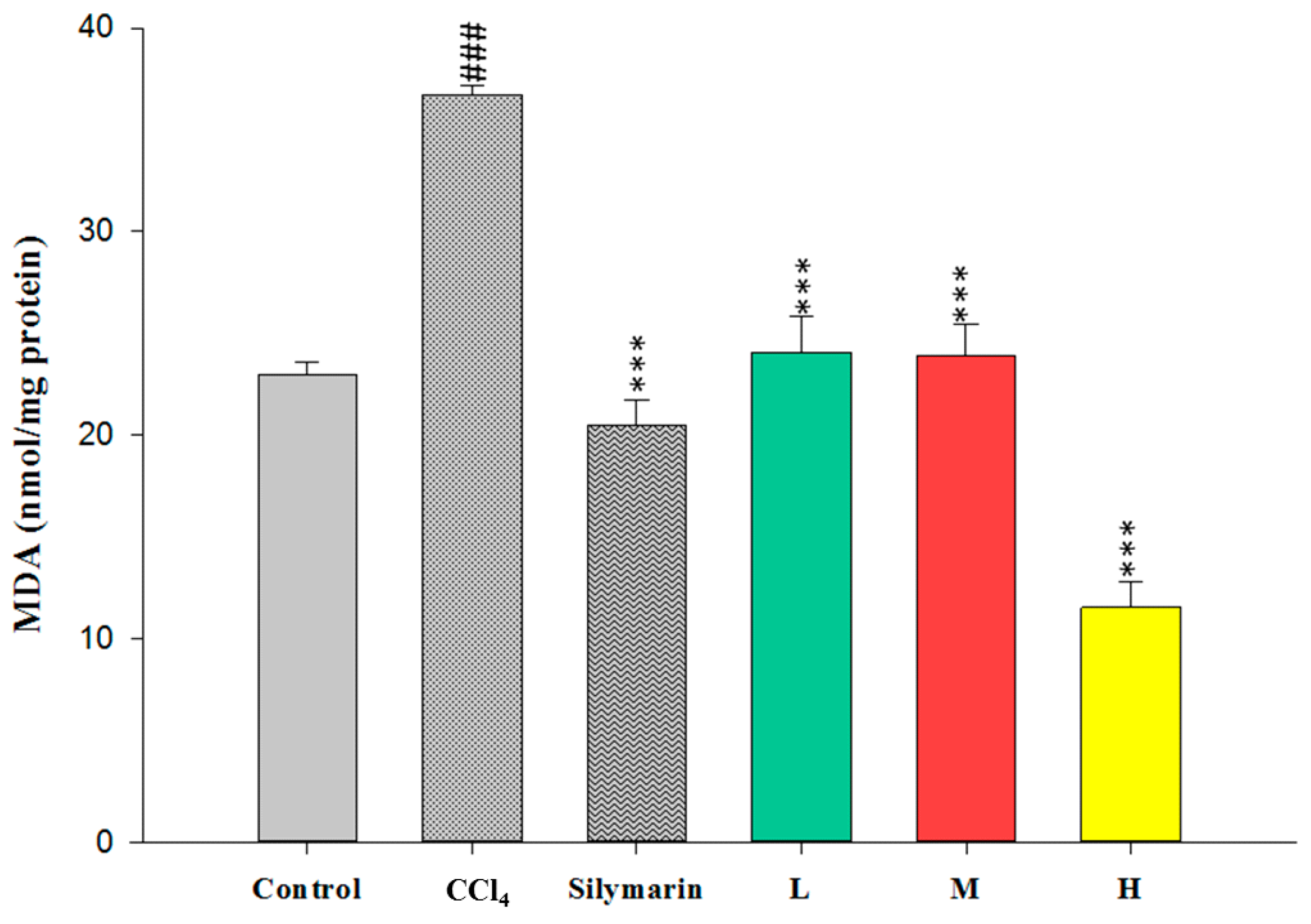
2.3. Effect of CCEtoH on MDA Level

As MDA level is usually used to elucidate the level of lipid peroxidation in liver, the effects of CCEtOH on CCl4-induced MDA production were examined. As shown in Figure 4, the level of MDA in the CCl4 group was dramatically increased (p < 0.001) compared with the control group, however, the levels of MDA were significantly reduced by treatment with CCEtOH (20, 100 and 500 mg/kg) (p < 0.001) and silymarin (200 mg/kg) (p < 0.001) compared with the CCl4 group. The results suggested that the CCl4-induced hepatic lipid peroxidation is reduced by CCEtOH.

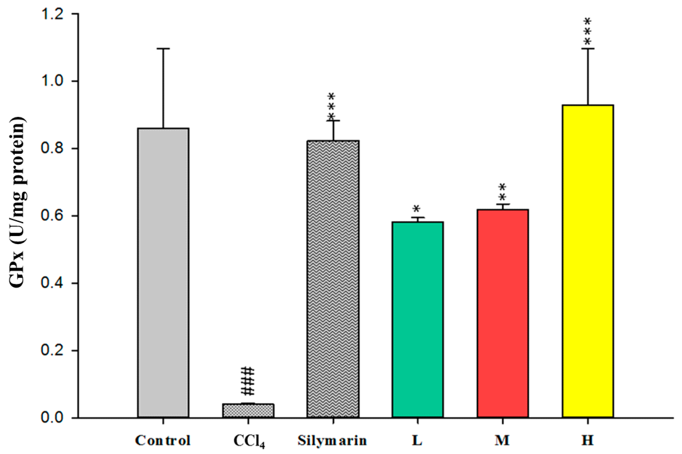
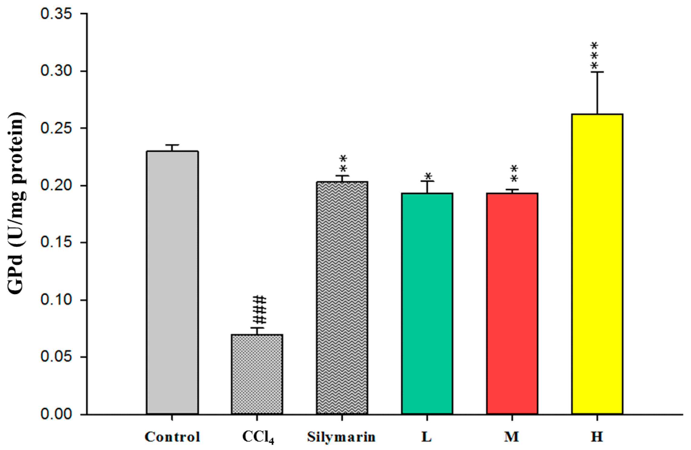
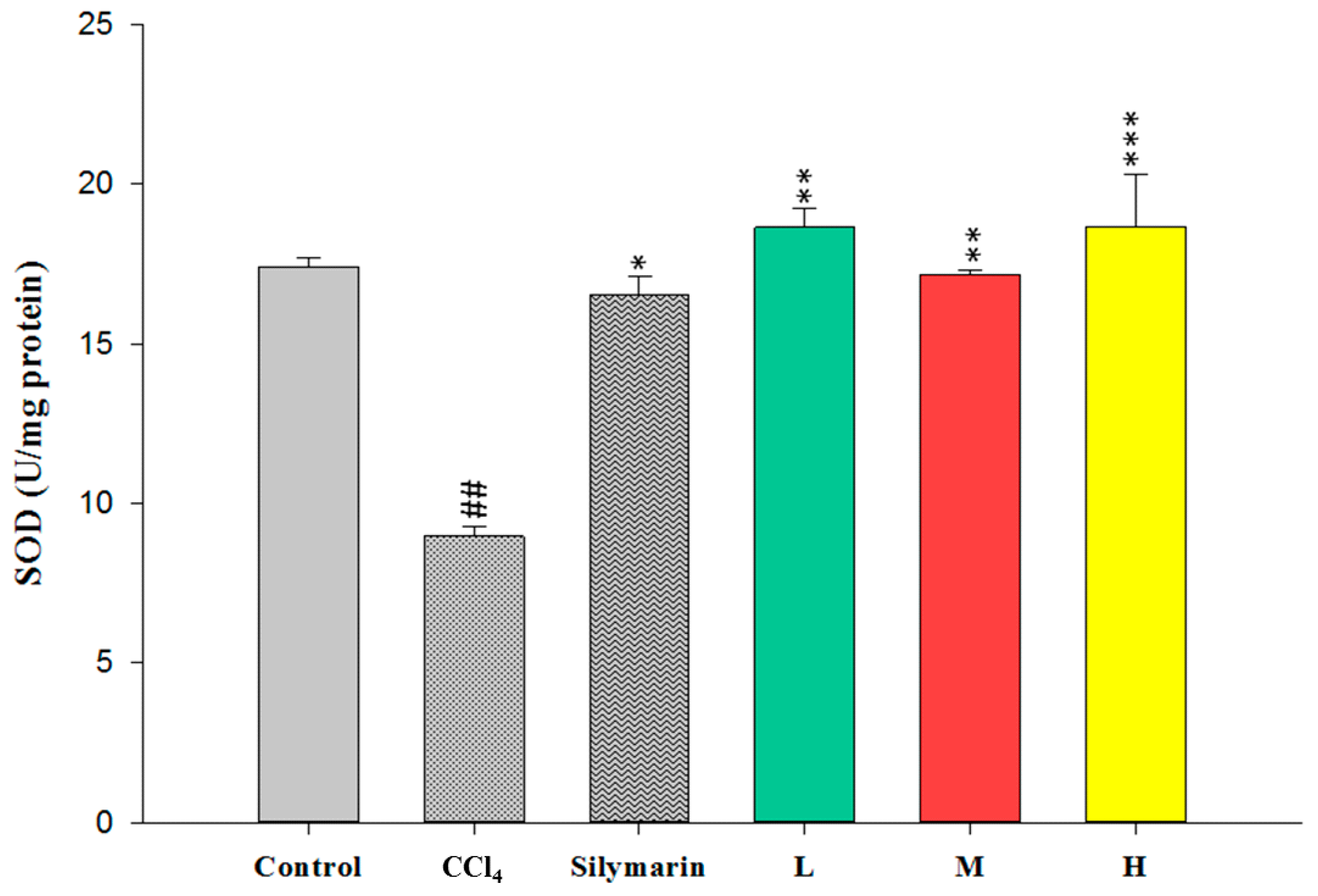
2.4. Effect of CCEtOH on Antioxidant Enzymatic Activities

To evaluate the antioxidant effects of CCEtOH, SOD, GPx and GRd were measured in the liver. The activities of these hepatic enzymes in the CCl4 group were dramatically decreased compared with the control group (Figure 5, Figure 6 and Figure 7). However, treatment with CCEtOH at the three doses and silymarin significantly increased the levels of SOD, GPx and GRd activities. The results suggested that the inhibitory effect of CCl4 on these hepatic enzymes was reversed by CCEtOH.

2.5. Phytochemical Analysis of CCEtOH

As shown in Figure S1, the HPLC chromatograms of CCEtOH and the standards showed that peaks at the retention times of 17.5 and 24.5 min were hyperoside and quercetin, respectively. Two peaks showing at the retention times of 17.5 and 24.5 min were detected in the CCEtOH chromatogram. The results revealed that hyperoside, quercetin and their glycosides are present in CCEtOH.
Cuscuta whole plant has also been used as a folk medicinal material to treat adiposity or as a substitute for the Cuscuta seeds, which have been widely used to nourish the liver and kidney in Chinese medicine. In this study, we demonstrated in the first time that the whole plant of C. campestris exhibits a hepatoprotective activity.
CCl4 has been commonly employed for the evaluation of hepatoprotective activity of different kinds of herbal extracts and drugs [8,15]. CCl4 is thought to be transformed into trichloromethyl radicals, which are hepatotoxic metabolites. These radicals are able to react with sulfhydryl groups of glutathione (GSH) and protein. In addition, they can trigger protein oxidation and lipid peroxidation, which result in hepatocellular damage [7,12,16]. In this study, CCl4 was used to induce chronic hepatic injury and our results revealed that the injury was significantly reduced by CCEtOH, clearly demonstrating that Cuscuta campestris possesses hepatoprotective activity.
Previous studies have shown that hepatic damage increases AST and ALT activities in the hepatocytes [16] and the levels of ALT, AST, triglyceride and cholesterol in serum are increased by administering CCl4 to mice [13,17,18]. In this study, serum ALT, AST, triglyceride and cholesterol levels were increased after CCl4 administration and these increases were all significantly decreased by treatment with CCEtOH at three concentrations (Figure 1). In addition, histological analyses showed that hepatic cell injury induced by CCl4 was accompanied by fibrosis and such injury was attenuated by CCEtOH (Figure 3). The quantitative histopathologic score of the fibrosis of hepatocytes showed that the fibrosis levels were significantly decreased by CCEtOH (100 and 500 mg/kg; Table 1). These results indicated that CCEtOH can prevent liver against fibrosis.
Lipid peroxidation has been shown to be an important cause of CCl4-induced liver injury [10]. Malondialdehyde (MDA) is the end product of the lipid peroxidation and thus commonly used as an indicator of the CCl4-induced liver injury [19]. In this study, the increased hepatic MDA levels induced by CCl4 were significantly decreased by treatment of CCl4 (Figure 4). Therefore, these results indicated that CCl4 can protect the liver against CCl4-induced injury through inhibiting MDA production. Superoxide dismutase (SOD), glutathione peroxidase (GPx) and glutathione reductase (GRd) are anti-oxidative enzymes which are easily inactivated by reactive oxygen species (ROS) and lipid peroxides which are caused by CCl4 [16]. In this study, the activities of SOD, GPx and GRd from the CCl4-induced injury livers were measured. The results showed that their activities were promoted by treatment with CCEtOH (Figure 5, Figure 6 and Figure 7), suggesting that CCEtOH is able to reduce ROS production by increasing hepatic anti-oxidative enzymes activities and thus prevent the development of CCl4-induced liver damage. To confirm the anti-oxidative activity of the extract used in this study, the catechin-equivalent phenolics and quercetin-equivalent flavonoid concentrations of the extract were examined and determined as 58.61 ± 0.8 and 15.032 ± 1.3 mg/g CCEtOH, respectively. In addition, 1,1-Diphenyl-2-picrylhydrazyl (DPPH) scavenging of the extract was examined. The catechin equivalent DPPH scavenging capability was determined as 25.19 ± 0.54 mg/g CCEtOH and the IC50 of CCEtOH for DPPH scavenging is approximately 1.71 mg/mL. These results support that CCEtOH containing hyperoside and quercetin has a capability to increase the anti-oxidant systems in liver. Moreover, the inhibitory effect on MDA production was also likely due to the increase in SOD, GPx and GRd activities.
Phytochemical analyses by HPLC showed that the major compounds in CCEtOH are hyperoside, quercetin and flavonoid glycosides (Figure S1). Although hyperoside and quercetin have been detected in both of seeds and whole plant of C. campestris, their amounts and the other flavonoids are different [4,20]. Hyperoside have been shown to increase the level of heme oxygenase-1, an important enzyme in antioxidant defense systems to reduce oxidative stress [21]. Quercetin has shown a high antioxidant activity by reducing the production of reactive oxygen species and nitric oxide [20,22]. The methanol extract of the seeds of Cuscuta chinensis containing hyperoside, quercetin and kaempferol have been reported to increase the activities of SOD, GPx and GRd in the liver [4]. Hyperoside and quercetin have been shown to exhibit hepatoprotective effect against CCl4-induced liver injury [23,24]. Therefore, hyperoside and quercetin can be the major active constituents in CCEtOH which contribute to the hepatoprotective effect.

3. Materials and Methods

3.1. Plant Materials and Preparation of Plant Extract

Cuscuta campestris Yunck. grown on Bidens pilosa var. radiata was collected from Miaoli County, Taiwan. They were authenticated by Ming-Kuem Lin and Wen-Huang Peng in several aspects, including the morphology of its flowers and the chemical compositions of its seeds [20]. The whole plants of C. campestris Yunck. were dried in a circulating air oven, and then ground. The powder (1.05 kg) was extracted with 75% ethanol three times. The filtrates were collected and concentrated with a rotary evaporator under reduced pressure. The concentrated extract was then lyophilized and weighted. The yield ratio of CCEtOH (91 g) was 8.7% (w/w). The extract was stored in −20 °C before the experiments.

3.2. Chemicals

Silymarin, quercetin and kampferol were purchased from Sigma-Aldrich Chemical Co. (Saint Louis, MO, USA). Carboxymethylcellulose (CMC) and carbon tetrachloride (CCl4) was purchased from Merck Co. (Munchen, Germany). CCl4 was dissolved into olive oil as a 40% (v/v) solution. All other reagents used were of analytical grades (Merck Co., Munchen, Germany).

3.3. Experimental Animals

ICR male mice (18–22 g) were purchased from BioLASCO Taiwan Co., Ltd. (Taipei, Taiwan). These mice were maintained in standard cages with a 12-h:12-h light-dark cycle, relative humidity 55% ± 5%, and 22 ± 1 °C for seven days before the experiment. Food and water ad libitum were supplied by following the NIH Guide for the Care and Use of Laboratory Animals. The experimental protocol conducted in this study has been approved by the Institutional Animal Care and Use Committee, China Medical University (104-70-N; 18 December 2014).

3.4. Experimental Design of CCl4-Induced Hepatotoxicity

Sixty experimental mice were randomly separated into 6 groups. For the control group and the CCl4 group, mice were orally administered 1% carboxymethyl cellulose (CMC). For the silymarin group, mice were orally administered silymarin (200 mg/kg in 1% CMC). For the CCEtOH groups, mice were orally administered CCEtOH (20, 100 and 500 mg/kg in 1% CMC). Theses oral administrations were conducted using a feeding tube with 100 μL/10 g Body Weight every day for 9 consecutive weeks. After one week of the administration of the silymarin and experimental drugs, CCl4 (40 μL/kg BW, 40% in olive oil) was started to inject intraperitoneally into all mice except for mice in the control group at one hour before the administration of the experimental drugs for every 3.5 days (twice a week) and 8 consecutive weeks. Thus, there were sixteen times of CCl4 treatment for the five CCl4 treated groups. The control mice received an equivalent volume of olive oil. One week after the last administration of the experimental drugs, the mice were sacrificed under anesthesia and their blood was collected for evaluation of the biochemical parameters (AST, ALT, triglyceride and cholesterol). Their liver tissues were obtained for MDA assay, histological analysis and antioxidant enzymatic activity measurements.

3.5. Serum Biochemistry

The serum was obtained as descripted previously [4]. Serum ALT, AST, triglyceride and cholesterol were measured using spectrophotometric diagnostic kits (Roche, Berlin, Germany).

3.6. Histological Analysis

Histological Analysis was performed according to the method of the previous report by staining with hematoxylin and eosin [8,11] and with Sirius Red [25], and then observed under light microscopy (Olympus, Tokyo, Japan). For quantitative scoring of the hepatic fibrosis, the values were used according to the published method [14], as the following: none: normal liver, score 0; slight: increase of collagen without formation of septa, score 1; mild: septa do not connect with each other and incomplete septa formation from portal tract to central vein, score 2; moderate: septa interconnecting completely but thin (incomplete cirrhosis), score 3; and remarkable: with thick septa (complete cirrhosis), score 4.

3.7. MDA Level as Well as Antioxidant Enzymatic Activity Measurement

MDA level was determined as descripted previously using the thiobarbituric acid reacting substance method [26]. SOD, GPx and GRd enzymatic activities were determined according to the published methods [27,28,29]. MDA, SOD, GPx and GRd assay kits were purchased from Randox Laboratory Ltd. (Antrim, UK).

3.8. Statistical Analyses

All data were shown as mean ± SEM. SPSS statistics software program was used to do the statistical data analyses. One-way ANOVA followed by Scheffe’s multiple range test was used to perform the statistical analyses. For the histological analyses, non-parametric Kruskal–Wallis test followed by the Mann–Whitney U-test was used to carry out the statistical analyses. The criterion for statistical significance was p < 0.05.

3.9. Phytochemical Analysis of CCEtOH by HPLC

The HPLC profile of CCEtOH was determined and compared with the standard (hyperoside and quercetin), which was conducted as descripted previously [20]. Quantification was performed by comparing the sample peak with the corresponding standard compound.

4. Conclusions

The present study clearly elucidated that CCEtOH exhibited a hepatoprotective activity against CCl4-induced chronic liver injury in mice. The underlying mechanisms were likely the decreasing in MDA level through enhancing the activities of hepatic anti-oxidative enzymes such as GPx, GRd and SOD, and thereby the significant decrease of serum ALT, AST, triglyceride and cholesterol. In addition, fibrosis of liver was significantly reduced by CCEtOH. Therefore, C. campestris can be developed into pharmacological agents to prevent some liver disorders.

Supplementary Materials

Supplementary materials can be found at www.mdpi.com/1422-0067/17/12/2056/s1.

Acknowledgments

This work is supported by grants from National Science Council (MOST 105-2320-B-039-012-MY3 and MOHW103-CMAP-M-114-122408) and China Medical University (CMU104-TC-02). We would like to express our thanks to Tun-Jen Cheng for his help with the anti-oxidation experiment and Jiunn-Wang Liao from National Chung Hsing University for his help with the histological analyses.

Author Contributions

Conceived and designed the experiments: Wen-Huang Peng, and Ming-Kuem Lin. Performed the experiments: Yi-Wen Chen and Jen-Chieh Tsai. Analyzed the data: Meng-Shiou Lee, Wen-Te Chang, and Ming-Kuem Lin. Contributed reagents/materials/analysis tools: Ying-Chih Lin. Wrote the paper: Wen-Huang Peng and Ming-Kuem Lin.

Conflicts of Interest

The authors declare no conflict of interest.

References

  1. Donnapee, S.; Li, J.; Yang, X.; Ge, A.H.; Donkor, P.O.; Gao, X.M.; Chang, Y.X. Cuscuta chinensis Lam.: A systematic review on ethnopharmacology, phytochemistry and pharmacology of an important traditional herbal medicine. J. Ethnopharmacol. 2014, 157, 292–308. [Google Scholar] [CrossRef] [PubMed]
  2. Yang, J.; Wang, Y.; Bao, Y.; Guo, J. The total flavones from Semen cuscutae reverse the reduction of testosterone level and the expression of androgen receptor gene in kidney-yang deficient mice. J. Ethnopharmacol. 2008, 119, 166–171. [Google Scholar] [CrossRef] [PubMed]
  3. Yen, F.L.; Wu, T.H.; Lin, L.T.; Lin, C.C. Hepatoprotective and antioxidant effects of Cuscuta chinensis against acetaminophen-induced hepatotoxicity in rats. J. Ethnopharmacol. 2007, 111, 123–128. [Google Scholar] [CrossRef] [PubMed]
  4. Liao, J.C.; Chang, W.T.; Lee, M.S.; Chiu, Y.J.; Chao, W.K.; Lin, Y.C.; Lin, M.K.; Peng, W.H. Antinociceptive and anti-inflammatory activities of Cuscuta chinensis seeds in mice. Am. J. Chin. Med. 2014, 42, 223–242. [Google Scholar] [CrossRef] [PubMed]
  5. Bao, X.; Wang, Z.; Fang, J.; Li, X. Structural features of an immunostimulating and antioxidant acidic polysaccharide from the seeds of Cuscuta chinensis. Planta Med. 2002, 68, 237–243. [Google Scholar] [CrossRef] [PubMed]
  6. Lee, S.H.; Heo, S.I.; Li, L.; Lee, M.J.; Wang, M.H. Antioxidant and hepatoprotective activities of Cirsium setidens NAKAI against CCl4-induced liver damage. Am. J. Chin. Med. 2008, 36, 107–114. [Google Scholar] [CrossRef] [PubMed]
  7. Wang, T.; Sun, N.L.; Zhang, W.D.; Li, H.L.; Lu, G.C.; Yuan, B.J.; Jiang, H.; She, J.H.; Zhang, C. Protective effect of dehydrocavidine on carbon tetrachloride-induced acute hepatotoxicity in rats. J. Ethnopharmacol. 2008, 117, 300–308. [Google Scholar] [CrossRef] [PubMed]
  8. Tsai, J.C.; Peng, W.H.; Chiu, T.H.; Huang, S.C.; Huang, T.H.; Lai, S.C.; Lai, Z.R.; Lee, C.Y. Hepatoprotective effect of Scoparia dulcis on carbon tetrachloride induced acute liver injury in mice. Am. J. Chin. Med. 2010, 38, 761–775. [Google Scholar] [CrossRef] [PubMed]
  9. Brattin, W.J.; Glende, E.A., Jr.; Recknagel, R.O. Pathological mechanisms in carbon tetrachloride hepatotoxicity. J. Free Radic. Biol. Med. 1985, 1, 27–38. [Google Scholar] [CrossRef]
  10. Basu, S. Carbon tetrachloride-induced lipid peroxidation: Eicosanoid formation and their regulation by antioxidant nutrients. Toxicology 2003, 189, 113–127. [Google Scholar] [CrossRef]
  11. Lee, C.Y.; Peng, W.H.; Cheng, H.Y.; Chen, F.N.; Lai, M.T.; Chiu, T.H. Hepatoprotective effect of Phyllanthus in Taiwan on acute liver damage induced by carbon tetrachloride. Am. J. Chin. Med. 2006, 34, 471–482. [Google Scholar] [CrossRef] [PubMed]
  12. Weber, L.W.; Boll, M.; Stampfl, A. Hepatotoxicity and mechanism of action of haloalkanes: Carbon tetrachloride as a toxicological model. Crit. Rev. Toxicol. 2003, 33, 105–136. [Google Scholar] [CrossRef] [PubMed]
  13. Adewole, S.O.; Salako, A.A.; Doherty, O.W.; Naicker, T. Effect of melatonin on carbon tetrachloride-induced kidney injury in Wistar rats. Afr. J. Biomed. Res. 2007, 10, 153–164. [Google Scholar] [CrossRef]
  14. Ruwart, M.J.; Wilkinson, K.F.; Rush, B.D.; Vidmar, T.J.; Peters, K.M.; Henley, K.S.; Appelman, H.D.; Kim, K.Y.; Schuppan, D.; Hahn, E.G. The integrated value of serum procollagen III peptide over time predicts hepatic hydroxyproline content and stainable collagen in a model of dietary cirrhosis in the rat. Hepatology 1989, 10, 801–806. [Google Scholar] [CrossRef] [PubMed]
  15. Manibusan, M.K.; Odin, M.; Eastmond, D.A. Postulated carbon tetrachloride mode of action: A review. J. Environ. Sci. Health C Environ. Carcinog. Ecotoxicol. Rev. 2007, 25, 185–209. [Google Scholar] [CrossRef] [PubMed]
  16. Recknagel, R.O.; Glende, E.A., Jr.; Dolak, J.A.; Waller, R.L. Mechanisms of carbon tetrachloride toxicity. Pharmacol. Ther. 1989, 43, 139–154. [Google Scholar] [CrossRef]
  17. Lee, C.H.; Park, S.W.; Kim, Y.S.; Kang, S.S.; Kim, J.A.; Lee, S.H.; Lee, S.M. Protective mechanism of glycyrrhizin on acute liver injury induced by carbon tetrachloride in mice. Biol. Pharm. Bull. 2007, 30, 1898–1904. [Google Scholar] [CrossRef] [PubMed]
  18. Tseng, S.H.; Chien, T.Y.; Tzeng, C.F.; Lin, Y.H.; Wu, C.H.; Wang, C.C. Prevention of hepatic oxidative injury by Xiao-Chen-Chi-Tang in mice. J. Ethnopharmacol. 2007, 111, 232–239. [Google Scholar] [CrossRef] [PubMed]
  19. Hung, M.Y.; Fu, T.Y.; Shih, P.H.; Lee, C.P.; Yen, G.C. Du-Zhong (Eucommia ulmoides Oliv.) leaves inhibit CCl4-induced hepatic damage in rats. Food Chem. Toxicol. 2006, 44, 1424–1431. [Google Scholar] [CrossRef] [PubMed]
  20. Lee, M.S.; Chen, C.J.; Wan, L.; Koizumi, A.; Chang, W.T.; Yang, M.J.; Lin, W.H.; Tsai, F.J.; Lin, M.K. Quercetin is increased in heat-processed Cuscuta campestris seeds, which enhances the seed’s anti-inflammatory and anti-cancer activities. Process Biochem. 2011, 46, 2248–2254. [Google Scholar] [CrossRef]
  21. Park, J.Y.; Han, X.; Piao, M.J.; Oh, M.C.; Fernando, P.M.; Kang, K.A.; Ryu, Y.S.; Jung, U.; Kim, I.G.; Hyun, J.W. Hyperoside induces endogenous antioxidant system to alleviate oxidative stress. J. Cancer Prev. 2016, 21, 41–47. [Google Scholar] [CrossRef] [PubMed]
  22. Zhang, M.; Swarts, S.G.; Yin, L.; Liu, C.; Tian, Y.; Cao, Y.; Swarts, M.; Yang, S.; Zhang, S.B.; Zhang, K.; et al. Antioxidant properties of quercetin. Adv. Exp. Med. Biol. 2011, 701, 283–289. [Google Scholar] [PubMed]
  23. Choi, J.H.; Kim, D.W.; Yun, N.; Choi, J.S.; Islam, M.N.; Kim, Y.S.; Lee, S.M. Protective effects of hyperoside against carbon tetrachloride-induced liver damage in mice. J. Nat. Prod. 2011, 74, 1055–1060. [Google Scholar] [CrossRef] [PubMed]
  24. Domitrović, R.; Jakovac, H.; Vasiljev Marchesi, V.; Vladimir-Knežević, S.; Cvijanović, O.; Tadić, Z.; Romić, Z.; Rahelić, D. Differential hepatoprotective mechanisms of rutin and quercetin in CCl4-intoxicated BALB/cN mice. Acta Pharmacol. Sin. 2012, 33, 1260–1270. [Google Scholar] [CrossRef] [PubMed]
  25. Junqueira, L.C.; Bignolas, G.; Brentani, R.R. Picrosirius staining plus polarization microscopy, a specific method for collagen detection in tissue sections. Histochem. J. 1979, 11, 447–455. [Google Scholar] [CrossRef] [PubMed]
  26. Draper, H.H.; Hadley, M. Malondialdehyde determination as index of lipid peroxidation. Methods Enzymol. 1990, 186, 421–431. [Google Scholar] [PubMed]
  27. Misra, H.P.; Fridovich, I. The role of superoxide anion in the autooxidation of epinephrine and a simple assay for superoxide dismutase. J. Biol. Chem. 1972, 247, 3170–3175. [Google Scholar] [PubMed]
  28. Carlberg, I.; Mannervik, B. Glutathione reductase. Methods Enzymol. 1984, 113, 484–490. [Google Scholar]
  29. Flohe, L.; Gunzler, W.A. Assays of glutathione peroxidase. Methods Enzymol. 1984, 105, 114–121. [Google Scholar] [PubMed]
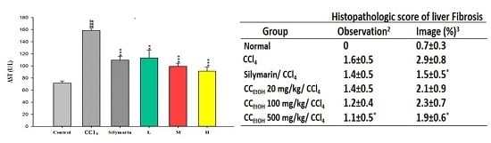
Figure 1. The effects of silymarin and the ethanol extracts of Cuscuta campestris whole plant at low (L), middle (M) and high (H) concentrations (20, 100 and 500 mg/kg, respectively) on serum: aspartate aminotransferase (AST) (A); and alanine aminotransferase (ALT) (B) activities; and cholesterol (C); and triglyceride (D) levels in mice treated with CCl4. Values are mean ± SEM (n = 10). # indicates significant difference from the control group (### p < 0.001). * indicates significant difference from the CCl4 group (* p < 0.05, ** p < 0.01 and *** p < 0.001).
Ijms 17 02056 g001
Figure 2. Hepatic histological analyses of the effects of silymarin and the ethanol extracts of Cuscuta campestris whole plant (CCEtOH) on CCl4-induced liver damage in mice using H&E staining (40× (A,C,E,G,I,K) and 200× (B,D,F,H,J,L) magnification): (A,B) control group; (C,D) animals treated with CCl4; (E,F) animals treated with silymarin (200 mg/kg) and CCl4; and (GL) animals treated with CCEtOH (20, 100 and 500 mg/kg) and CCl4, respectively.
Ijms 17 02056 g002
Figure 3. Hepatic histological analyses of the effects of silymarin and the ethanol extracts of Cuscuta campestris whole plant (CCEtOH) on CCl4-induced liver damage in mice using Sirius Red staining (40× (A,C,E,G,I,K) and 200× (B,D,F,H,J) magnification): (A,B) control group; (C,D) animals treated with CCl4; (E,F) animals treated with silymarin (200 mg/kg) and CCl4; and (GL) animals treated with CCEtOH (20, 100 and 500 mg/kg) and CCl4, respectively. Arrows indicate the fibrosis.
Ijms 17 02056 g003
Figure 4. The effects of silymarin and the ethanol extracts of Cuscuta campestris whole plant on malondialdehyde (MDA) content in mice treated with CCl4. Values are mean ± SEM (n = 10). # indicates significant difference from the control group (### p < 0.001). * indicates significant difference from the CCl4 group (*** p < 0.001).
Ijms 17 02056 g004
Figure 5. The effects of silymarin and the ethanol extracts of Cuscuta campestris whole plant on superoxide dismutase (SOD) activity in mice treated with CCl4. Values are mean ± SEM (n = 10). # indicates significant difference from the control group (## p < 0.01). * indicates significant difference from the CCl4 group (* p < 0.05, ** p < 0.01 and *** p < 0.001).
Ijms 17 02056 g005
Figure 6. The effects of silymarin and the ethanol extracts of Cuscuta campestris whole plant on glutathione peroxidase (GPx) activity in mice treated with CCl4. Values are mean ± SEM (n = 10). # indicates significant difference from the control group (### p < 0.001). * indicates significant difference from the CCl4 group (* p < 0.05, ** p < 0.01 and *** p < 0.001).
Ijms 17 02056 g006
Figure 7. The effects of silymarin and the ethanol extracts of Cuscuta campestris whole plant on glutathione reductase (GRd) activity in mice treated with CCl4. Values are mean ± SEM (n = 10). # indicates significant difference from the control group (### p < 0.001). * indicates significant difference from the CCl4 group (* p < 0.05, ** p < 0.01 and *** p < 0.001).
Ijms 17 02056 g007
Table 1. Quantitative the protective effects of silymarin and CCEtOH on CCl4-induced hepatic fibrosis based on histological analyses using Sirius Red staining 1.
GroupHistopathologic Score of Liver Fibrosis
Observation 2Image (%) 3
Normal00.7 ± 0.3
CCl41.6 ± 0.52.9 ± 0.8
Silymarin/CCl41.4 ± 0.51.5 ± 0.5 *
CCEtOH 20 mg/kg/CCl41.4 ± 0.52.1 ± 0.9
CCEtOH 100 mg/kg/CCl41.2 ± 0.42.3 ± 0.7
CCEtOH 500 mg/kg/CCl41.1 ± 0.5 *1.9 ± 0.6 *
1 Hepatic fibrosis was scored 0–4 according to the method of Ruwart et al. [14] as mentioned in the Materials and Methods; 2 The scores were obtained by the following calculation: the sum of the number per grade of affected mice/the total number of examined mice (n = 9–10); 3 The final Sirius Red positive area (%) was calculated by Image-Plus and was divided the sum of the number per SR positive area (%) of affected mice by the total number of examined mice (n = 9–10). * Statistically significant difference between CCl4 group and drug-treated groups at p < 0.05.

