Review

Differential Impacts of Alternative Splicing Networks on Apoptosis

1
School of Medical Laboratory Science and Biotechnology, College of Medical Science and Technology, Taipei Medical University, Taipei 11031, Taiwan
2
Department of Laboratory Medicine, Taipei Medical University Hospital, Taipei 11031, Taiwan
3
School of Chinese Medicine, China Medical University, Taichung 404, Taiwan
*
Author to whom correspondence should be addressed.
Int. J. Mol. Sci. 2016, 17(12), 2097; https://doi.org/10.3390/ijms17122097
Submission received: 14 October 2016 / Revised: 26 November 2016 / Accepted: 2 December 2016 / Published: 14 December 2016
(This article belongs to the Collection Programmed Cell Death and Apoptosis)

Abstract

:
Apoptosis functions as a common mechanism to eliminate unnecessary or damaged cells during cell renewal and tissue development in multicellular organisms. More than 200 proteins constitute complex networks involved in apoptotic regulation. Imbalanced expressions of apoptosis-related factors frequently lead to malignant diseases. The biological functions of several apoptotic factors are manipulated through alternative splicing mechanisms which expand gene diversity by generating discrete variants from one messenger RNA precursor. It is widely observed that alternatively-spliced variants encoded from apoptosis-related genes exhibit differential effects on apoptotic regulation. Alternative splicing events are meticulously regulated by the interplay between trans-splicing factors and cis-responsive elements surrounding the regulated exons. The major focus of this review is to highlight recent studies that illustrate the influences of alternative splicing networks on apoptotic regulation which participates in diverse cellular processes and diseases.

1. Introduction

Apoptosis is defined as a death modality of damaged or unnecessary cells [1,2], which is executed by the caspase pathway [3]. The presence of proteolytic caspases triggers nuclear fragmentation, chromatin condensation, and cell rounding, and apoptotic cells are taken apart in membrane-bound vesicles [4]. Apoptotic bodies are rapidly phagocytosed by resident macrophages or neutrophils [5]. Impaired clearance of apoptotic cells leads to the exposure of intracellular organelles which frequently sensitize the innate immune system [6]. In eukaryotes, apoptosis participates in diverse processes, including immune responses [7], embryonic development [8], and maintenance of tissue homeostasis [9]. Depending on the environmental stress or cell types, apoptosis is differentially executed through extrinsic and intrinsic pathways [10,11]. The intrinsic pathway is activated in the presence of numerous intracellular stimuli, including DNA damage, endoplasmic reticular stress, oxidative stress, and breakage of mitochondrial membranes [12,13]. Activation of multiple death receptors or the withdrawal of cytokines induces extrinsic pathway-mediated apoptosis [14,15]. Both the intrinsic and extrinsic pathways lead to the release of cytochrome C and other apoptosis-inducing factors, which subsequently activate downstream caspases [16].
Alternative splicing constitutes a posttranscriptional mechanism to expand the proteomic diversity of a single gene in eukaryotes [17]. Accurate alternative splicing profiles and regulation determine cellular fates and functions [18]. It was documented that over 90% of human genes produce more than one transcript by undergoing alternative splicing mechanisms [19]. Alternative splicing profiles are meticulously regulated by the interplay among spliceosomes, splice sites, cis-regulatory elements, and corresponding splicing regulators, the expression profiles of which occur in a spatial-temporal manner [20]. The development of high-throughput approaches, including proteome and transcriptome analyses, has been very helpful in understanding alternative splicing mechanisms involved in cell homeostasis and pathological causes [21].

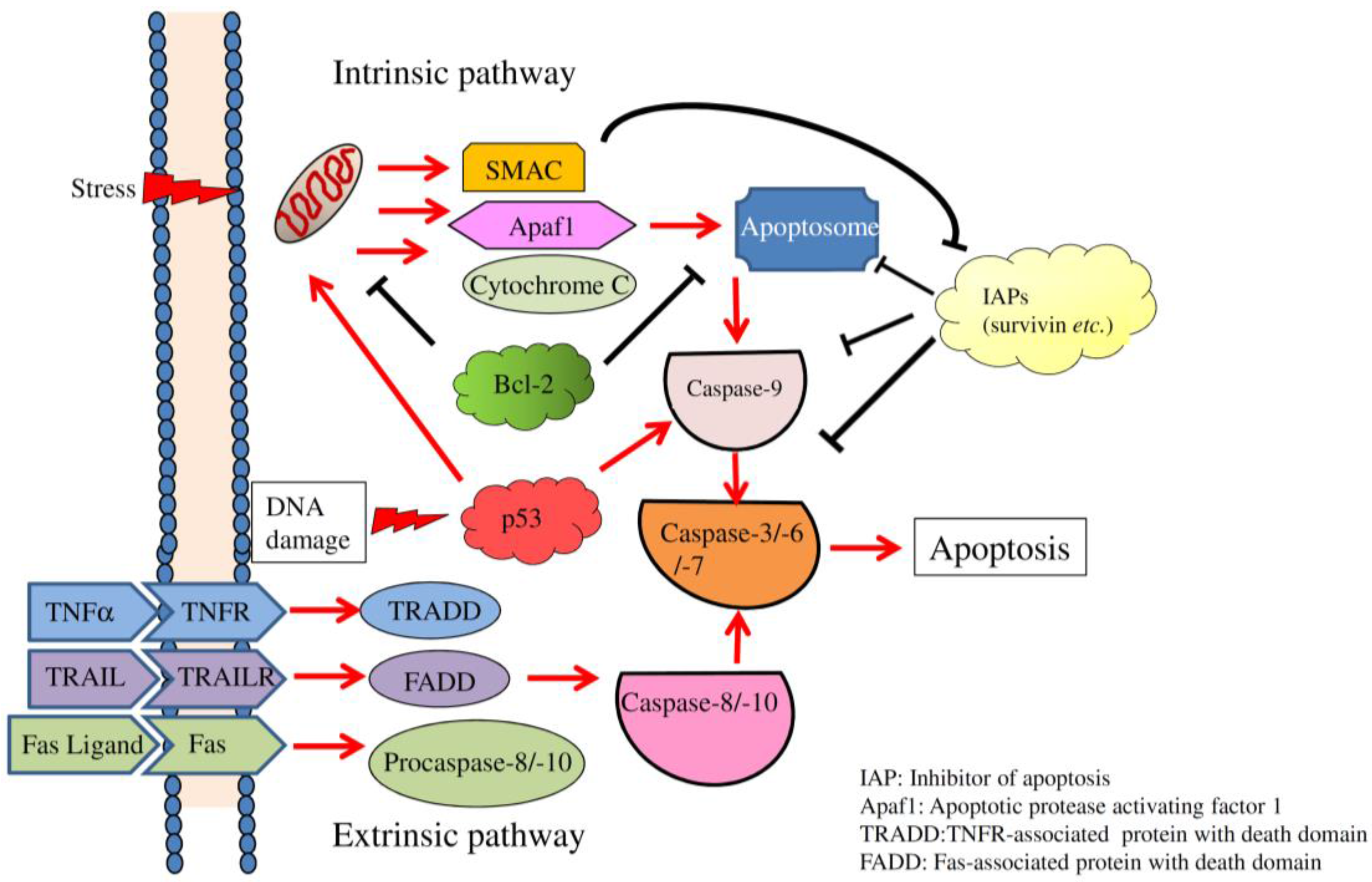
2. Overview of Apoptosis

Apoptosis, necroptosis, and autophagy are classified as programmed cell death, an integral process to maintain a homeostatic circumstance in organisms [22]. The extrinsic and intrinsic pathways are two well-studied mechanisms that contribute to the execution of apoptosis (Figure 1; [10,11,12,13,14,15]). The binding between death ligands, including Fas ligand (Fas L), tumor necrosis factor (TNF)-related apoptosis-inducing ligand (TRAIL), and TNF-α, and corresponding death receptors result in the assembly of death-inducing signaling complexes which initiate the extrinsic pathway by activating caspase-8 [23]. DNA breakage, endoplasmic reticular stress, and growth factor withdrawal are functional signals for activating the release of intrinsic factors, such as cytochrome C and Similar to Mothers Against Decapentaplegic (SMAD), from the inner membrane of mitochondria to trigger the intrinsic pathway [24,25]. The B-cell lymphoma (Bcl)-2 family is composed of pro-apoptotic and anti-apoptotic factors, which manipulate the activity of intrinsic pathway [26]. Several Bcl-2 family genes encode alternatively spliced variants which exhibit pro- or anti-apoptotic activity [27]. The intrinsic pathway-specific apoptosome is assembled to participate in recruiting and processing procaspase-9 [28]. Eventually, processed caspase-9 activates the downstream caspases-3, -6, and -7, which leads to cell apoptosis [29].
Evasion of apoptosis constitutes one mechanism mediating the acquired resistance of cancer cells during treatment with chemotherapeutic agents [30]. Much higher doses of agents are required to achieve efficacy due to the inherent resistance to apoptosis, which induces off-target adverse effects. Therefore, targeting apoptosis toward cancer cells by inducing the extrinsic pathway through TRAIL signaling or eliminating the anti-apoptotic activities of inhibitors of apoptosis, such as Bcl-2, is considered a potential strategy [31,32]. More death receptor-ligand complexes, including TNF receptor (TNFR)-TNF-α, FAS-Fas ligand, TRAIL receptor (TRAILR)1/2 (also referred to DR4/5)-TRAIL were identified as functioning as apoptosis-inducing signal molecules [33,34]. Upon interactions between death receptors and the corresponding ligands, oligomerization and conformation changes of the same receptors expose the cytoplasmic death domain (DD) which is involved in the interaction with other DD-containing proteins [35], subsequently mediating the processing and activation of procaspases [36]. Two alternative splice variants of TRAIL, TRAIL-β, and TRAIL-γ, were identified in neoplastic cells [37]. The lack of exon 3 in TRAIL-β and of exons 2 and 3 in TRAIL-γ, which encode the truncated extracellular binding domain, results in loss of their pro-apoptotic activity [38]. In addition, Bcl-2 family members exhibit both pro- and anti-apoptotic activities on modulating the intrinsic apoptosis pathway [39]. The balance between Bcl-2 family members is meticulously determined by cell survival or apoptosis [40]. Moreover, impaired splicing profiles of Bcl-2 family members result in differential or opposite effects in regulating cell viability [27]. A growing body of studies has demonstrated that predominant expressions of anti-apoptotic members or isoforms participate in the evasion of cancer cells from programmed cell death.

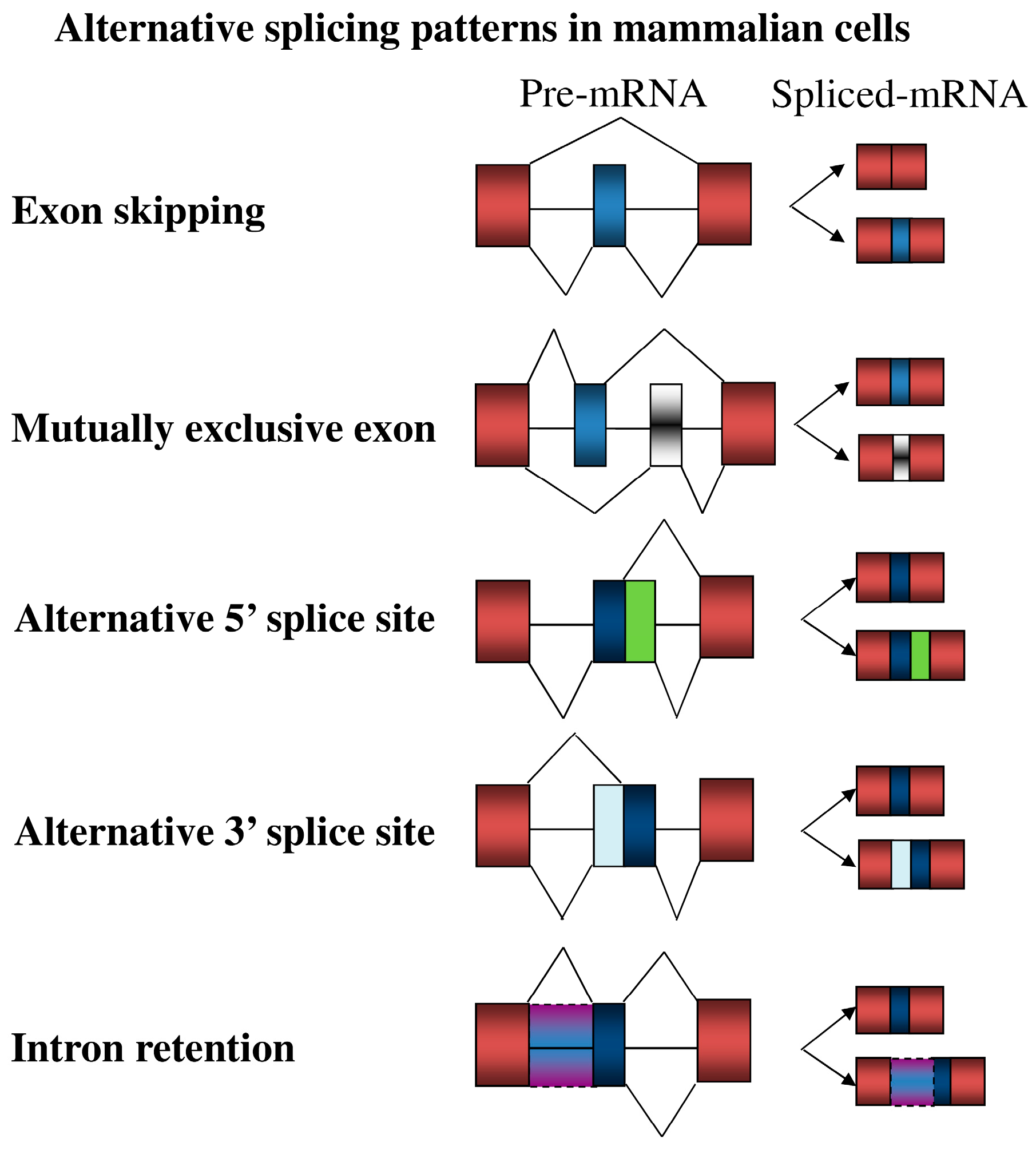
3. Overview of Alternative Splicing

Recognition of 5′ and 3′ splice sites is the critical step in the definition of intron in the mammalian genome. The spliceosome is composed of five small nuclear (sn)RNAs and more than 150 associated proteins, which contributes to the splicing of defined introns [41]. However, the utilization of 5′ or 3′ splice sites is widely strengthened or weakened by the interplay between trans-splicing factors and the corresponding cis-elements within regulated exon and surrounding intron, leading to the alternative splicing regulation in mammalian cells (Figure 2, [42]). The regulatory elements are classified into exonic and intronic splicing enhancers (ESEs and ISEs) or silencers (ESSs and ISSs) according to their impact on the alternatively-spliced exons [43]. Interplay between splicing factors and the binding elements in turn manipulate utilization of 5′ or 3′ splice sites by facilitating or interfering with the assembly of spliceosomes. Heterogeneous nuclear ribonucleoproteins (hnRNPs) and serine/arginine-rich (SR) proteins are two major groups of splicing factors [44]. HnRNPs generally abolish the exon inclusion by binding to the exonic pyrimidine-rich element, whereas the interplay between hnRNPs and intronic binding elements exhibited differential effect on the alternatively-spliced exons [45], (Figure 3). Many studies demonstrated that SR proteins enhance utilization of most alternatively spliced exons by binding to the exonic purine-rich element [45]. However, the interplay between SR proteins and the adjacent exons subsequently mediated the exclusion of internal exons [45], (Figure 3). Collectively, the potential effect of splicing factor on individual splicing event is modulated by sequence or position context that should be further defined. Moreover, splicing profiles are spatial-temporally reprogrammed by the relative expressions of numerous splicing factors in the nucleus [46].

4. Impacts of Alternative Splicing Events on Apoptosis

4.1. Apoptosis-Related Alternative Splicing Events
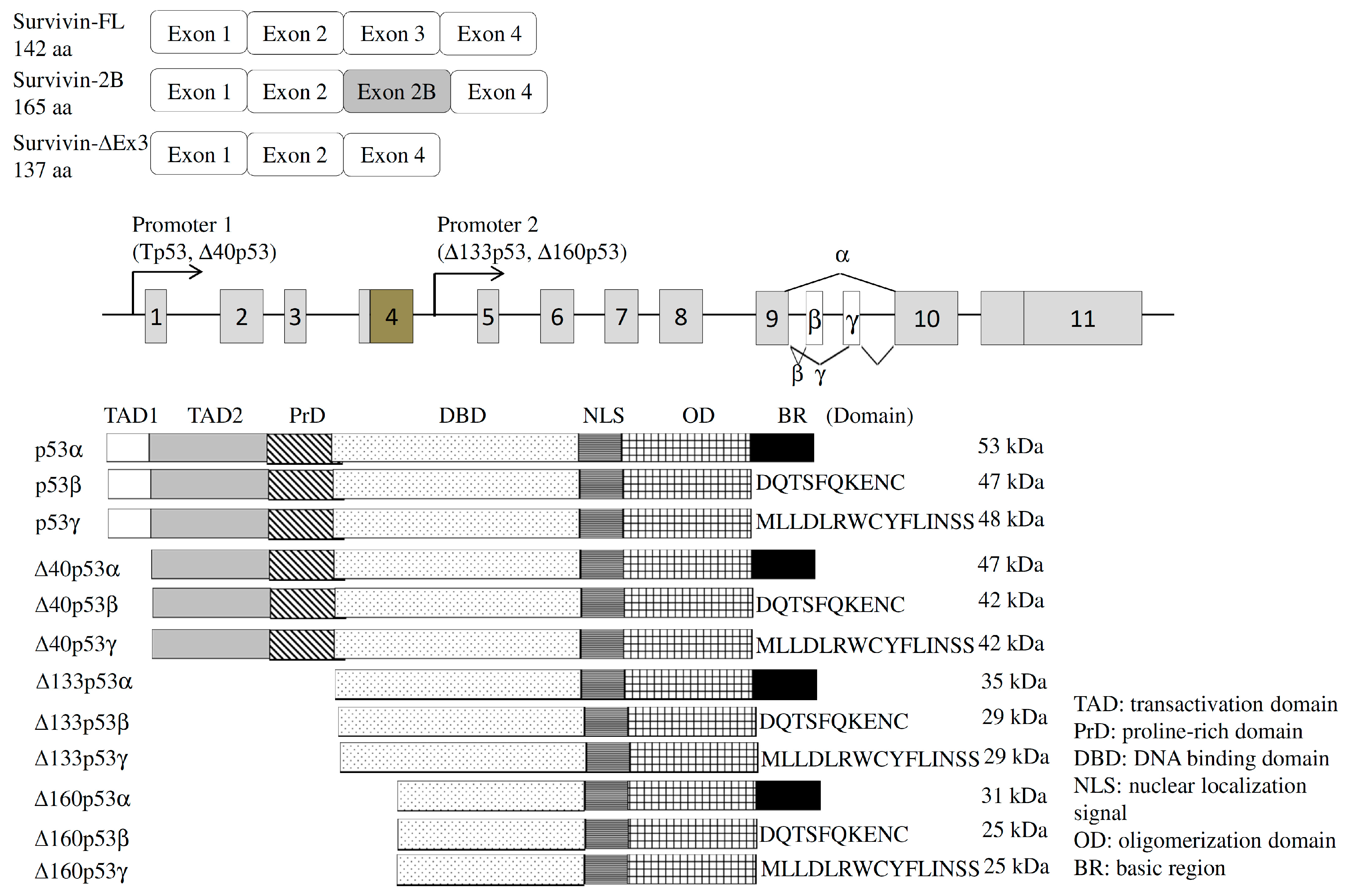
4.1.1. Survivin

Temporal expression of the survivin protein is highly correlated with the transition from the G2/M to the G1 phase [47]. Several reports indicated that ubiquitous expression of the survivin protein in most malignant cells, but not well-differentiated cells, functions as an inhibitor of the apoptosis protein (IAP, [48]). The survivin gene produces three alternatively spliced variants, including full length, delta Ex3 (∆Ex3), and 2B transcripts (Figure 4, [49]), which exhibit differential effects on the apoptotic process [50]. Several studies demonstrated the cytoprotective effects of survivin ΔEx3 variants inhibiting the apoptotic process [51]. In contrast, the presence of the survivin 2B isoforms was reported to confer pro-apoptotic properties on cancer cells [51]. Accordingly, relatively high expressions of full length and survivin ∆Ex3 variants are especially correlated with the active progression or poor prognoses of breast, gastric, thyroid, and pituitary cancers [52]. The molecular mechanism involved in programming of the survivin gene is largely unclear.

4.1.2. Estrogen Receptor (ER)

The imbalanced stimulation of sexual hormones, including estrogen, frequently leads to breast cancer which is the most common malignancy in females worldwide [53]. ERα and ERβ proteins transmit the action of estrogens into target cells [54,55]. Although generated by individual genes, ERα and ERβ have almost 100% amino acid homology in their DNA-binding domain and about 60% amino acid homology in their protein-interacting domains [56]. Interestingly, in vivo and in vitro experiments demonstrated differential or opposite effects of ERα and ERβ on biological features of breast cancer cells [57]. ERα-regulated gene expressions facilitate the growth and survival of breast cancer cells in response to estrogens [58], whereas the impact of ERβ on breast cancer cells is controversial [59]. hnRNP G and Tra2-β1 proteins were recently demonstrated to modulate the selection of ERα exon 7 [60]. Overexpressing Tra2-β1 induced relative levels of ERα+ex7 transcripts, whereas overexpressing hnRNP G exhibited an antagonistic effect on inducing ERα−ex7 levels [60]. Statistical analyses of several cohort studies suggested positive correlations between ERα+7 variants and tumor grades in breast cancer [61]. In addition, several orphan receptors that share structural similarities to ERs were characterized as estrogen-related receptors (ERRs). Two alternatively-spliced variants, short-form ERRβ (ERRβsf) and ERRβ2, were recently identified as being involved in cell cycle regulation and survival [62]. Silencing of ERRβ2-suppressed p53 signaling-mediated apoptosis, whereas overexpression of ERRβsf enhanced p21 activity which facilitates cell proliferation [63]. It is widely noted that splice variants often exhibit antagonistic effects. Taken together, these results may bring new insights into the clinical treatment of breast cancer.

4.1.3. Transient Receptor Potential Melastatin (TRPM)

TRPM family members share similar structural signatures, including transmembrane domains and the cytosolic terminus [64]. TRPM members assemble hetero-oligomers to function as a Ca2+-permeable cation channel that is related to the progression of malignancies, such as prostate cancer [64]. Among these members, TRPM3 and TRPM8 genes reportedly encode variants through an alternative splicing mechanism [65,66]. In addition to full-length transcripts, short sM8α and sM8β were generated from the TRPM8 gene through alternative splicing regulation [65]. Spatial expressions of TRPM8 isoforms were noted in lung tissues and prostate cancer [67]. These short transcripts encode the N-terminus region of the TRPM8 protein, and were demonstrated to manipulate the activity and sensitivity of authentic TRPM8 [67]. Overexpression of sTRPM8α, but not sTRPM8β, substantially abolished starvation-induced apoptosis of several prostate cancer cells [66]. Moreover, the presence of sTRPM8α overexpression largely enhanced the activity of metalloproteinase-2, which subsequently induced progression of LNCaP prostate cancer cells [67]. In contrast, the influence of sTRPM8β is largely uncharacterized.

4.1.4. Interleukin (IL)-15

Expression of IL-15 mediates the secretion of inflammatory cytokines which lessens apoptosis of CD8+ T cells [68]. Recently, an exon 6-excluded IL-15 (IL-15ΔE6) transcript was identified in lipopolysaccharide-stimulated macrophages and B cells [69]. An in vitro proliferation assay showed that IL-15ΔE6 overexpression interfered with IL-15-induced proliferation of T cells by mediating cell apoptosis [70]. Alternatively-spliced IL-15 variants compete with full-length IL-15 protein for their binding to the IL-15 receptor α [70]. The association between IL15ΔE6 variants and IL-15Rα reduced the maturation and function of macrophages and activated T cells, subsequently reducing the activity of the innate immune system in the central nerve system [70]. Therefore, the presence of IL-15Rα potentially constitutes a regulatory mechanism for manipulating the immune response toward exogenous stimuli.

4.2. Alternative Splicing of Apoptotic Factors

4.2.1. Tumor Protein p53 (TP53)

TP53 was documented to be a master factor involved in cell cycle arrest, DNA repair, and apoptosis [71]. Loss of TP53 function was widely discovered in about 50% of human malignancies [71]. In addition to expression levels, alternative splicing regulation constitutes another mechanism for manipulating the effect of the TP53 gene [72]. The human TP53 gene was reported to generate 12 isoforms by use of a distinct promoter, translation start site, and alternative exons in normal cells [73]. In brief, three N-terminus variants of human TP53, Δ40TP53, Δ133TP53, and Δ160TP53, were encoded using distinct translation start sites. Three C-terminus domains (α, β, and γ) were differentially selected with four N-terminus regions to generate 12 TP53 variants (Figure 4, [73]). The TP53β variant was documented to enhance the activity of the p21 protein as did TP53β [74]. Moreover, TP53β mediated cell apoptosis through both TP53-dependent and -independent pathways [75]. In contrast, the effect of TP53γ on cell apoptosis was not reported. The association between Δ133TP53α and TP53α was demonstrated to lessen cell apoptosis and cell cycle arrest, which is highly relevant to cancer progression [76].
Reactivation of TP53 is considered a potential gene therapy for TP53-deficient malignancies. A spliceosome-mediated RNA trans-splicing (SMaRT) strategy was recently reported to correct the mutant TP53 gene to the wild-type TP53 gene through a trans-splicing mechanism which involves splicing between two individual transcripts [77]. In brief, the expression plasmid containing a pre-trans-spliced exon that encodes the correct TP53 fragment is delivered into TP53-defective hepatocellular carcinoma (HCC) cells. Mutant TP53 transcripts were corrected by replacing the mutant exon with the trans-spliced exon, which encoded the functional TP53 protein in TP53-defective HCC cells [77]. Introduction of the pre-trans-spliced TP53 exon mediated activation of TP53-responsive genes and subsequently suppressed the progression of HCC cells in vitro.

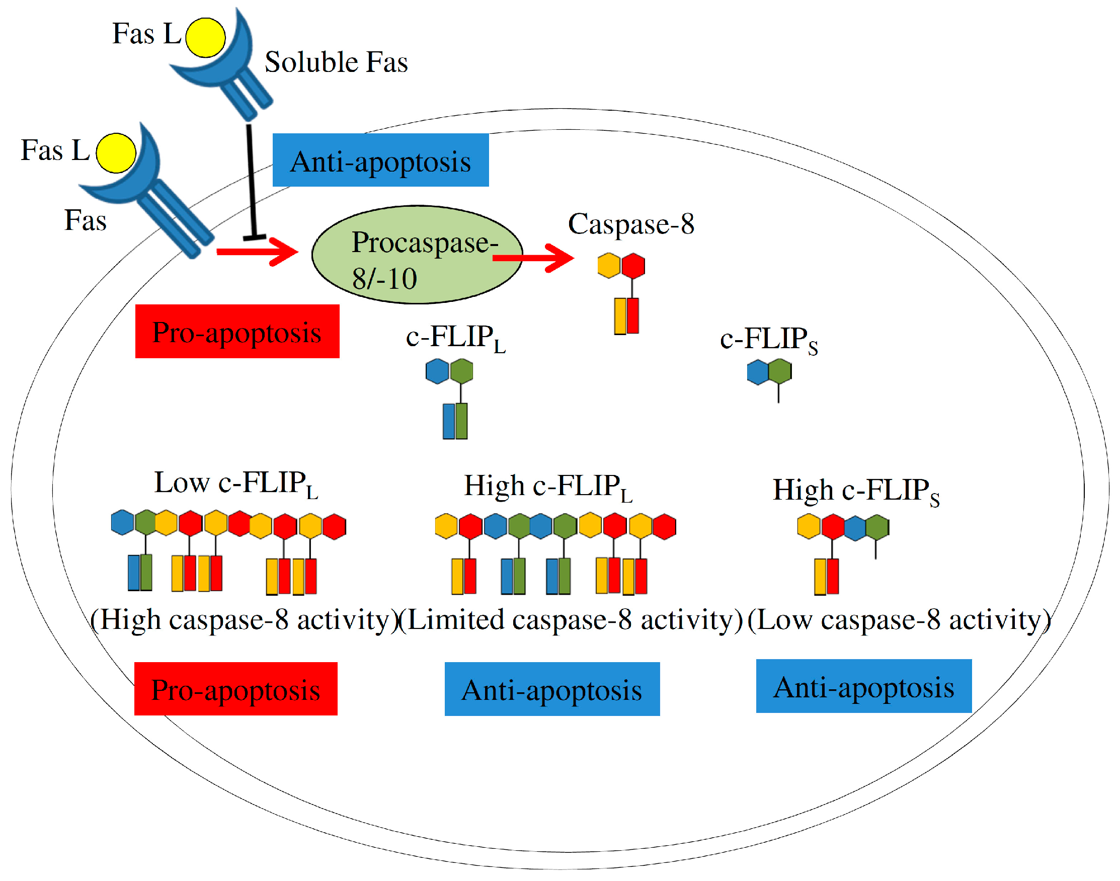
4.2.2. Fas Signaling

Fas (also referred as Apo-1/CD95) is a well-studied member of the TNF receptor superfamily which mediates extrinsic pathway-induced apoptosis upon interaction with the Fas ligand or agonistic antibodies [78]. Alternative splicing of Fas exon 6 generates membrane-bound or soluble isoforms that exhibit opposite activities on cellular apoptosis [79]. Neoplastic cells are frequently noted to reduce Fas expression or induce relative levels of soluble Fas proteins, encoded by Fas−exon 6, to evade Fas/Fas L-mediated apoptosis [80]. The direct interaction between T-cell intracellular antigen-1 (TIA-1) and the Uridine-rich stretch next to Fas exon 6 facilitated the recognition of U1snRNP for the 5′ splice site of Fas intron 6 and also enhanced the binding of U2AF to the 3′ splice site of Fas intron 5, which led to the definition of Fas exon 6 [81]. The binding of polypyrimidine tract binding protein 1 (PTBP1) and the U-rich element (URE) within Fas exon 6 exhibited the antagonistic effect on the TIA-1-enhanced inclusion of Fas exon 6 [81]. In addition to PTBP1, the direct binding of Hu antigen R (HuR) and the URE within Fas exon 6 was demonstrated to reduce the association of U2AF and the 3′ splice site of Fas intron 5, subsequently resulting in the skipping of Fas exon 6 [82]. The binding of hnRNP C and URE within Fas exon 6 cooperatively facilitated the repressive effect of PTBP1 and HuR on interfering with the interaction between TIA-1/TIAR and the 5′ splice site next to Fas exon 6 [83]. In addition to the exonic element, recent study indicated that over 90% of single nucleotide mutation (58/63 positions) mediated distinct effect on the usage of Fas exon 6 [84], which potentially constituted a novel mechanism regarding the exon definition. Results of genome-wide screening indicated that elimination of more than 200 splicing regulators, including SR proteins, hnRNP family and splicing factor 45, changed the splicing profile of Fas in mammalian cells [85]. In addition, the splicing profile of the Fas gene is regulated by natural antisense RNA (Fas-AS1 or saf) which manipulates utilization of Fas exon 6 and, therefore, manipulates the proapoptotic activity of Fas signaling [86]. Despite this, the molecular mechanism involved in antisense RNA-regulated splicing is still largely unclear.
Cellular FLICE inhibitory protein (c-FLIP), a caspase-8 homolog, functions as a crucial factor in manipulating apoptotic activity of the Fas/Fas L-mediated pathway [87]. By usage of alternative 5′ splice site within c-FLIP exon 5, the c-FLIP gene generates alternative transcripts which encode three variants, including 55 kDa c-FLIP long (c-FLIPL), 26 kDa c-FLIPS, and 24 kDa c-FLIPR, in human cells [88]. The c-FLIPL isoform shares a high homology with procaspase-8 except for a cysteine residue within the catalytic center, whereas short c-FLIP variants are truncated isoforms which lack the dimerization motif of procaspase-8 and only contains the tandem death effector domain (DED) [87]. c-FLIP isoforms exhibit differential effects on restricting activation of procaspase-8. Relatively high expressions of short c-FLIP isoforms substantially interfere with the oligomerization of procaspase-8, resulting in its inactivation and evasion of apoptosis. Overexpressed c-FLIPL isoform assembles heterodimers with procaspase-8, but block its activation [87]. In contrast, at physiological levels, c-FLIPL forms heterodimers with procaspase-8 within the death-inducing signaling complex (DISC), facilitating procaspase-8 activation and subsequent programmed cell death (Figure 5, [87]). Nevertheless, understanding the molecular mechanism involved in the regulation of c-FLIP splicing still requires further investigation.

4.2.3. Bcl-2 Family

Bcl-2 family members, including Bcl-2, Bax, Bcl-x, Bcl-g, Bcl-rambo, Bim, Bfl-1, Bid, Mcl-1, and PUMA, are well-characterized factors which exhibit both pro- and anti-apoptotic activities [89]. These proteins form homodimers or heterodimers through Bcl-2 homologous (BH) domains. Therefore, relative levels of Bcl-2 family members are critical in fine-tuning a cell’s fate [90]. In addition, Bcl-2-related genes encode protein isoforms with differential or opposite functions through alternative splicing mechanisms [27].
The Bcl-x gene was characterized as generating alternative transcripts by using the alternative 5′ splice site within exon 2, encoding the anti-apoptotic Bcl-xL and pro-apoptotic Bcl-xS isoforms [91]. Relative expressions of these two isoforms manipulate sensitization of mammalian cells under apoptotic conditions [92]. The splicing profiles of Bcl-x gene were widely modulated by SR proteins, hnRNP family and RNA binding motif proteins serine/arginine-rich splicing factor 1 (SRSF1), polypyrimidine tract binding protein 1 (PTBP1), RNA binding motif protein 4 (RBM4), RBM5, RBM10, and RBM11, were documented to program the splicing profile of Bcl-x [93,94,95]. For example, overexpression of the RBM4 protein mediates relatively high levels of Bcl-xS transcripts, leading to processing of procaspase-3 and poly(ADP ribose) polymerase (PARP) which function as apoptotic markers [95]. In addition to a splicing regulator, the presence of a splice-switching oligonucleotide (SSO) was also demonstrated to reprogram Bcl-x splicing from Bcl-xL to Bcl-xS and subsequently induce apoptosis of human hepatic stellate cells [96]. The antisense oligonucleotide may function as a better therapeutic Bcl-x SSO than other apoptotic inducers that can only focus on splicing mechanisms [96].
Bax was reported to exhibit pro-apoptotic activity [97]. Targeting of dimerized Bax to the mitochondrial membrane resulted in the release of cytochrome C and sequentially induced caspase-9/3-mediated cell apoptosis [98]. It was recently reported that a single guanosine deletion (G8 to G7) within Bax exon 3 resulted in the skipping of Bax exon 3, which generated the BaxΔ2 transcript [99]. Interestingly, BaxΔ2-positive colorectal cancer (CRC) cells were much more sensitive to adriamycin and 5-FU compared to BaxΔ2-negative CRC clones [100]. Moreover, the presence of the BaxΔ2 protein further mediated activation of procaspase-8 and downstream apoptotic signaling [100]. However, both CRC clones showed similar sensitivities to treatment with daunorubicin, which shares a structure similar to that of adriamycin. These results suggested that BaxΔ2-positive cells exhibit a preference for specific chemotherapeutic drugs [100].
Mutual utilization of BIM exon 3 or 4 constitutes the molecular mechanism involved in the generation of two distinct transcripts [101]. BIM−exon 4 transcripts encode the BH3 domain-absence variant which exhibited the antagonistic effect toward the activity of anti-apoptotic Bcl-2 proteins [102].The splicing profile of the BIM gene is programmed by the interplay between cis-acting elements and trans-splicing regulators [101]. For example, up-regulated expression of the SRSF1 protein induced a relatively high level of the BIM+exon 3 isoform in breast cancer cells [103]. Accordingly, overexpression of the SRSF1 protein preferentially lessened the sensitivity of neoplastic cells to chemotherapeutic compound-mediated cell death [103]. In addition, a cytosine-to-thymidine mutation (rs724710) within BIM exon 4 was noted to reduce the selection of exon 4 in lymphoblastic leukemia cells, which contributed to drug resistance [104]. Therefore, alternative splicing patterns of the BIM gene are considered an emerging mediator of the immortality of cancerous cells. The apoptosis-related splicing events are listed in Table 1.

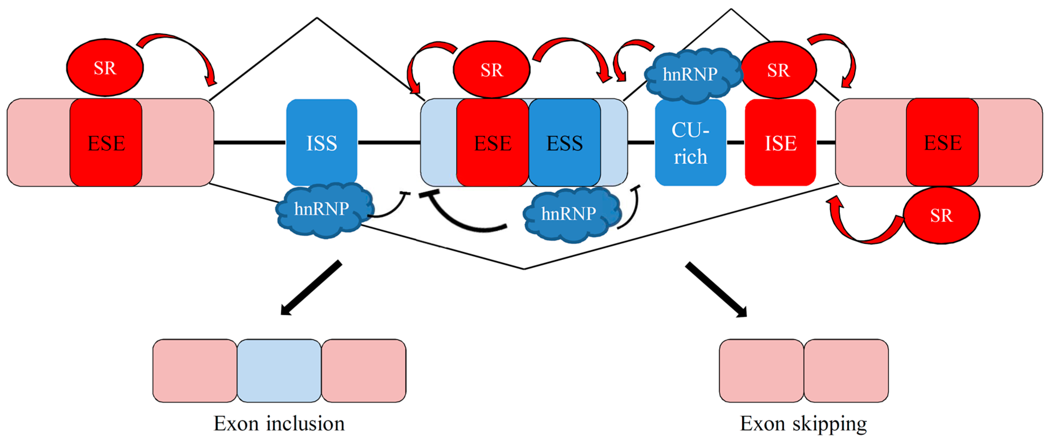
4.3. Splicing Factors Involved in Apoptosis-Related Splicing Events

4.3.1. Serine/Arginine (SR)-Rich Splicing Factors

The serine/arginine-rich splicing factors are widely involved in numerous alternative splicing events [105]. Homozygous knockout embryos of most SR proteins, such as SRSF2, are lethal to embryos, suggesting the important functions that these factors exert in tissue development and organogenesis [106]. Down-regulation of SRSF2 substantially led to cell cycle arrest and destabilization of the genome [107]. Recent reports documented that imbalanced expression of SRSF2 resulted in the reduced growth and imbalanced apoptosis of hematopoietic cells, especially bone marrow cells [108]. Deep RNA-seq results indicated that SRSF2 depletion mediated the aberrant splicing of hematopoiesis-related genes, including MEIS1, UPF38, PRKAA1, RBM23, PDK1, PDE4DIP, MLL, and RNF34, which are closely related to the homeostasis of myeloid progenitors [108].
SRSF3 reportedly modulated the alternative splicing of several apoptosis-related genes, such as caspase-2 (Casp2), programmed cell death 4 (PDCD4), and homeodomain-interacting protein kinase-2 (HIPK2) in distinct cancer cells [109,110,111]. Caspase-2 was demonstrated to act the early initiator in the intrinsic apoptosis pathway [112]. The utilization of caspase-2 exon 9 leads to the generation of two caspase-2 isoforms which exert opposite effect on the apoptotic process [112]. The direct interaction between overexpressing SRSF3 and the CU-rich element within Casp2 exon 8 was reported to promote the skipping of CASP2 exon 9, which increased the relative expression of anti-apoptotic Casp2S isoform [112]. In distinct colorectal cancer cells, SRSF3 silencing was demonstrated to enhance the selection of alternative 3′ splice site within the HIPK2 exon 8, generating the 81 nucleotides-deleted HIPK2 Δe8 isoform [110]. HIPK2 Δe8 isoform was resistant to the proteasome-mediated degradation in the absence of E3 ligase binding site which was encoded the deleted 5′ region of HIPK2 exon 8. The relatively high level of HIPK2 Δe8 isoform profoundly induced the phosphorylation of p53 protein and downstream apoptotic pathway [110]. Moreover, SRSF3 was also reported to modulate the expression of PDCD4 gene through the alternative splicing and translational mechanisms [111]. PDCD4 reportedly acted the tumor suppressor in repressing the transformation and immortality of cancer cells [113]. SRSF3 silencing was noted to reduce the relative level of PDCD4 isoform 2, containing the partial PDCD4 intron 3 in distinct cancer cells. The premature stop codon-harboring PDCD4 isoform 2 was considered as the potential substrate of nonsense-mediated decay mechanism [46]. Moreover, the preferential binding of SRSF3 with the 5′ UTR of PDCD4 transcript drove its enrichment to the processing body more than repressed the translational activity of PDCD4 mRNA. Collectively, elevated SRSF3 expression enhanced the anti-apoptotic signature of cancer cells by reprogramming the splicing profiles of related genes.
SRSF1 (also referred as ASE/SF2) is the well-studied protein involved in most posttranscriptional regulations, including alternative splicing [114]. The manipulated expression of SRSF1 is frequently observed during the organogenesis and carcinogenesis [115]. More than 500 potential candidates, including insulin receptor (INSR) were identified as the potential candidates of SRSF1 by using the RNA-seq approach [116]. INSR gene was demonstrated to generate two alternatively-spliced variants in a spatial-temporal manner [116].The exon 11-excluded INSR (INSR-A) transcripts were predominantly expressed in embryonic and cancerous cells [116], whereas the exon 11-included INSR (INSR-B) transcripts were widely noted in the pancreatic β-cells, skeletal muscle, and adipocytes [117]. Overexpression of SRSF1 was demonstrated to constitute a molecular mechanism in enhancing the relative level of exon 11-included INSR–B, which subsequently lessened the sensitivity of pancreatic progenitors to stress-induced apoptosis [116]. In addition, the up-regulated SRSF1 expression with a concomitant increase in the relative level of exon-9-included cancer susceptibility candidate 4 (CASC4) transcripts was revealed in breast cancer cells compared to the normal ductal cells [115]. SRSF1 overexpressing cells generated the relatively high level of full-length CASC4 transcripts which contained the 168-nucleotides long exon 9, encoding the long CASC4 variants [115]. The epithelial MCF-10A ductal cells exhibited progressive and anti-apoptotic activity in the presence of exogenous CASC4-FL protein, whereas the overexpressing CASC4-Δ9 variant exerted limited effect on these signatures [115].

4.3.2. Heterogeneous Nuclear Ribonucleoprotein (hnRNP) Family

The hnRNP family is composed of about 20 members that play significant roles in transcriptional, post-transcriptional, and translational regulation [118]. Several hnRNP proteins are considered to be proto-oncogenes according to recent studies [119]. Among these members, hnRNP A1, hnRNP K, and PTBP1 (hnRNP I) are involved in apoptosis-related splicing events. The expression of hnRNP A1, an hnRNP family member, is abundant and ubiquitously generated [120]. A bioinformatics analysis and RNA-protein binding assay indicated a direct interaction between hnRNP A1 and Fas exon 5, which subsequently facilitated inclusion of Fas exon 6 [121]. Due to the proapoptotic activity of Fas+exon 6, using hnRNP A1 and other splicing regulators may be considered a potential strategy to reduce the immortality of cancer cells. HnRNP K was reported to strengthen utilization of the 5′ splice site by interacting with an exonic enhancer and subsequently interfered with the generation of Bcl-xS transcripts, which led to evasion of apoptosis by cancerous cells [122]. In addition, the association of hnRNP K with the Sam68 protein abrogated its effect in inducing relative levels of Bcl-xS transcripts [123]. Nevertheless, the hnRNP K protein diminishes apoptotic activity through multi-layer mechanisms [124]. The PTBP1 was demonstrated to facilitate the Warburg effect in colorectal cancer cells by reprogramming splicing profiles of the PKM gene [125]. Recent studies indicated the emerging role of PTBP1-modulated regulation in diminishing cell apoptosis with treatment with an antitubulin agent [126]. The presence of PTBP1 reduced the stability of Mcl-1 transcripts by binding to its 3′ UTR [126]. Moreover, the ablation of PTBP1 induced the apoptotic evasion of antitubulin-treated cells in a Mcl-1-dependent manner [126]. However, the influence of PTBP1 on other post-transcriptional mechanisms, such as alternative splicing regulation, in terms of Mcl-1 isoform expressions is worthy of further investigation. In addition, overexpressing PTBP1 functioned as a splicing silencer of Bim exon 3 by directly targeting the responsive element within Bim intron 2 [101].

4.3.3. RNA-Binding Motif Proteins (RBMPs)

RBMPs constitute another family that participates in diverse gene regulation. Individual RBMPs contain multiple RNA recognition motifs (RRMs) which are the most common class of RNA-binding domains. RNA-binding motifs are multi-functional and have been implicated in nucleotide- and protein-protein interactions of RBMPs. The binding surface of RRMs is composed of 80–90 residues which are folded in four-strand anti-parallel β-sheets [127]. In addition, the serine/arginine rich elements were widely noted as well in various RBMPs. Among the RBMPs, RBM4, 5, 10, and 11 were demonstrated to modulate apoptosis-related splicing events in various malignancies [95,128,129]. RBM4 was shown to program splicing cascades which are closely relevant to the development of the mesodermal lineage, including skeletal muscles and brown adipocytes [130]. Recent reports indicated the tumor-suppressive effect of RBM4 through regulating splicing profiles of apoptosis-related genes [95]. Relatively low levels of RBM4 were noted in cancerous tissues compared to adjacent normal tissues which were dissected from non-small cell lung cancer (NSCLC) and breast cancer (BC) patients [95]. Overexpression of RBM4 increased the relative ratio of Bcl-xS transcripts and subsequently induced apoptosis of several lung cancer cell lines [95]. Moreover, the association of overexpression of RBM4 with Mcl-1 exon 2 and intron 2 shifted Mcl-1L to Mcl-1S transcripts, which, in part, deprived breast cancer cells of apoptotic resistance against chemotherapeutic treatment [131]. RBM5 and RBM10 are highly similar homologues which share about 50% amino acid identity [132]. Previous studies showed the regulatory effect of RBM5 on modulating splicing profiles of c-FLIP, Fas, and caspase-2 [129,133]. Reduced expressions of RBM5 and RBM10 were noted in cancerous tissues of NSCLS, prostate cancer, and BC patients compared to adjacent normal counterparts [132]. Overexpression of RBM5 or RBM10 both consistently resulted in relatively high levels of Fas−exon 6 transcripts which encoded soluble and antiapoptotic isoforms in different cells [134]. Recent studies showed that RBM5 and RBM10 exhibited similar effects on the same apoptosis-related splicing events, such as c-FLIP, caspase-2, caspase-3, caspase-9, and Bcl-x genes [129,133]. However, the differential influence of each RBM5/10-modulated splicing event on cell apoptosis was individually characterized. The endocytic adaptor protein (NUMB) gene was identified as a novel candidate of RBM5, RBM6, and RBM10 [132]. NUMB has been reported to participate in the activation of p53 protein by regulating the Notch pathway [135]. Intriguingly, depletion of RBM5 and RBM10 showed opposite effects on inclusion of NUMB exon 9. Subsequently, RBM5 and RBM10 exhibited differential influences on the radiosensitivity and proliferation of lung adenocarcinoma cells through NUMB-mediated Notch signaling [132]. Collectively, cell apoptosis and proliferation are meticulously controlled processes which are regulated through multilayer mechanisms. The apoptosis-related splicing regulators are summarized in Table 2.

5. Conclusions and Perspectives

Alternative splicing was demonstrated to be an important molecular mechanism that is widely involved in the homeostasis of mammalian cells. Dysregulated splicing events were widely demonstrated to be molecular hallmarks of developmental and malignant diseases. In this review, we attempted to summarize recent studies regarding the influence of alternatively spliced transcripts on cell apoptosis, which is highly relevant to organogenesis and carcinogenesis. The impacts of splicing regulators on apoptosis-related splicing events were discussed as well. Along with the development of high-throughput approaches, including deep RNA sequencing and proteomics analyses, new insights will be brought to the identification of disease-associated splicing networks on a genome-wide scale. A thorough realization of the mechanisms underlying development- and cancer-related splicing networks will function as a convincing source of therapeutic strategies for treating inherited and malignant diseases.

Acknowledgments

This work was supported by a grant (MOST 105-2311-B-038-003) from the Ministry of Science and Technology of Taiwan. All authors declare no conflict of interest exists.

Conflicts of Interest

The authors declare no conflict of interest.

References

  1. Yao, Y.; Gao, Z.; Liang, W.; Kong, L.; Jiao, Y.; Li, S.; Tao, Z.; Yan, Y.; Yang, J. Osthole promotes neuronal differentiation and inhibits apoptosis via Wnt/β-catenin signaling in an Alzheimer’s disease model. Toxicol. Appl. Pharmacol. 2015, 289, 474–481. [Google Scholar] [CrossRef] [PubMed]
  2. Filatova, E.N.; Anisenkova, E.V.; Presnyakova, N.B.; Utkin, O.V. DR3 regulation of apoptosis of naive T-lymphocytes in children with acute infectious mononucleosis. Acta Microbiol. Immunol. Hung. 2016, 63, 339–357. [Google Scholar] [CrossRef] [PubMed]
  3. Kiraz, Y.; Adan, A.; Kartal Yandim, M.; Baran, Y. Major apoptotic mechanisms and genes involved in apoptosis. Tumour Biol. 2016, 37, 8471–8486. [Google Scholar] [CrossRef] [PubMed]
  4. Flusberg, D.A.; Sorger, P.K. Surviving apoptosis: Life-death signaling in single cells. Trends Cell Biol. 2015, 25, 446–458. [Google Scholar] [CrossRef] [PubMed]
  5. Liang, Y.Y.; Rainprecht, D.; Eichmair, E.; Messner, B.; Oehler, R. Serum-dependent processing of late apoptotic cells and their immunogenicity. Apoptosis 2015, 20, 1444–1456. [Google Scholar] [CrossRef] [PubMed]
  6. Choi, S.J.; Kim, M.H.; Jeon, J.; Kim, O.Y.; Choi, Y.; Seo, J.; Hong, S.W.; Lee, W.H.; Jeon, S.G.; Gho, Y.S.; et al. Active immunization with extracellular vesicles derived from Staphylococcus aureus effectively protects against Staphylococcal Lung infections, mainly via Th1 cell-mediated immunity. PLoS ONE 2015, 10, e0136021. [Google Scholar] [CrossRef] [PubMed]
  7. Chu, J.Y.; Dransfield, I.; Rossi, A.G.; Vermeren, S. Non-canonical PI3K-Cdc42-Pak-Mek-Erk signaling promotes immune-complex-induced apoptosis in human neutrophils. Cell Rep. 2016, 17, 374–386. [Google Scholar] [CrossRef] [PubMed]
  8. Singh, A.P.; Foley, J.F.; Rubino, M.; Boyle, M.C.; Tandon, A.; Shah, R.; Archer, T.K. Brg1 enables rapid growth of the early embryo by suppressing genes that regulate apoptosis and cell growth arrest. Mol. Cell. Biol. 2016, 36, 1990–2010. [Google Scholar] [CrossRef] [PubMed]
  9. Moon, Y.J.; Yun, C.Y.; Choi, H.; Ka, S.O.; Kim, J.R.; Park, B.H.; Cho, E.S. Smad4 controls bone homeostasis through regulation of osteoblast/osteocyte viability. Exp. Mol. Med. 2016, 48, e256. [Google Scholar] [CrossRef] [PubMed]
  10. Liu, X.F.; Xiang, L.; Zhou, Q.; Carralot, J.P.; Prunotto, M.; Niederfellner, G.; Pastan, I. Actinomycin D enhances killing of cancer cells by immunotoxin RG7787 through activation of the extrinsic pathway of apoptosis. Proc. Natl. Acad. Sci. USA 2016, 113, 10666–10671. [Google Scholar] [CrossRef] [PubMed]
  11. Jo, A.R.; Jeong, H.S.; Kim, M.K.; Yun, H.Y.; Baek, K.J.; Kwon, N.S.; Kim, D.S. Geranylgeranylacetone induces apoptosis via the intrinsic pathway in human melanoma cells. Biomed. Pharmacother. 2016, 82, 15–19. [Google Scholar] [CrossRef] [PubMed]
  12. Montagnani Marelli, M.; Marzagalli, M.; Moretti, R.M.; Beretta, G.; Casati, L.; Comitato, R.; Gravina, G.L.; Festuccia, C.; Limonta, P. Vitamin E δ-tocotrienol triggers endoplasmic reticulum stress-mediated apoptosis in human melanoma cells. Sci. Rep. 2016, 6, 30502. [Google Scholar] [CrossRef] [PubMed]
  13. Lee, M.H.; Hong, S.H.; Park, C.; Kim, G.Y.; Leem, S.H.; Choi, S.H.; Keum, Y.S.; Hyun, J.W.; Kwon, T.K.; Hong, S.H.; et al. Hwang-Heuk-San induces apoptosis in HCT116 human colorectal cancer cells through the ROS-mediated activation of caspases and the inactivation of the PI3K/Akt signaling pathway. Oncol. Rep. 2016, 36, 205–214. [Google Scholar] [CrossRef] [PubMed]
  14. Yanagi, T.; Shi, R.; Aza-Blanc, P.; Reed, J.C.; Matsuzawa, S. PCTAIRE1-knockdown sensitizes cancer cells to TNF family cytokines. PLoS ONE 2015, 10, e0119404. [Google Scholar] [CrossRef] [PubMed]
  15. Liu, B.; Meng, D.; Wei, T.; Zhang, S.; Hu, Y.; Wang, M. Apoptosis and pro-inflammatory cytokine response of mast cells induced by influenza A viruses. PLoS ONE 2014, 9, e100109. [Google Scholar] [CrossRef] [PubMed]
  16. Shao, L.W.; Huang, L.H.; Yan, S.; Jin, J.D.; Ren, S.Y. Cordycepin induces apoptosis in human liver cancer HepG2 cells through extrinsic and intrinsic signaling pathways. Oncol. Lett. 2016, 12, 995–1000. [Google Scholar] [CrossRef] [PubMed]
  17. Nilsen, T.W.; Graveley, B.R. Expansion of the eukaryotic proteome by alternative splicing. Nature 2010, 463, 457–463. [Google Scholar] [CrossRef] [PubMed]
  18. Quesnel-Vallières, M.; Irimia, M.; Cordes, S.P.; Blencowe, B.J. Essential roles for the splicing regulator nSR100/SRRM4 during nervous system development. Genes Dev. 2015, 29, 746–759. [Google Scholar] [CrossRef] [PubMed]
  19. Pan, Q.; Shai, O.; Lee, L.J.; Frey, B.J.; Blencowe, B.J. Deep surveying of alternative splicing complexity in the human transcriptome by high-throughput sequencing. Nat. Genet. 2008, 40, 1413–1415. [Google Scholar] [CrossRef] [PubMed]
  20. Matera, A.G.; Wang, Z. A day in the life of the spliceosome. Nat. Rev. Mol. Cell Biol. 2014, 15, 108–121. [Google Scholar] [CrossRef] [PubMed]
  21. Arrigoni, A.; Ranzani, V.; Rossetti, G.; Panzeri, I.; Abrignani, S.; Bonnal, R.J.; Pagani, M. Analysis RNA-seq and Noncoding RNA. Methods Mol. Biol. 2016, 1480, 125–135. [Google Scholar] [PubMed]
  22. Li, J.; Yang, Z.; Li, Y.; Xia, J.; Li, D.; Li, H.; Ren, M.; Liao, Y.; Yu, S.; Chen, Y.; et al. Cell apoptosis, autophagy and necroptosis in osteosarcoma treatment. Oncotarget 2016, 7, 44763–44778. [Google Scholar] [PubMed]
  23. Kallenberger, S.M.; Beaudouin, J.; Claus, J.; Fischer, C.; Sorger, P.K.; Legewie, S.; Eils, R. Intra- and interdimeric caspase-8 self-cleavage controls strength and timing of CD95-induced apoptosis. Sci. Signal. 2014, 7, ra23. [Google Scholar] [CrossRef] [PubMed]
  24. Wang, X.; Chen, E.; Tang, M.; Yang, X.; Wang, Y.; Quan, Z.; Wu, X.; Luo, C. The SMAD2/3 pathway is involved in hepaCAM-induced apoptosis by inhibiting the nuclear translocation of SMAD2/3 in bladder cancer cells. Tumour Biol. 2016, 37, 10731–10743. [Google Scholar] [CrossRef] [PubMed]
  25. Baluchamy, S.; Ravichandran, P.; Periyakaruppan, A.; Ramesh, V.; Hall, J.C.; Zhang, Y.; Jejelowo, O.; Gridley, D.S.; Wu, H.; Ramesh, G.T. Induction of cell death through alteration of oxidants and antioxidants in lung epithelial cells exposed to high energy protons. J. Biol. Chem. 2010, 285, 24769–24774. [Google Scholar] [CrossRef] [PubMed]
  26. Zhang, N.; Ye, F.; Zhu, W.; Hu, D.; Xiao, C.; Nan, J.; Su, S.; Wang, Y.; Liu, M.; Gao, K.; et al. Cardiac ankyrin repeat protein attenuates cardiomyocyte apoptosis by upregulation of Bcl-2 expression. Biochim. Biophys. Acta 2016, 4889, 30254. [Google Scholar] [CrossRef] [PubMed]
  27. Akgul, C.; Moulding, D.A.; Edwards, S.W. Alternative splicing of Bcl-2-related genes: Functional consequences and potential therapeutic applications. Cell. Mol. Life Sci. 2004, 61, 2189–2199. [Google Scholar] [CrossRef] [PubMed]
  28. Würstle, M.L.; Laussmann, M.A.; Rehm, M. The central role of initiator caspase-9 in apoptosis signal transduction and the regulation of its activation and activity on the apoptosome. Exp. Cell Res. 2012, 318, 1213–1220. [Google Scholar] [CrossRef] [PubMed]
  29. Lee, N.R.; Park, B.S.; Kim, S.Y.; Gu, A.; Kim da, H.; Lee, J.S.; Kim, I.S. Cytokine secreted by S100A9 via TLR4 in monocytes delays neutrophil apoptosis by inhibition of caspase 9/3 pathway. Cytokine 2016, 86, 53–63. [Google Scholar] [CrossRef] [PubMed]
  30. Gai, W.T.; Yu, D.P.; Wang, X.S.; Wang, P.T. Anti-cancer effect of ursolic acid activates apoptosis through ROCK/PTEN mediated mitochondrial translocation of cofilin-1 in prostate cancer. Oncol. Lett. 2016, 12, 2880–2885. [Google Scholar] [CrossRef] [PubMed]
  31. Zhu, J.; Zhou, Q.; Tan, S. Targeting miRNAs associated with surface expression of death receptors to modulate TRAIL resistance in breast cancer. Cancer Lett. 2016. [Google Scholar] [CrossRef] [PubMed]
  32. Ling, G.; Zhang, T.; Zhang, P.; Sun, J.; He, Z. Synergistic and complete reversal of the multidrug resistance of mitoxantrone hydrochloride by three-in-one multifunctional lipid-sodium glycocholate nanocarriers based on simultaneous BCRP and Bcl-2 inhibition. Int. J. Nanomed. 2016, 11, 4077–4091. [Google Scholar]
  33. Yang, A.; Wilson, N.S.; Ashkenazi, A. Proapoptotic DR4 and DR5 signaling in cancer cells: Toward clinical translation. Curr. Opin. Cell Biol. 2010, 22, 837–844. [Google Scholar] [CrossRef] [PubMed]
  34. Mohammadpour, H.; Pourfathollah, A.A.; Nikougoftar Zarif, M.; Shahbazfar, A.A. Irradiation enhances susceptibility of tumor cells to the antitumor effects of TNF-α activated adipose derived mesenchymal stem cells in breast cancer model. Sci. Rep. 2016, 6, 28433. [Google Scholar] [CrossRef] [PubMed]
  35. Liu, Y.; Hawkins, O.E.; Vilgelm, A.E.; Pawlikowski, J.S.; Ecsedy, J.A.; Sosman, J.A.; Kelley, M.C.; Richmond, A. Combining an aurora kinase inhibitor and a death receptor ligand/agonist antibody triggers apoptosis in melanoma cells and prevents tumor growth in preclinical mouse models. Clin. Cancer Res. 2015, 21, 5338–5348. [Google Scholar] [CrossRef] [PubMed]
  36. Dickens, L.S.; Boyd, R.S.; Jukes-Jones, R.; Hughes, M.A.; Robinson, G.L.; Fairall, L.; Schwabe, J.W.; Cain, K.; Macfarlane, M. A death effector domain chain DISC model reveals a crucial role for caspase-8 chain assembly in mediating apoptotic cell death. Mol. Cell 2012, 47, 291–305. [Google Scholar] [CrossRef] [PubMed]
  37. Krieg, A.; Krieg, T.; Wenzel, M.; Schmitt, M.; Ramp, U.; Fang, B.; Gabbert, H.E.; Gerharz, C.D.; Mahotka, C. TRAIL-β and TRAIL-γ: Two novel splice variants of the human TNF-related apoptosis-inducing ligand (TRAIL) without apoptotic potential. Br. J. Cancer 2003, 88, 918–927. [Google Scholar] [CrossRef] [PubMed]
  38. Picarda, G.; Surget, S.; Guiho, R.; Téletchéa, S.; Berreur, M.; Tirode Pellat-Deceunynck, C.; Heymann, D.; Trichet, V.; Rédini, F. A functional, new short isoform of death receptor 4 in Ewing’s sarcoma cell lines may be involved in TRAIL sensitivity/resistance mechanisms. Mol. Cancer Res. 2012, 10, 336–346. [Google Scholar] [CrossRef] [PubMed]
  39. Rautureau, G.J.; Day, C.L.; Hinds, M.G. Intrinsically disordered proteins in Bcl-2 regulated apoptosis. Int. J. Mol. Sci. 2010, 11, 1808–1824. [Google Scholar] [CrossRef] [PubMed]
  40. Zhou, X.; Li, X.; Cheng, Y.; Wu, W.; Xie, Z.; Xi, Q.; Han, J.; Wu, G.; Fang, J.; Feng, Y. BCLAF1 and its splicing regulator SRSF10 regulate the tumorigenic potential of colon cancer cells. Nat. Commun. 2014, 5, 4581. [Google Scholar] [CrossRef] [PubMed]
  41. Chen, W.; Moore, M.J. The spliceosome: Disorder and dynamics defined. Curr. Opin. Struct. Biol. 2014, 24, 141–149. [Google Scholar] [CrossRef] [PubMed]
  42. Papasaikas, P.; Tejedor, J.R.; Vigevani, L.; Valcárcel, J. Functional splicing network reveals extensive regulatory potential of the core spliceosomal machinery. Mol. Cell 2015, 57, 7–22. [Google Scholar] [CrossRef] [PubMed]
  43. Cyphert, T.J.; Suchanek, A.L.; Griffith, B.N.; Salati, L.M. Starvation actively inhibits splicing of glucose-6-phosphate dehydrogenase mRNA via a bifunctional ESE/ESS element bound by hnRNP K. Biochim. Biophys. Acta 2013, 1829, 905–915. [Google Scholar] [CrossRef] [PubMed]
  44. Erkelenz, S.; Mueller, W.F.; Evans, M.S.; Busch, A.; Schöneweis, K.; Hertel, K.J.; Schaal, H. Position-dependent splicing activation and repression by SR and hnRNP proteins rely on common mechanisms. RNA 2013, 19, 96–102. [Google Scholar] [CrossRef] [PubMed]
  45. Fu, X.D.; Ares, M., Jr. Context-dependent control of alternative splicing by RNA-binding proteins. Nat. Rev. Genet. 2014, 15, 689–701. [Google Scholar] [CrossRef] [PubMed]
  46. Lin, J.C.; Tarn, W.Y. RBM4 down-regulates PTB and antagonizes its activity in muscle cell–specific alternative splicing. J. Cell Biol. 2011, 193, 509–520. [Google Scholar] [CrossRef] [PubMed]
  47. Martini, E.; Schneider, E.; Neufert, C.; Neurath, M.F.; Becker, C. Survivin is a guardian of the intestinal stem cell niche and its expression is regulated by TGF-β. Cell Cycle 2016, 7, 1–7. [Google Scholar] [CrossRef] [PubMed]
  48. Lee, S.H.; Lee, J.Y.; Jung, C.L.; Bae, I.H.; Suh, K.H.; Ahn, Y.G.; Jin, D.H.; Kim, T.W.; Suh, Y.A.; Jang, S.J. A novel antagonist to the inhibitors of apoptosis (IAPs) potentiates cell death in EGFR-overexpressing non-small-cell lung cancer cells. Cell Death Dis. 2014, 5, e1477. [Google Scholar] [CrossRef] [PubMed]
  49. Turkkila, M.; Andersson, K.M.; Amu, S.; Brisslert, M.; Erlandsson, M.C.; Silfverswärd, S.; Bokarewa, M.I. Suppressed diversity of survivin splicing in active rheumatoid arthritis. Arthritis Res. Ther. 2015, 17, 175. [Google Scholar] [CrossRef] [PubMed]
  50. Faversani, A.; Vaira, V.; Moro, G.P.; Tosi, D.; Lopergolo, A.; Schultz, D.C.; Rivadeneira, D.; Altieri, D.C.; Bosari, S. Survivin family proteins as novel molecular determinants of doxorubicin resistance in organotypic human breast tumors. Breast Cancer Res. 2014, 16, R55. [Google Scholar] [CrossRef] [PubMed]
  51. Tazo, Y.; Hara, A.; Onda, T.; Saegusa, M. Bifunctional roles of survivin-ΔEx3 and survivin-2B for susceptibility to apoptosis in endometrial carcinomas. J. Cancer Res. Clin. Oncol. 2014, 140, 2027–2037. [Google Scholar] [CrossRef] [PubMed]
  52. Waligórska-Stachura, J.; Andrusiewicz, M.; Sawicka-Gutaj, N.; Kubiczak, M.; Jankowska, A.; Liebert, W.; Czarnywojtek, A.; Waśko, R.; Blanco-Gangoo, A.R.; Ruchała, M. Evaluation of survivin splice variants in pituitary tumors. Pituitary 2015, 18, 410–416. [Google Scholar] [CrossRef] [PubMed]
  53. Morgan, M.; Deoraj, A.; Felty, Q.; Roy, D. Environmental estrogen-like endocrine disrupting chemicals and breast cancer. Mol. Cell. Endocrinol. 2016, 7207, 30411–30417. [Google Scholar] [CrossRef] [PubMed]
  54. Zhang, C.; Wang, H.J.; Bao, Q.C.; Wang, L.; Guo, T.K.; Chen, W.L.; Xu, L.L.; Zhou, H.S.; Bian, J.L.; Yang, Y.R.; et al. NRF2 promotes breast cancer cell proliferation and metastasis by increasing RhoA/ROCK pathway signal transduction. Oncotarget 2016. [Google Scholar] [CrossRef] [PubMed]
  55. Divekar, S.D.; Tiek, D.M.; Fernandez, A.; Riggins, R.B. Estrogen-related receptor β (ERRβ)—Renaissance receptor or receptor renaissance? Nucl. Recept. Signal. 2016, 14, e002. [Google Scholar] [PubMed]
  56. Huang, P.C.; Kuo, W.W.; Shen, C.Y.; Chen, Y.F.; Lin, Y.M.; Ho, T.J.; Padma, V.V.; Lo, J.F.; Huang, C.Y.; Huang, C.Y. Anthocyanin attenuates doxorubicin-induced cardiomyotoxicity via estrogen receptor-α/β and stabilizes HSF1 to inhibit the IGF-IIR apoptotic pathway. Int. J. Mol. Sci. 2016, 17, 1588. [Google Scholar] [CrossRef] [PubMed]
  57. Zhao, Z.; Wang, L.; James, T.; Jung, Y.; Kim, I.; Tan, R.; Hoffmann, F.M.; Xu, W. Reciprocal regulation of ERα and ERβ stability and activity by Diptoindonesin G. Chem. Biol. 2015, 22, 1608–1621. [Google Scholar] [CrossRef] [PubMed]
  58. Diao, Y.; Azatyan, A.; Rahman, M.F.; Zhao, C.; Zhu, J.; Dahlman-Wright, K.; Zaphiropoulos, P.G. Blockade of the Hedgehog pathway downregulates estrogen receptor α signaling in breast cancer cells. Oncotarget 2016. [Google Scholar] [CrossRef] [PubMed]
  59. Piperigkou, Z.; Bouris, P.; Onisto, M.; Franchi, M.; Kletsas, D.; Theocharis, A.D.; Karamanos, N.K. Estrogen receptor β modulates breast cancer cells functional properties, signaling and expression of matrix molecules. Matrix Biol. 2016. [Google Scholar] [CrossRef] [PubMed]
  60. Hirschfeld, M.; Ouyang, Y.Q.; Jaeger, M.; Erbes, T.; Orlowska-Volk, M.; Zur Hausen, A.; Stickeler, E. HNRNP G and HTRA2-Β1 regulate estrogen receptor α expression with potential impact on endometrial cancer. BMC Cancer 2015, 15, 86. [Google Scholar] [CrossRef] [PubMed]
  61. Backes, F.J.; Walker, C.J.; Goodfellow, P.J.; Hade, E.M.; Agarwal, G.; Mutch, D.; Cohn, D.E.; Suarez, A.A. Estrogen receptor-α as a predictive biomarker in endometrioid endometrial cancer. Gynecol. Oncol. 2016, 141, 312–317. [Google Scholar] [CrossRef] [PubMed]
  62. Zhou, W.; Liu, Z.; Wu, J.; Liu, J.H.; Hyder, S.M.; Antoniou, E.; Lubahn, D.B. Identification and characterization of two novel splicing isoforms of human estrogen-related receptor β. J. Clin. Endocrinol. Metab. 2006, 91, 569–579. [Google Scholar] [CrossRef] [PubMed]
  63. Yu, S.; Wong, Y.C.; Wang, X.H.; Ling, M.T.; Ng, C.F.; Chen, S.; Chan, F.L. Orphan nuclear receptor estrogen-related receptor-β suppresses in vitro and in vivo growth of prostate cancer cells via p21(WAF1/CIP1) induction and as a potential therapeutic target in prostate cancer. Oncogene 2008, 27, 3313–3328. [Google Scholar] [CrossRef] [PubMed]
  64. Komiya, Y.; Runnels, L.W. TRPM channels and magnesium in early embryonic development. Int. J. Dev. Biol. 2015, 59, 281–288. [Google Scholar] [CrossRef] [PubMed]
  65. Frühwald, J.; Camacho Londoño, J.; Dembla, S.; Mannebach, S.; Lis, A.; Drews, A.; Wissenbach, U.; Oberwinkler, J.; Philipp, S.E. Alternative splicing of a protein domain indispensable for function of transient receptor potential melastatin 3 (TRPM3) ion channels. J. Biol. Chem. 2012, 287, 36663–36672. [Google Scholar] [CrossRef] [PubMed]
  66. Bidaux, G.; Beck, B.; Zholos, A.; Gordienko, D.; Lemonnier, L.; Flourakis, M.; Roudbaraki, M.; Borowiec, A.S.; Fernández, J.; Delcourt, P.; et al. Regulation of activity of transient receptor potential melastatin 8 (TRPM8) channel by its short isoforms. J. Biol. Chem. 2012, 287, 2948–2962. [Google Scholar] [CrossRef] [PubMed]
  67. Peng, M.; Wang, Z.; Yang, Z.; Tao, L.; Liu, Q.; Yi, L.U.; Wang, X. Overexpression of short TRPM8 variant α promotes cell migration and invasion, and decreases starvation-induced apoptosis in prostate cancer LNCaP cells. Oncol. Lett. 2015, 10, 1378–1384. [Google Scholar] [CrossRef] [PubMed]
  68. Lin, S.J.; Huang, Y.C.; Cheng, P.J.; Lee, P.T.; Hsiao, H.S.; Kuo, M.L. Interleukin-15 enhances the expansion and function of natural killer T cells from adult peripheral and umbilical cord blood. Cytokine 2015, 76, 348–355. [Google Scholar] [CrossRef] [PubMed]
  69. Nishimura, H.; Washizu, J.; Nakamura, N.; Enomoto, A.; Yoshikai, Y. Translational efficiency is up-regulated by alternative exon in murine IL-15 mRNA. J. Immunol. 1998, 160, 936–942. [Google Scholar] [PubMed]
  70. Zhao, L.; Hu, B.; Zhang, Y.; Song, Y.; Lin, D.; Liu, Y.; Mei, Y.; Sandikin, D.; Sun, W.; Zhuang, M.; et al. An activation-induced IL-15 isoform is a natural antagonist for IL-15 function. Sci. Rep. 2016, 6, 25822. [Google Scholar] [CrossRef] [PubMed]
  71. Laptenko, O.; Tong, D.R.; Manfredi, J.; Prives, C. The tail that wags the dog: How the disordered C-terminal domain controls the transcriptional activities of the p53 tumor-suppressor protein. Trends Biochem. Sci. 2016, 41, 1022–1034. [Google Scholar] [CrossRef] [PubMed]
  72. Chen, J.; Weiss, W.A. Alternative splicing in cancer: Implications for biology and therapy. Oncogene 2015, 34, 1–14. [Google Scholar] [CrossRef] [PubMed]
  73. Kim, S.; An, S.S. Role of p53 isoforms and aggregations in cancer. Medicine 2016, 95, e3993. [Google Scholar] [CrossRef] [PubMed]
  74. Marcel, V.; Fernandes, K.; Terrier, O.; Lane, D.P.; Bourdon, J.C. Modulation of p53β and p53γ expression by regulating the alternative splicing of TP53 gene modifies cellular response. Cell Death Differ. 2014, 21, 1377–1387. [Google Scholar] [CrossRef] [PubMed]
  75. Fujita, K.; Mondal, A.M.; Horikawa, I.; Nguyen, G.H.; Kumamoto, K.; Sohn, J.J.; Bowman, E.D.; Mathe, E.A.; Schetter, A.J.; Pine, S.R. p53 Isoforms Δ133p53 and p53β are endogenous regulators of replicative cellular senescence. Nat. Cell Biol. 2009, 11, 1135–1142. [Google Scholar] [CrossRef] [PubMed]
  76. Silden, E.; Hjelle, S.M.; Wergeland, L.; Sulen, A.; Andresen, V.; Bourdon, J.C.; Micklem, D.R.; McCormack, E.; Gjertsen, B.T. Expression of TP53 isoforms p53β or p53γ enhances chemosensitivity in TP53null cell lines. PLoS ONE 2013, 8, e56276. [Google Scholar] [CrossRef] [PubMed]
  77. He, X.; Liu, F.; Yan, J.; Zhang, Y.; Yan, J.; Shang, H.; Dou, Q.; Zhao, Q.; Song, Y. Trans-splicing repair of mutant p53 suppresses the growth of hepatocellular carcinoma cells in vitro and in vivo. Sci. Rep. 2015, 5, 8705. [Google Scholar] [CrossRef] [PubMed]
  78. Liu, W.; Xu, C.; Zhao, H.; Xia, P.; Song, R.; Gu, J.; Liu, X.; Bian, J.; Yuan, Y.; Liu, Z. Osteoprotegerin induces apoptosis of osteoclasts and osteoclast precursor cells via the fas/fas ligand pathway. PLoS ONE 2015, 10, e0142519. [Google Scholar] [CrossRef] [PubMed]
  79. Paronetto, M.P.; Bernardis, I.; Volpe, E.; Bechara, E.; Sebestyén, E.; Eyras, E.; Valcárcel, J. Regulation of FAS exon definition and apoptosis by the Ewing sarcoma protein. Cell Rep. 2014, 7, 1211–1226. [Google Scholar] [CrossRef] [PubMed]
  80. Proussakova, O.V.; Rabaya, N.A.; Moshnikova, A.B.; Telegina, E.S.; Turanov, A.; Nanazashvili, M.G.; Beletsky, I.P. Oligomerization of soluble Fas antigen induces its cytotoxicity. J. Biol. Chem. 2003, 278, 36236–36241. [Google Scholar] [CrossRef] [PubMed]
  81. Izquierdo, J.M.; Majós, N.; Bonnal, S.; Martínez, C.; Castelo, R.; Guigó, R.; Bilbao, D.; Valcárcel, J. Regulation of Fas alternative splicing by antagonistic effects of TIA-1 and PTB on exon definition. Mol. Cell 2005, 19, 475–484. [Google Scholar] [CrossRef] [PubMed]
  82. Izquierdo, J.M. Hu antigen R (HuR) functions as an alternative pre-mRNA splicing regulator of Fas apoptosis-promoting receptor on exon definition. J. Biol. Chem. 2008, 283, 19077–19084. [Google Scholar] [CrossRef] [PubMed]
  83. Izquierdo, J.M. Heterogeneous ribonucleoprotein C displays a repressor activity mediated by T-cell intracellular antigen-1-related/like protein to modulate Fas exon 6 splicing through a mechanism involving Hu antigen R. Nucleic Acids Res. 2010, 38, 8001–8014. [Google Scholar] [CrossRef] [PubMed]
  84. Julien, P.; Miñana, B.; Baeza-Centurion, P.; Valcárcel, J.; Lehner, B. The complete local genotype-phenotype landscape for the alternative splicing of a human exon. Nat. Commun. 2016, 7, 11558. [Google Scholar] [CrossRef] [PubMed]
  85. Tejedor, J.R.; Papasaikas, P.; Valcárcel, J. Genome-wide identification of Fas/CD95 alternative splicing regulators reveals links with iron homeostasis. Mol. Cell 2015, 57, 23–38. [Google Scholar] [CrossRef] [PubMed]
  86. Villamizar, O.; Chambers, C.B.; Riberdy, J.M.; Persons, D.A.; Wilber, A. Long noncoding RNA Saf and splicing factor 45 increase soluble Fas and resistance to apoptosis. Oncotarget 2016, 7, 13810–13826. [Google Scholar] [PubMed]
  87. Hughes, M.A.; Powley, I.R.; Jukes-Jones, R.; Horn, S.; Feoktistova, M.; Fairall, L.; Schwabe, J.W.; Leverkus, M.; Cain, K.; MacFarlane, M. Co-operative and hierarchical binding of c-FLIP and caspase-8: A unified model defines how c-FLIP isoforms differentially control cell fate. Mol. Cell 2016, 61, 834–849. [Google Scholar] [CrossRef] [PubMed]
  88. Ram, D.R.; Ilyukha, V.; Volkova, T.; Buzdin, A.; Tai, A.; Smirnova, I.; Poltorak, A. Balance between short and long isoforms of cFLIP regulates Fas-mediated apoptosis in vivo. Proc. Natl. Acad. Sci. USA 2016, 113, 1606–1611. [Google Scholar] [CrossRef] [PubMed]
  89. Hatok, J.; Racay, P. Bcl-2 family proteins: Master regulators of cell survival. Biomol. Concepts 2016, 7, 259–270. [Google Scholar] [CrossRef] [PubMed]
  90. Risberg, K.; Redalen, K.R.; Sønstevold, L.; Bjørnetrø, T.; Sølvernes, J.; Ree, A.H. Pro-survival responses to the dual inhibition of anti-apoptotic Bcl-2 family proteins and mTOR-mediated signaling in hypoxic colorectal carcinoma cells. BMC Cancer 2016, 16, 531. [Google Scholar] [CrossRef] [PubMed]
  91. Shkreta, L.; Toutant, J.; Durand, M.; Manley, J.L.; Chabot, B. SRSF10 connects DNA damage to the alternative splicing of transcripts encoding apoptosis, cell-cycle control, and DNA repair factors. Cell Rep. 2016, 17, 1990–2003. [Google Scholar] [CrossRef] [PubMed]
  92. Wu, L.; Mao, C.; Ming, X. Modulation of Bcl-x alternative splicing induces apoptosis of human hepatic stellate cells. BioMed Res. Int. 2016, 2016, 7478650. [Google Scholar] [CrossRef] [PubMed]
  93. Pedrotti, S.; Busà, R.; Compagnucci, C.; Sette, C. The RNA recognition motif protein RBM11 is a novel tissue-specific splicing regulator. Nucleic Acids Res. 2012, 40, 1021–1032. [Google Scholar] [CrossRef] [PubMed]
  94. Bielli, P.; Bordi, M.; di Biasio, V.; Sette, C. Regulation of BCL-X splicing reveals a role for the polypyrimidine tract binding protein (PTBP1/hnRNP I) in alternative 5′ splice site selection. Nucleic Acids Res. 2014, 42, 12070–12081. [Google Scholar] [CrossRef] [PubMed]
  95. Wang, Y.; Chen, D.; Qian, H.; Tsai, Y.S.; Shao, S.; Liu, Q.; Dominguez, D.; Wang, Z. The splicing factor RBM4 controls apoptosis, proliferation, and migration to suppress tumor progression. Cancer Cell 2014, 26, 374–389. [Google Scholar] [CrossRef] [PubMed]
  96. Li, Z.; Li, Q.; Han, L.; Tian, N.; Liang, Q.; Li, Y.; Zhao, X.; Du, C.; Tian, Y. Pro-apoptotic effects of splice-switching oligonucleotides targeting Bcl-x pre-mRNA in human glioma cell lines. Oncol. Rep. 2016, 35, 1013–1019. [Google Scholar] [CrossRef] [PubMed]
  97. Reyna, D.E.; Gavathiotis, E. Self-regulation of BAX-induced cell death. Oncotarget 2016. [Google Scholar] [CrossRef] [PubMed]
  98. Bleicken, S.; Jeschke, G.; Stegmueller, C.; Salvador-Gallego, R.; García-Sáez, A.J.; Bordignon, E. Structural model of active Bax at the membrane. Mol. Cell 2014, 56, 496–505. [Google Scholar] [CrossRef] [PubMed]
  99. Haferkamp, B.; Zhang, H.; Lin, Y.; Yeap, X.; Bunce, A.; Sharpe, J.; Xiang, J. BaxΔ2 is a novel Bax isoform unique to microsatellite unstable tumors. J. Biol. Chem. 2012, 287, 34722–34729. [Google Scholar] [CrossRef] [PubMed]
  100. Zhang, H.; Lin, Y.; Mañas, A.; Zhao, Y.; Denning, M.F.; Ma, L.; Xiang, J. BaxΔ2 promotes apoptosis through caspase-8 activation in microsatellite-unstable colon cancer. Mol. Cancer Res. 2014, 12, 1225–1232. [Google Scholar] [CrossRef] [PubMed]
  101. Juan, W.C.; Roca, X.; Ong, S.T. Identification of cis-acting elements and splicing factors involved in the regulation of BIM Pre-mRNA splicing. PLoS ONE 2014, 9, e95210. [Google Scholar] [CrossRef] [PubMed]
  102. Miao, J.; Chen, G.G.; Yun, J.P.; Chun, S.Y.; Zheng, Z.Z.; Ho, R.L.; Chak, E.C.; Xia, N.S.; Lai, P.B. Identification and characterization of BH3 domain protein Bim and its isoforms in human hepatocellular carcinomas. Apoptosis 2007, 12, 1691–1701. [Google Scholar] [CrossRef] [PubMed]
  103. Leu, S.; Lin, Y.M.; Wu, C.H.; Ouyang, P. Loss of Pnn expression results in mouse early embryonic lethality and cellular apoptosis through SRSF1-mediated alternative expression of Bcl-xS and ICAD. J. Cell Sci. 2012, 125, 3164–3172. [Google Scholar] [CrossRef] [PubMed]
  104. Augis, V.; Airiau, K.; Josselin, M.; Turcq, B.; Mahon, F.X.; Belloc, F. A single nucleotide polymorphism in cBIM is associated with a slower achievement of major molecular response in chronic myeloid leukaemia treated with imatinib. PLoS ONE 2013, 8, e78582. [Google Scholar] [CrossRef] [PubMed]
  105. Cáceres, J.F.; Kornblihtt, A.R. Alternative splicing: Multiple control mechanisms and involvement in human disease. Trends Genet. 2002, 18, 186–193. [Google Scholar] [CrossRef]
  106. Pandit, S.; Zhou, Y.; Shiue, L.; Coutinho-Mansfield, G.; Li, H.; Qiu, J.; Huang, J.; Yeo, G.W.; Ares, M., Jr.; Fu, X.D. Genome-wide analysis reveals SR protein cooperation and competition in regulated splicing. Mol. Cell 2013, 50, 223–235. [Google Scholar] [CrossRef] [PubMed]
  107. Skrdlant, L.; Stark, J.M.; Lin, R.J. Myelodysplasia-associated mutations in serine/arginine-rich splicing factor SRSF2 lead to alternative splicing of CDC25C. BMC Mol. Biol. 2016, 17, 18. [Google Scholar] [CrossRef] [PubMed]
  108. Komeno, Y.; Huang, Y.J.; Qiu, J.; Lin, L.; Xu, Y.; Zhou, Y.; Chen, L.; Monterroza, D.D.; Li, H.; DeKelver, R.C.; et al. SRSF2 is essential for hematopoiesis, and its myelodysplastic syndrome-related mutations dysregulate alternative pre-mRNA splicing. Mol. Cell. Biol. 2015, 35, 3071–3082. [Google Scholar] [CrossRef] [PubMed]
  109. Jang, H.N.; Lee, M.; Loh, T.J.; Choi, S.W.; Oh, H.K.; Moon, H.; Cho, S.; Hong, S.E.; Kim, D.H.; Sheng, Z.; et al. Exon 9 skipping of apoptotic caspase-2 pre-mRNA is promoted by SRSF3 through interaction with exon 8. Biochim. Biophys. Acta 2014, 1839, 25–32. [Google Scholar] [CrossRef] [PubMed]
  110. Kim, J.; Park, R.Y.; Chen, J.K.; Kim, J.; Jeong, S.; Ohn, T. Splicing factor SRSF3 represses the translation of programmed cell death 4 mRNA by associating with the 5′-UTR region. Cell Death Differ. 2014, 21, 481–490. [Google Scholar] [CrossRef] [PubMed]
  111. Kurokawa, K.; Akaike, Y.; Masuda, K.; Kuwano, Y.; Nishida, K.; Yamagishi, N.; Kajita, K.; Tanahashi, T.; Rokutan, K. Downregulation of serine/arginine-rich splicing factor 3 induces G1 cell cycle arrest and apoptosis in colon cancer cells. Oncogene 2014, 33, 1407–1417. [Google Scholar]
  112. Shen, X.; Li, J.; Liao, W.; Wang, J.; Chen, H.; Yao, Y.; Liu, H.; Ding, K. microRNA-149 targets caspase-2 in glioma progression. Oncotarget 2016, 7, 26388–26399. [Google Scholar] [CrossRef] [PubMed]
  113. Li, Y.; Jiang, D.; Zhang, Q.; Liu, X.; Cai, Z. Ubiquitin-specific protease 4 inhibits breast cancer cell growth through the upregulation of PDCD4. Int. J. Mol. Med. 2016, 38, 803–811. [Google Scholar] [CrossRef] [PubMed]
  114. Gonçalves, V.; Jordan, P. Posttranscriptional regulation of splicing factor SRSF1 and its role in cancer cell biology. BioMed Res. Int. 2015, 2015, 287048. [Google Scholar] [CrossRef] [PubMed]
  115. Anczuków, O.; Akerman, M.; Cléry, A.; Wu, J.; Shen, C.; Shirole, N.H.; Raimer, A.; Sun, S.; Jensen, M.A.; Hua, Y.; et al. SRSF1-regulated alternative splicing in breast cancer. Mol. Cell 2015, 60, 105–117. [Google Scholar] [CrossRef] [PubMed]
  116. Malakar, P.; Chartarifsky, L.; Hija, A.; Leibowitz, G.; Glaser, B.; Dor, Y.; Karni, R. Insulin receptor alternative splicing is regulated by insulin signaling and modulates β cell survival. Sci. Rep. 2016, 16, 31222. [Google Scholar] [CrossRef] [PubMed]
  117. Lin, J.C.; Tarn, W.Y.; Hsieh, W.K. Emerging role for RNA binding motif protein 4 in the development of brown adipocytes. Biochim. Biophys. Acta 2014, 1843, 769–779. [Google Scholar] [CrossRef] [PubMed]
  118. Chaudhury, A.; Chander, P.; Howe, P.H. Heterogeneous nuclear ribonucleoproteins (hnRNPs) in cellular processes: Focus on hnRNP E1’s multifunctional regulatory roles. RNA 2010, 16, 1449–1462. [Google Scholar] [CrossRef] [PubMed]
  119. Gallardo, M.; Hornbaker, M.J.; Zhang, X.; Hu, P.; Bueso-Ramos, C.; Post, S.M. Aberrant hnRNP K expression: All roads lead to cancer. Cell Cycle 2016, 15, 1552–1557. [Google Scholar] [CrossRef] [PubMed]
  120. Jean-Philippe, J.; Paz, S.; Caputi, M. HnRNP A1: The Swiss army knife of gene expression. Int. J. Mol. Sci. 2013, 14, 18999–19024. [Google Scholar] [CrossRef] [PubMed]
  121. Oh, H.k.; Lee, E.; Jang, H.N.; Lee, J.; Moon, H.; Sheng, Z.; Jun, Y.; Loh, T.J.; Cho, S.; Zhou, J.; et al. HnRNP A1 contacts exon 5 to promote exon 6 inclusion of apoptotic Fas gene. Apoptosis 2013, 18, 825–835. [Google Scholar] [CrossRef] [PubMed]
  122. Revil, T.; Pelletier, J.; Toutant, J.; Cloutier, A.; Chabot, B. Heterogeneous nuclear ribonucleoprotein K represses the production of pro-apoptotic Bcl-xS splice isoform. J. Biol. Chem. 2009, 284, 21458–21467. [Google Scholar] [CrossRef] [PubMed]
  123. Paronetto, M.P.; Achsel, T.; Massiello, A.; Chalfant, C.E.; Sette, C. The RNA-binding protein Sam68 modulates the alternative splicing of Bcl-x. J. Cell Biol. 2007, 176, 929–939. [Google Scholar] [CrossRef] [PubMed]
  124. Eder, S.; Lamkowski, A.; Priller, M.; Port, M.; Steinestel, K. Radiosensitization and downregulation of heterogeneous nuclear ribonucleoprotein K (hnRNP K) upon inhibition of mitogen/extracellular signal-regulated kinase (MEK) in malignant melanoma cells. Oncotarget 2015, 6, 17178–17191. [Google Scholar] [CrossRef] [PubMed]
  125. Calabretta, S.; Bielli, P.; Passacantilli, I.; Pilozzi, E.; Fendrich, V.; Capurso, G.; Fave, G.D.; Sette, C. Modulation of PKM alternative splicing by PTBP1 promotes gemcitabine resistance in pancreatic cancer cells. Oncogene 2016, 35, 2031–2039. [Google Scholar] [CrossRef] [PubMed]
  126. Cui, J.; Placzek, W.J. PTBP1 modulation of MCL1 expression regulates cellular apoptosis induced by antitubulin chemotherapeutics. Cell Death Differ. 2016, 23, 1681–1690. [Google Scholar] [CrossRef] [PubMed]
  127. Stefl, R.; Skrisovska, L.; Allain, F.H. RNA sequence- and shape-dependent recognition by proteins in the ribonucleoprotein particle. EMBO Rep. 2005, 6, 33–38. [Google Scholar] [CrossRef] [PubMed]
  128. Lv, X.J.; Du, Y.W.; Hao, Y.Q.; Su, Z.Z.; Zhang, L.; Zhao, L.J.; Zhang, J. RNA-binding motif protein 5 inhibits the proliferation of cigarette smoke-transformed BEAS-2B cells through cell cycle arrest and apoptosis. Oncol. Rep. 2016, 35, 2315–2327. [Google Scholar] [CrossRef] [PubMed]
  129. Wang, K.; Bacon, M.L.; Tessier, J.J.; Rintala-Maki, N.D.; Tang, V.; Sutherland, L.C. RBM10 modulates apoptosis and influences TNF-α gene expression. J. Cell Death 2012, 5, 1–19. [Google Scholar] [PubMed]
  130. Lin, J.C.; Chi, Y.L.; Peng, H.Y.; Lu, Y.H. RBM4-Nova1-SRSF6 splicing cascade modulates the development of brown adipocytes. Biochim. Biophys. Acta 2016, 1859, 1368–1379. [Google Scholar] [CrossRef] [PubMed]
  131. Lin, J.C.; Lin, C.Y.; Tarn, W.Y.; Li, F.Y. Elevated SRPK1 lessens apoptosis in breast cancer cells through RBM4-regulated splicing events. RNA 2014, 20, 1621–1631. [Google Scholar] [CrossRef] [PubMed]
  132. Bechara, E.G.; Sebestyén, E.; Bernardis, I.; Eyras, E.; Valcárcel, J. RBM5, 6, and 10 differentially regulate NUMB alternative splicing to control cancer cell proliferation. Mol. Cell 2013, 52, 720–733. [Google Scholar] [CrossRef] [PubMed]
  133. Fushimi, K.; Ray, P.; Kar, A.; Wang, L.; Sutherland, L.C.; Wu, J.Y. Up-regulation of the proapoptotic caspase 2 splicing isoform by a candidate tumor suppressor, RBM5. Proc. Natl. Acad. Sci. USA 2008, 105, 15708–15713. [Google Scholar] [CrossRef] [PubMed]
  134. Inoue, A.; Yamamoto, N.; Kimura, M.; Nishio, K.; Yamane, H.; Nakajima, K. RBM10 regulates alternative splicing. FEBS Lett. 2014, 588, 942–947. [Google Scholar] [CrossRef] [PubMed]
  135. Pece, S.; Confalonieri, S.R.; Romano, P.; di Fiore, P.P. NUMB-ing down cancer by more than just a NOTCH. Biochim. Biophys. Acta 2011, 1815, 26–43. [Google Scholar] [CrossRef] [PubMed]
Figure 1. Intrinsic and extrinsic apoptosis pathways in mammalian cells. Environmental stimuli induce DNA damage or other cell stress which induces the release of second mitochondria-derived activator of caspase (SMAC), Apaf1, and cytochrome C from damaged mitochondria to form apoptosome. The presence of SMAC counteracts the repressive effect of inhibitor of apoptosis proteins, such as survivin on activate caspase-3 which acts the executer of intrinsic pathway. The extrinsic pathway is triggered by the binding of pro-apoptotic receptors and corresponding ligands that leads to the formation death-inducing signaling complex and subsequent activation of the downstream procaspases-8 and -10. TNF, tumor necrosis factor; TNFR, TNF receptor; TRAIL, TNF-related apoptosis-inducing ligand; TRAILR, TRAIL-receptor.
Ijms 17 02097 g001
Figure 2. The diagram presents the major alternative splicing modes in mammalian cells.
Ijms 17 02097 g002
Figure 3. Molecular mechanism involved in alternative splicing of pre-mRNA. The interplay between serine/arginine-rich (SR) proteins and exonic splicing enhancer (ESE) or intronic splicing enhancer (ISE) mostly strengthens the utilization of splice sites. In contrast, the binding of hnRNPs to exonic splicing silencer (ESS) or intronic splicing silencer (ISS) exerts a differential effect on the utilization of splice sites.
Ijms 17 02097 g003
Figure 4. Survivin and p53 genes generated different isoforms from the alternative splicing, usage of alternative promoters, and alternative initiation of translation. Schemes represent the exon composition of surviving transcripts (upper) and the functional domains of p53 variants (lower). DQTSFQKENC and MLLDLRWCYFLINSS: the amino acid sequences of C-terminus of p53β and p53γ isoforms.
Ijms 17 02097 g004
Figure 5. The relative expressions of c-FLIP variants differentially regulate the activity of the caspase-8 pathway, directing the cell fate. Fas L, Fas ligand; c-FLIP, Cellular FLICE inhibitory protein.
Ijms 17 02097 g005
Table 1. Exon/intron usage and biological relevance of apoptosis-related alternative splicing (AS) events.
GeneAS RegionAS TypeSplicing RegulatorBiological SignaturesReference
SurvivinExon 3
Exon 2B
Exon skipping
Exon skipping
uncharacterizedanti-apoptosis (Survivin ∆Ex3 and 3β)
pro-apoptosis (Survivin 2β and 2α)
[49,50,51,52]
ERa
ERb
Exon 7
Exon 10/11
Exon skipping
Exon skipping
hnRNP G, Tra2b
unclear
anti-apoptosis (ERα+7)
pro-apoptosis (ERRb2)
anti-apoptosis (ERRβsf)
[60,61,62,63]
Transient receptor potential melastatin 8Exon 5aExon skippinguncharacterizedanti-apoptosis (sTRPM8α)[65,66,67]
Interleukin-15Exon 6Exon skippinguncharacterizedpro-apoptosis (IL-15ΔE6)[69,70]
p53Exon β/γExon skippinguncharacterizedpro-apoptosis (p53β variants)[72,73,74,75,76]
FasExon 6Exon skippingTIA-1/TIAR, PTBP1, HuR, hnRNP Canti-apoptosis (Fas−exon 6)
pro-apoptosis (Fas)
[79,80,81,82,83,84,85]
c-FLIPExon 5Alternative 5′ SSRBM5/10pro-apoptosis (c-FLIPL)
anti-apoptosis (c-FLIPS)
[87,88]
Bcl-xExon 2Alternative 5′ SSSRSF1, PTBP1, RBM4, RBM5, RBM10, and RBM11anti-apoptosis (Bcl-xL)
pro-apoptosis (Bcl-xS)
[91,92,93,94,95,96]
BaxExon 3Exon skippinguncharacterizedpro-apoptosis (Bax and BaxΔ2)[99,100]
BIMExon 3/4Mutual selectionSRSF1pro-apoptosis (BIM+exon 3)
anti-apoptosis (BIM+exon 4)
[101,102,103,104]
Table 2. Distinct splicing factors modulate a set of apoptosis-related alternative splicing (AS) events.
Splicing RegulatorSpecific CandidateImpact on ASBiological SignaturesReference
SRSF1INSRExon 11 inclusion (INSR-B)anti-apoptosis[115,116,117]
CASC4Exon 9 inclusionanti-apoptosis
SRSF3Casp2Exon 9 skippinganti-apoptosis[109,110,111,112,113]
HIPK2Alternative 3′ SS selection (Exon 8)pro-apoptosis
PDCD4Intron retention (Intron 3)anti-apoptosis
HnRNP A1FasExon 6 inclusionanti-apoptosis[121]
HnRNP KBcl-xAuthentic 5' SS selection (Bcl-xL)anti-apoptosis[122,123,124]
HnRNP IMcl-1Exon 2 inclusion (Mcl-1L)anti-apoptosis[126]
BimExon 4 inclusionanti-apoptosis[101]
RBM4Bcl-xAlternative 5′ SS selection (Bcl-xS)pro-apoptosis[95]
Mcl-1Exon 2 skipping (Mcl-1S)pro-apoptosis[131]
RBM5/10c-FLIPAlternative 5′ SS selectionanti-apoptosis (c-FLIPS)[129]
FasExon 6 skippinganti-apoptosis[134]
Casp2Exon 9 inclusionpro-apoptosis[133]
NUMBExon 9 inclusion/exclusionuncharacterized[132]

