Review

Astrocytic Pathological Calcium Homeostasis and Impaired Vesicle Trafficking in Neurodegeneration

by
Nina Vardjan
1,2,*,
Alexej Verkhratsky
1,2,3,4,* and
Robert Zorec
1,2,*
1
Laboratory of Cell Engineering, Celica BIOMEDICAL, 1000 Ljubljana, Slovenia
2
Laboratory of Neuroendocrinology & Molecular Cell Physiology, Institute of Pathophysiology, Faculty of Medicine, University of Ljubljana, 1000 Ljubljana, Slovenia
3
Faculty of Life Sciences, University of Manchester, Manchester M13 9PT, UK
4
Achucarro Center for Neuroscience, IKERBASQUE, Basque Foundation for Science, 48011 Bilbao, Spain
*
Authors to whom correspondence should be addressed.
Int. J. Mol. Sci. 2017, 18(2), 358; https://doi.org/10.3390/ijms18020358
Submission received: 6 November 2016 / Revised: 30 January 2017 / Accepted: 31 January 2017 / Published: 8 February 2017
(This article belongs to the Special Issue Calcium Regulation and Sensing)

Abstract

:
Although the central nervous system (CNS) consists of highly heterogeneous populations of neurones and glial cells, clustered into diverse anatomical regions with specific functions, there are some conditions, including alertness, awareness and attention that require simultaneous, coordinated and spatially homogeneous activity within a large area of the brain. During such events, the brain, representing only about two percent of body mass, but consuming one fifth of body glucose at rest, needs additional energy to be produced. How simultaneous energy procurement in a relatively extended area of the brain takes place is poorly understood. This mechanism is likely to be impaired in neurodegeneration, for example in Alzheimer’s disease, the hallmark of which is brain hypometabolism. Astrocytes, the main neural cell type producing and storing glycogen, a form of energy in the brain, also hold the key to metabolic and homeostatic support in the central nervous system and are impaired in neurodegeneration, contributing to the slow decline of excitation-energy coupling in the brain. Many mechanisms are affected, including cell-to-cell signalling. An important question is how changes in cellular signalling, a process taking place in a rather short time domain, contribute to the neurodegeneration that develops over decades. In this review we focus initially on the slow dynamics of Alzheimer’s disease, and on the activity of locus coeruleus, a brainstem nucleus involved in arousal. Subsequently, we overview much faster processes of vesicle traffic and cytosolic calcium dynamics, both of which shape the signalling landscape of astrocyte-neurone communication in health and neurodegeneration.

1. Introduction

Locus coeruleus (LC) is a brainstem nucleus located in the pons in the lateral floor of the fourth ventricle, which contains neurones with neuromelanin granules. These give LC colouring and an alternative name of nucleus pigmentosus pontis or “heavily pigmented nucleus of the pons” [1]. This coloration results from the polymerization of noradrenaline (NA) and is analogous to the black dopamine-based neuromelanin. The LC thus is the primary source of NA in the central nervous system (CNS) [2,3,4]. When the LC neurones fire, this results in an activation of astrocytes in the cortex [5,6] and this likely fails to happen when LC is degenerated [2,7]. The LC, which contains a relatively small number of neurones (50,000 in adult humans [8]); the axons of these neurones project [3,9,10] to the brain stem, the spinal cord, the cerebellum, the hypothalamus, the hippocampus, the thalamic relay nuclei, the amygdala, the basal telencephalon, and the cortex along distinct bands [10]. These diffusely distributed nerve endings serve as an anatomical platform supporting synchronous and spatially homogeneous activation of neural networks in several regions of the brain and spinal cord [11]. This anatomical arrangement likely mediates the functional “reset” for many brain networks [12,13], as well as the most fundamental LC-mediated functions including arousal, attention, awareness, the sleep–wake cycle, memory formation, behavioural flexibility, behavioural inhibition and stress, cognitive control, emotions, neuroplasticity, posture, and balance [9].
LC nucleus is abundantly vascularised [14], indicating that LC neurones are metabolically demanding. They exhibit relatively high autonomous spiking rate even when glutamate and γ aminobutyric acid (GABA) transmission is blocked [15]. Reach vascularisation makes LC neurones exposed to circulating toxic substances, including viruses. Toxin accumulation in LC neurones may occur over long periods even when toxins are present at low concentrations and with limited blood–brain barrier penetration. Moreover, toxins could be taken up in sufficient quantities by LC terminal axons to be subsequently retrogradely transported to the cell body [16,17]. The LC proximal position to the fourth ventricle may further facilitate the exposure of LC neurones to toxins present in the cerebrospinal fluid (CSF) [18]. Therefore, LC nucleus is vulnerable to environmental conditions, which may lead over decades to the development of neurological and neurodegenerative disorders, including Alzheimer’s diseases (AD), Parkinson’s diseases (PD), and other diseases [2,17].

2. Locus Coeruleus Contributes to the Development and Plasticity of Neocortex

The mechanisms that facilitate or prevent neurodegeneration are poorly understood, however they may be related to the global coordinating function of LC that starts in prenatal period when the nucleus is already formed. It has been noted that LC efferents are associated with the development of various brain areas, especially the neocortex [3]. In rats, neurodevelopmental influences of LC neurones on neocortex start at 10–13 days of gestation [19], much earlier than neurones in the brain areas innervated by LC appear [3]. Using tyrosine hydroxylase (TH) immunocytochemistry, the LC neurones are detected, in humans, at 12 weeks of gestation, whereas they are absent at 5 weeks. At this age, labelled axons are also visible in catecholamine (CA) terminal areas. By 17–21 weeks of gestation, LC and other CA cell groups have coalesced into distinct cell clusters, and neuronal perikarya and processes have become more differentiated [20]. Morphological identification of LC nerve endings in the neocortex indicates that NA is involved in the development [21]. Axon terminals with NA are first located in the lower part of the cortical marginal zone, the site of tangential axons of Cajal-Retzius cells. These principal cells provide cues for the migration of cells born later in development and are involved in the neocortical lamination [22,23]. Early NA input appears to target Cajal-Retzius neurones [24], since removal of the NA system after birth resulted in an altered number of Cajal-Retzius cells [25].
Developing brain is growing in mass, which is associated with a problem for cell-to-cell signalling. Distances between cells become far greater than those reachable by diffusion–based signalling [26]. To bypass these obstacles, two mechanisms operate in the developing brain. The first is represented by convection-based signalling, where substances in the extracellular solution are transported by the bulk flow. This flow varies diurnally appearing to be the strongest during sleep, when the flux of CSF increases, thus helping to remove the extracellular debris [27]. Changes in the flux of CSF and the tortuosity of extracellular space are regulated by adrenergic receptors (ARs) [27,28], which also regulate astroglial morphologic plasticity [29,30]. Second mechanism for distant cell-to-cell signalling is represented by propagating action potentials. During development the propagation of action potentials along the ensembles of branching LC neurones may play a prominent role [3], because of their wide innervation of brain structures [9].
Activity of LC neurones affects the tortuosity of the extracellular space in the CNS, as well as volume and CSF flux acting primarily on astroglia. Several studies support this notion. An early electron microscopy report [31] demonstrated that there is a nine-fold greater number of β-ARs on astrocytic processes compared to neuronal processes in adult cortex, indicating that non-neuronal structures are more sensitive to NA. In support of this conclusion a recent study using brain slices and monitoring calcium (Ca2+) changes revealed robust NA-mediated signalling predominantly through α1-ARs in neocortical astrocytes but not in neurones [32]. Moreover, in vivo studies monitoring cytosolic Ca2+ activity in astroglia revealed that the primary target of NA appears to be a synchronous response in the most, if not in all of astrocytes in the microscope field [5,6]. Additionally, it is important to note that β-ARs are expressed on astrocytic processes [31], which may play a function in regulating the shape of astrocytes via cytosolic cAMP [29]. These astrocytic processes may get removed from the synapse upon memory formation [33].
The aforementioned action of NA released from LC neurones, which primarily affects astroglia [32], likely gets altered during neurodegeneration. In addition to the impaired morphological plasticity and signalling, which will be discussed below, it is relevant to note that astroglia is an important energy producing hub [34]. Developing tissues, where cells divide and undergo morphological plasticity, consume substantial energy [35]. Aerobic glycolysis, a non-oxidative metabolism of glucose which proceeds despite the presence of adequate levels of oxygen, known also as “the Warburg effect” [36] is an adaptation in growing tissues. While being inefficient to generate large amounts of ATP, this mode of metabolism provides intermediates for the biosynthesis of lipids, nucleic acids and amino acids [37]. Aerobic glycolysis is also associated with tissue plasticity in the adult CNS, especially in the frontal cortex [38]. Glycolytic intermediates are essential for biomass growth in cancer [35], indicating that cells exhibiting Warburg-effect are a universal feature of health and disease. The hallmark of aerobic glycolysis is the synthesis of L-lactate. Production of L-lactate and its release are up-regulated in the brain in states of alertness, attention, sensory stimulation, exercise, and in pathological conditions. Although the mechanisms are still unclear, it appears that at the cellular level, these processes likely depend on activation of astroglia by NA released from LC projections [11,39].

3. Atrophy of Locus Coeruleus and Neurodegeneration

A deficit in LC was initially proposed to be associated with the pathogenesis of idiopathic PD; subsequently views on neurodegeneration widened and PD is now viewed as a member of a family of diseases, which are neurological in nature, neurodegenerative in their progressive pathomorphological and functional characteristics and generally associated with ageing [2,4,17]. A sufficient plastic reserve appears to resist the ageing-associated deficits in LC [7]. However, the cellular mechanisms of the loss of LC neurones in neurodegeneration are unclear.
Perhaps the most compelling evidence of the role of the integrity of the LC in neurodegeneration comes from a longitudinal cohort study [7], where 165 patients were evaluated annually by 19 cognitive tests used for a composite measure of global cognition. After death, brain autopsy and neuropathological examination was performed to determine the density of neurones in the LC and in other brainstem nuclei. Measures of neuronal neurofibrillary tangles and Lewy bodies (likely due to the accumulation of α-synuclein [40]) from these nuclei as well as from medial temporal lobe and neocortex were obtained. The presence of pathological changes such as tangles and Lewy bodies in the brainstem nuclei were associated with accelerated cognitive decline [7]. This study confirmed the hypothesis that higher neuronal density in LC is a structural indicator of plastic reserve that limits the impact of neurodegenerative lesions on cognitive function. Therefore, a strategy of preserving the viability of neurones in LC and/or mimicking the action of NA in targeted areas is a valid and sensible strategy to mitigate neurodegeneration and age-dependent cognitive decline. This strategy also considers astroglial cells as signal integrators and energy providing entities in the CNS [34]. These functions, however, depend on morphological and signalling integrity of astrocytes. In both of these processes vesicle traffic is of particular importance.

4. Astrocytic Morphologic Dynamics and Neurodegeneration

Neuronal networks are connected through (mostly) chemical synapses, many of which are enwrapped by astroglial processes, with the single astrocyte associating with many synapses. In the CA1 area of the adult rat hippocampal synaptic density is ~213 synapses/100 µm3 [41]. Since the estimated volume of a rat astrocyte is ~66,000 µm3, an individual astrocyte in rat hippocampus can be linked to ~140,000 synapses [42]. Human hippocampal astrocytes are larger and a single human astrocyte can cover up to ~2 million synapses [43]. The NA released from LC projections predominantly activates astroglia through α- and β-ARs, which are abundantly expressed in white and grey matter astrocytes [5,31,44,45,46]. Activation of ARs also instigates changes in cell morphology [47]. That is, stimulation of β-AR increases intracellular cAMP [29], which induces stellation of cultured astrocytes, i.e., transformation from a flattened irregular morphology to a star-like shape [29,48,49]. In vivo inhibition of β-ARs suppresses reactive gliosis [50,51], indicating the involvement of noradrenergic stimulation and cAMP signalling in the transformation of resting astrocytes into reactive ones. Morphological association between a synapse and a perisynaptic astroglial process can be dynamically modified during memory formation [33], that also requires NA and β-AR activation [52]. Morphological remodelling of astrocytes is also associated with pathological changes (e.g., astrocyte swelling during brain oedema formation). Recently, it was shown that NA, likely acting through β-ARs, attenuates acute cytotoxic oedema of astrocytes in response to hypotonicity and neurotrauma [30].
Morphological changes of astrocytes in neurodegeneration likewise occur at a slow time-scale. In neurodegenerative disorders this leads to a progressive loss of CNS function, due to a decrease in number and deterioration in structure of neural cells, ultimately resulting in the atrophy of the brain and in profound cognitive deficits. This is associated with aberrant protein synthesis reflected by accumulation of pathological proteins (such as β-amyloid or α-synuclein) either inside the cells or in the brain parenchyma; these alterations are accompanied with pathological changes in astroglia [53]. Signs of astroglial atrophy and astrogliotic activation have been observed at the presymptomatic phase of AD in humans even before the formation of β-amyloid deposits [54]. Occurrence of atrophic astrocytes appears to precede astrogliosis that develops in response to disease-specific lesions [55,56]. Similarly, in amyotrophic lateral sclerosis (ALS), astrodegeneration and astroglial atrophy occur before clinical symptoms and may be a key factor instigating neuronal death. In the animal model of ALS, expressing human mutant superoxide dismutase 1 (Tg(SOD1*G93A)1Gur mice), atrophic astrocytes appear to be the earliest pathological signature [57,58]. These atrophic astrocytes have a reduced ability to remove glutamate, hence extracellular glutamate accumulates with ensuing excitotoxicity [59]. At the later stages of ALS, a sub-populations of astrocytes also becomes reactive, albeit atrophic forms remain. The importance of astrocytes in ALS pathogenesis is further corroborated by the observation that silencing the ALS-related mutant SOD1 gene specifically in astrocytes delayed the appearance of disease symptoms in the transgenic mouse model [60]. Astroglial degeneration with loss of function characterized by a significant down-regulation of astroglial glutamate transporters resulting in prominent excitotoxicity is manifest in Wernicke encephalopathy, a thalamo-cortical neurodegeneration that represents the morphological substrate of Korsakoff syndrome [61,62]. Similarly, in Huntington disease (HD), a decreased astroglial glutamate uptake as well as an aberrant release of glutamate from astrocytes contributes to neurotoxicity [63]. Suppression of astrogliotic response by inhibition of JAK/STAT3 signalling cascade increases the number of huntingtin aggregates [64], thus exacerbating pathological progression. In the context of PD, astrocytes are supposed to play a neuroprotective role [65,66]. Astrocytes were also shown to convert l-3,4-dihydroxyphenylalanine (l-DOPA) to dopamine [67]. In the striatum, astrocytes act as a reservoir for L-DOPA, which they release to be subsequently transported to neurones [68]. Expression of glial fibrillary acidic protein (GFAP) was decreased in astrocytes in PD human tissue [69], indicating astroglial atrophy and reduced astrogliotic response, which may reflect compromised astroglial neuroprotection.

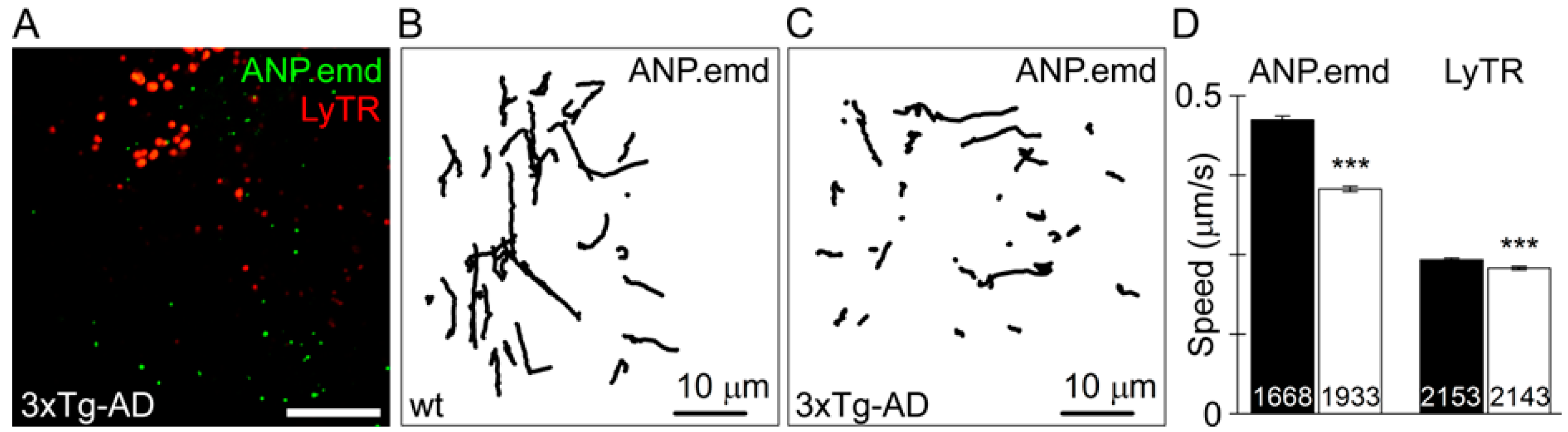
5. Vesicle Traffic, Surface Signalling Landscape of Astrocytes, and Neurodegeneration

Vesicle traffic, with a dynamics in minutes, contributes to morphological plasticity of all cells, also in pathology, since membrane added to the plasmalemma during exocytosis and then taken away by endocytosis may change drastically with disease. The consequences of imbalanced exo- and endocytotic vesicle traffic alters cell surface signalling landscape of astrocytes [70]. Vesicles, carrying signalling molecules in their lumen as well as receptors and transporters in their membrane, usually originate in the secretory pathway, from the Golgi complex, deep in the cytoplasm, and are then trafficked to the cell surface. Once the vesicle membrane merges completely with the plasmalemma, this vesicle incorporated membrane may, at a later stage, give birth to an endocytotic vesicle which can travel into the cytoplasm or may recycle back to the plasma membrane.
In astrocytes vesicle traffic is maintained by an elaborated system regulated by fluctuations in [Ca2+]i [70,71]. The complexity of vesicle traffic regulation in astrocytes is characterized by two typical, yet opposing, properties of vesicles that contain peptides, such as atrial natriuretic peptide (ANP), and those that carry amino acid transmitters and are labelled by the vesicular glutamate transporter VGLUT1 [70,71,72]. Glutamatergic vesicle mobility is accelerated by an increase in [Ca2+]i [73], whereas the same increase in [Ca2+]i slows down peptidergic vesicles and endolysosomes [74]. Similar regulation also applies to recycling peptidergic vesicles, which have merged with the plasma membrane and subsequently entered back into the cytoplasm. The mobility of recycling peptidergic vesicles was studied in astrocytes in culture [75] and in the brain slices [76]. In these two preparations, at rest, peptidergic vesicles moved faster and more directionally, being linked to cytoskeletal elements [75]. The effect of increased [Ca2+]i was remarkable: the movement of vesicles was almost halted, with only a jitter remaining (that was associated with random diffusional movement). At least some of the peptidergic vesicles carry ATP and a similar attenuation of their mobility was observed when astrocytes were stimulated [77].
When vesicle mobility is suppressed, their increased residency at the plasma membrane may increase probability that the vesicle membrane interacts with the plasma membrane. This is facilitated by the n-ethylmaleimide-sensitive fusion factor attachment protein receptor (SNARE) complex formation [78]. By using a dominant-negative domain of synaptobrevin 2 protein (dnSNARE), a vesicle-based SNARE protein, it was shown, at the level of single astrocytic vesicle, that disassembly of the SNARE complex mediates the full merger between the vesicle and the plasma membranes [79]. In other words, when a vesicle interacts with the plasma membrane, for which SNARE complex formation is needed, and when the SNARE complex cannot be disassembled, fusion pore maintains in open and stable configuration while the vesicle membrane is unable to merge completely with the plasmalemma. This process may be regulated by endogenous lipids, such as sphingosine, which can be generated on the outer leaflet of the plasmalemma, followed by subsequent internalization across the plasma membrane into the cell [80,81]. Sphingosine is considered to facilitate regulated exocytosis by recruiting vesicle synaptobrevin 2 for the SNARE complex formation [82]. It appeared that sphingosine modulates the formation of the fusion pore, the latter being a channel that forms between the vesicle and the plasma membrane, a process that is also strongly influenced by vesicle size in endocrine cells [83] and in astrocytes [79]. In astrocytes, sphingosine analogue FTY720 (fingolimod), currently used for treatment of multiple sclerosis, strongly affects vesicle mobility and the release of peptides from a single vesicle [84]. This is associated with a change in Ca2+ homeostasis in astrocytes [85].
The process of vesicle interaction with the plasma membrane is also a target of ketamine, an anaesthetic and an antidepressant. Unlike classical antidepressants, ketamine demonstrates both fast and sustained effects [86], indicative of a fundamentally different mechanism of action that may alter synaptic efficacy [87]. Ketamine affects astrocytes by inhibiting the release of brain derived neurotrophic factor BDNF [88]. Membrane capacitance measurements (used to monitor the interaction of a single astrocytic vesicle membrane with the plasmalemma) revealed that sub-anaesthetic doses of ketamine stabilize the fusion pore in a narrow flickering state [89], the latter being too narrow to allow the discharge of vesicle content into the extracellular medium [88]. Therefore, vesicle traffic and the fusion pore can be a target for therapy, possibly not only for treating depression and neurodegeneration in multiple sclerosis, but also for other neurological conditions.
In neurodegeneration, as in AD, a vesicular traffic deficit could be a primary mechanism of the early, pre-symptomatic stage of the disease, possibly associated with alterations in the signalling profile of astrocytes [70] and the removal of β-amyloid from the extracellular space [90]. For example, endolysosomes store and release proteolytic enzymes, such as insulin degrading enzyme (IDE), one of the major proteases of β-amyloid peptide, which may contribute to the development of AD. When secreted to the extracellular space IDE may degrade β-amyloid. While IDE is secreted primarily from neurones in the healthy brain [91], in AD astrocytes become the major cell type secreting IDE [90,92]. It has been proposed that in AD the capacity of astrocytes to secrete IDE is reduced when compared to healthy astrocytes. This leads to an increase in β-amyloid accumulation, which involves a reduction in autophagy-based lysosomal secretion of IDE [90]. Why this reduction occurs is not clear, but it may relate to a general vesicle traffic impairment that has been observed in AD and may involve lysosomal capacity to repair injured astrocytes [93].
Astrocytes from 3xTg-AD mice isolated in the pre-symptomatic phase of the disease exhibit alterations in vesicle traffic. Spontaneous motility of peptidergic and endolysosomal vesicles as well as the ATP-evoked, Ca2+-dependent, vesicle mobility are all diminished in diseased astrocytes (Figure 1). Similar impairment of peptidergic vesicle trafficking was observed in healthy rat astrocytes transfected with familial AD-associated mutated presenilin 1 (PS1M146V). The stimulation-dependent peptide discharge from single vesicles was less efficient in 3xTg-AD and PS1M146V-expressing astrocytes than in respective controls. The impaired vesicle dynamics and reduced evoked secretion of the signalling peptides may contribute to the development of AD [94]. Although in this study ANP-containing vesicles were examined, it is likely that all peptidergic vesicles exhibit similar changes. Moreover, peptidergic secretion appears to be impaired also in HD by mutated huntingtin [95].

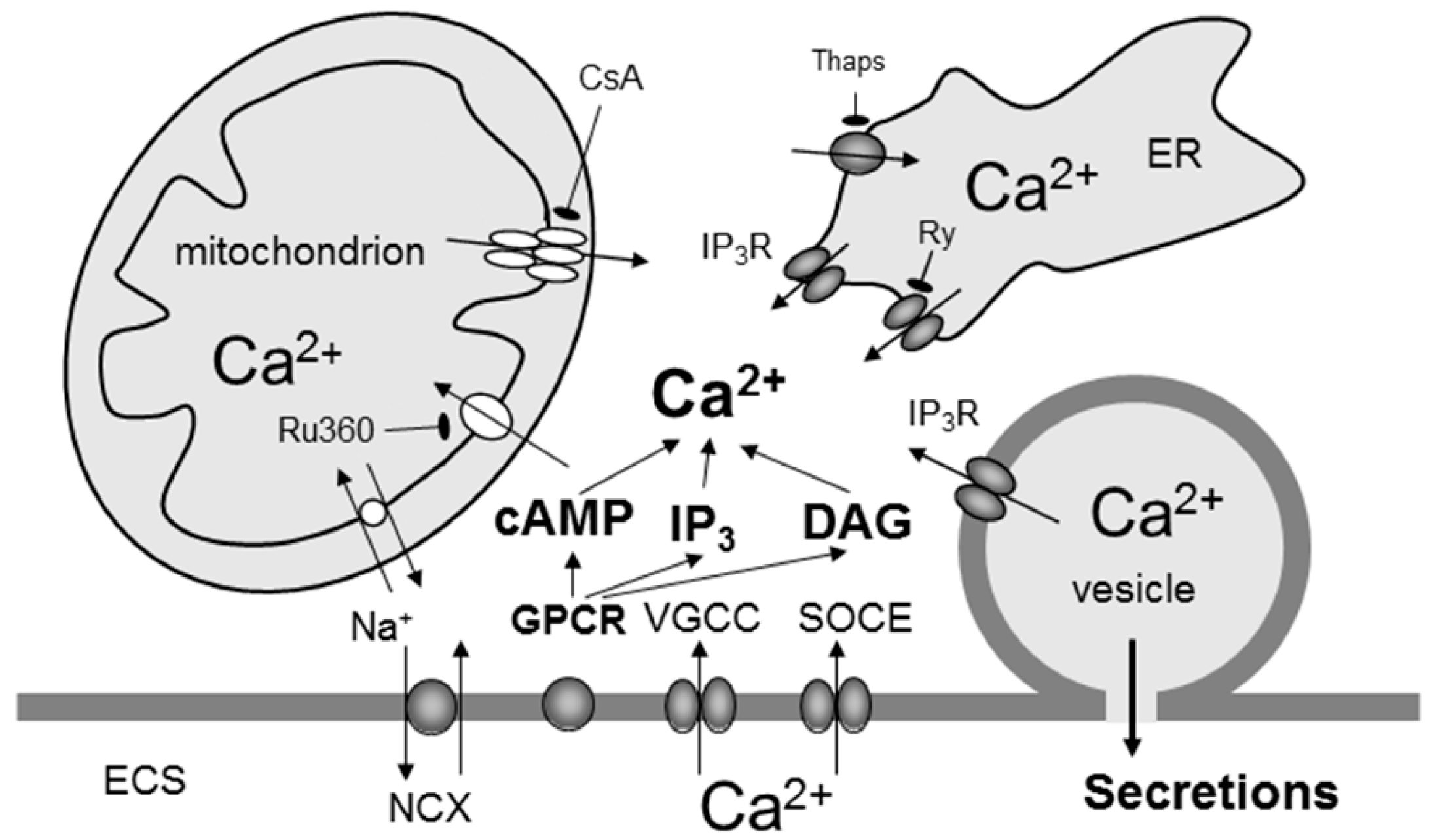
6. Calcium and cAMP Signalling in Astrocytes

Unlike neurones, which exhibit electrical excitability (firing action potentials generated at the level of the plasmalemma) that triggers neurotransmitter release from synaptic terminals, astrocytes are electrically silent and display intracellular excitability. The hallmark of cytosolic excitability is an increase in cytosolic levels of second messengers such as cyclic adenosine monophosphate (cAMP), Ca2+ and sodium (Na+) [96,97]. The overall evidence for Ca2+ as a second messengers in astrocytes exceeds that for cAMP, because Ca2+ recordings have been possible since 1990 [98], whereas real-time measurements of cAMP emerged only recently with the development of genetically encoded cAMP nanosensors [29]. It is generally acknowledged that changes in cytosolic Ca2+ follow a more phasic pattern, a consequence of the complex activation of various receptors, pumps and transporters; whereas cAMP levels change more tonically, following activation of cAMP-synthesizing enzyme adenylate cyclase [96,99].
Astrocytes as neuronal partners in the multipartite synapse use various receptors to sense neurotransmitters released during synaptic activity, but they also detect a host of other signalling molecules present in the brain parenchyma, being for example transported with the convective flow of CSF, which is a part of the glymphatic system [100]. Many of these molecules are released by astrocytes themselves [101]. Binding of signalling molecules to their receptors affects not only [Ca2+]i but also other cytosolic second messengers such as cAMP (Figure 2). For instance, binding of NA to astrocytic ARs triggers simultaneous, although temporally distinct, elevation of both second messengers in astrocytes. Such receptor mediated cytosolic excitability may lead to astroglial secretion of gliosignalling molecules, which in turn can interact with the receptors on synaptic terminals modulating neuronal excitability [102,103] or affect receptors on other neighbouring cells in a paracrine or autocrine manner [96,101].
Many of astroglial membrane receptors are high affinity metabotropic G protein-coupled receptors (GPCRs) [102,105,106]. Extracellular ligands activate GPCRs, which elicit a conformational change transmitting the signal to an attached intracellular heterotrimeric G protein complex. G protein subunit isoforms trigger distinct signalling pathways. For example, Gq subunit activation results in the stimulation of phospholipase C, which increases the concentration of diacylglycerol and inositol triphosphate (InsP3). In astrocytes, binding of InsP3 to InsP3Rs located on the endoplasmic reticulum (ER) [107] or on secretory vesicles [108] increases [Ca2+]i through the release of Ca2+ from these organelles. Activation of ryanodine receptors on the ER may also increase cytosolic Ca2+ levels through Ca2+-induced release of Ca2+ from the ER [107]. The Ca2+ signal arising from the generation of InsP3 may be amplified by activating further Ca2+ release from InsP3Rs and ryanodine receptors [109]. In addition, mitochondria, otherwise acting as metabolic furnaces, have a role in Ca2+ buffering in astrocytes through Ca2+ uptake and release [110,111]. The Ca2+ can also enter astrocytes from the extracellular space through plasmalemmal Ca2+ channels [112,113,114], ionotropic receptors [115], and the Na+/Ca2+ exchanger [116] as well as through the transient receptor potential canonical type 1-channel [117] acting as a substrate for store-operated Ca2+ entry that, along with store-specific Ca2+-ATPase, replenish the depleted ER store. Although the mechanisms that can contribute to the elevation in cytosolic Ca2+ are many, it is universally accepted that the main pathway for global Ca2+ signalling is represented by GPCR activation and release of Ca2+ from InsP3-sensitive internal stores. Unlike Gq GPCR activation, stimulation of Gs GPCR subunits in astrocytes triggers adenylyl cyclase to catalyse the conversion of ATP to cAMP [29,118]. This cAMP activates intracellular effectors, primarily cAMP-dependent protein kinase A, which, by phosphorylating cytoplasmic and nuclear targets, mediates functional responses. Signalling via cAMP-activated GTP-exchange protein [119], cAMP-gated ion channels, and Popeye domain-containing proteins [120] may also contribute [121].
In astrocytes, Gq-induced cytosolic Ca2+ increases occur as either oscillations or sustained elevations [102,105,122]. Astroglial Ca2+ excitability has been observed in culture [98], in brain slices in situ [123], and in vivo [124] and may occur spontaneously or in response to neuroligands, including neurotransmitters [98]. Ca2+ excitability can propagate from an excited astrocyte to its neighbouring unstimulated astrocytes in the form of intercellular Ca2+ waves, which are carried by diffusion of InsP3 through gap junctions [125] or via astrocytic release of ATP and subsequent receptor-mediated activation of adjacent astrocytes [126]. These waves travel at 10–20 μm/s [109]. In contrast, Gs activation induces persistent cAMP elevations [29,96]. It is currently unclear whether Gs-induced cAMP excitability can be propagated in astroglial syncytium, as was observed for Ca2+ excitability. The Gq- and Gs-mediated pathways in astrocytes may interact, as activation of the Gs-signalling pathway may potentiate Gq-mediated Ca2+ responses [127] and vice versa [128,129].
Both Gq and Gs GPCR signalling pathways were shown to be involved in exocytotic release of chemical messengers from astrocytes. It is well established that Ca2+ elevation in astrocytes triggers the exocytotic release of glutamate [130,131,132,133,134], ATP [135,136], secretogranin II [103], ANP [137], and d-serine [138]. The role of cAMP in vesicle-based release of gliosignalling molecules from astrocytes is much less studied. Limited results indicate that increases in cAMP can trigger the discharge of secretogranin II from peptidergic vesicles [103]. In agreement with this, augmented Ca2+-triggered release of ANP was measured under conditions when membrane permeable cAMP analogue dibutyryl cAMP was introduced prior to stimulating astrocytes [139]. The mechanism of cAMP modulation of fusion machinery is not known. It may affect the discharge of gliosignalling vesicles de novo. It may also modulate the fusion pore dynamics as described in neuroendocrine cells [140].
Activation of Ca2+ and cAMP is involved, among others, in the regulation of energy needed for the morphological plasticity in the CNS. Although synapses are the main energy consumers in the brain, glycogen, the only CNS energy storage system, is present mainly, if not exclusively, in astrocytes. Memory consolidation associated with morphological plasticity of neurones and astrocytes requires (in young chickens) glycogenolysis [141,142]. The release of NA is required for memory consolidation [143]. Since NA, released from noradrenergic LC neurones, stimulates relatively rapid changes in astrocyte morphology following changes in Ca2+ and cAMP, this indicates that astrocytes act as signal integrators; they coordinate morphological and metabolic functions [144]. It was reported that NA is the main neurotransmitter that triggers astroglial Ca2+ signalling in the adult awake brain [5]. In addition it may trigger astroglial cAMP signalling via β-AR activation. Thus, astrocytes are hubs of NA mediated excitation-energy coupling in the CNS [144].

7. Altered Astroglial Calcium Homeostasis in Alzheimer’s Disease

Given that knowledge about Ca2+ signalling is far greater than that about cAMP signalling, it is logical that current views about alterations of astrocyte excitability focus mainly into Ca2+ homeostasis. In human astrocytes, isolated from the post-mortem temporal cortex obtained from three groups with different degrees of pathology in PD and AD (different Braak scores) 32 genes associated with Ca2+ signalling and homeostasis were identified to be abnormally expressed [145].
When astrocytes are exposed to β-amyloid, complex effects on intracellular Ca2+ signalling are observed [146,147] which may increase glutamate release from human astrocytes [148]. Resting [Ca2+]i was increased several fold in astroglial cultures treated, for several hours, with oligomeric β-amyloid1–42 [149,150]. This was, however, not confirmed, when longer treatment times were used (e.g., 48 h exposure) and other amyloid peptides such as β-amyloid1–40 [151] or β-amyloid25–35 [152] were employed. In dissociated and organotypic brain cultures astrocytes may respond to β-amyloid treatment with the acute intracellular Ca2+ elevations, sometimes Ca2+ oscillations [153,154,155,156,157]. This was however not confirmed in some other studies [150,151,152]. The observed differences may be due to different species and concentrations of β-amyloid used (higher concentrations trigger Ca2+ elevations more readily). It has been suggested that β-amyloid can generally increase astroglial Ca2+ excitability [146,147] by increasing intracellular Ca2+ release and store-operated Ca2+ entry, SOCE [158,159].
An increased astroglial Ca2+ signalling has been reported in several AD animal models. In APP/PS1 mice [160] harbouring the mutant human β-amyloid precursor protein (APPswe) and mutant presenilin 1 (PS1ΔE9) an increase in the resting Ca2+ concentrations and Ca2+ hyperactivity with abnormal long-projecting intercellular Ca2+ waves were detected in astrocytes surrounding senile plaques. Similar high-frequency intercellular Ca2+ waves were detected in astrocytes at the pre-plaque stages in APPSwe mice model [161]. The underlying cause of increased Ca2+ excitability of astrocytes from AD model mice may be an elevated ATP release from reactive astrocytes that leads to over-activation of P2Y1 purinergic receptors [162].
Abnormal Ca2+ signalling could be associated with the mutated presenilin 1 as observed in primary astroglial cultures from neonatal 3xTg-AD mice [150,159]. In cultured hippocampal astrocytes from 3xTg-AD mice exposure to ATP triggered larger Ca2+ oscillations compared to the control cells [163]. The SOCE was also increased in cultured 3xTg-AD astrocytes [159]. Moreover, expression of mutant presenilin 1 (M146V) impaired vesicular trafficking and secretion [94]. The above mentioned abnormalities are part of early pathological remodelling of astroglia and may be involved in development of AD pathology. In addition, deletion of amyloid precursor protein inhibited astroglial SOCE, likely via down-regulation of TRPC1 and Orai 1 Ca2+ channel expression [164]. On the contrary the over-expression of APP did not affect Ca2+ transients and SOCE in primary cultured astroglial cells from Tg5469 AD mice [164]. Resting Ca2+ levels were elevated two-fold compared to the controls also in astrocytes obtained from a Down syndrome mouse model (Trisomy 16 mice) that shares several key features with AD [165]. When these astrocytes were exposed to a sarco-endoplasmic reticulum Ca2+ transport ATPase (SERCA) inhibitor cyclopiazonic acid, which unmasks leakage of Ca2+ from the ER, large intracellular Ca2+ elevations were observed, reflecting higher ER Ca2+ content compared to controls. A positive correlation between the amplitude of cyclopiazonic acid-induced intracellular Ca2+ elevations and the resting Ca2+ levels was observed [165].
Chronic long-term exposure to β-amyloid changes the expression of ionotropic and metabotropic receptors, Ca2+-dependent enzymes, intracellular Ca2+ channels and SOCE. Upregulation of metabotropic glutamate receptor mGluR5 has been observed in vitro upon 24–72 h exposure of cultured astrocytes to oligomeric β-amyloid (100 nM to 20 μM) [150,151,163] and also in the brains of patients with Down’s syndrome [166], in cortical astrocytes found in the APPswe/PS1ΔE9 mice senile plaques [167], in post-mortem hippocampi of Braak V–VI stage AD patients [150] and late-stage sporadic AD cases [151]. Chronic exposure to low concentrations of β-amyloid1–42 (0.1–100 nM; [168]) increased expression of Ca2+ permeable α7 nicotinic cholinoreceptors in cultured astrocytes. This receptor has been found post-mortem in astrocytes of sporadic AD patients and in patients carrying the Swedish β-amyloid precursor protein mutation [169]. Another possible mechanism for observed abnormalities in astroglial Ca2+ signalling in AD pathology could result from direct β-amyloid activation of metabotropic receptors [170,171].

8. Conclusions

There are conditions where relatively large areas of the CNS need to be synchronously activated. This involves the stimulation of LC neurones and NA release, which primarily excites astrocytes, since the density of adrenergic receptors is almost an order of magnitude higher in astroglia vs. neurones [31,32]. Thus, primarily astrocytes respond to noradrenergic activation with elevations of intracellular Ca2+ and cAMP. Increased Ca2+ and cAMP excitability that occurs at short time-scale regulates astrocyte cellular metabolism, morphology and vesicle traffic. The latter controls the surface signalling properties of astrocytes and their capacity to provide sufficient amounts of energy via augmented aerobic glycolysis. These processes may be altered during decades in neurodegeneration, such as taking place in AD. If LC neurones, which are vulnerable due to their relatively high metabolic rate, are reduced in numbers, e.g., during neurodegeneration, NA-mediated synchronous brain “reset” is impaired. On the other hand, even if the LC neurones are intact, alterations in vesicle traffic of astrocytes, affecting signalling landscape of these cells, together with impaired astroglial Ca2+ and possibly cAMP systems may also play an additional role in facilitating the long-term course of neurodegeneration. Since the excitation-energy coupling occurs mainly in the astrocytes, more emphasis is needed to understand how astrocytes interact with the noradrenergic signals at the level of both Ca2+ and cAMP signalling, and how they cope with the demand of coherent spatio-temporal signalling in the relatively large brain areas taking place in a long-time domain.

Acknowledgments

This work was supported by the Slovenian Research Agency; grant numbers P3 310, J3 3632, J3 4051, J3-4146, J3 6790, J3 7605, EU COST Action CM1207-GLISTEN.

Author Contributions

Robert Zorec wrote the first draft of the review; Nina Vardjan and Alexei Verkhratsky made comments and wrote additional sections of the manuscript.

Conflicts of Interest

The authors declare no conflict of interest.

Abbreviations

CNScentral nervous system
CSFcerebrospinal fluid
MSmultiple sclerosis
ADAlzheimer’s disease
PDParkinson’s disease
HDHuntington disease
ALSamyotrophic lateral sclerosis
PKAprotein kinase A
PKCprotein kinase C
IP3inositol triphosphate
GFAPglial fibrillary acidic protein
SNAREsoluble n-ethylmaleimide-sensitive fusion factor attachment protein receptor
cAMPadenosine monophosphate
ERendoplasmic reticulum
ANPatrial natriuretic peptide
LClocus coeruleus
GPCRG protein-coupled receptors
SOCEstore-operated Ca2+ entry

References

  1. Jacobsohn, L. Über die Kerne des Menschlichen Hirnstamms. (Meddulla Oblongata, Pons und Pedunculus Cerebri); Verlag der Konigl. Akademie der Wissenschaften: Berlin, Germany, 1909; p. 70. (In German) [Google Scholar]
  2. Feinstein, D.L.; Kalinin, S.; Braun, D. Causes, consequences, and cures for neuroinflammation mediated via the locus coeruleus: Noradrenergic signaling system. J. Neurochem. 2016, 139, 154–178. [Google Scholar] [CrossRef] [PubMed]
  3. Foote, S.L.; Bloom, F.E.; Aston-Jones, G. Nucleus locus coeruleus: New evidence of anatomical and physiological specificity. Physiol. Rev. 1983, 63, 844–914. [Google Scholar] [PubMed]
  4. Marien, M.R.; Colpaert, F.C.; Rosenquist, A.C. Noradrenergic mechanisms in neurodegenerative diseases: A theory. Brain Res. Brain Res. Rev. 2004, 45, 38–78. [Google Scholar] [CrossRef] [PubMed]
  5. Ding, F.; O’Donnell, J.; Thrane, A.S.; Zeppenfeld, D.; Kang, H.; Xie, L.; Wang, F.; Nedergaard, M. α1-Adrenergic receptors mediate coordinated Ca2+ signaling of cortical astrocytes in awake, behaving mice. Cell Calcium 2013, 54, 387–394. [Google Scholar] [CrossRef] [PubMed]
  6. Paukert, M.; Agarwal, A.; Cha, J.; Doze, V.A.; Kang, J.U.; Bergles, D.E. Norepinephrine controls astroglial responsiveness to local circuit activity. Neuron 2014, 82, 1263–1270. [Google Scholar] [CrossRef] [PubMed]
  7. Wilson, R.S.; Nag, S.; Boyle, P.A.; Hizel, L.P.; Yu, L.; Buchman, A.S.; Schneider, J.A.; Bennett, D.A. Neural reserve, neuronal density in the locus coeruleus, and cognitive decline. Neurology 2013, 80, 1202–1208. [Google Scholar] [CrossRef] [PubMed]
  8. Mouton, P.R.; Pakkenberg, B.; Gundersen, H.J.; Price, D.L. Absolute number and size of pigmented locus coeruleus neurons in young and aged individuals. J. Chem. Neuroanat. 1994, 7, 185–190. [Google Scholar] [CrossRef]
  9. Benarroch, E.E. The locus coeruleus norepinephrine system: Functional organization and potential clinical significance. Neurology 2009, 73, 1699–1704. [Google Scholar] [CrossRef] [PubMed]
  10. Chandler, D.J.; Gao, W.J.; Waterhouse, B.D. Heterogeneous organization of the locus coeruleus projections to prefrontal and motor cortices. Proc. Natl. Acad. Sci. USA 2014, 111, 6816–6821. [Google Scholar] [CrossRef] [PubMed]
  11. Zorec, R.; Vardjan, N.; Verkhratsky, A. Locus coeruleus noradrenergic neurons and astroglia in health and disease. In Noradrenergic Signaling and Astroglia; Vardjan, N., Zorec, R., Eds.; Elsevier: New York, NY, USA, 2017; Volume 1, pp. 1–10. [Google Scholar]
  12. Sara, S.J. Locus coeruleus in time with the making of memories. Curr. Opin. Neurobiol. 2015, 35, 87–94. [Google Scholar] [CrossRef] [PubMed]
  13. Bouret, S.; Sara, S.J. Network reset: A simplified overarching theory of locus coeruleus noradrenaline function. Trends Neurosci. 2005, 28, 574–582. [Google Scholar] [CrossRef] [PubMed]
  14. Findley, K.H.; Cobb, S. The capillary bed of the locus coeruleus. J. Comp. Neurol. 1940, 73, 49–58. [Google Scholar] [CrossRef]
  15. Sanchez-Padilla, J.; Guzman, J.N.; Ilijic, E.; Kondapalli, J.; Galtieri, D.J.; Yang, B.; Schieber, S.; Oertel, W.; Wokosin, D.; Schumacker, P.T.; et al. Mitochondrial oxidant stress in locus coeruleus is regulated by activity and nitric oxide synthase. Nat. Neurosci. 2014, 17, 832–840. [Google Scholar] [CrossRef] [PubMed]
  16. Pamphlett, R. Uptake of environmental toxicants by the locus coeruleus: A potential trigger for neurodegenerative, demyelinating and psychiatric disorders. Med. Hypotheses 2014, 82, 97–104. [Google Scholar] [CrossRef] [PubMed]
  17. Mather, M.; Harley, C.W. The locus coeruleus: Essential for maintaining cognitive function and the aging brain. Trends Cogn. Sci. 2016, 20, 214–226. [Google Scholar] [CrossRef] [PubMed]
  18. Mravec, B.; Lejavova, K.; Cubinkova, V. Locus coeruleus minoris resistentiae in pathogenesis of Alzheimer’s disease. Curr. Alzheimer Res. 2014, 11, 992–1001. [Google Scholar] [CrossRef] [PubMed]
  19. Lauder, J.M.; Bloom, F.E. Ontogeny of monoamine neurons in the locus coeruleus, Raphe nuclei and substantia nigra of the rat. I. Cell differentiation. J. Comp. Neurol. 1974, 155, 469–481. [Google Scholar] [CrossRef] [PubMed]
  20. Pickel, V.M.; Specht, L.A.; Sumal, K.K.; Joh, T.H.; Reis, D.J.; Hervonen, A. Immunocytochemical localization of tyrosine hydroxylase in the human fetal nervous system. J. Comp. Neurol. 1980, 194, 465–474. [Google Scholar] [CrossRef] [PubMed]
  21. Latsari, M.; Dori, I.; Antonopoulos, J.; Chiotelli, M.; Dinopoulos, A. Noradrenergic innervation of the developing and mature visual and motor cortex of the rat brain: A light and electron microscopic immunocytochemical analysis. J. Comp. Neurol. 2002, 445, 145–158. [Google Scholar] [CrossRef] [PubMed]
  22. D’Arcangelo, G.; Miao, G.G.; Chen, S.C.; Soares, H.D.; Morgan, J.I.; Curran, T. A protein related to extracellular matrix proteins deleted in the mouse mutant reeler. Nature 1995, 374, 719–723. [Google Scholar] [CrossRef] [PubMed]
  23. Frotscher, M. Cajal–Retzius cells, Reelin, and the formation of layers. Curr. Opin. Neurobiol. 1998, 8, 570–575. [Google Scholar] [CrossRef]
  24. Marin-Padilla, M. Cajal–Retzius cells and the development of the neocortex. Trends Neurosci. 1998, 21, 64–71. [Google Scholar] [CrossRef]
  25. Naqui, S.Z.; Harris, B.S.; Thomaidou, D.; Parnavelas, J.G. The noradrenergic system influences the fate of Cajal–Retzius cells in the developing cerebral cortex. Brain Res. Dev. Brain Res. 1999, 113, 75–82. [Google Scholar] [CrossRef]
  26. Guček, A.; Vardjan, N.; Zorec, R. Exocytosis in astrocytes: Transmitter release and membrane signal regulation. Neurochem. Res. 2012, 37, 2351–2363. [Google Scholar] [CrossRef] [PubMed]
  27. Xie, L.; Kang, H.; Xu, Q.; Chen, M.J.; Liao, Y.; Thiyagarajan, M.; O’Donnell, J.; Christensen, D.J.; Nicholson, C.; Iliff, J.J.; et al. Sleep drives metabolite clearance from the adult brain. Science 2013, 342, 373–377. [Google Scholar] [CrossRef] [PubMed]
  28. Sherpa, A.D.; Xiao, F.; Joseph, N.; Aoki, C.; Hrabetova, S. Activation of β-adrenergic receptors in rat visual cortex expands astrocytic processes and reduces extracellular space volume. Synapse 2016, 70, 307–316. [Google Scholar] [CrossRef] [PubMed]
  29. Vardjan, N.; Kreft, M.; Zorec, R. Dynamics of β-adrenergic/cAMP signaling and morphological changes in cultured astrocytes. Glia 2014, 62, 566–579. [Google Scholar] [CrossRef] [PubMed]
  30. Vardjan, N.; Horvat, A.; Anderson, J.E.; Yu, D.; Croom, D.; Zeng, X.; Luznik, Z.; Kreft, M.; Teng, Y.D.; Kirov, S.A.; et al. Adrenergic activation attenuates astrocyte swelling induced by hypotonicity and neurotrauma. Glia 2016, 64, 1034–1049. [Google Scholar] [CrossRef] [PubMed]
  31. Aoki, C. β-Adrenergic receptors: Astrocytic localization in the adult visual cortex and their relation to catecholamine axon terminals as revealed by electron microscopic immunocytochemistry. J. Neurosci. 1992, 12, 781–792. [Google Scholar] [PubMed]
  32. Pankratov, Y.; Lalo, U. Role for astroglial α1-adrenoreceptors in gliotransmission and control of synaptic plasticity in the neocortex. Front. Cell. Neurosci. 2015, 9, 230. [Google Scholar] [CrossRef] [PubMed]
  33. Ostroff, L.E.; Manzur, M.K.; Cain, C.K.; Ledoux, J.E. Synapses lacking astrocyte appear in the amygdala during consolidation of Pavlovian threat conditioning. J. Comp. Neurol. 2014, 522, 2152–2163. [Google Scholar] [CrossRef] [PubMed]
  34. Vardjan, N.; Kreft, M.; Zorec, R. Regulated exocytosis in astrocytes is as slow as the metabolic availability of gliotransmitters: Focus on glutamate and ATP. Adv. Neurobiol. 2014, 11, 81–101. [Google Scholar] [PubMed]
  35. Salcedo-Sora, J.E.; Caamano-Gutierrez, E.; Ward, S.A.; Biagini, G.A. The proliferating cell hypothesis: A metabolic framework for Plasmodium growth and development. Trends Parasitol. 2014, 30, 170–175. [Google Scholar] [CrossRef] [PubMed] [Green Version]
  36. Vander Heiden, M.G.; Cantley, L.C.; Thompson, C.B. Understanding the Warburg effect: The metabolic requirements of cell proliferation. Science 2009, 324, 1029–1033. [Google Scholar] [CrossRef] [PubMed]
  37. Tech, K.; Gershon, T.R. Energy metabolism in neurodevelopment and medulloblastoma. Transl. Pediatr. 2015, 4, 12–19. [Google Scholar] [PubMed]
  38. Goyal, M.S.; Hawrylycz, M.; Miller, J.A.; Snyder, A.Z.; Raichle, M.E. Aerobic glycolysis in the human brain is associated with development and neotenous gene expression. Cell. Metab. 2014, 19, 49–57. [Google Scholar] [CrossRef] [PubMed]
  39. Dienel, G.A.; Cruz, N.F. Aerobic glycolysis during brain activation: Adrenergic regulation and influence of norepinephrine on astrocytic metabolism. J. Neurochem. 2016. [Google Scholar] [CrossRef] [PubMed]
  40. Lee, H.J.; Cho, E.D.; Lee, K.W.; Kim, J.H.; Cho, S.G.; Lee, S.J. Autophagic failure promotes the exocytosis and intercellular transfer of α-synuclein. Exp. Mol. Med. 2013, 45, e22. [Google Scholar] [CrossRef] [PubMed]
  41. Kirov, S.A.; Sorra, K.E.; Harris, K.M. Slices have more synapses than perfusion-fixed hippocampus from both young and mature rats. J. Neurosci. 1999, 19, 2876–2886. [Google Scholar] [PubMed]
  42. Bushong, E.A.; Martone, M.E.; Jones, Y.Z.; Ellisman, M.H. Protoplasmic astrocytes in CA1 stratum radiatum occupy separate anatomical domains. J. Neurosci. 2002, 22, 183–192. [Google Scholar] [PubMed]
  43. Oberheim, N.A.; Wang, X.; Goldman, S.; Nedergaard, M. Astrocytic complexity distinguishes the human brain. Trends Neurosci. 2006, 29, 547–553. [Google Scholar] [CrossRef] [PubMed]
  44. Sutin, J.; Shao, Y. Resting and reactive astrocytes express adrenergic receptors in the adult rat brain. Brain Res. Bull. 1992, 29, 277–284. [Google Scholar] [CrossRef]
  45. Zeinstra, E.; Wilczak, N.; de Keyser, J. [3H]dihydroalprenolol binding to β-adrenergic receptors in multiple sclerosis brain. Neurosci. Lett. 2000, 289, 75–77. [Google Scholar] [CrossRef]
  46. Catus, S.L.; Gibbs, M.E.; Sato, M.; Summers, R.J.; Hutchinson, D.S. Role of β-adrenoceptors in glucose uptake in astrocytes using β-adrenoceptor knockout mice. Br. J. Pharmacol. 2011, 162, 1700–1715. [Google Scholar] [CrossRef] [PubMed]
  47. Hatton, G.I.; Luckman, S.M.; Bicknell, R.J. Adrenalin activation of β2-adrenoceptors stimulates morphological changes in astrocytes (pituicytes) cultured from adult rat neurohypophyses. Brain Res. Bull. 1991, 26, 765–769. [Google Scholar] [CrossRef]
  48. Shain, W.; Forman, D.S.; Madelian, V.; Turner, J.N. Morphology of astroglial cells is controlled by β-adrenergic receptors. J. Cell. Biol. 1987, 105, 2307–2314. [Google Scholar] [CrossRef] [PubMed]
  49. Bicknell, R.J.; Luckman, S.M.; Inenaga, K.; Mason, W.T.; Hatton, G.I. β-Adrenergic and opioid receptors on pituicytes cultured from adult rat neurohypophysis: Regulation of cell morphology. Brain Res. Bull. 1989, 22, 379–388. [Google Scholar] [CrossRef]
  50. Griffith, R.; Sutin, J. Reactive astrocyte formation in vivo is regulated by noradrenergic axons. J. Comp. Neurol. 1996, 371, 362–375. [Google Scholar] [CrossRef]
  51. Sutin, J.; Griffith, R. β-adrenergic receptor blockade suppresses glial scar formation. Exp. Neurol. 1993, 120, 214–222. [Google Scholar] [CrossRef] [PubMed]
  52. Johansen, J.P.; Diaz-Mataix, L.; Hamanaka, H.; Ozawa, T.; Ycu, E.; Koivumaa, J.; Kumar, A.; Hou, M.; Deisseroth, K.; Boyden, E.S.; et al. Hebbian and neuromodulatory mechanisms interact to trigger associative memory formation. Proc. Natl. Acad. Sci. USA 2014, 111, 5584–5592. [Google Scholar] [CrossRef] [PubMed]
  53. Verkhratsky, A.; Zorec, R.; Rodríguez, J.J.; Parpura, V. Astroglia dynamics in ageing and Alzheimer’s disease. Curr. Opin. Pharmacol. 2016, 26, 74–79. [Google Scholar] [CrossRef] [PubMed]
  54. Rodriguez-Vieitez, E.; Saint-Aubert, L.; Carter, S.F.; Almkvist, O.; Farid, K.; Scholl, M.; Chiotis, K.; Thordardottir, S.; Graff, C.; Wall, A.; et al. Diverging longitudinal changes in astrocytosis and amyloid PET in autosomal dominant Alzheimer’s disease. Brain 2016, 139, 922–936. [Google Scholar] [CrossRef] [PubMed]
  55. Anderson, M.A.; Burda, J.E.; Ren, Y.; Ao, Y.; O’Shea, T.M.; Kawaguchi, R.; Coppola, G.; Khakh, B.S.; Deming, T.J.; Sofroniew, M.V. Astrocyte scar formation aids central nervous system axon regeneration. Nature 2016, 532, 195–200. [Google Scholar] [CrossRef] [PubMed]
  56. Pekny, M.; Pekna, M.; Messing, A.; Steinhauser, C.; Lee, J.M.; Parpura, V.; Hol, E.M.; Sofroniew, M.V.; Verkhratsky, A. Astrocytes: A central element in neurological diseases. Acta Neuropathol. 2016, 131, 323–345. [Google Scholar] [CrossRef] [PubMed]
  57. Rossi, D.; Brambilla, L.; Valori, C.F.; Roncoroni, C.; Crugnola, A.; Yokota, T.; Bredesen, D.E.; Volterra, A. Focal degeneration of astrocytes in amyotrophic lateral sclerosis. Cell. Death Differ. 2008, 15, 1691–1700. [Google Scholar] [CrossRef] [PubMed]
  58. Valori, C.F.; Brambilla, L.; Martorana, F.; Rossi, D. The multifaceted role of glial cells in amyotrophic lateral sclerosis. Cell. Mol. Life Sci. 2014, 71, 287–297. [Google Scholar] [CrossRef] [PubMed]
  59. Rossi, D. Astrocyte physiopathology: At the crossroads of intercellular networking, inflammation and cell death. Prog. Neurobiol. 2015, 130, 86–120. [Google Scholar] [CrossRef] [PubMed]
  60. Yamanaka, K.; Chun, S.J.; Boillee, S.; Fujimori-Tonou, N.; Yamashita, H.; Gutmann, D.H.; Takahashi, R.; Misawa, H.; Cleveland, D.W. Astrocytes as determinants of disease progression in inherited amyotrophic lateral sclerosis. Nat. Neurosci. 2008, 11, 251–253. [Google Scholar] [CrossRef] [PubMed]
  61. Hazell, A.S. Astrocytes are a major target in thiamine deficiency and Wernicke’s encephalopathy. Neurochem. Int. 2009, 55, 129–135. [Google Scholar] [CrossRef] [PubMed]
  62. Hazell, A.S.; Sheedy, D.; Oanea, R.; Aghourian, M.; Sun, S.; Jung, J.Y.; Wang, D.; Wang, C. Loss of astrocytic glutamate transporters in Wernicke encephalopathy. Glia 2009, 58, 148–156. [Google Scholar] [CrossRef] [PubMed]
  63. Lee, W.; Reyes, R.C.; Gottipati, M.K.; Lewis, K.; Lesort, M.; Parpura, V.; Gray, M. Enhanced Ca2+-dependent glutamate release from astrocytes of the BACHD Huntington’s disease mouse model. Neurobiol. Dis. 2013, 58, 192–199. [Google Scholar] [CrossRef] [PubMed]
  64. Ben Haim, L.; Ceyzeriat, K.; Carrillo de Sauvage, M.A.; Aubry, F.; Auregan, G.; Guillermier, M.; Ruiz, M.; Petit, F.; Houitte, D.; Faivre, E.; et al. The JAK/STAT3 pathway is a common inducer of astrocyte reactivity in Alzheimer’s and Huntington’s diseases. J. Neurosci. 2015, 35, 2817–2829. [Google Scholar] [CrossRef] [PubMed]
  65. Mena, M.A.; de Bernardo, S.; Casarejos, M.J.; Canals, S.; Rodriguez-Martin, E. The role of astroglia on the survival of dopamine neurons. Mol. Neurobiol. 2002, 25, 245–263. [Google Scholar] [CrossRef]
  66. Mena, M.A.; Garcia de Yebenes, J. Glial cells as players in parkinsonism: The “good”, the “bad”, and the “mysterious” glia. Neuroscientist 2008, 14, 544–560. [Google Scholar] [CrossRef] [PubMed]
  67. Mena, M.A.; Casarejos, M.J.; Carazo, A.; Paino, C.L.; Garcia de Yebenes, J. Glia conditioned medium protects fetal rat midbrain neurones in culture from L-DOPA toxicity. NeuroReport 1996, 7, 441–445. [Google Scholar] [CrossRef] [PubMed]
  68. Asanuma, M.; Miyazaki, I.; Murakami, S.; Diaz-Corrales, F.J.; Ogawa, N. Striatal astrocytes act as a reservoir for L-DOPA. PLoS ONE 2014, 9, e106362. [Google Scholar] [CrossRef] [PubMed]
  69. Tong, J.; Ang, L.C.; Williams, B.; Furukawa, Y.; Fitzmaurice, P.; Guttman, M.; Boileau, I.; Hornykiewicz, O.; Kish, S.J. Low levels of astroglial markers in Parkinson’s disease: Relationship to α-synuclein accumulation. Neurobiol. Dis. 2015, 82, 243–253. [Google Scholar] [CrossRef] [PubMed]
  70. Vardjan, N.; Verkhratsky, A.; Zorec, R. Pathologic potential of astrocytic vesicle traffic: New targets to treat neurologic diseases? Cell Transplant. 2015, 24, 599–612. [Google Scholar] [CrossRef] [PubMed]
  71. Potokar, M.; Vardjan, N.; Stenovec, M.; Gabrijel, M.; Trkov, S.; Jorgacevski, J.; Kreft, M.; Zorec, R. Astrocytic vesicle mobility in health and disease. Int. J. Mol. Sci. 2013, 14, 11238–11258. [Google Scholar] [CrossRef] [PubMed]
  72. Potokar, M.; Kreft, M.; Pangrsic, T.; Zorec, R. Vesicle mobility studied in cultured astrocytes. Biochem. Biophys. Res. Commun. 2005, 329, 678–683. [Google Scholar] [CrossRef] [PubMed]
  73. Stenovec, M.; Kreft, M.; Grilc, S.; Potokar, M.; Kreft, M.E.; Pangrsic, T.; Zorec, R. Ca2+-dependent mobility of vesicles capturing anti-VGLUT1 antibodies. Exp. Cell Res. 2007, 313, 3809–3818. [Google Scholar] [CrossRef] [PubMed]
  74. Potokar, M.; Stenovec, M.; Gabrijel, M.; Li, L.; Kreft, M.; Grilc, S.; Pekny, M.; Zorec, R. Intermediate filaments attenuate stimulation-dependent mobility of endosomes/lysosomes in astrocytes. Glia 2010, 58, 1208–1219. [Google Scholar] [CrossRef] [PubMed]
  75. Potokar, M.; Stenovec, M.; Kreft, M.; Kreft, M.E.; Zorec, R. Stimulation inhibits the mobility of recycling peptidergic vesicles in astrocytes. Glia 2008, 56, 135–144. [Google Scholar] [CrossRef] [PubMed]
  76. Potokar, M.; Kreft, M.; Lee, S.Y.; Takano, H.; Haydon, P.G.; Zorec, R. Trafficking of astrocytic vesicles in hippocampal slices. Biochem. Biophys. Res. Commun. 2009, 390, 1192–1196. [Google Scholar] [CrossRef] [PubMed]
  77. Pangrsic, T.; Potokar, M.; Stenovec, M.; Kreft, M.; Fabbretti, E.; Nistri, A.; Pryazhnikov, E.; Khiroug, L.; Giniatullin, R.; Zorec, R. Exocytotic release of ATP from cultured astrocytes. J. Biol. Chem. 2007, 282, 28749–28758. [Google Scholar] [CrossRef] [PubMed]
  78. Jorgacevski, J.; Potokar, M.; Grilc, S.; Kreft, M.; Liu, W.; Barclay, J.W.; Buckers, J.; Medda, R.; Hell, S.W.; Parpura, V.; et al. Munc18-1 tuning of vesicle merger and fusion pore properties. J. Neurosci. 2011, 31, 9055–9066. [Google Scholar] [CrossRef] [PubMed]
  79. Gucek, A.; Jorgacevski, J.; Singh, P.; Geisler, C.; Lisjak, M.; Vardjan, N.; Kreft, M.; Egner, A.; Zorec, R. Dominant negative SNARE peptides stabilize the fusion pore in a narrow, release-unproductive state. Cell. Mol. Life Sci. 2016, 73, 3719–3731. [Google Scholar] [CrossRef] [PubMed]
  80. Tani, M.; Sano, T.; Ito, M.; Igarashi, Y. Mechanisms of sphingosine and sphingosine 1-phosphate generation in human platelets. J. Lipid Res. 2005, 46, 2458–2467. [Google Scholar] [CrossRef] [PubMed]
  81. Hannun, Y.A.; Obeid, L.M. Principles of bioactive lipid signaling: Lessons from sphingolipids. Nat. Rev. Mol. Cell Biol. 2008, 9, 139–150. [Google Scholar] [CrossRef] [PubMed]
  82. Darios, F.; Wasser, C.; Shakirzyanova, A.; Giniatullin, A.; Goodman, K.; Munoz-Bravo, J.L.; Raingo, J.; Jorgacevski, J.; Kreft, M.; Zorec, R.; et al. Sphingosine facilitates SNARE complex assembly and activates synaptic vesicle exocytosis. Neuron 2009, 62, 683–694. [Google Scholar] [CrossRef] [PubMed]
  83. Flasker, A.; Jorgacevski, J.; Calejo, A.I.; Kreft, M.; Zorec, R. Vesicle size determines unitary exocytic properties and their sensitivity to sphingosine. Mol. Cell. Endocrinol. 2013, 376, 136–147. [Google Scholar] [CrossRef] [PubMed]
  84. Trkov, S.; Stenovec, M.; Kreft, M.; Potokar, M.; Parpura, V.; Davletov, B.; Zorec, R. Fingolimod—A sphingosine-like molecule inhibits vesicle mobility and secretion in astrocytes. Glia 2012, 60, 1406–1416. [Google Scholar] [CrossRef] [PubMed]
  85. Stenovec, M.; Trkov, S.; Kreft, M.; Zorec, R. Alterations of calcium homoeostasis in cultured rat astrocytes evoked by bioactive sphingolipids. Acta Physiol. 2014, 212, 49–61. [Google Scholar] [CrossRef] [PubMed]
  86. Henderson, T. Practical application of the neuroregenerative properties of ketamine: Real world treatment experience. Neur. Regen. Res. 2016, 11, 195–200. [Google Scholar] [CrossRef] [PubMed]
  87. Kavalali, E.T.; Monteggia, L.M. How does ketamine elicit a rapid antidepressant response? Curr. Opin. Pharmacol. 2014, 20, 35–39. [Google Scholar] [CrossRef] [PubMed]
  88. Stenovec, M.; Lasic, E.; Bozic, M.; Bobnar, S.T.; Stout, R.F., Jr.; Grubisic, V.; Parpura, V.; Zorec, R. Ketamine inhibits ATP-evoked exocytotic release of brain-derived neurotrophic factor from vesicles in cultured rat astrocytes. Mol. Neurobiol. 2015, 53, 6882–6896. [Google Scholar] [CrossRef] [PubMed]
  89. Lasic, E.; Rituper, B.; Jorgacevski, J.; Kreft, M.; Stenovec, M.; Zorec, R. Subanesthetic doses of ketamine stabilize the fusion pore in a narrow flickering state in astrocytes. J. Neurochem. 2016, 138, 909–917. [Google Scholar] [CrossRef] [PubMed]
  90. Son, S.M.; Cha, M.Y.; Choi, H.; Kang, S.; Lee, M.S.; Park, S.A.; Mook-Jung, I. Insulin-degrading enzyme secretion from astrocytes is mediated by an autophagy-based unconventional secretory pathway in Alzheimer’s disease. Autophagy 2016, 12, 784–800. [Google Scholar] [CrossRef] [PubMed]
  91. Vekrellis, K.; Ye, Z.; Qiu, W.Q.; Walsh, D.; Hartley, D.; Chesneau, V.; Rosner, M.R.; Selkoe, D.J. Neurons regulate extracellular levels of amyloid β-protein via proteolysis by insulin-degrading enzyme. J. Neurosci. 2000, 20, 1657–1665. [Google Scholar] [PubMed]
  92. Dorfman, V.B.; Pasquini, L.; Riudavets, M.; Lopez-Costa, J.J.; Villegas, A.; Troncoso, J.C.; Lopera, F.; Castano, E.M.; Morelli, L. Differential cerebral deposition of IDE and NEP in sporadic and familial Alzheimer’s disease. Neurobiol. Aging 2010, 31, 1743–1757. [Google Scholar] [CrossRef] [PubMed]
  93. Sreetama, S.C.; Takano, T.; Nedergaard, M.; Simon, S.M.; Jaiswal, J.K. Injured astrocytes are repaired by Synaptotagmin XI-regulated lysosome exocytosis. Cell Death Differ. 2016, 23, 596–607. [Google Scholar] [CrossRef] [PubMed]
  94. Stenovec, M.; Trkov, S.; Lasic, E.; Terzieva, S.; Kreft, M.; Rodriguez Arellano, J.J.; Parpura, V.; Verkhratsky, A.; Zorec, R. Expression of familial Alzheimer’s disease presenilin 1 gene attenuates vesicle traffic and reduces peptide secretion in cultured astrocytes devoid of pathologic tissue environment. Glia 2016, 64, 317–329. [Google Scholar] [CrossRef] [PubMed]
  95. Hong, Y.; Zhao, T.; Li, X.J.; Li, S. Mutant huntingtin impairs BDNF release from astrocytes by disrupting conversion of Rab3a-GTP into Rab3a-GDP. J. Neurosci. 2016, 36, 8790–8801. [Google Scholar] [CrossRef] [PubMed]
  96. Vardjan, N.; Zorec, R. Excitable Astrocytes: Ca2+- and cAMP-regulated exocytosis. Neurochem. Res. 2015, 40, 2414–2424. [Google Scholar] [CrossRef] [PubMed]
  97. Rose, C.R.; Verkhratsky, A. Principles of sodium homeostasis and sodium signaling in astroglia. Glia 2016, 64, 1611–1627. [Google Scholar] [CrossRef] [PubMed]
  98. Cornell-Bell, A.H.; Finkbeiner, S.M.; Cooper, M.S.; Smith, S.J. Glutamate induces calcium waves in cultured astrocytes: Long-range glial signaling. Science 1990, 247, 470–473. [Google Scholar] [CrossRef] [PubMed]
  99. Horvat, A.; Zorec, R.; Vardjan, N. Adrenergic stimulation of single rat astrocytes results in distinct temporal changes in intracellular Ca2+ and cAMP-dependent PKA responses. Cell Calcium 2016, 59, 156–163. [Google Scholar] [CrossRef] [PubMed]
  100. Thrane, A.S.; Rangroo Thrane, V.; Nedergaard, M. Drowning stars: Reassessing the role of astrocytes in brain edema. Trends Neurosci. 2014, 37, 620–628. [Google Scholar] [CrossRef] [PubMed]
  101. Verkhratsky, A.; Matteoli, M.; Parpura, V.; Mothet, J.P.; Zorec, R. Astrocytes as secretory cells of the central nervous system: Idiosyncrasies of vesicular secretion. EMBO J. 2016, 35, 239–257. [Google Scholar] [CrossRef] [PubMed]
  102. Parpura, V.; Verkhratsky, A. The astrocyte excitability brief: From receptors to gliotransmission. Neurochem. Int. 2012, 61, 610–621. [Google Scholar] [CrossRef] [PubMed]
  103. Calegari, F.; Coco, S.; Taverna, E.; Bassetti, M.; Verderio, C.; Corradi, N.; Matteoli, M.; Rosa, P. A regulated secretory pathway in cultured hippocampal astrocytes. J. Biol. Chem. 1999, 274, 22539–22547. [Google Scholar] [CrossRef] [PubMed]
  104. Vardjan, N.; Parpura, V.; Zorec, R. Loose excitation-secretion coupling in astrocytes. Glia 2016, 64, 655–667. [Google Scholar] [CrossRef] [PubMed]
  105. Zorec, R.; Araque, A.; Carmignoto, G.; Haydon, P.G.; Verkhratsky, A.; Parpura, V. Astroglial excitability and gliotransmission: An appraisal of Ca2+ as a signaling route. ASN Neuro 2012. [Google Scholar] [CrossRef] [PubMed]
  106. Agulhon, C.; Petravicz, J.; McMullen, A.B.; Sweger, E.J.; Minton, S.K.; Taves, S.R.; Casper, K.B.; Fiacco, T.A.; McCarthy, K.D. What is the role of astrocyte calcium in neurophysiology? Neuron 2008, 59, 932–946. [Google Scholar] [CrossRef] [PubMed]
  107. Hua, X.; Malarkey, E.B.; Sunjara, V.; Rosenwald, S.E.; Li, W.H.; Parpura, V. Ca2+-dependent glutamate release involves two classes of endoplasmic reticulum Ca2+ stores in astrocytes. J. Neurosci. Res. 2004, 76, 86–97. [Google Scholar] [CrossRef] [PubMed]
  108. Hur, Y.S.; Kim, K.D.; Paek, S.H.; Yoo, S.H. Evidence for the existence of secretory granule (dense-core vesicle)-based inositol 1,4,5-trisphosphate-dependent Ca2+ signaling system in astrocytes. PLoS ONE 2010, 5, e11973. [Google Scholar] [CrossRef] [PubMed]
  109. Leybaert, L.; Sanderson, M.J. Intercellular Ca2+ waves: Mechanisms and function. Physiol. Rev. 2012, 92, 1359–1392. [Google Scholar] [CrossRef] [PubMed]
  110. Reyes, R.C.; Parpura, V. Mitochondria modulate Ca2+-dependent glutamate release from rat cortical astrocytes. J. Neurosci. 2008, 28, 9682–9691. [Google Scholar] [CrossRef] [PubMed]
  111. Simpson, P.B.; Russell, J.T. Role of mitochondrial Ca2+-regulation in neuronal and glial cell signaling. Brain Res. Rev. 1998, 26, 72–81. [Google Scholar] [CrossRef]
  112. MacVicar, B.A. Voltage-dependent calcium channels in glial cells. Science 1984, 226, 1345–1347. [Google Scholar] [CrossRef] [PubMed]
  113. Latour, I.; Hamid, J.; Beedle, A.M.; Zamponi, G.W.; Macvicar, B.A. Expression of voltage-gated Ca2+ channel subtypes in cultured astrocytes. Glia 2003, 41, 347–353. [Google Scholar] [CrossRef] [PubMed]
  114. Parri, H.R.; Crunelli, V. Pacemaker calcium oscillations in thalamic astrocytes in situ. NeuroReport 2001, 12, 3897–3900. [Google Scholar] [CrossRef] [PubMed]
  115. Lalo, U.; Pankratov, Y.; Parpura, V.; Verkhratsky, A. Ionotropic receptors in neuronal-astroglial signaling: What is the role of “excitable” molecules in non-excitable cells. Biochim. Biophys. Acta 2011, 1813, 992–1002. [Google Scholar] [CrossRef] [PubMed]
  116. Reyes, R.C.; Verkhratsky, A.; Parpura, V. Plasmalemmal Na+/Ca2+ exchanger modulates Ca2+-dependent exocytotic release of glutamate from rat cortical astrocytes. ASN Neuro 2012. [Google Scholar] [CrossRef] [PubMed]
  117. Malarkey, E.B.; Ni, Y.; Parpura, V. Ca2+ entry through TRPC1 channels contributes to intracellular Ca2+ dynamics and consequent glutamate release from rat astrocytes. Glia 2008, 56, 821–835. [Google Scholar] [CrossRef] [PubMed]
  118. Rathbone, M.P.; Middlemiss, P.J.; DeLuca, B.; Jovetich, M. Extracellular guanosine increases astrocyte cAMP: Inhibition by adenosine A2 antagonists. NeuroReport 1991, 2, 661–664. [Google Scholar] [CrossRef] [PubMed]
  119. De Rooij, J.; Zwartkruis, F.J.; Verheijen, M.H.; Cool, R.H.; Nijman, S.M.; Wittinghofer, A.; Bos, J.L. Epac is a Rap1 guanine-nucleotide-exchange factor directly activated by cyclic AMP. Nature 1998, 396, 474–477. [Google Scholar] [PubMed]
  120. Froese, A.; Breher, S.S.; Waldeyer, C.; Schindler, R.F.; Nikolaev, V.O.; Rinné, S.; Wischmeyer, E.; Schlueter, J.; Becher, J.; Simrick, S.; et al. Popeye domain containing proteins are essential for stress-mediated modulation of cardiac pacemaking in mice. J. Clin. Investig. 2012, 122, 1119–1130. [Google Scholar] [CrossRef] [PubMed]
  121. Beavo, J.A.; Brunton, L.L. Cyclic nucleotide research—Still expanding after half a century. Nat. Rev. Mol. Cell Biol. 2002, 3, 710–718. [Google Scholar] [CrossRef] [PubMed]
  122. Volterra, A.; Liaudet, N.; Savtchouk, I. Astrocyte Ca²⁺ signaling: An unexpected complexity. Nat. Rev. Neurosci. 2014, 15, 327–335. [Google Scholar] [CrossRef] [PubMed]
  123. Pasti, L.; Volterra, A.; Pozzan, T.; Carmignoto, G. Intracellular calcium oscillations in astrocytes: A highly plastic, bidirectional form of communication between neurons and astrocytes in situ. J. Neurosci. 1997, 17, 7817–7830. [Google Scholar]
  124. Hirase, H.; Qian, L.; Barthó, P.; Buzsáki, G. Calcium dynamics of cortical astrocytic networks in vivo. PLoS Biol. 2004, 2, 96. [Google Scholar] [CrossRef] [PubMed] [Green Version]
  125. Scemes, E.; Suadicani, S.O.; Spray, D.C. Intercellular communication in spinal cord astrocytes: Fine tuning between gap junctions and P2 nucleotide receptors in calcium wave propagation. J. Neurosci. 2000, 20, 1435–1445. [Google Scholar] [PubMed]
  126. Bowser, D.N.; Khakh, B.S. Vesicular ATP is the predominant cause of intercellular calcium waves in astrocytes. J. Gen. Physiol. 2007, 129, 485–491. [Google Scholar] [CrossRef] [PubMed]
  127. Jiménez, A.I.; Castro, E.; Mirabet, M.; Franco, R.; Delicado, E.G.; Miras-Portugal, M.T. Potentiation of ATP calcium responses by A2B receptor stimulation and other signals coupled to Gs proteins in type-1 cerebellar astrocytes. Glia 1999, 26, 119–128. [Google Scholar] [CrossRef]
  128. Balázs, R.; Miller, S.; Chun, Y.; O’Toole, J.; Cotman, C.W. Metabotropic glutamate receptor agonists potentiate cyclic AMP formation induced by forskolin or β-adrenergic receptor activation in cerebral cortical astrocytes in culture. J. Neurochem. 1998, 70, 2446–2458. [Google Scholar] [CrossRef] [PubMed]
  129. Hansson, E.; Simonsson, P.; Alling, C. Interactions between cyclic AMP and inositol phosphate transduction systems in astrocytes in primary culture. Neuropharmacology 1990, 29, 591–598. [Google Scholar] [CrossRef]
  130. Parpura, V.; Basarsky, T.A.; Liu, F.; Jeftinija, K.; Jeftinija, S.; Haydon, P.G. Glutamate-mediated astrocyte-neuron signaling. Nature 1994, 369, 744–747. [Google Scholar] [CrossRef] [PubMed]
  131. Pasti, L.; Zonta, M.; Pozzan, T.; Vicini, S.; Carmignoto, G. Cytosolic calcium oscillations in astrocytes may regulate exocytotic release of glutamate. J. Neurosci. 2001, 21, 477–484. [Google Scholar] [PubMed]
  132. Bezzi, P.; Gundersen, V.; Galbete, J.L.; Seifert, G.; Steinhäuser, C.; Pilati, E.; Volterra, A. Astrocytes contain a vesicular compartment that is competent for regulated exocytosis of glutamate. Nat. Neurosci. 2004, 7, 613–620. [Google Scholar] [CrossRef] [PubMed]
  133. Bezzi, P.; Carmignoto, G.; Pasti, L.; Vesce, S.; Rossi, D.; Rizzini, B.L.; Pozzan, T.; Volterra, A. Prostaglandins stimulate calcium-dependent glutamate release in astrocytes. Nature 1998, 391, 281–285. [Google Scholar] [PubMed]
  134. Zhang, Q.; Pangrsic, T.; Kreft, M.; Krzan, M.; Li, N.; Sul, J.Y.; Halassa, M.; van Bockstaele, E.; Zorec, R.; Haydon, P.G. Fusion-related release of glutamate from astrocytes. J. Biol. Chem. 2004, 279, 12724–12733. [Google Scholar] [CrossRef] [PubMed]
  135. Bal-Price, A.; Moneer, Z.; Brown, G.C. Nitric oxide induces rapid, calcium-dependent release of vesicular glutamate and ATP from cultured rat astrocytes. Glia 2002, 40, 312–323. [Google Scholar] [CrossRef] [PubMed]
  136. Coco, S.; Calegari, F.; Pravettoni, E.; Pozzi, D.; Taverna, E.; Rosa, P.; Matteoli, M.; Verderio, C. Storage and release of ATP from astrocytes in culture. J. Biol. Chem. 2003, 278, 1354–1362. [Google Scholar] [CrossRef] [PubMed]
  137. Krzan, M.; Stenovec, M.; Kreft, M.; Pangrsic, T.; Grilc, S.; Haydon, P.G.; Zorec, R. Calcium-dependent exocytosis of atrial natriuretic peptide from astrocytes. J. Neurosci. 2003, 23, 1580–1583. [Google Scholar] [PubMed]
  138. Mothet, J.P.; Pollegioni, L.; Ouanounou, G.; Martineau, M.; Fossier, P.; Baux, G. Glutamate receptor activation triggers a calcium-dependent and SNARE protein-dependent release of the gliotransmitter d-serine. Proc. Natl. Acad. Sci. USA 2005, 102, 5606–5611. [Google Scholar] [CrossRef] [PubMed]
  139. Paco, S.; Margelí, M.A.; Olkkonen, V.M.; Imai, A.; Blasi, J.; Fischer-Colbrie, R.; Aguado, F. Regulation of exocytotic protein expression and Ca2+-dependent peptide secretion in astrocytes. J. Neurochem. 2009, 110, 143–156. [Google Scholar] [CrossRef] [PubMed]
  140. Calejo, A.I.; Jorgacevski, J.; Kucka, M.; Kreft, M.; Goncalves, P.P.; Stojilkovic, S.S.; Zorec, R. cAMP-mediated stabilization of fusion pores in cultured rat pituitary lactotrophs. J. Neurosci. 2013, 33, 8068–8078. [Google Scholar] [CrossRef] [PubMed]
  141. Gibbs, M.E.; Anderson, D.G.; Hertz, L. Inhibition of glycogenolysis in astrocytes interrupts memory consolidation in young chickens. Glia 2006, 54, 214–222. [Google Scholar] [CrossRef] [PubMed]
  142. Hertz, L.; Gibbs, M.E. What learning in day-old chickens can teach a neurochemist: Focus on astrocyte metabolism. J. Neurochem. 2009, 109, 10–16. [Google Scholar] [CrossRef] [PubMed]
  143. Gibbs, M.E.; Hutchinson, D.S.; Summers, R.J. Noradrenaline release in the locus coeruleus modulates memory formation and consolidation; roles for α- and β-adrenergic receptors. Neuroscience 2010, 170, 1209–1222. [Google Scholar] [CrossRef] [PubMed]
  144. Zorec, R.; Horvat, A.; Vardjan, N.; Verkhratsky, A. Memory formation shaped by astroglia. Front. Integr. Neurosci. 2015, 9, 56. [Google Scholar] [CrossRef] [PubMed]
  145. Simpson, J.E.; Ince, P.G.; Shaw, P.J.; Heath, P.R.; Raman, R.; Garwood, C.J.; Gelsthorpe, C.; Baxter, L.; Forster, G.; Matthews, F.E.; et al. Microarray analysis of the astrocyte transcriptome in the aging brain: Relationship to Alzheimer’s pathology and APOE genotype. Neurobiol. Aging 2011, 32, 1795–1807. [Google Scholar] [CrossRef] [PubMed]
  146. Lim, D.; Ronco, V.; Grolla, A.A.; Verkhratsky, A.; Genazzani, A.A. Glial calcium signaling in Alzheimer's disease. Rev. Physiol. Biochem. Pharmacol. 2014, 167, 45–65. [Google Scholar] [PubMed]
  147. Lim, D.; Rodriguez-Arellano, J.J.; Parpura, V.; Zorec, R.; Zeidan-Chulia, F.; Genazzani, A.A.; Verkhratsky, A. Calcium signaling toolkits in astrocytes and spatio-temporal progression of Alzheimer’s disease. Curr. Alzheimer Res. 2016, 13, 359–369. [Google Scholar] [CrossRef] [PubMed]
  148. Sanz-Blasco, S.; Pina-Crespo, J.C.; Zhang, X.; McKercher, S.R.; Lipton, S.A. Levetiracetam inhibits oligomeric Abeta-induced glutamate release from human astrocytes. Neuroreport 2016, 27, 705–709. [Google Scholar] [CrossRef] [PubMed]
  149. Haughey, N.J.; Mattson, M.P. Alzheimer’s amyloid β-peptide enhances ATP/gap junction-mediated calcium-wave propagation in astrocytes. Neuromol. Med. 2003, 3, 173–180. [Google Scholar] [CrossRef]
  150. Lim, D.; Iyer, A.; Ronco, V.; Grolla, A.A.; Canonico, P.L.; Aronica, E.; Genazzani, A.A. Amyloid β deregulates astroglial mGluR5-mediated calcium signaling via calcineurin and NF-κB. Glia 2013, 61, 1134–1145. [Google Scholar] [CrossRef] [PubMed]
  151. Casley, C.S.; Lakics, V.; Lee, H.G.; Broad, L.M.; Day, T.A.; Cluett, T.; Smith, M.A.; O’Neill, M.J.; Kingston, A.E. Up-regulation of astrocyte metabotropic glutamate receptor 5 by amyloid-β peptide. Brain Res. 2009, 1260, 65–75. [Google Scholar] [CrossRef] [PubMed]
  152. Toivari, E.; Manninen, T.; Nahata, A.K.; Jalonen, T.O.; Linne, M.L. Effects of transmitters and amyloid-β peptide on calcium signals in rat cortical astrocytes: Fura-2AM measurements and stochastic model simulations. PLoS ONE 2011, 6, e17914. [Google Scholar] [CrossRef] [PubMed]
  153. Abramov, A.Y.; Canevari, L.; Duchen, M.R. Changes in intracellular calcium and glutathione in astrocytes as the primary mechanism of amyloid neurotoxicity. J. Neurosci. 2003, 23, 5088–5095. [Google Scholar] [PubMed]
  154. Abramov, A.Y.; Canevari, L.; Duchen, M.R. Calcium signals induced by amyloid β-peptide and their consequences in neurons and astrocytes in culture. Biochim. Biophys. Acta 2004, 1742, 81–87. [Google Scholar] [CrossRef] [PubMed]
  155. Alberdi, E.; Wyssenbach, A.; Alberdi, M.; Sanchez-Gomez, M.V.; Cavaliere, F.; Rodriguez, J.J.; Verkhratsky, A.; Matute, C. Ca2+-dependent endoplasmic reticulum stress correlates with astrogliosis in oligomeric amyloid β-treated astrocytes and in a model of Alzheimer’s disease. Aging Cell 2013, 12, 292–302. [Google Scholar] [CrossRef] [PubMed]
  156. Chow, S.K.; Yu, D.; Macdonald, C.L.; Buibas, M.; Silva, G.A. Amyloid β-peptide directly induces spontaneous calcium transients, delayed intercellular calcium waves and gliosis in rat cortical astrocytes. ASN Neuro 2010, 2, e00026. [Google Scholar] [CrossRef] [PubMed]
  157. Jalonen, T.O.; Charniga, C.J.; Wielt, D.B. β-Amyloid peptide-induced morphological changes coincide with increased K+ and Cl channel activity in rat cortical astrocytes. Brain Res. 1997, 746, 85–97. [Google Scholar] [CrossRef]
  158. Grolla, A.A.; Fakhfouri, G.; Balzaretti, G.; Marcello, E.; Gardoni, F.; Canonico, P.L.; DiLuca, M.; Genazzani, A.A.; Lim, D. Aβ leads to Ca2+ signaling alterations and transcriptional changes in glial cells. Neurobiol. Aging 2013, 34, 511–522. [Google Scholar] [CrossRef] [PubMed]
  159. Ronco, V.; Grolla, A.A.; Glasnov, T.N.; Canonico, P.L.; Verkhratsky, A.; Genazzani, A.A.; Lim, D. Differential deregulation of astrocytic calcium signaling by amyloid-β, TNFα, IL-1β and LPS. Cell Calcium 2014, 55, 219–229. [Google Scholar] [CrossRef] [PubMed]
  160. Kuchibhotla, K.V.; Lattarulo, C.R.; Hyman, B.T.; Bacskai, B.J. Synchronous hyperactivity and intercellular calcium waves in astrocytes in Alzheimer mice. Science 2009, 323, 1211–1215. [Google Scholar] [CrossRef] [PubMed]
  161. Takano, T.; Han, X.; Deane, R.; Zlokovic, B.; Nedergaard, M. Two-photon imaging of astrocytic Ca2+ signaling and the microvasculature in experimental mice models of Alzheimer’s disease. Ann. N. Y. Acad. Sci. 2007, 1097, 40–50. [Google Scholar] [CrossRef] [PubMed]
  162. Delekate, A.; Fuchtemeier, M.; Schumacher, T.; Ulbrich, C.; Foddis, M.; Petzold, G.C. Metabotropic P2Y1 receptor signaling mediates astrocytic hyperactivity in vivo in an Alzheimer’s disease mouse model. Nat. Commun. 2014, 5, 5422. [Google Scholar] [CrossRef] [PubMed]
  163. Grolla, A.A.; Sim, J.A.; Lim, D.; Rodriguez, J.J.; Genazzani, A.A.; Verkhratsky, A. Amyloid-β and Alzheimer’s disease type pathology differentially affects the calcium signaling toolkit in astrocytes from different brain regions. Cell Death Dis. 2013, 4, e623. [Google Scholar] [CrossRef] [PubMed]
  164. Linde, C.I.; Baryshnikov, S.G.; Mazzocco-Spezzia, A.; Golovina, V.A. Dysregulation of Ca2+ signaling in astrocytes from mice lacking amyloid precursor protein. Am. J. Physiol. Cell Physiol. 2011, 300, 1502–1512. [Google Scholar] [CrossRef] [PubMed]
  165. Bambrick, L.L.; Golovina, V.A.; Blaustein, M.P.; Yarowsky, P.J.; Krueger, B.K. Abnormal calcium homeostasis in astrocytes from the trisomy 16 mouse. Glia 1997, 19, 352–358. [Google Scholar] [CrossRef]
  166. Iyer, A.M.; van Scheppingen, J.; Milenkovic, I.; Anink, J.J.; Lim, D.; Genazzani, A.A.; Adle-Biassette, H.; Kovacs, G.G.; Aronica, E. Metabotropic glutamate receptor 5 in down’s syndrome hippocampus during development: Increased expression in astrocytes. Curr. Alzheimer Res. 2014, 11, 694–705. [Google Scholar] [CrossRef] [PubMed]
  167. Shrivastava, A.N.; Kowalewski, J.M.; Renner, M.; Bousset, L.; Koulakoff, A.; Melki, R.; Giaume, C.; Triller, A. β-Amyloid and ATP-induced diffusional trapping of astrocyte and neuronal metabotropic glutamate type-5 receptors. Glia 2013, 61, 1673–1686. [Google Scholar] [CrossRef] [PubMed]
  168. Xiu, J.; Nordberg, A.; Zhang, J.T.; Guan, Z.Z. Expression of nicotinic receptors on primary cultures of rat astrocytes and up-regulation of the α7, α4 and β2 subunits in response to nanomolar concentrations of the β-amyloid peptide1–42. Neurochem. Int. 2005, 47, 281–290. [Google Scholar] [CrossRef] [PubMed]
  169. Yu, W.F.; Guan, Z.Z.; Bogdanovic, N.; Nordberg, A. High selective expression of α7 nicotinic receptors on astrocytes in the brains of patients with sporadic Alzheimer’s disease and patients carrying Swedish APP 670/671 mutation: A possible association with neuritic plaques. Exp. Neurol. 2005, 192, 215–225. [Google Scholar] [CrossRef] [PubMed]
  170. Chiarini, A.; Gardenal, E.; Whitfield, J.F.; Chakravarthy, B.; Armato, U.; Dal Pra, I. Preventing the spread of Alzheimer’s disease neuropathology: A role for calcilytics? Curr. Pharm. Biotechnol. 2015, 16, 696–706. [Google Scholar] [CrossRef] [PubMed]
  171. Dal Pra, I.; Chiarini, A.; Pacchiana, R.; Gardenal, E.; Chakravarthy, B.; Whitfield, J.F.; Armato, U. Calcium-sensing receptors of human astrocyte-neuron teams: Amyloid-β-driven mediators and therapeutic targets of Alzheimer’s disease. Curr. Neuropharm. 2014, 12, 353–364. [Google Scholar]
Figure 1. Diminished mobility of peptidergic (ANP.emd) and acidic vesicles in astrocytes from an animal model of Alzheimer’s disease (3xTg-AD). (A) A double fluorescent confocal image of the 3xTg-AD astrocyte expressing ANP.emd stored in individual vesicles observed as bright green fluorescent puncta and LysoTracker-labeled (LyTR) vesicles observed as red fluorescent puncta; scale bar, 10 µm; (B) Vesicle tracks (n = 45) obtained in a 15-s epoch of imaging representative control (wt); and (C) 3xTg-AD astrocytes expressing ANP.emd. Note less elongated vesicle tracks in the 3xTg-AD astrocyte. (D) Speed of ANP-loaded vesicles and LyTR-labelled vesicles in wt (black bars; mean ± SEM) and 3xTg-AD astrocytes (white bars). Note substantially diminished speed of peptidergic vesicles and modestly diminished speed of LyTR-labeled vesicles in 3xTg-AD astrocytes. The numbers at the bottom of the bars indicate the number of vesicles analyzed. *** p < 0.001 versus wt (Mann-Whitney U test). Modified with permission [94].
Ijms 18 00358 g001
Figure 2. Interactions between Ca2+ and cAMP signalling for regulated vesicle-based secretion from astrocytes. The accumulation of Ca2+ in the cytosol may occur (1) following the entry of Ca2+ from the extracellular space (ECS) through L-type voltage-gated channels (VGCC), store-operated Ca2+ entry (SOCE) via transient receptor potential canonical type 1-containing channels, and the plasma membrane Na+/Ca2+ exchanger (NCX), and (2) via G protein-coupled receptor (GPCR) activation, which can generate the additional second messengers cAMP, inositol 1,4,5 triphosphate (IP3), and diacylglycerol (DAG). T bar denotes inhibition, arrows Ca2+ flux direction and interactions between second messengers. The GPCR activation in astrocytes retrieves Ca2+ from the endoplasmic reticulum (ER) internal stores that possess IP3 receptors (IP3R) as well as from ryanodine (Ry)-sensitive channels acting as conduits for Ca2+ delivery to the cytosol. The ER store is (re)filled by Ca2+-ATPase (i.e., SERCA pumps), which can be blocked by thapsigargin (Thaps). Cytosolic Ca2+ levels are modulated by mitochondria. These organelles take up Ca2+ via the Ca2+ uniporter, which is blocked by ruthenium 360 (Ru360), during the cytosolic Ca2+ increase. As cytosolic Ca2+ decreases due to the extrusion mechanisms, Ca2+ is slowly released by mitochondria into the cytosol via the mitochondrial Na+/Ca2+ exchanger as well as by the transient opening of the mitochondrial permeability transition pore. This transient opening is indirectly blocked by cyclosporin A (CsA), which binds cyclophilin D (not shown). The increase in cytosolic Ca2+ levels is sufficient and necessary to cause the fusion of secretory vesicles (which themselves can act as IP3-sensitive stores for Ca2+) with the plasma membrane, mediating the exit of gliosignalling molecules (such as amino acids, peptides, and ATP) from the vesicle lumen into the ECS. The cAMP-mediated modulation of Ca2+ homeostasis may occur at the level of Ca2+ entry or extrusion from the cytosol. Moreover, cAMP-mediated mechanisms may directly affect the fusion pore and the extrusion of gliosignals from the vesicle lumen. Drawing is not to scale. Reproduced with permission [104].
Ijms 18 00358 g002

