Article

Specific MicroRNA Pattern in Colon Tissue of Young Children with Eosinophilic Colitis

1
1st Department of Pediatrics, Semmelweis University, Budapest H-1083, Hungary
2
MTA-SE Pediatrics and Nephrology Research Group, Budapest H-1083, Hungary
3
2nd Department of Pathology, Semmelweis University, Budapest H-1091, Hungary
*
Author to whom correspondence should be addressed.
Int. J. Mol. Sci. 2017, 18(5), 1050; https://doi.org/10.3390/ijms18051050
Submission received: 14 March 2017 / Revised: 15 April 2017 / Accepted: 5 May 2017 / Published: 12 May 2017
(This article belongs to the Collection Regulation by Non-coding RNAs)

Abstract

:
Eosinophilic colitis (EC) is a common cause of haematochezia in infants and young children. The exact pathomechanism is not understood, and the diagnosis is challenging. The role of microRNAs as key class of regulators of mRNA expression and translation in patients with EC has not been explored. Therefore, the aim of the present study was to explore the miRNA profile in EC with respect to eosinophilic inflammation. Patients enrolled in the study (n = 10) had persistent rectal bleeding, and did not respond to elimination dietary treatment. High-throughput microRNA sequencing was carried out on colonic biopsy specimens of children with EC (EC: n = 4) and controls (C: n = 4) as a preliminary screening of the miRNA profile. Based on the next-generation sequencing (NGS) results and literature data, a potentially relevant panel of miRNAs were selected for further measurements by real-time reverse transcription (RT)-PCR (EC: n = 14, C: n = 10). Validation by RT-PCR resulted in significantly altered expression of miR-21, -31, -99b, -125a, -146a, -184, -221, -223, and -559 compared to controls (p ≤ 0.05). Elevation in miR-21, -99b, -146a, -221, and -223 showed statistically significant correlation to the extent of tissue eosinophilia. Based on our results, we conclude that the dysregulated miRNAs have a potential role in the regulation of apoptosis by targeting Protein kinase B/Mechanistic target of rapamycin (AKT/mTOR)-related pathways in inflammation by modulating Nuclear factor kappa-light-chain-enhancer of activated B cells (NF-κB)-related signalling and eosinophil cell recruitment and activation, mainly by regulating the expression of the chemoattractant eotaxin and the adhesion molecule CD44. Our results could serve as a basis for further extended research exploring the pathomechanism of EC.

1. Introduction

Eosinophilic colitis (EC) is a frequent cause of lower gastrointestinal bleeding in the early years of life. The main clinical manifestations of EC are diarrhoea, and the presence of small amounts of mucus and fresh blood in the faeces of otherwise healthy infants [1]. The endoscopic features are generally intact macroscopic structures with swollen erythematous lymphoid follicles (LNH), patchy granularity, and aphthous lesions. Histologically, EC is characterized with eosinophil infiltrate [2,3]. The disease affects both breast-fed and soy or cow’s milk-based formula-receiving children. It has been suggested that the underlying cause could be a non-IgE-mediated hypersensitivity against unidentified dietary antigens in the maternal diet [4]. The most recent study addressing this condition postulated that the recruitment of eosinophils in the colonic mucosa is affected by the expression of eotaxin-2, and degranulation of the perineural mast cells are frequent in EC; however, the exact pathomechanism of EC is unknown, and non-invasive diagnostic tests currently are not available [5,6,7,8,9]. Recently there has been an exponential rise in EC recognition, with an estimated prevalence of 3.3/100,000 [9].
Therapy of EC is based mostly on dietary alterations in the maternal diet or exclusive administration of amino acid-based formulas. Our study focused on patients unresponsive to dietary intervention therapy (both maternal diet restrictions and formula feeding). In these types of cases, persistent bleeding, weight loss, iron deficiency, and anaemia with thrombocytosis can occur; therefore, endoscopy can be considered. However, due to the overlapping symptoms of EC with Crohn’s disease (CD) and several other inflammatory conditions or physical damages of the colon, it is difficult to establish the final diagnosis [10,11,12,13]. This unique and clinically more relevant patient group were previously characterized in the context of endoscopic mucosal histology [10]. Our recent study is a continuation of efforts to investigate the characteristics of these patients.
Regarding the genetic background of EC patients, there are only very limited data available—mostly due to the lack of appropriate tissue specimens [14,15]. However, epigenetic factors gained more importance in recent years, also exerting remarkable effects in diseases with less significant genetic exposure. In some cases, these epigenetic regulators are more closely related to the phenotype; therefore, they have been proposed as suitable biomarkers [16,17,18].
One of the most evolving research areas in epigenetics is that of microRNAs (miRs), the small (20–22 bp) noncoding sequences capable of repressing mRNA translation and affecting biological processes at mRNA and protein level [19]. An increasing number of studies report specific expression alterations of miRs in different gastrointestinal diseases, including eosinophil-associated disorders such as airway allergies or eosinophilic esophagitis (EOE) [16,17,20,21]. However, to the best of our knowledge, data regarding the involvement of miRs in the pathomechanism of EC has not yet been published [20,21,22].
Therefore, the major aim of our present study was to identify disease-specific miR expression in a group of patients with persistent haematochezia after maternal exclusion diet (Figure 1). Differences may contribute to the diagnostic confirmation of EC in cases with poor progression. Moreover, we aimed to investigate the pool of potential target genes of the altered miRs to gain insight into the pathomechanism of EC.

2. Results

2.1. EC-Specific miR Expression Profile by Next-Generation Sequencing (NGS)

In the EC biopsies, 456 miRs were dysregulated in some extent, of which 79 miRs had a fold change ≥1.5 in EC patients compared to controls (p ≤ 0.05). Seventy-one of these miRs were upregulated, and eight miRs showed lower expression compared to the controls (Table S1). Based on the results of NGS, but with molecular biological relevance as a higher priority, nine dysregulated miRs previously connected to tissue eosinophilia, eosinophil cell-related signalling apoptosis, and inflammation by previous studies were selected for method validation and further analysis by real-time reverse transcription (RT)-PCR [20,21,23,24,25,26,27,28,29,30,31,32].

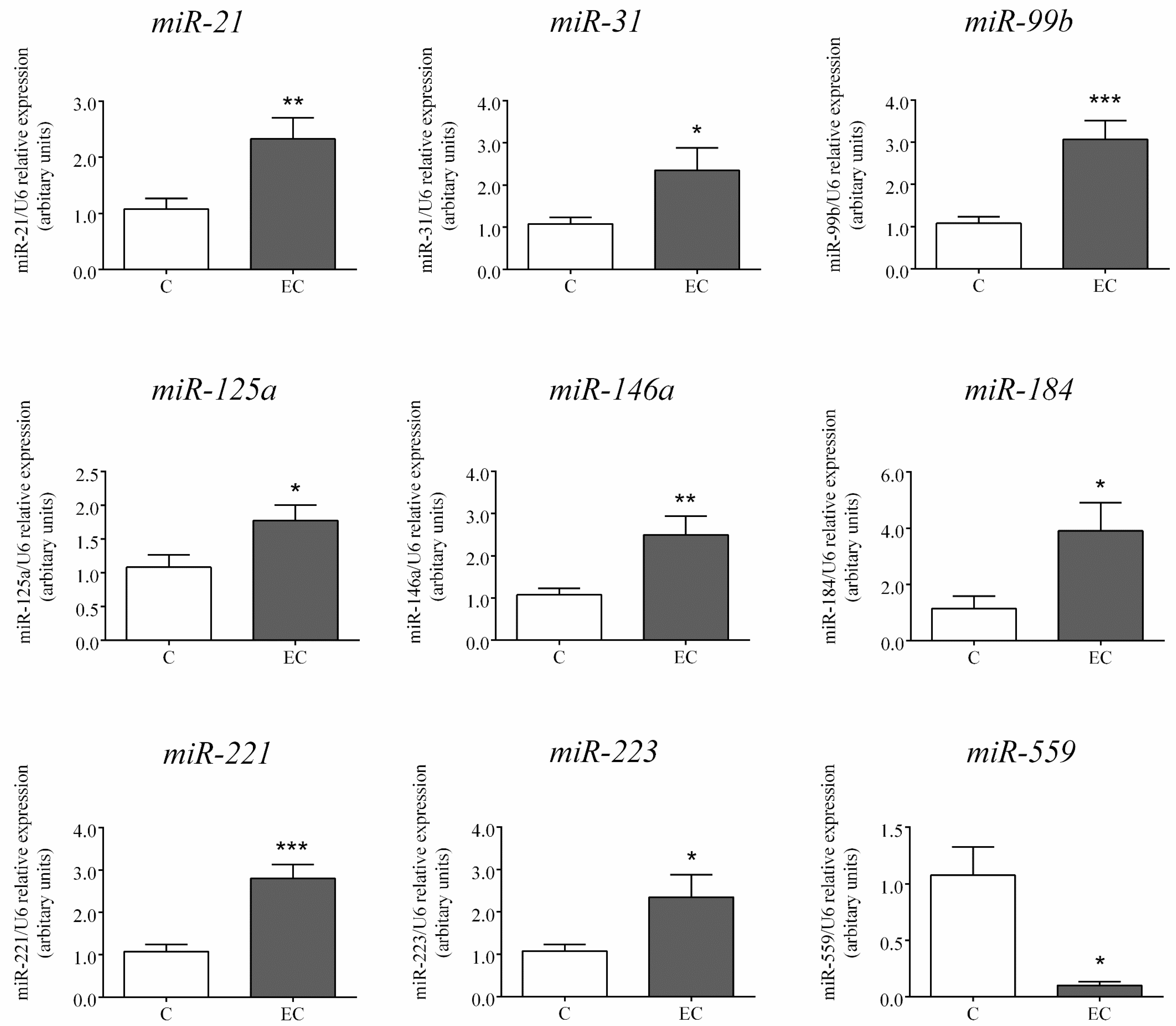
2.2. Expression of miRs Selected for Further Analysis by RT-PCR

Similar to the changes found by NGS, the expression of the nine miRs selected for method validation and further analysis by RT-PCR showed statistically significant differences in the colonic mucosa of paediatric patients with EC compared to controls (p ≤ 0.05). The expression of miR-21, -31, -99b, -125a, -146a, -184, -221, and -223 was significantly elevated, and that of miR-559 significantly decreased in the colonic mucosa of children with EC compared to controls (Figure 2).

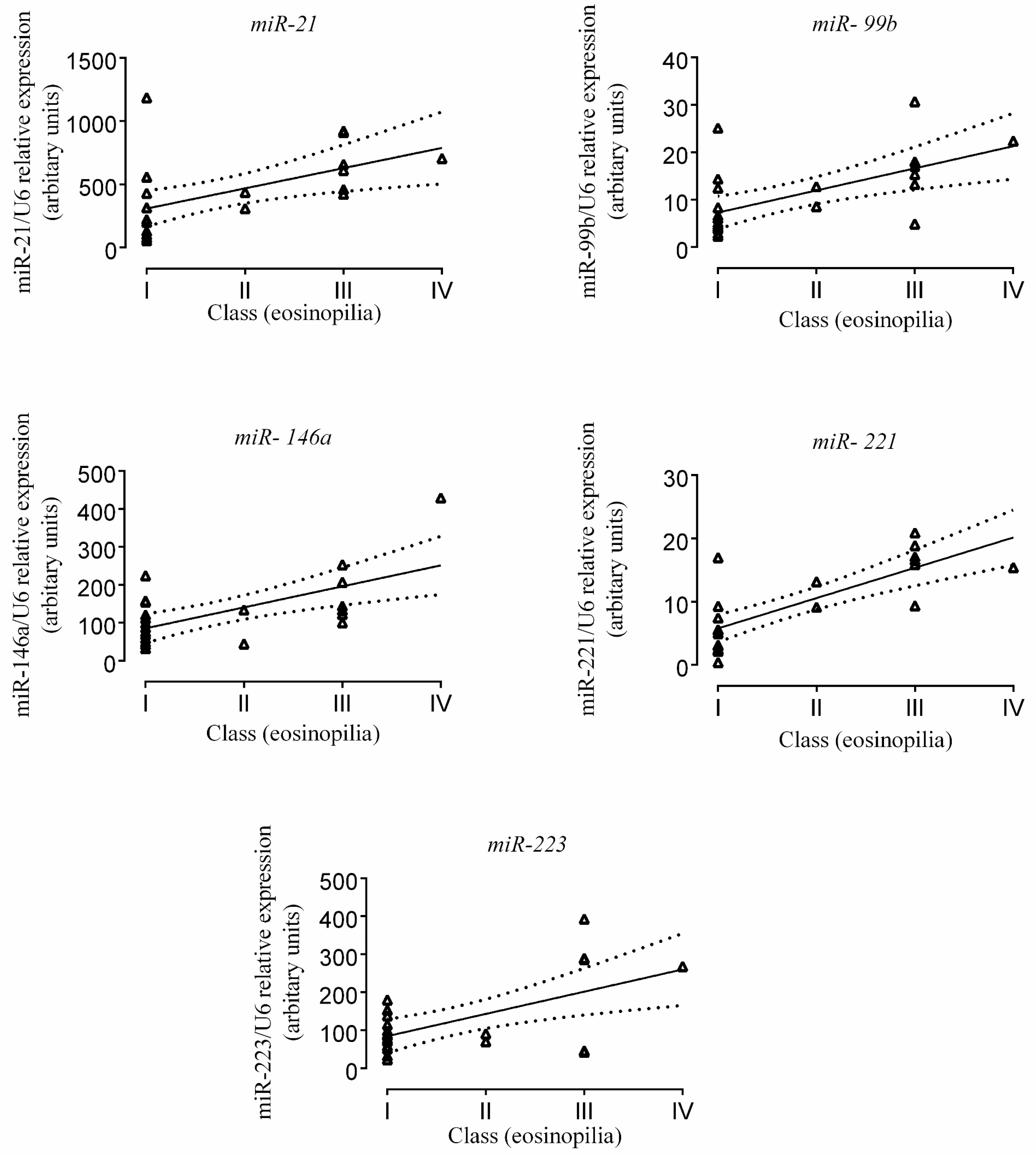
2.3. Correlation of miR Expression with Tissue Eosinophil Counts

From the PCR-validated miR panel, expression of miR-21 (r = 0.51; 95% CI = 0.15–0.77; p = 0.009), -99b (r = 0.60; 95% CI = 0.25–0.81, p = 0.002), -146a (r = 0.62, 95% CI = 0.29–0.82, p = 0.001), -221 (r = 0.78, 95% CI = 0.78, p = 0.53–0.90, p < 0.0001) and -223 (r = 0.59. 95% CI = 0.22–0.81, p = 0.0041) showed a significantly positive correlation with the number of eosinophil granulocytes in paediatric patients with EC. MiR-125a, -31, -184, and 559 showed similar tendency, but the correlations with eosinophil numbers were statistically not significant (Figure 3).

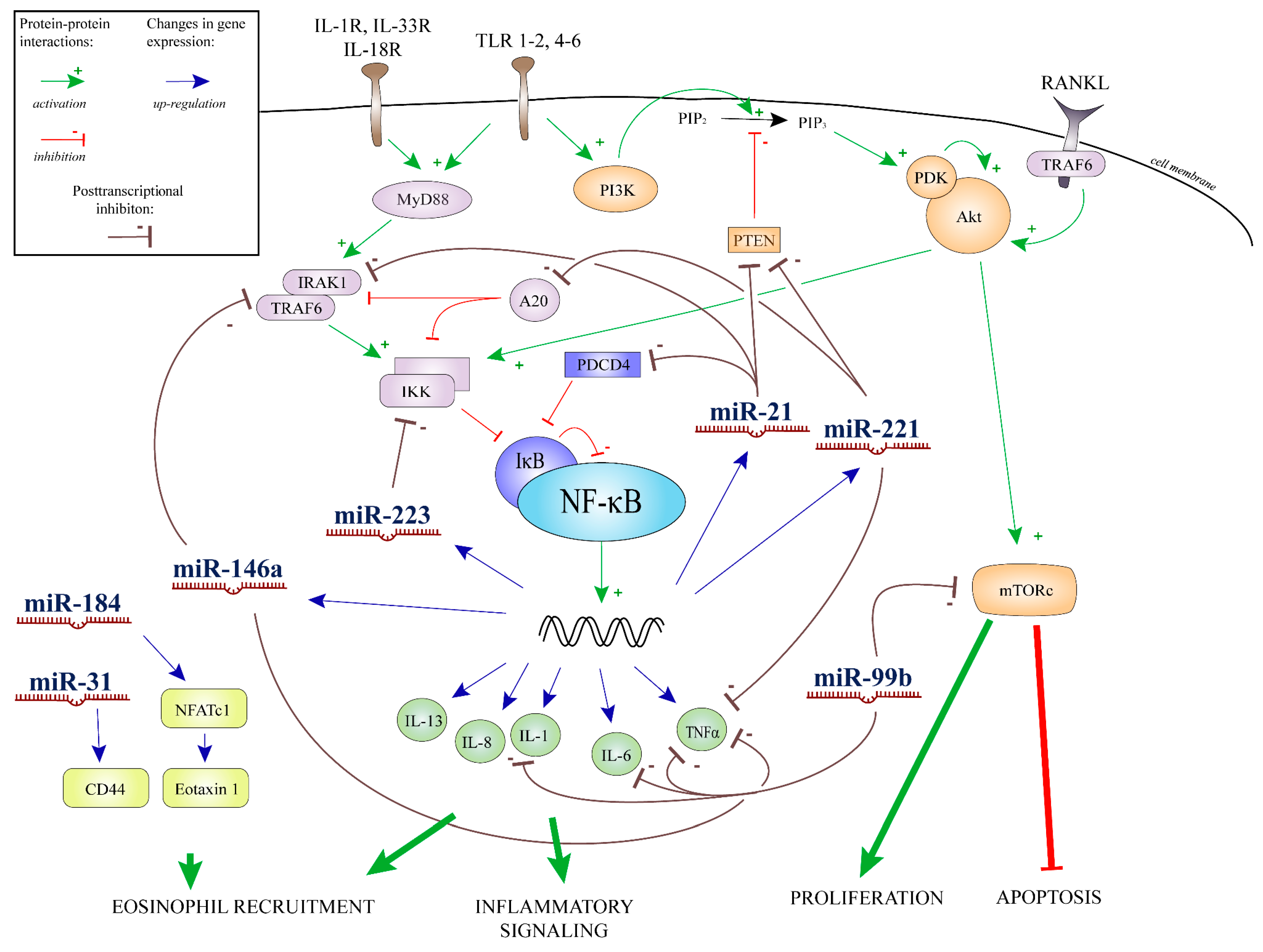
2.4. Results of Bioinformatics Analysis

MiRTarBase database contained 386 reporter assay validated targets of the NGS miR profile found in EC. From this list expression of 100 entities were reported in epithelial tissue. Annotation of these 100 entities with gene ontology (GO) terms (biological process domain), and enrichment analysis resulted in 24 GO term categories. The most abundant terms were connected with immune response, regulation of apoptosis, transcription regulation, and cell proliferation (Table S2).

3. Discussion

EC in infancy is in most cases a benign self-limiting disease associated with abdominal pain, intestinal dismotility, and mucosal eosinophilia occurring predominantly in the first six months of life [2]. However, several cases with overall poor progression and persistent rectal bleeding were reported (including in our study population). It is of high importance to investigate the pathomechanism and explore the diagnostic potential regarding these particularly sensitive patients. The exact pathomechanism of EC is not understood, and its causative therapy is not available in many cases [10,11,33].
Recently, the role of epigenetic factors came into focus as key determinants in the pathogenesis of different gastrointestinal diseases, including IBD and EOE, frequent forms of eosinophil-associated gastrointestinal disorders (EGID), and bronchial asthma. The expression of several miRs showed a strong correlation with the extent of eosinophil infiltration in the biopsy samples of patients with EOE, and were also dysregulated in their serum [21]. In the current study, we screened the miRNA profile on low patient count in EC with NGS, and based on these preliminary results, we selected a panel of miRNAs strongly connected with eosinophilic inflammation for further analysis. We analysed the selected miRNA with RT-PCR methods on a higher patient number. The panel consisted of nine miRs: miR-21, -31, -99b, -125a, -146a, -184, -221, -223, and -559. Dysregulation of six of these miRs—miR-21, -99b, -125a, -146a, -221, and -223—were previously reported in EOE [20,21]. We selected three additional miRs with strong connection to eosinophil cell recruitment and activation, as described earlier in other conditions. [28,29,32]. The selected miRs also have several potential effects in the modulation of inflammatory pathways not directly affecting eosinophil cells. In the following paragraphs, we will discuss the potential target points, starting with the general pathways of apoptosis followed by the closely relevant molecular events of eosinophilic inflammation.
From the selected miR panel, miR-21, -99b, and -221 are closely related to apoptosis by the potential regulation of the PI3K/Akt/mTOR pathway [34,35,36]. Negative regulation of apoptosis in lymphoid cells and eosinophils could contribute to their accumulation and prolonged survival in the colonic tissue [37]. This mechanism could also be an important factor in tissue infiltration and the subsequent formation of LNH—a frequent endoscopic finding in EC. MiR-559 is an inhibitor of ERBB2 expression. Eosinophil peroxidase—a secreted effector molecule of activated eosinophil cells—is a ligand of ERBB2, and the downstream signalisation originating from ERBB2 has significant proliferative effects; hence, it could contribute to the dwelling caused by prolonged accumulation and activation of eosinophils on inflammatory sites [28].
On the other hand, positive regulation of apoptosis—on which the dysregulated miRs could also have a potential effect according to our present analysis—could slow down the turnover and proliferation of epithelial cells in the colonic tissue, leading to the impairment of intestinal barrier function and the facilitation of inflammatory processes [38,39,40]. This phenomenon as a histologic finding has been reported in connection with EC. Apoptotic cell-specific histochemical TUNEL assay on biopsies labelled with epithelial cell-specific antibodies identified an increased number of apoptotic epithelial cells in the lamina propria of rectosigmoid biopsy specimens originating from infants with haematochezia previously diagnosed with allergic/eosinophilic colitis [40].
Similar to the observations in EOE, in the present study we demonstrated significantly elevated expression of miR-21, -146a, and -223 in the colonic mucosa of paediatric patients with EC compared to controls, and it was strongly correlated with the extent of tissue eosinophilia. Furthermore, we found a positive correlation between the expression changes of miR-99b and -221 and the number of tissue eosinophils in the colonic mucosa of EC patients [41,42] (Figure 3). The correlations suggest that these miRs could be actively connected to the pathomechanism of EC. A composite graph of the possible interacting points of these miRs with respect to inflammation and tissue eosinophilia is presented in Figure 4.
Certain dysregulated miRs in the colonic mucosa of children with EC (e.g., miR-125a, -146a, and -223 as identified in our present experiments) are also related to eosinophil cell line differentiation, activation, and recruitment [37]. Lu et al. found a connection between miR-223 deficiency and increased eosinophil progenitor cell proliferation in an ex vivo bone marrow-derived eosinophil culture system [43]. It is inferred from bone marrow culture experiments that miR-125a and -146a are also parts of the regulation network of eosinophil cell maturation [41]. To further investigate the relationship between miRs and eosinophil function in EC, additional studies would be needed on eosinophil granulocytes freshly isolated from the colonic tissues of EC patients.
MiR-21 has been reported to be the most frequently upregulated miR in eosinophil-related diseases [21,23]. It is a complex regulator of eosinophil cell fate, activation and survival. MiR-21 overexpression is able to increase eosinophil progenitor cell growth, it has a pro-survival effect on eosinophils via granulocyte-macrophage colony-stimulating factor (GM-CSF), and acts towards Th2 polarization of the adaptive immune processes [23,25,26,27].
The correlation of miR-221 expression with airway tissue eosinophilia has been reported in a murine model of bronchial asthma. In this model, the level of miR-221 was also correlated with IL-4 secretion, presumably via phosphatase and tensin homolog (PTEN) and nuclear factor kappa-light-chain-enhancer of activated B cells (NF-κB) protein related pathways that are potent inductors of tissue eosinophil infiltration [42]. Increased prevalence of EC has been reported amongst patients with PTEN hamartoma tumour syndromes [44]. MiR-21 and -221 are two miRs which showed significant elevation according to our measurements, and have been connected to the regulation of PTEN gene expression [36,45]. PTEN protein is a mon-redundant master regulator of the PI3K signalling pathway. Humans are highly sensitive of PTEN dosage, and alterations in the PI3K/Akt/mTOR pathway has been connected to eosinophil cell-related conditions [46,47,48]. MiR-99b is a member of the miR-99 family, and is also a potent regulator of the PI3K/Akt/mTOR pathway. Besides the effects on eosinophil cell survival, this pathway is active in the tissues of inflammation sites, acting as a mediator of wound healing [35].
MiR-184 targets the NFAT1 transcript directly. Deficiency in the NFAT1 protein product could lead to elevated IL-5, IL-2, and IL-13 production. These cytokines promote eosinophil cell survival and recruit via upregulation of eotaxin levels [28].
MiR-31 levels are positively correlated with the expression of CD44 adhesion molecule, a promoter of tissue eosinophilia. Expression of CD44 in epithelial cells could promote eosinophil migration and activation [31]. It has been demonstrated that changes in CD44 adhesion molecule levels in epithelial cells is a major component of tissue alterations in EOE, and promotes eosinophil and leukocyte migration and activation on the site of inflammation [32].
A limitation of our present study is that biopsies from age-matched healthy subjects could not be obtained. However, we enrolled the youngest possible patients to the control group in order to minimize these effects on the results. To completely eliminate the possible effect of age-related differences, only samples originating from autopsy procedures of infants involved in accidents or who suffered sudden infant death would be completely applicable.
In summary, to the best of our knowledge, we conducted the first experiments in order to define the miR profile specific to EC. Using bioinformatics tools, we assessed the pool of target genes potentially affected by the miRs dysregulated in the colon of children with EC having persistent rectal bleeding, unresponsive to dietary therapies. Our results defined several possible target point for miRs on the molecular biological events of eosinophilic inflammation, and therefore could serve as a base for further investigations of the pathomechanism of EC.

4. Materials and Methods
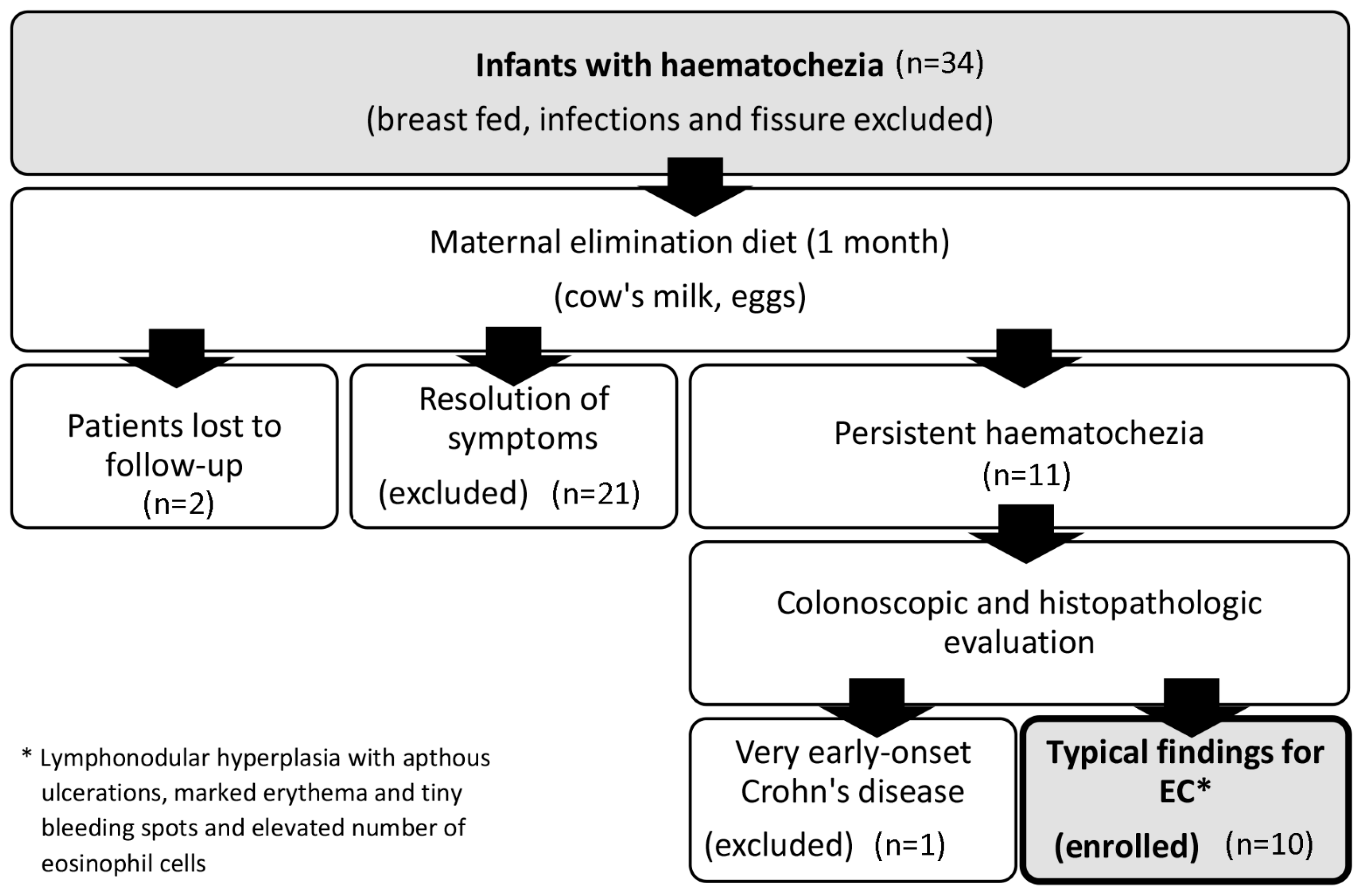
4.1. Patients

Samples from all patients enrolled in the study were obtained at the 1st Department of Pediatrics, Semmelweis University, Budapest, Hungary. Colon pinch biopsies were taken from 10 children with EC and 14 controls. Clinical characteristics and laboratory parameters of the patients are shown in Table 1. Patients enrolled in the study had persistent rectal bleeding, and they were unresponsive to dietary intervention therapies. Patients underwent colonoscopy in order to exclude IBD and other organic causes of long-lasting haematochezia. The overall symptoms, long term follow-up, and endoscopic features of the patients did not justify the consideration of monogenic origin. Subsequent diagnosis of CD (without monogenic cause) was obtained in one patient, who was therefore excluded from the study. Diagnosis of EC was based on clinical history, physical examination (no perianal fissure), laboratory findings (infections excluded), endoscopic findings (focal erythema, LNH, and aphta), and pathological evaluation of tissue specimens (average eosinophil count/five high power field (eo/HPF) > 20, HPF area = 0.2 mm2) with long-term follow-up of the patients. Microscopic evaluation was open-label conducted by a skilled pathologist as a part of diagnostic processes. Control children underwent colonoscopy to assess the cause of failure to thrive or suspicion of polyp or Meckel-diverticulum. All specimens in the control group showed normal macroscopic and histologic features. Biopsies were immediately snap-frozen and stored at −80 °C until further experiments. Written informed consent was obtained from the parents prior to the procedure, and the study was approved by the Semmelweis University Regional and Institutional Committee for Research Ethics (TUKEB No.: 10408/2012, Date: 30 March 2012).

4.2. RNA Isolation

Total RNA isolation from the colonic biopsies was conducted with TRIzol reagent (Ambion, Austin, TX, USA) for the next-generation sequencing (NGS) and Quick-RNA MiniPrep isolation kit (Zymo Research, Irvine, CA, USA) for the real-time reverse transcription polymerase chain reaction (RT-PCR) measurements, according to the manufacturer’s protocol.

4.3. cDNA Library Preparation and NGS

A cDNA library for small RNA-Seq was generated from 1 μg total RNA samples derived from the colonic mucosa of paediatric EC patients (EC: n = 4) and controls (C: n = 4) using TruSeq Small RNA Sample Preparation Kit (Illumina, San Diego, CA, USA) according to the manufacturer’s protocol. Fragment size distribution and molarity of libraries were checked on Agilent BioAnalyzer DNA1000 chip (Agilent Technologies, Santa Clara, CA, USA). Concentration of small RNA libraries were set to 10 nM, and cluster generation was done using TruSeq SR Cluster kit v3-cBot-HS kit on cBot instrument, then a single read 50 bp sequencing run was performed on Illumina HiScan SQ instrument (Illumina, San Diego, CA, USA) carried out by UD-GenoMed Medical Genomic Technologies Ltd. (Debrecen, Hungary).

4.4. RT-PCR

RT-PCRs were performed on colonic biopsy samples of EC patients (n = 14) and controls (n = 10). Total RNA was reverse-transcribed using TaqMan MicroRNA Reverse Transcription Kit (Life Technologies, Carlsbad, CA, USA). MiRs were selected for validation according to the NGS results, and their expression was determined by RT-PCR using TaqMan Universal PCR Master Mix No AmpErase UNG (Life Technologies). Measurements were carried out on a LightCycler 480 instrument (Roche, Basel, Switzerland). Relative expression level was calculated by the ΔΔCq formula, using U6 as an internal standard. TaqMan miR assays (miR-21: ID: 000397, miR-31: ID: 002279, miR-99b: ID: 000436, miR-125a: ID: 002198, miR-146a: ID: 000468, miR-184: ID: 000485, miR-221: ID: 000524, miR-223: ID: 002295, miR-559: ID: 001527, U6: ID: 001973) were used according to the manufacturer’s instructions.

4.5. Bioinformatics Analysis of the Whole NGS miR Profile

Experimentally validated (reporter assay) target genes were selected from the MiRTarBase database (Available online: http://mirtarbase.mbc.nctu.edu.tw) [49]. Target genes were filtered for site of expression reported according to the tissue expression database of UniProt (Available online: http://www.uniprot.org) [50]. The Database for Annotation, Visualization and Integrated Discovery (DAVID) bioinformatics tool was used to annotate the resulting list in the Biological process domain of the Gene Ontology (GO) database [51,52,53,54]. Enrichment analysis was carried out on the results with an established 0.01 threshold of the modified Fisher Exact p-value (EASE) scores of the enriched categories adjusted with the Benjamini–Hochberg correction method, and 10 entities threshold for group count.

4.6. Determination of Eosinophil Counts in the Colonic Tissue of EC Patients

Colonic tissue eosinophil counts were assessed in the microscopic slides by a pathologist with an assumption of representativeness. Evaluation of the specimens was open-label and conducted as a part of the normal diagnostic procedure. Paraffin-embedded tissue samples and fresh-frozen specimens were taken within close vicinity of each other. Eosinophil cells were detected by standard Hematoxylin and Eosin (H&E) staining (ST5020 Multistainer, Leica Microsystems GmbH, Wetzlar, Germany). Eosinophils in the lamina propria were counted in five random high-power fields (HPF: area of tissue covered by a ×40 light microscope objective: 0.2 mm2) selected from an eosinophil-dense part of the slides, on optical microscopy (BX51TF, Olympus, Tokyo, Japan). More than 20 eo/HPF was the cut-off number for EC (reference of histological evaluation presented in Figure 5). Based on the average number of eosinophil granulocytes in five HPFs (HPF area = 0.2 mm2), patients were divided into four categories (Class I: 0–10 eo/HPF, Class II: 10–20 eo/HPF, Class III: 20–40 eo/HPF, Class IV: >40 eo/HPF).
Correlation between tissue eosinophil counts and miR expressions was calculated using a Spearman method assuming Gaussian distribution of the data.

4.7. Statistical Analysis

Statistical analysis was performed using the GraphPad statistical software package (GraphPad Software, La Jolla, CA, USA). Shapiro–Wilk test was used to test normality. Data were analysed by unpaired, two-tailed t-test and presented as mean ± standard error of the mean (SEM) and normalized to the control group. Two-tailed p-values were estimated in all the statistical calculations, and results were filtered after a 0.05 p-value threshold for significance.

Supplementary Materials

Supplementary materials can be found at www.mdpi.com/1422-0067/18/5/1050/s1.

Acknowledgments

We are grateful to Maria Bernáth for her skilful technical assistance. This work was supported by grants OTKA-K105530, -K108688, -K116928, -PD105361, and LP008/2016.

Author Contributions

Gábor Veres was the gastroenterologist providing the mucosal biopsies for the study; Zoltán Kiss, Nóra Béres and Gábor Veres designed the research; András Arató, Attila J. Szabó coordinated the research; Nóra Béres, Zoltán Kiss, Bálint Tél performed the research; Katalin Borka performed the pathological evaluations of the biopsies; Nóra Béres, Bálint Tél and Zoltán Kiss analysed the data; Zoltán Kiss, Nóra Judit Béres, and Erna Sziksz wrote the paper.

Conflicts of Interest

The authors declare no conflict of interest. The founding sponsors had no role in the design of the study; in the collection, analyses, or interpretation of data; in the writing of the manuscript, and in the decision to publish the results.

References

  1. Fox, V.L. Gastrointestinal bleeding in infancy and childhood. Gastroenterol. Clin. N. Am. 2000, 29, 37–66. [Google Scholar] [CrossRef]
  2. Machida, H.M.; Catto Smith, A.G.; Gall, D.G.; Trevenen, C.; Scott, R.B. Allergic colitis in infancy: Clinical and pathologic aspects. J. Pediatr. Gastroenterol. Nutr. 1994, 19, 22–26. [Google Scholar] [CrossRef] [PubMed]
  3. Rothenberg, M.E.; Mishra, A.; Brandt, E.B.; Hogan, S.P. Gastrointestinal eosinophils. Immunol. Rev. 2001, 179, 139–155. [Google Scholar] [CrossRef] [PubMed]
  4. Alfadda, A.A.; Storr, M.A.; Shaffer, E.A. Eosinophilic colitis: An update on pathophysiology and treatment. Br. Med. Bull. 2011, 100, 59–72. [Google Scholar] [CrossRef] [PubMed]
  5. Uppal, V.; Kreiger, P.; Kutsch, E. Eosinophilic gastroenteritis and colitis: A comprehensive review. Clin. Rev. Allergy Immunol. 2016, 50, 175–188. [Google Scholar] [CrossRef] [PubMed]
  6. Ohtsuka, Y.; Jimbo, K.; Inage, E.; Mori, M.; Yamakawa, Y.; Aoyagi, Y.; Suzuki, M.; Kudo, T.; Suzuki, R.; Shimizu, T. Microarray analysis of mucosal biopsy specimens in neonates with rectal bleeding: Is it really an allergic disease? J. Allergy Clin. Immunol. 2012, 129, 1676–1678. [Google Scholar] [CrossRef] [PubMed]
  7. Cseh, A.; Molnar, K.; Pinter, P.; Szalay, B.; Szebeni, B.; Treszl, A.; Arato, A.; Vasarhelyi, B.; Veres, G. Regulatory T cells and T helper subsets in breast-fed infants with hematochezia caused by allergic colitis. J. Pediatr. Gastroenterol. Nutr. 2010, 51, 675–677. [Google Scholar] [CrossRef] [PubMed]
  8. Torrente, F.; Barabino, A.; Bellini, T.; Murch, S.H. Intraepithelial lymphocyte eotaxin-2 expression and perineural mast cell degranulation differentiate allergic/eosinophilic colitis from classic IBD. J. Pediatr. Gastroenterol. Nutr. 2014, 59, 300–307. [Google Scholar] [CrossRef] [PubMed]
  9. Jensen, E.T.; Martin, C.F.; Kappelman, M.D.; Dellon, E.S. Prevalence of eosinophilic gastritis, gastroenteritis, and colitis: Estimates from a national administrative database. J. Pediatr. Gastroenterol. Nutr. 2016, 62, 36–42. [Google Scholar] [CrossRef] [PubMed]
  10. Molnar, K.; Pinter, P.; Gyorffy, H.; Cseh, A.; Muller, K.E.; Arato, A.; Veres, G. Characteristics of allergic colitis in breast-fed infants in the absence of cow’s milk allergy. World J. Gastroenterol. 2013, 19, 3824–3830. [Google Scholar] [CrossRef] [PubMed]
  11. Cannioto, Z.; Berti, I.; Martelossi, S.; Bruno, I.; Giurici, N.; Crovella, S.; Ventura, A. IBD and IBD mimicking enterocolitis in children younger than 2 years of age. Eur. J. Pediatr. 2009, 168, 149–155. [Google Scholar] [CrossRef] [PubMed]
  12. Katsanos, K.H.; Zinovieva, E.; Lambri, E.; Tsianos, E.V. Eosinophilic–Crohn overlap colitis and review of the literature. J. Crohns Colitis 2011, 5, 256–261. [Google Scholar] [CrossRef] [PubMed]
  13. Bala, G.; Swincow, G.; Rytarowska, A.; Czerwionka-Szaflarska, M. Non-specific inflammatory bowel disease or allergic colitis in small children—What is the final diagnosis. Med. Wieku Rozwoj. 2006, 10, 475–482. [Google Scholar] [PubMed]
  14. Ozen, A.; Gulcan, E.M.; Saricoban, H.E.; Ozkan, F.; Cengizlier, R. Food protein-induced non-immunoglobulin E-mediated allergic colitis in infants and older children: What cytokines are involved? Int. Arch. Allergy Immunol. 2015, 168, 61–68. [Google Scholar] [CrossRef] [PubMed]
  15. Bischoff, S.C. Food allergy and eosinophilic gastroenteritis and colitis. Curr. Opin. Allergy Clin. Immunol. 2010, 10, 238–245. [Google Scholar] [CrossRef] [PubMed]
  16. Kalla, R.; Ventham, N.T.; Kennedy, N.A.; Quintana, J.F.; Nimmo, E.R.; Buck, A.H.; Satsangi, J. MicroRNAs: New players in IBD. Gut 2015, 64, 504–517. [Google Scholar] [CrossRef] [PubMed]
  17. Xiao, L.; Wang, J.Y. RNA-binding proteins and microRNAs in gastrointestinal epithelial homeostasis and diseases. Curr. Opin. Pharmacol. 2014, 19, 46–53. [Google Scholar] [CrossRef] [PubMed]
  18. Wang, J.; Chen, J.; Sen, S. MicroRNA as biomarkers and diagnostics. J. Cell. Physiol. 2016, 231, 25–30. [Google Scholar] [CrossRef] [PubMed]
  19. Hammond, S.M. An overview of microRNAs. Adv. Drug Deliv. Rev. 2015, 87, 3–14. [Google Scholar] [CrossRef] [PubMed]
  20. Zahm, A.M.; Menard-Katcher, C.; Benitez, A.J.; Tsoucas, D.M.; le Guen, C.L.; Hand, N.J.; Friedman, J.R. Pediatric eosinophilic esophagitis is associated with changes in esophageal microRNAs. Am. J. Physiol. Gastrointest. Liver Physiol. 2014, 307, G803–G812. [Google Scholar] [CrossRef] [PubMed]
  21. Lu, T.X.; Sherrill, J.D.; Wen, T.; Plassard, A.J.; Besse, J.A.; Abonia, J.P.; Franciosi, J.P.; Putnam, P.E.; Eby, M.; Martin, L.J.; et al. MicroRNA signature in patients with eosinophilic esophagitis, reversibility with glucocorticoids, and assessment as disease biomarkers. J. Allergy Clin. Immunol. 2012, 129, 1064–1075. [Google Scholar] [CrossRef] [PubMed]
  22. Pua, H.H.; Ansel, K.M. MicroRNA regulation of allergic inflammation and asthma. Curr. Opin. Immunol. 2015, 36, 101–108. [Google Scholar] [CrossRef] [PubMed]
  23. Lu, T.X.; Munitz, A.; Rothenberg, M.E. MicroRNA-21 is up-regulated in allergic airway inflammation and regulates IL-12p35 expression. J. Immunol. 2009, 182, 4994–5002. [Google Scholar] [CrossRef] [PubMed]
  24. Wu, F.; Zikusoka, M.; Trindade, A.; Dassopoulos, T.; Harris, M.L.; Bayless, T.M.; Brant, S.R.; Chakravarti, S.; Kwon, J.H. MicroRNAs are differentially expressed in ulcerative colitis and alter expression of macrophage inflammatory peptide-2α. Gastroenterology 2008, 135, 1624–1635. [Google Scholar] [CrossRef] [PubMed]
  25. Lu, T.X.; Lim, E.J.; Itskovich, S.; Besse, J.A.; Plassard, A.J.; Mingler, M.K.; Rothenberg, J.A.; Fulkerson, P.C.; Aronow, B.J.; Rothenberg, M.E. Targeted ablation of miR-21 decreases murine eosinophil progenitor cell growth. PLoS ONE 2013, 8, e59397. [Google Scholar] [CrossRef] [PubMed]
  26. Wong, C.K.; Lau, K.M.; Chan, I.H.; Hu, S.; Lam, Y.Y.; Choi, A.O.; Lam, C.W. MicroRNA-21* regulates the prosurvival effect of GM-CSF on human eosinophils. Immunobiology 2013, 218, 255–262. [Google Scholar] [CrossRef] [PubMed]
  27. Lu, T.X.; Hartner, J.; Lim, E.J.; Fabry, V.; Mingler, M.K.; Cole, E.T.; Orkin, S.H.; Aronow, B.J.; Rothenberg, M.E. MicroRNA-21 limits in vivo immune response-mediated activation of the IL-12/IFN-γ pathway, Th1 polarization, and the severity of delayed-type hypersensitivity. J. Immunol. 2011, 187, 3362–3373. [Google Scholar] [CrossRef] [PubMed]
  28. Hennigan, K.; Conroy, P.J.; Walsh, M.T.; Amin, M.; O’Kennedy, R.; Ramasamy, P.; Gleich, G.J.; Siddiqui, Z.; Glynn, S.; McCabe, O.; et al. Eosinophil peroxidase activates cells by HER2 receptor engagement and β1-integrin clustering with downstream MAPK cell signaling. Clin. Immunol. 2016, 171, 1–11. [Google Scholar] [CrossRef] [PubMed]
  29. Fonseca, B.P.; Olsen, P.C.; Coelho, L.P.; Ferreira, T.P.; Souza, H.S.; Martins, M.A.; Viola, J.P. NFAT1 transcription factor regulates pulmonary allergic inflammation and airway responsiveness. Am. J. Respir. Cell Mol. Biol. 2009, 40, 66–75. [Google Scholar] [CrossRef] [PubMed]
  30. Weitzel, R.P.; Lesniewski, M.L.; Haviernik, P.; Kadereit, S.; Leahy, P.; Greco, N.J.; Laughlin, M.J. 184 regulates expression of NFAT1 in umbilical cord blood CD4+ T cells. Blood 2009, 113, 6648–6657. [Google Scholar] [CrossRef] [PubMed]
  31. Li, L.; Hui, Y.; Xing, C.; Guo, Y.; Wang, Q.; Shu, J.; Qian, J.; Zhou, G. MicroRNA-31 affects the expression of asthma-related cytokines via regulation of CD44. Int. J. Clin. Exp. Med. 2016, 9, 11. [Google Scholar]
  32. Abdulnour-Nakhoul, S.M.; Al-Tawil, Y.; Gyftopoulos, A.A.; Brown, K.L.; Hansen, M.; Butcher, K.F.; Eidelwein, A.P.; Noel, R.A.; Rabon, E.; Posta, A.; et al. Alterations in junctional proteins, inflammatory mediators and extracellular matrix molecules in eosinophilic esophagitis. Clin. Immunol. 2013, 148, 265–278. [Google Scholar] [CrossRef] [PubMed]
  33. Hua, S.; Cook, D.; Walker, M.M.; Talley, N.J. Pharmacological treatment of eosinophilic gastrointestinal disorders. Expert Rev. Clin. Pharmacol. 2016, 9, 1195–1209. [Google Scholar] [CrossRef] [PubMed]
  34. Ou, H.; Li, Y.; Kang, M. Activation of miR-21 by STAT3 induces proliferation and suppresses apoptosis in nasopharyngeal carcinoma by targeting PTEN gene. PLoS ONE 2014, 9, e109929. [Google Scholar] [CrossRef] [PubMed]
  35. Jin, Y.; Tymen, S.D.; Chen, D.; Fang, Z.J.; Zhao, Y.; Dragas, D.; Dai, Y.; Marucha, P.T.; Zhou, X. MicroRNA-99 family targets AKT/mTOR signaling pathway in dermal wound healing. PLoS ONE 2013, 8, e64434. [Google Scholar] [CrossRef] [PubMed]
  36. Zhu, A.; Kang, N.; He, L.; Li, X.; Xu, X.; Zhang, H. MiR-221 and miR-26b regulate chemotactic migration of MSCs toward HGF through activation of Akt and FAK. J. Cell. Biochem. 2016, 117, 1370–1383. [Google Scholar] [CrossRef] [PubMed]
  37. Araki, Y.; Mukaisyo, K.; Sugihara, H.; Fujiyama, Y.; Hattori, T. Increased apoptosis and decreased proliferation of colonic epithelium in dextran sulfate sodium-induced colitis in mice. Oncol. Rep. 2010, 24, 869–874. [Google Scholar] [CrossRef] [PubMed]
  38. Sun, H.; Lou, Y.; Porturas, T.; Morrissey, S.; Luo, G.; Qi, J.; Ruan, Q.; Shi, S.; Chen, Y.H. Exacerbated experimental colitis in TNFAIP8-deficient mice. J. Immunol. 2015, 194, 5736–5742. [Google Scholar] [CrossRef] [PubMed]
  39. Chen, P.; Li, Y.C.; Toback, F.G. AMP-18 Targets p21 to maintain epithelial homeostasis. PLoS ONE 2015, 10, e0125490. [Google Scholar] [CrossRef] [PubMed]
  40. Kumagai, H.; Masuda, T.; Maisawa, S.; Chida, S. Apoptotic epithelial cells in biopsy specimens from infants with streaked rectal bleeding. J. Pediatr. Gastroenterol. Nutr. 2001, 32, 428–433. [Google Scholar] [CrossRef] [PubMed]
  41. Yang, M.; Eyers, F.; Xiang, Y.; Guo, M.; Young, I.G.; Rosenberg, H.F.; Foster, P.S. Expression profiling of differentiating eosinophils in bone marrow cultures predicts functional links between microRNAs and their target mRNAs. PLoS ONE 2014, 9, e97537. [Google Scholar] [CrossRef] [PubMed]
  42. Zhou, Y.; Yang, Q.; Xu, H.; Zhang, J.; Deng, H.; Gao, H.; Yang, J.; Zhao, D.; Liu, F. miRNA-221-3p Enhances the secretion of interleukin-4 in mast cells through the phosphatase and tensin homolog/p38/nuclear factor-κB pathway. PLoS ONE 2016, 11, e0148821. [Google Scholar] [CrossRef] [PubMed]
  43. Lu, T.X.; Lim, E.J.; Besse, J.A.; Itskovich, S.; Plassard, A.J.; Fulkerson, P.C.; Aronow, B.J.; Rothenberg, M.E. MiR-223 deficiency increases eosinophil progenitor proliferation. J. Immunol. 2013, 190, 1576–1582. [Google Scholar] [CrossRef] [PubMed]
  44. Henderson, C.J.; Ngeow, J.; Collins, M.H.; Martin, L.J.; Putnam, P.E.; Abonia, J.P.; Marsolo, K.; Eng, C.; Rothenberg, M.E. Increased prevalence of eosinophilic gastrointestinal disorders in pediatric PTEN hamartoma tumor syndromes. J. Pediatr. Gastroenterol. Nutr. 2014, 58, 553–560. [Google Scholar] [CrossRef] [PubMed]
  45. Gui, F.; Hong, Z.; You, Z.; Wu, H.; Zhang, Y. MiR-21 inhibitor suppressed the progression of retinoblastoma via the modulation of PTEN/PI3K/AKT pathway. Cell Biol. Int. 2016, 40, 1294–1302. [Google Scholar] [CrossRef] [PubMed]
  46. Hua, W.; Liu, H.; Xia, L.X.; Tian, B.P.; Huang, H.Q.; Chen, Z.Y.; Ju, Z.Y.; Li, W.; Chen, Z.H.; Shen, H.H. Rapamycin inhibition of eosinophil differentiation attenuates allergic airway inflammation in mice. Respirology 2015, 20, 1055–1065. [Google Scholar] [CrossRef] [PubMed]
  47. Chen, F.; Cao, A.; Yao, S.; Evans-Marin, H.L.; Liu, H.; Wu, W.; Carlsen, E.D.; Dann, S.M.; Soong, L.; Sun, J.; Zhao, Q.; Cong, Y. mTOR Mediates IL-23 induction of neutrophil IL-17 and IL-22 production. J. Immunol. 2016, 196, 4390–4399. [Google Scholar] [CrossRef] [PubMed]
  48. Saito, Y.; Takeda, M.; Nishikawa, J.; Konno, Y.; Tamaki, M.; Itoga, M.; Kobayashi, Y.; Moritoki, Y.; Ito, W.; et al. The effect of pharmacological PI3Kγ inhibitor on eotaxin-induced human eosinophil functions. Pulm. Pharmacol. Ther. 2014, 27, 164–169. [Google Scholar] [CrossRef] [PubMed]
  49. Chou, C.H.; Chang, N.W.; Shrestha, S.; Hsu, S.D.; Lin, Y.L.; Lee, W.H.; Yang, C.D.; Hong, H.C.; Wei, T.Y.; Tu, S.J.; et al. miRTarBase 2016: Updates to the experimentally validated miRNA-target interactions database. Nucleic Acids Res. 2016, 44, D239–D247. [Google Scholar] [CrossRef] [PubMed]
  50. UniProt Consortium. UniProt: A hub for protein information. Nucleic Acids Res. 2015, 43, D204–D212. [Google Scholar]
  51. Huang da, W.; Sherman, B.T.; Lempicki, R.A. Systematic and integrative analysis of large gene lists using DAVID bioinformatics resources. Nat. Protoc. 2009, 4, 44–57. [Google Scholar] [CrossRef] [PubMed]
  52. Huang da, W.; Sherman, B.T.; Lempicki, R.A. Bioinformatics enrichment tools: Paths toward the comprehensive functional analysis of large gene lists. Nucleic Acids Res. 2009, 37, 1–13. [Google Scholar] [CrossRef] [PubMed]
  53. Ashburner, M.; Ball, C.A.; Blake, J.A.; Botstein, D.; Butler, H.; Cherry, J.M.; Davis, A.P.; Dolinski, K.; Dwight, S.S.; Eppig, J.T.; et al. Gene ontology: Tool for the unification of biology. Gene Ontol. Consort. Nat. Genet. 2000, 25, 25–29. [Google Scholar] [CrossRef] [PubMed]
  54. Gene Ontology Consortium. Gene Ontology Consortium: Going forward. Nucleic Acids Res. 2015, 43, D1049–D1056. [Google Scholar]
Figure 1. Patient selection chart. EC: eosinophilic colitis.
Ijms 18 01050 g001
Figure 2. Expression of selected miRs in the colonic mucosa of children with eosinophilic colitis (EC) and controls (C). MiRs were selected based on the results of next-generation sequencing (NGS), and their colonic expression was validated by real-time reverse transcription polymerase chain reaction (RT-PCR). Data are presented as mean ± SEM. * p < 0.05, ** p < 0.01, *** p < 0.001 vs. C.
Ijms 18 01050 g002
Figure 3. Correlation of miR-21, -99b, -146a, -221, and -223 with the extent of tissue eosinophilia in the colonic mucosa of paediatric patients with eosinophilic colitis. Class I: 0–10 eosinophil/high power field (eo/HPF), Class. II: 10–20 eo/HPF, Class III: 20–40 eo/HPF, Class IV: >40 eo/HPF, area of HPF = 0.2 mm2. Solid lines represent a regression line with dotted lines depicting 95% CI.
Ijms 18 01050 g003
Figure 4. Composite summary graph of the potential target points of the miRs correlated with tissue eosinophil level. Abbreviations: inhibitor of kappa B kinase (IKK), interleukin 1, 6, 8, 13 (IL-1, -6, -8, -13), interleukin receptor-1R, -33R, 18R (IL-1R, -33R, -18R), interleukin-1 receptor-associated kinase 1 (IRAK1), mammalian target of rapamycin complex (mTORc), microRNA-21, -31, -99b, -146a, -184, -221, -223 (miR-21, -31, -99b, -146a, -184, -221, -223), myeloid differentiation primary response 88 (MyD88), nuclear factor kappa-light-chain-enhancer of activated B cells (NF-κB), nuclear factor of activated T-cells, cytoplasmic 1 (NFATc1), nuclear factor of kappa light polypeptide gene enhancer in B cells inhibitor (IκB), phosphatase and tensin homolog (PTEN), phosphatidylinositol (3,4,5)-trisphosphate (PIP3), phosphatidylinositol 4,5-bisphosphate (PIP2), phosphatidylinositol-3-kinase (PI3K), phosphoinositide dependent protein kinase (PDK), programmed cell death protein 4 (PDCD4), protein kinase B (Akt), receptor activator of nuclear factor kappa-B ligand (RANKL), TNF receptor associated factor-6 (TRAF6), Toll-like receptor-1-2, 4-6 (TLR 1-2, 4-6), tumor necrosis factor alpha (TNFα), tumor necrosis factor, α-induced protein 3 (A20).
Ijms 18 01050 g004
Figure 5. Reference of the histologic assessment. Detection of eosinophil cells with Hematoxylin and Eosin (H&E) staining: 3–4/HPF eosinophil cells were detectable in normal biopsy (A, B). In a patient with eosinophil colitis, 100–120 eosinophils per HPF are seen (C, D).
Ijms 18 01050 g005
Table 1. Clinical characteristics and laboratory parameters of patients enrolled in the study (A) in the next-generation sequencing groups; and (B) in the real-time reverse transcription polymerase chain reaction validation groups. BMI: body mass index, CRP: C-reactive protein; Data are presented as mean ± SD * p ≤ 0.05 vs. Control, *** p ≤ 0.0001 vs. Control.
AControlEosinophilic Colitis
n44
Male/Female2/23/1
Age (months)136.5 ± 35.788.88 ± 5.14 *
BMI (kg/m2)-18.66 ± 3.79
Iron (µmol/L)10.75 ± 2.513.25 ± 3.57
Albumin (g/L)51.75 ± 8.81-
Haemoglobin (g/L)134.3 ± 7.76121.8 ± 3.64
Haematocrit (%)0.39 ± 0.020.34 ± 0.013
Platelet count (Giga/L)325.8 ± 21.10440.5 ± 57.81
CRP (mg/L)1.25 ± 1.40
BControlEosinophilic Colitis
n1410
Male/Female6/88/2
Age (months)29.82 ± 17.085.7 ± 4.76 ***
BMI (kg/m2)16.15 ± 1.616.21 ± 1.74
Length-weight percentile (%)-42.25 ± 34.92
1/10 overweight
1/10 underweight
Iron (µmol/L)15.70 ± 4.99.89 ± 5.62 *
Albumin (g/L)45.8 ± 2.0144 ± 4.24
Haemoglobin (g/L)122.1 ± 12.26112.6 ± 8.32 *
Haematocrit (%)35.18 ± 2.832 ± 1.94
Platelet count (Giga/L)364.6 ± 87.09394.5 ± 104.1
CRP (mg/L)0.79 ± 1.43.44 ± 8.49

