Article

Effects of Melatonin on Early Pregnancy in Mouse: Involving the Regulation of StAR, Cyp11a1, and Ihh Expression

1
College of Wildlife Resources, Northeast Forestry University, No. 26, Hexing Road, Xiangfang District, Harbin 150036, China
2
National Engineering Laboratory for Animal Breeding, Key Laboratory of Animal Genetics and Breeding of the Ministry of Agriculture, Beijing Key Laboratory for Animal Genetic Improvement, College of Animal Science and Technology, China Agricultural University, Beijing 100193, China
*
Author to whom correspondence should be addressed.
These authors contributed equally to this work.
Int. J. Mol. Sci. 2017, 18(8), 1637; https://doi.org/10.3390/ijms18081637
Submission received: 3 April 2017 / Revised: 18 July 2017 / Accepted: 21 July 2017 / Published: 27 July 2017
(This article belongs to the Special Issue Melatonin and Its Analogues: Experimental and Clinical Aspects)

Abstract

:
To test whether melatonin plays an important role in the process of early pregnancy, melatonin was given in drinking water to pregnant mice at different gestation stages. These included mice who were given melatonin 14 days prior to their successful mating (confirmed by vaginal plug) (Group A), after successful mating (Group B), and 14 days prior to and until after successful mating (Group C). Melatonin administration significantly enhanced serum as well as ovarian melatonin levels in the mice. It was observed that melatonin did not affect the natural estrous of mice. On day 0.5 of gestation (D0.5), melatonin not only elevated progesterone (P) secretion, but also upregulated expressions of StAR and Cyp11a1, the two marker genes of corpus luteum in ovaries (p < 0.05). Group A had a significantly lower estradiol (E2) secretion and a higher number of implantation sites as well as litter size than controls (p < 0.05) and also had an increased Ihh expression in endometrium of D7.5 gestation. Melatonin treatment after successful mating improved the progesterone (P) secretion at D7.5 of gestation (p < 0.05) and significantly induced leukaemia inhibitory factor (LIF) expression (p < 0.05). Our study indicates that melatonin treatment up-regulated the genes involved in pregnenolone synthesis in ovary and Ihh expression in uterine endometrium. The mechanisms of melatonin to improve embryo implantation related to their actions on promoting the development of corpus luteum before gestation and helping to specify uterine receptivity in early pregnant mice.

Graphical Abstract

1. Introduction

In mammals, reproduction is a complex process with several critical steps: sexual maturity, estrus, early embryo development, embryo implantation, and post implantation fetal development [1]. Many factors influence this process including ovarian hormones, cytokines, growth factor, homeotic proteins, and morphogens [2]. Among them, melatonin (MT) is believed to play a critical role in animal reproductive physiology. Melatonin, the major pineal secretory product, is an amphiphilic molecule which readily enters all subcellular compartments including membrane, cytosol, nucleus, and mitochondrion. Melatonin is widely distributed in almost any tissue, organ, and cell [3,4]. It participates in multiple physiological processes [4,5,6]. It regulates the secretion of gonadotropin in the hypothalamus through the hypothalamus–pituitary–gonadal (HPG) axis, thus, influencing biorhythms, sexual maturation, seasonal estrus, reproductive behavior, and gamete protection [6,7,8,9,10,11]. Pévet and Haldar-Misra [12] reported that injection or oral melatonin had the same effect in the suppression of gonadal function of the golden hamster, and oral melatonin effectively inhibited the endogenous activity of the ovary [13]. After pinealectomy, rats show an increase in estradiol (E2) and a decrease in the progesterone (P) levels during the estrous phase [14].
As a powerful antioxidant, melatonin has the ability to scavenge reactive oxygen species (ROS) and reactive nitrogen species (RNS) via receptor-independent actions [15,16,17]. In addition, melatonin also acts on its membrane receptors (MT1/MT2) to stimulate cascading events which increase the transcriptional activity of antioxidant enzymes, suppresses the pro-oxidant enzymes, and reduce the toxic cytokines [18,19,20]. Thus, melatonin treatment protects ovaries from oxidative stress during ovulation [21] and delays the process of ovarian aging [22,23,24]. In addition, melatonin reduced oxidative DNA damage and balanced the expressions of apoptosis genes Bcl-2 and Bax [24]; therefore, it protected oocytes from oxidative stress [25,26].
Numerous studies have investigated the functions of melatonin on female reproduction [27,28,29]. For example, in out-of-season breeding of New Zealand Romney composite ewes, melatonin implantation can increase the number of lambs born [11]. The AANAT (aralkylamine N-acetyltransferase) and ASMT (acetylserotonin O-methyltransferase) were reported to be present in the rat ovary; and the kinetics of these enzymes, for example the apparent Km (Michaelis constant), are similar to those found in the pineal gland of this species. The observation highly supports melatonin synthesis in ovaries. The local synthesized melatonin improves the quality of oocytes [30]. Our previous studies showed that melatonin administration significantly improved uterine receptivity and promoted embryo implantation [29].
However, there are few studies that report the effects of melatonin on early pregnancy. In the current study, the effects of melatonin on early pregnancy were explored in mice by use of different time schedules of melatonin treatment.

2. Results

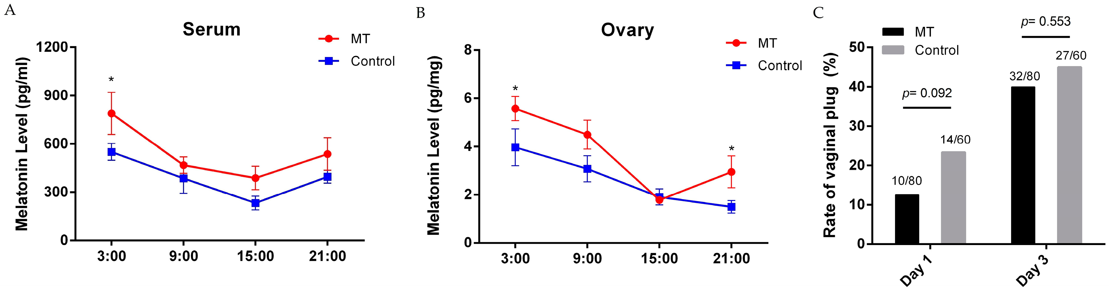
2.1. Effects of Melatonin on Mice Estrus

The serum melatonin levels in the melatonin treatment group and in the control group at 3:00 were 790 ± 131.6 vs. 550 ± 52.79, respectively; at 9:00 they were 469 ± 51.6 vs. 386 ± 93.8, respectively; at 15:00 they were 289 ± 72.8 vs. 232 ± 42.5, respectively; at 21:00 were 538 ± 101.3 pg/mL vs. 396 ± 106.3 pg/mL, respectively. Melatonin administration elevated serum levels of mice at all-time points tested. The highest serum melatonin level occurred at 3:00 (p = 0.031, n = 7) (Figure 1A). The ovarian melatonin levels in the treatment group and in the control group at 3:00 were 5.6 ± 0.50 vs. 4.0 ± 0.77, respectively; at 9:00 they were 4.5 ± 0.60 vs. 3.1 ± 0.54, respectively; at 15:00 they were 1.9 ± 0.11 vs. 1.9 ± 0.33, respectively; at 21:00 they were 3.0 ± 0.66 pg/mg vs. 1.5 ± 0.26 pg/mg tissue, respectively. The ovarian melatonin levels in the melatonin treated group significantly increased at 3:00 (p = 0.31, n = 7) and 21:00 (Figure 1B) compared to that of the control.
Melatonin-treated mice were allowed to mate at night for three days and were evaluated for successful mating each morning. Once the females were found to have vaginal plugs, they were separated immediately. The vaginal plug rate was recorded during the period of three days. The results showed that there were no significant differences on vaginal plug rate between the control and MT-treated mice at day one or in the accumulated rate during all three days (Figure 1C).

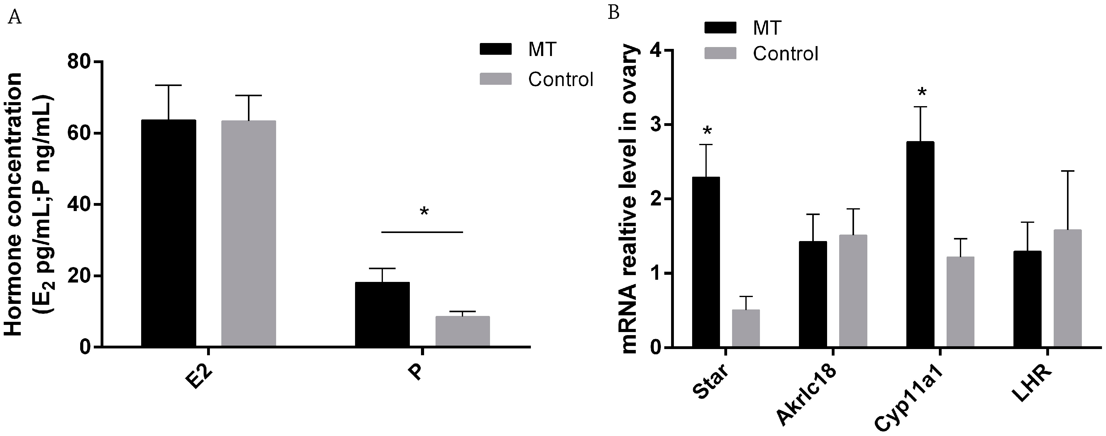
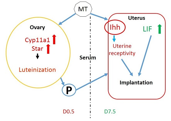
2.2. Effects of Melatonin on E2 and P Secretion and Gene Expression in Ovaries on 0.5 Day Gestation

P and E2 were measured by radioimmunoassay with 10 individuals in each group. There was no significant difference in serum E2 content between control and melatonin treated mice (p > 0.05), while P level was significantly increased in the melatonin treated group compared to the that of the control group (18.15 ± 3.96 vs. 8.60 ± 1.44 ng/mL, respectively, p < 0.05 Figure 2A). The results of RT-qPCR show that the expressions of two corpus luteum marker genes, StAR and Cyp11a1, were significantly higher in melatonin treated mice than those of controls (Figure 2B).

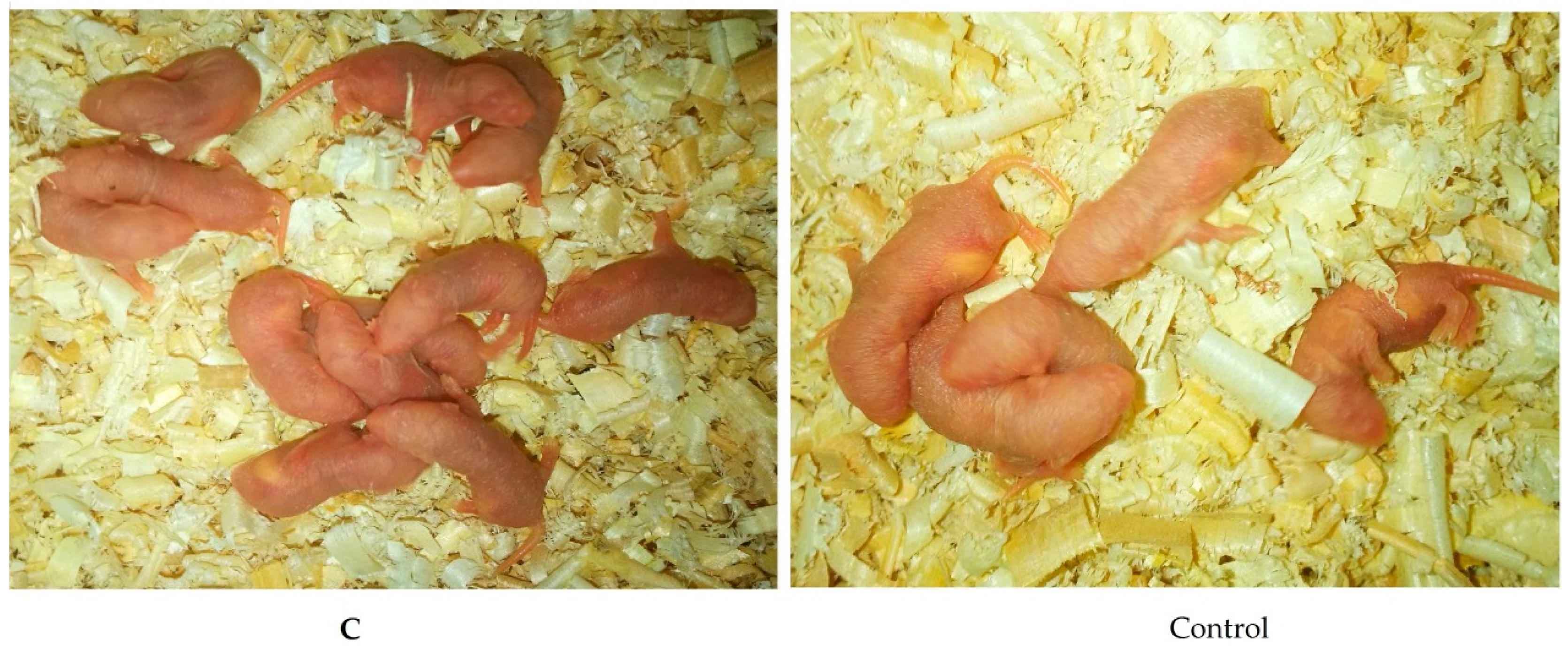
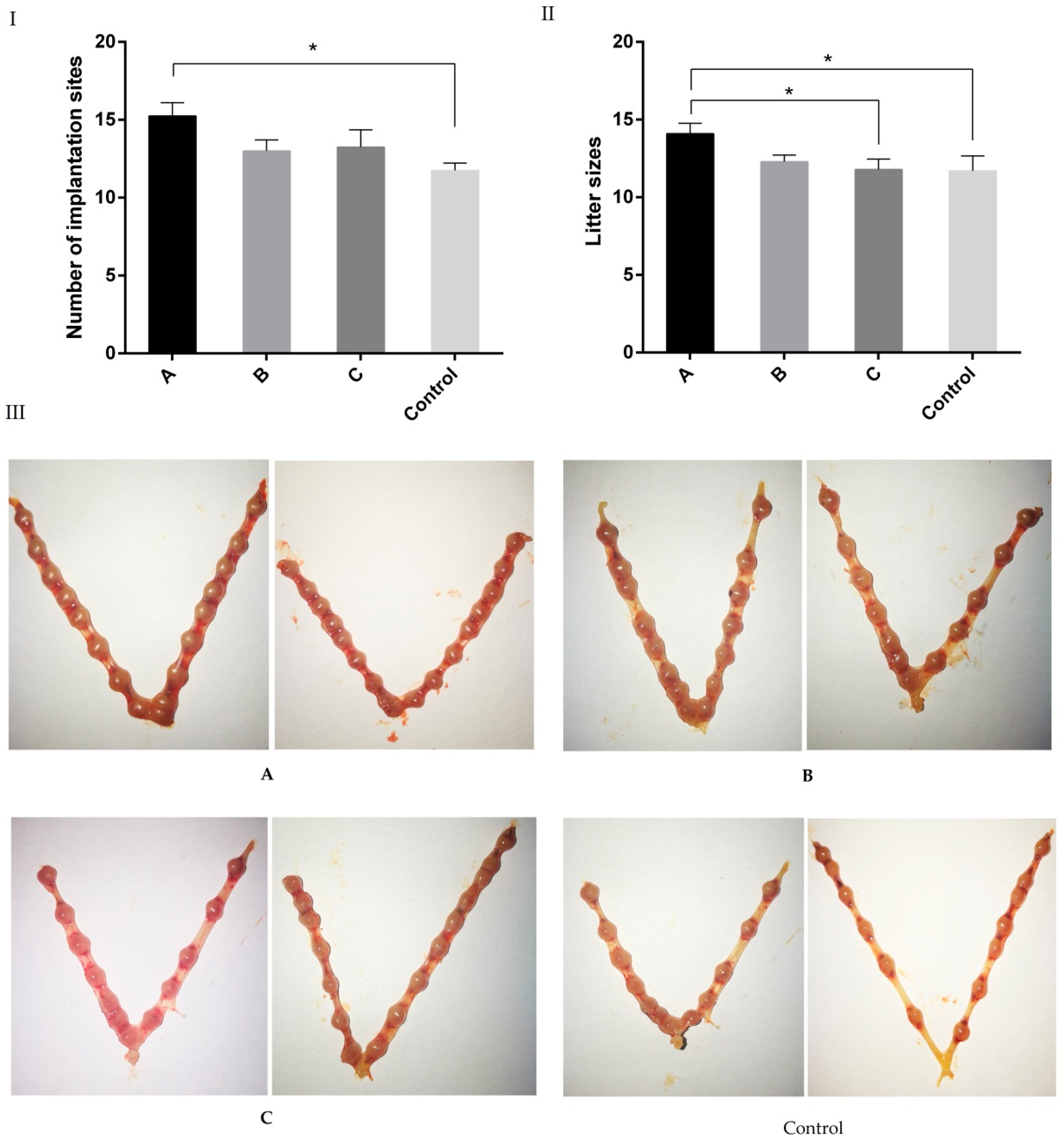
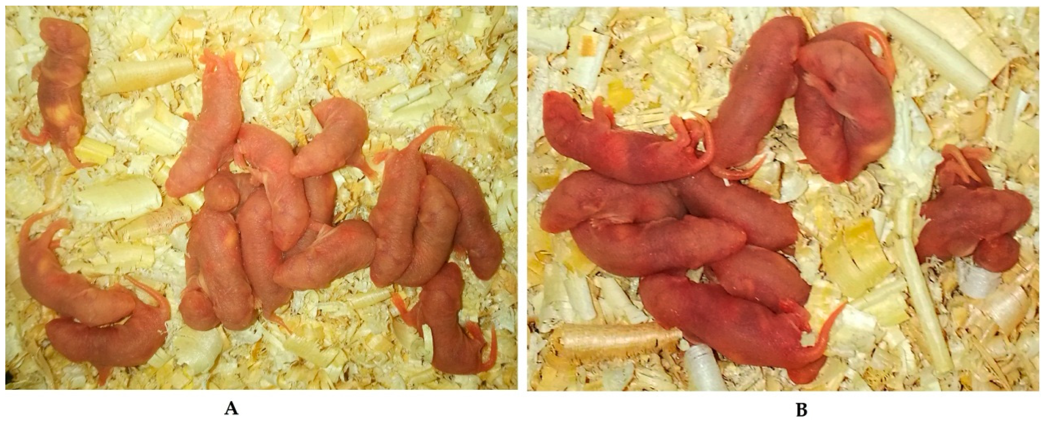
2.3. Effects of Melatonin on Implantation and Litter Size on 7.5 Day of Gestation Mice

The implantation sites 7.5 days after gestation were evaluated (Figure 3). The number of implantation sites in group A (15.3 ± 0.85) was significantly higher than that in the control group (11.7 ± 0.48, p < 0.05); the implantation sites in group B (13.0 ± 0.71) and group C (13.3 ± 1.11) were slightly higher than that of controls, however, this increase did not reach a significant difference (p > 0.05).
The litter size was analyzed after natural delivery. The results showed that the litter size of group A (14.1 ± 0.66) was significantly higher than that of other groups (Figure 4).

2.4. Effects of Melatonin on Serum E2 and P Levels on Day 7.5 of Early Pregnant Mice

The results showed that E2 in group A (52.4 ± 9.05 pg/mL) was significantly lower than that in group B (82.4 ± 9.39 pg/mL, p < 0.05). P level in group B (78.82 ± 8.14 ng/mL) was significantly higher than that in other groups (Figure 5).

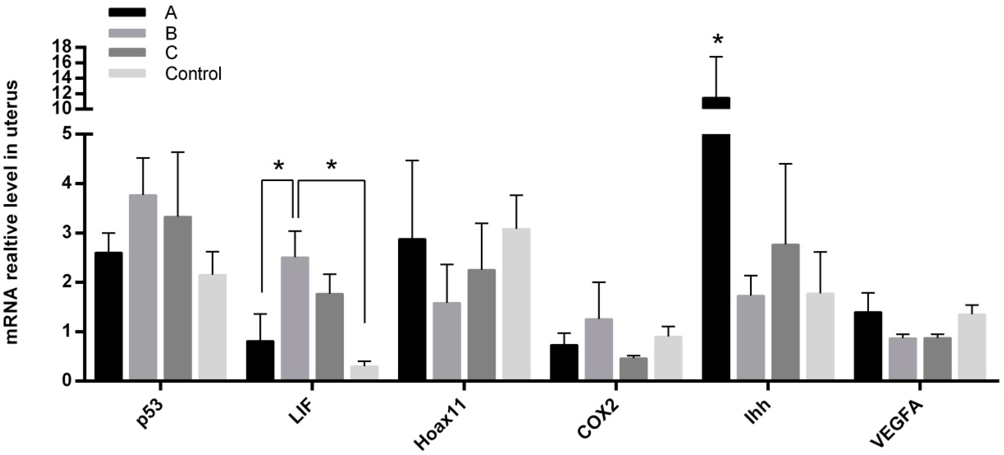
2.5. Effects of Melatonin on Gene Expressions in Mice Endometrium
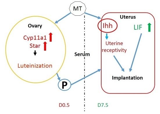
The gene expressions of p53, Hoxa11, COX2, and VEGFA in the endometrium of the uteri were detected among different groups. The mRNA expression levels of p53, Hoxa11, COX2, and VEGFA showed no significant differences among the groups. The expression level of LIF in group B was significantly higher than that in group A and in controls (p < 0.05); in contrast, the expression of Ihh in group A was significantly higher than that of other groups (Figure 6).

3. Discussion

Melatonin has been effectively used to combat oxidative stress, inflammation, cellular apoptosis, and to restore many tissues’ functions. The activities of melatonin are related to its pleiotropic functions. These include its direct antioxidant cascade reactions and its stimulation of antioxidant enzymes while suppressing pro-oxidant enzymes via membrane receptor (MT1/MT2) signal transduction [20]. In the current study, the effects of melatonin on early pregnant mice were investigated. It was reported that, in rats, the melatonin levels alternated periodically in the estrous cycle, the highest melatonin levels occurred during the evenings of metestrus and diestrus, while the minimum levels were detected in the evening of estrus [31]. Melatonin directly affects ovarian function to serve as a local regulator [32,33]. Many studies have tested the effects of melatonin on animal reproduction [34,35,36], and in the majority of these studies, melatonin was given either by intraperitoneal injection or implantation [37,38]. In the current study, the melatonin delivery route was through drinking water. Since the majority of water consumption by mice occurs during the night [39], the melatonin delivered though drinking water more or less resembled the physiological melatonin circadian rhythm. This aspect might render additional benefits of melatonin in reproductive physiology compared to other delivery methods. Indeed, in the study, we observed a significantly elevated melatonin level in serum as well as in the ovarian tissue in mice treated with melatonin in their drinking water compared to that of controls (Figure 1A,B). This melatonin elevation is similar to its physiological circadian rhythm. This was due to the melatonin delivered in drinking water since majority of the water was consumed by mice during the night. It was observed that only small portion of the exogenously supplied melatonin reached the ovaries. This demonstrated a high melatonin metabolism rate in animals [17]. Melatonin added to drinking water did not affect vaginal plug rate of mice, but significantly improved the implantation sites and litter size, especially in the mice given melatonin before mating.
The results revealed that early melatonin treatment significantly upregulated the expression of the two corpus luteum marker genes, StAR and Cyp11a1, in ovaryies [40,41]. Cyp11a1 protein localizes to the mitochondrial inner membrane and catalyzes the conversion of cholesterol to pregnenolone, the first and rate-limiting step in the synthesis of the steroid hormones. The function of StAR is to transport large amounts of cholesterol from the outer to inner mitochondrial membrane [42]. Both StAR and Cyp11a1 were upregulated under melatonin treatment as early as on the 0.5 day of gestation. Since these genes involve pregnenolone formation, it was not a surprise that the level of P was also significantly increased with melatonin treatment in Experiment I. Ovarian P and E2 are essential for successful implantation in mice [1]. The P can promote endometrial luminal epithelial differentiation, stromal cell proliferation, and decidualization [2]. E2/ERα (estrogen receptors α) are critical for blastocyst attachment, but dispensable for subsequent decidualization [43,44]. Melatonin was reported to regulate E2 and P in mammals [45]. The results indicated that during early pregnancy, melatonin could improve the development of corpus luteum, promote synthesis, and release of P in ovaries which is beneficial for the early pregnancy in mice of other species.
In order to find out whether melatonin also affected the post-implantation process, melatonin was given at different gestation stages as mentioned above. The results showed that melatonin given before mating significantly increased implantation sites and litter size (Figure 3) and reduced the E2 level at D7.5 (Figure 5). Melatonin given after mating, for example in group B, elevated the serum P level in mice. Generally, the progesterone/estradiol (P/E2) ratio was reduced by melatonin treatment. The P/E2 ratio is crucial to implantation, and it determines pregnancy outcomes of animals [46,47]. High levels of estrogen shorten the duration window for uterine receptivity [48]. It is possible that the early melatonin treatment in pregnant mice influenced the uterine receptivity mediation.
Several genes that are crucial for implantation and endometrial development were tested in the current study. These include p53 (a regulator of LIF), cyclooxygenase-2 (COX2), abdominal-B-like Hox genes (Hoxa11), and vascular endothelial growth factor (VEGFA). It has been reported that these genes were regulated by melatonin in some tissues and organs [49,50,51,52]. However, this was not the case in our study, and we failed to find significant differences between treated and untreated groups. This may be tissue specific, and if it is, the results indicate that these genes in the uterine might not be influenced by melatonin in the early gestation stage. The expression level of leukaemia inhibitory factor (LIF) in melatonin treated animals, for example in group B, was significantly higher than that of the control group. Epithelial Ihh is a paracrine growth factor for stromal cells and involves epithelial–mesenchymal signaling transduction [53,54]. The significantly increased expression of Ihh with melatonin treatment before mating suggests that the improved uterine receptivity was via the Ihh signaling pathway. Collectively, the results indicate that early melatonin treatment before gestation promotes the development of corpus luteum after ovulation and regulates the differentiation and secretion of E2 and P. The increased ratio of P/E2 mediates endometrial development and improves endometrial receptivity.

4. Materials and Methods

4.1. Animals

All CD-1 mice (6 weeks old) were purchased from Vital River Laboratories Co., Ltd. (Beijing, China). Mice were housed at a temperature of 22–26 °C with a 12:12 light:dark cycle (darkness from 20:00 to 08:00) and had access to food and water ad libitum. All experimental procedures were approved by the Animal Welfare Committee of China Agricultural University (Permission Number: SYXK(Beijing)2015002; The period of valid days: 22 September 2015–22 September 2020). After a week of acclimation, those mice in good condition were randomized for the following experiments.

4.2. Experiment Designs

Female mice (body weight 27–30 g) were treated with melatonin in drinking water (4.3 μg/mL) for 7 days. The mice had free access to water and food ad libitum. The water consumption was 5.5 to 7.0 mL/mouse/day (roughly, melatonin 0.96 μg/g body weight). Blood samples were collected from the caudal vein at 3:00, 9:00, 15:00, and 21:00.
Estrus synchronization and mating: Female and male mice were placed into the same cage at 19:00 on day 1 of the estrous cycle and separated the next morning. Then, this process was repeated for the second and the third night. Successful mating was confirmed by the presence of a vaginal plug.
Experiment I: The mice were randomly divided into two groups (80 mice/group). Mice without treatment served as the control group while the other mice were treated with melatonin in their drinking water (4.3 μg/mL); the amount was based on our previous experimental results [24]. After 14 days of melatonin treatment, spontaneous estrus female mice from both control and MT groups were carefully selected. To ensure the consistency of gestation time, female mice were mated with male mice at night. On the following morning, females were examined for vaginal plugs and sacrificed by cervical dislocation after 12 h pregnancy (D0.5). Their blood, uterine, and ovaries were collected for the subsequent assays.
Experiment II: The mice were randomly divided into four groups (80 mice/group). Mice without treatment served as the control group while the other mice were treated with melatonin (4.3 μg/mL) as follows: 14 days before breeding (vaginal plugs was detected) as group A; mice treated with melatonin from the mating to the delivery as group B; mice treated with melatonin from 14 days before mating until the delivery as group C. Ten mice from each group (vaginal plugs were detected) were sacrificed by cervical dislocation in D7.5 pregnancy. Their blood and ovaries were collected for the subsequent assays. The other pregnant mice were kept for litter size calculation after delivery.

4.3. The Assays of Melatonin, Estradiol-17β, and Progesterone

The serum was separated from blood samples by centrifugation at 4 °C (800× g) for 10 min at room temperature, then serum was collected and stored at −80 °C for melatonin radioimmunoassays (RIAs). The procedures followed were from the instructions of the Melatonin Research RIA Kit (LDN GmbH & Co., KG, Nordhorn, Germany). The ovaries were weighed and homogenized immediately in 1 mL methyl alcohol and centrifuged at 12,000 rpm for 10 min at 4 °C. The supernatant was collected and dried by Pressure Blowing Concentrator, and then the residue was reconstituted in 100 μL double distilled water for RIA assays. The recovery of melatonin from the ovaries ranged from 81.7% to 93.1%.
On days 0.5 and 7.5 of pregnancy, mice from the Experiment I and Experiment II, respectively, were anesthetized with 2% pentobarbital sodium (0.15 mL/each) intraperitoneal injectionand the blood of each mouse (melatonin-treated and control group) was collected from the caudal vein and placed at 4 °C for 30 min and then centrifuged for 10 min at 4 °C (800× g). The serum was collected and stored at −80 °C for future estradiol-17β (E2) and progesterone radioimmunoassays (RIA). The procedures followed were from the instructions of the Progesterone Direct RIA Kit (ICN Biomedicals, Irvin, CA, USA) and the Estradiol-17b (E2) Direct RIA Kit (ICN Biomedicals). The RIAs involved chromatographic separation of the steroids to enhance specificity. The RIAs used a tracer and a charcoal–dextran method to separate the bound and free steroid. The kits included a specific antibody that had no significant cross-reactivity with other steroids.

4.4. Implantation Sites Counting

On day 7.5 of pregnancy, the mice (from the Experiment II) were given 2% pentobarbital sodium (0.15 mL/each) (intraperitoneal injection) and sacrificed by cervical dislocation. The uteri were collected and cleaned, and these uteri were placed on white paper for implantation site counting and recording.

4.5. Gene Expression Assay by Real-Time Q-PCR

Ovarian and uterine samples from mice of D0.5 and D7.5 of pregnancy were collected. The expression levels of cytochrome P450 family 11 subfamily A member 1 (Cyp11a), steroidogenic acute regulatory protein (StAR), luteinizing hormone receptor (LHR), and aldo-keto reductase family 1 member C18 (Akrlc18) genes in ovaries and the expression levels of transformation related protein 53 (P53), cyclooxygenase-2 (COX2), homeobox A11 (Hoxa11), leukaemia inhibitory factor (LIF), vascular endothelial growth factor (VEGFA), and indian hedgehog (Ihh) in uterine samples of 7.5 day pregnant mice were measured. Total RNA was extracted using the TRIzol reagent (Invitrogen Inc., Carlsbad, CA, USA) and immediately was reversely transcribed using PrimeScript™ RT reagent Kit with gDNA Eraser (TaKaRa Bio Inc., Tokyo, Japan). The expression levels of genes were detected by real-time PCR (Table 1). The reaction solution consisted of 10 μL SYBR Green, 30 mmol/L forward and reverse primers, 2 μL template, and ddH2O to a total volume of 20 μL. The procedure was as follows: 95 °C for 10 min; 35 cycles of 95 °C for 10 s, and 60 °C for 10 s; melting curve from 65 to 95 °C, increments of 0.5 °C every 5 s. Normalization was performed using the housekeeping gene glyceraldehyde-3-phosphate dehydrogenase (GAPDH). Primer sequences are listed in Table 1. Relative mRNA expression was calculated by the 2−ΔΔCt method.

4.6. Statistical Analyses

Data are expressed as mean ± SEM (standard error of mean). Statistical analyses were done using the one-way analysis of variance (ANOVA) with the aid of SPSS 19.0 statistical software followed by the Student t-test. p < 0.05 was considered significant, and p < 0.01 was considered highly significant.

5. Conclusions

In conclusion, exogenous melatonin promotes the development of corpus luteum after ovulation by up-regulating the expression of StAR and Cyp11a1. Melatonin delivered by drinking water is an effective way to enhance the physiological melatonin circadian rhythm. As a result, it increases the expression of genes involved in pregnenolone synthesis in ovary and endometrium Ihh expression, which mediates endometrial development and promotes embryo implantation.

Acknowledgments

This research was supported by the National Natural Science Foundation of China (31372306), National Program on Key Basic Research Project (973 Program) (2014CD138505), and Genetically Modified Organisms Breeding Major Projects of China (2014ZX08008002B).

Author Contributions

Shengyu Guan, Lu Xie and Teng Ma were responsible for mouse experiment, data analysis and paper writing. Dongying Lv and Wang Jing were responsible for collection of samples RNA isolation, qRT–PCR. Yukun Song and Xiuzhi Tian were responsible for melatonin and hormone assay. Guoshi Liu, Xianghong Xiao and Zhiping Liu responsible for mouse experimental design, supervised the experiments and revised the manuscript.

Conflicts of Interest

The authors declare no conflict of interest.

Abbreviations

MTMelatonin
AANATAralkylamine N-acetyltransferase
ASMTAcetylserotonin O-methyltransferase
HPG axisHypothalamus-pituitary-gonadal axis
KmMichaelis constant
Q-PCRQuantitative PCR
E2Estradiol-17β
PProgesterone
RIARadioimmunoassays
GAPDHGlyceraldehyde-3-phosphate dehydrogenase
Akr1c18Aldo-keto reductase family 1 member C18
p53Transformation related protein 53
StARSteroidogenic acute regulatory protein
Cyp11a1Cytochrome P450 family 11 subfamily A member 1
LHRLuteinizing hormone receptor
VEGFAVascular endothelial growth factor
Hoxa11Homeobox A11
IhhIndian hedgehog
COX2CYCLOOXYGENASE-2
LIFLeukaemia inhibitory factor

References

  1. Dey, S.K.; Lim, H.; Das, S.K.; Reese, J.; Paria, B.C.; Daikoku, T.; Wang, H. Molecular cues to implantation. Endocr. Rev. 2004, 25, 341–373. [Google Scholar] [CrossRef] [PubMed]
  2. Wang, H.; Dey, S.K. Roadmap to embryo implantation: Clues from mouse models. Nat. Rev. Genet. 2006, 7, 185–199. [Google Scholar] [CrossRef] [PubMed]
  3. Lerner, A.B.; Case, J.D.; Takahashi, Y. Isolation of melatonin, the pineal gland factor that lightens melanocyte. J. Am. Chem. Soc. 1958, 10, 2587. [Google Scholar] [CrossRef]
  4. Paria, B.C.; Reese, J.; Das, S.K.; Dey, S.K. Deciphering the cross-talk of implantation: Advances and challenges. Science 2002, 296, 2185–2188. [Google Scholar] [CrossRef] [PubMed]
  5. Tan, D.X.; Manchester, L.C.; Qin, L.; Reiter, R.J. Melatonin: A mitochondrial targeting molecule involving mitochondrial protection and dynamics. Int. J. Mol. Sci. 2016, 17, 2124. [Google Scholar] [CrossRef] [PubMed]
  6. Reiter, R.J.; Tan, D.X.; Galano, A. Melatonin: Exceeding expectations. Physiology 2014, 29, 325–333. [Google Scholar] [CrossRef] [PubMed]
  7. Shi, L.; Li, N.; Bo, L.; Xu, Z. Melatonin and hypothalamic-pituitary-gonadal axis. Curr. Med. Chem. 2013, 20, 2017–2031. [Google Scholar] [CrossRef] [PubMed]
  8. Renuka, K.; Joshi, B.N. Melatonin-induced changes in ovarian function in the freshwater fish Channa punctatus (Bloch) held in long days and continuous light. Gen. Comp. Endocr. 2010, 165, 42–46. [Google Scholar] [CrossRef] [PubMed]
  9. McCarter, S.J.; Boswell, C.L.; St. Louis, E.K.; Dueffert, L.G.; Slocumb, N.; Boeve, B.F.; Silber, M.H.; Olson, E.J; Tippmann-Peikert, M. Treatment outcomes in REM sleep behavior disorder. Sleep Med. 2013, 14, 237–242. [Google Scholar] [CrossRef] [PubMed]
  10. Barrett, P.; Bolborea, M. Molecular pathways involved in seasonal body weight and reproductive responses governed by melatonin. J. Pineal Res. 2012, 52, 376–388. [Google Scholar] [CrossRef] [PubMed]
  11. Tian, X.; Wang, F.; He, C.; Zhang, L.; Tan, D.; Reiter, R.J.; Xu, J.; Ji, P.; Liu, G. Beneficial effects of melatonin on bovine oocyte maturation: A mechanistic approach. J. Pineal Res. 2014, 57, 239–247. [Google Scholar] [CrossRef] [PubMed]
  12. DeNicolo, G.; Morris, S.T.; Kenyon, P.R.; Morel, P.C.; Parkinson, T.J. Melatonin-improved reproductive performance in sheep bred out of season. Anim. Reprod. Sci. 2008, 109, 124–133. [Google Scholar] [CrossRef] [PubMed]
  13. Pévet, P.; Haldar-Misra, C. Effect of orally administered melatonin on reproductive function of the golden hamster. Experientia 1982, 38, 1493–1494. [Google Scholar] [CrossRef] [PubMed]
  14. Graham, L.H.; Swanson, W.F.; Wildt, D.E. Influence of oral melatonin on natural and gonadotropin-induced ovarian function in the domestic cat. Theriogenology 2004, 61, 1061–1076. [Google Scholar] [CrossRef] [PubMed]
  15. Dair, E.L.; Simoes, R.S.; Simões, M.J.; Romeu, L.R.; Oliveira-Filho, R.M.; Haidar, M.A.; Baracat, E.C.; Soares, J.M., Jr. Effects of melatonin on the endometrial morphology and embryo implantation in rats. Fertil. Steril. 2008, 89, 1299–1305. [Google Scholar] [CrossRef] [PubMed]
  16. Galano, A.; Tan, D.X.; Reiter, R.J. On the free radical scavenging activities of melatonin’s metabolites, AFMK and AMK. J. Pineal Res. 2013, 54, 245–257. [Google Scholar] [CrossRef] [PubMed]
  17. Galano, A.; Tan, D.X.; Reiter, R.J. Melatonin as a natural ally against oxidative stress: A physicochemical examination. J. Pineal Res. 2011, 51, 1–16. [Google Scholar] [CrossRef] [PubMed]
  18. Manchester, L.C.; Coto-Montes, A.; Boga, J.A.; Andersen, L.P.; Zhou, Z.; Galano, A.; Vriend, J.; Tan, D.X.; Reiter, R.J. Melatonin: An ancient molecule that makes oxygen metabolically tolerable. J. Pineal Res. 2015, 59, 403–419. [Google Scholar] [CrossRef] [PubMed]
  19. Ding, K.; Wang, H.; Xu, J.; Li, T.; Zhang, L.; Ding, Y.; Zhu, L.; He, J.; Zhou, M. Melatonin stimulates antioxidant enzymes and reduces oxidative stress in experimental traumatic brain injury: The Nrf2-ARE signaling pathway as a potential mechanism. Free Radic. Biol. Med. 2014, 73, 1–11. [Google Scholar] [CrossRef] [PubMed]
  20. Reiter, R.J.; Mayo, J.C.; Tan, D.X.; Sainz, R.M.; Alatorre-Jimenez, M.; Qin, L. Melatonin as an antioxidant: Under promises but over delivers. J. Pineal Res. 2016, 61, 253–278. [Google Scholar] [CrossRef] [PubMed]
  21. Yang, Y.; Sun, Y.; Yi, W.; Li, Y.; Fan, C.; Xin, Z.; Jiang, S.; Di, S.; Qu, Y.; Reiter, R.J.; et al. A review of melatonin as a suitable antioxidant against myocardial ischemia-reperfusion injury and clinical heart diseases. J. Pineal Res. 2014, 57, 357–366. [Google Scholar] [CrossRef] [PubMed]
  22. Tamura, H.; Takasaki, A.; Miwa, I.; Taniguchi, K.; Maekawa, R.; Asada, H.; Taketani, T.; Matsuoka, A.; Yamagata, Y.; Shimamura, K.; et al. Oxidative stress impairs oocyte quality and melatonin protects oocytes from radical damage and improves fertilization rate. J. Pineal Res. 2008, 44, 280–287. [Google Scholar] [CrossRef] [PubMed]
  23. Tanabe, M.; Tamura, H.; Taketani, T.; Okada, M.; Lee, L.; Tamura, I.; Maekawa, R.; Asada, H.; Yamagata, Y.; Sugino, N. Melatonin protects the integrity of granulosa cells by reducing oxidative stress in nuclei, mitochondria, and plasma membranes in mice. J. Reprod. Dev. 2015, 61, 35–41. [Google Scholar] [CrossRef] [PubMed]
  24. García, J.J.; López-Pingarrón, L.; Almeida-Souza, P.; Tres, A.; Escudero, P.; García-Gil, F.A.; Tan, D.X.; Reiter, R.J.; Ramírez, J.M.; Bernal-Pérez, M. Protective effects of melatonin in reducing oxidative stress and in preserving the fluidity of biological membranes: A review. J. Pineal Res. 2014, 56, 225–237. [Google Scholar] [CrossRef] [PubMed]
  25. Reiter, R.J.; Tan, D.X. Melatonin: A novel protective agent against oxidative injury of the ischemic/reperfused heart. Cardiovasc. Res. 2003, 58, 10–19. [Google Scholar] [CrossRef]
  26. Zhang, H.M.; Zhang, Y. Melatonin: A well-documented antioxidant with conditional pro-oxidant actions. J. Pineal Res. 2014, 57, 131–146. [Google Scholar] [CrossRef] [PubMed]
  27. El Mouatassim, S.; Guérin, P.; Ménézo, Y. Expression of genes encoding antioxidant enzymes in human and mouse oocytes during the final stages of maturation. Mol. Hum. Reprod. 1999, 5, 720–725. [Google Scholar] [CrossRef] [PubMed]
  28. Tamura, H.; Kawamoto, M.; Sato, S.; Tamura, I.; Maekawa, R.; Taketani, T.; Aasada, H.; Takaki, E.; Nakai, A.; Reiter, R.J.; et al. Long-term melatonin treatment delays ovarian aging. J. Pineal Res. 2017, 62, e12381. [Google Scholar] [CrossRef] [PubMed]
  29. Moniruzzaman, M.; Hasan, K.N.; Maitra, S.K. Melatonin actions on ovaprim (synthetic GnRH and domperidone)-induced oocyte maturation in carp. Reproduction 2016, 151, 285–296. [Google Scholar] [CrossRef] [PubMed]
  30. He, C.; Wang, J.; Li, Y.; Zhu, K.; Xu, Z.; Song, Y.; Song, Y.; Liu, G. Melatonin-related genes expressed in the mouse uterus during early gestation promote embryo implantation. J. Pineal Res. 2015, 58, 300–309. [Google Scholar] [CrossRef] [PubMed]
  31. Ozaki, Y.; Wurtman, R.; Alonso, R.; Lynch, H.J. Melatonin secretion decreases during the proestrous stage of the rat estrous cycle. Proc. Natl. Acad. Sci. USA 1978, 75, 531–534. [Google Scholar] [CrossRef] [PubMed]
  32. Tamura, H.; Nakamura, Y.; Korkmaz, A.; Manchester, L.C.; Tan, D.X.; Sugino, N.; Reiter, R.J. Melatonin and the ovary: Physiological and pathophysiological implications. Fertil. Steril. 2009, 92, 328–343. [Google Scholar] [CrossRef] [PubMed]
  33. Tamura, H.; Takasaki, A.; Taketani, T.; Tanabe, M.; Lee, L.; Tamura, I.; Maekawa, R.; Aasada, H.; Yamagata, Y.; Sugino, N. Melatonin and female reproduction. J. Obstet. Gynaecol. Res. 2014, 40, 1–11. [Google Scholar] [CrossRef] [PubMed]
  34. Reiter, R.J.; Tamura, H.; Tan, D.X.; Xu, X. Melatonin and the circadian system: Contributions to successful female reproduction. Fertil. Steril. 2014, 102, 321–328. [Google Scholar] [CrossRef] [PubMed]
  35. Dai, X.; Lu, Y.; Zhang, M.; Miao, Y.; Zhou, C.; Cui, Z.; Xiong, B. Melatonin improves the fertilization ability of post-ovulatory aged mouse oocytes by stabilizing ovastacin and Juno to promote sperm binding and fusion. Hum. Reprod. 2017, 32, 598–606. [Google Scholar] [CrossRef] [PubMed]
  36. Pradip, M.; Kazi, N.H.; Palash, K.P.; Saumen, K.M. Influences of exogenous melatonin on the oocyte growth and oxidative status of ovary during different reproductive phases of an annual cycle in carp Catla catla. Theriogenology 2017, 87, 349–359. [Google Scholar]
  37. He, C.; Ma, T.; Shi, J.; Zhang, Z.; Wang, J.; Zhu, K.; Li, Y.; Yang, M.; Song, Y.; Liu, G. Melatonin and its receptor MT1 are involved in the downstream reaction to luteinizing hormone and participate in the regulation of luteinization in different species. J. Pineal Res. 2016, 61, 279–290. [Google Scholar] [CrossRef] [PubMed]
  38. Ramadan, T.A.; Sharma, R.K.; Phulia, S.K.; Balhara, A.K.; Ghuman, S.S.; Singh, I. Manipulation of reproductive performance of lactating buffaloes using melatonin and controlled internal drug release device treatment during out-of-breeding season under tropical conditions. Theriogenology 2016, 86, 1048–1053. [Google Scholar] [CrossRef] [PubMed]
  39. Andersen, L.P.; Werner, M.U.; Rosenkilde, M.M.; Harpsøe, N.G.; Fuglsang, H.; Rosenberg, J.; Gögenur, I. Pharmacokinetics of oral and intravenous melatonin in healthy volunteers. BMC Pharmacol. Toxicol. 2016, 19, 17–18. [Google Scholar] [CrossRef] [PubMed]
  40. Jimenezkrassel, F.; Folger, J.K.; Ireland, J.L.; Smith, G.W.; Hou, X.; Davis, J.S. Evidence that high variation in ovarian reserves of healthy young adults has a negative impact on the corpus luteum and endometrium during estrous cycles in cattle. Biol. Reprod. 2009, 80, 1272–1281. [Google Scholar] [CrossRef] [PubMed]
  41. Arakane, F.; Kallen, C.B.; Watari, H.; Foster, J.A.; Sepuri, N.B.; Pain, D.; Stayrook, S.E.; Lewis, M.; Gerton, G.L.; Strauss, J.F. The mechanism of action of steroidogenic acute regulatory protein (stAR). stAR acts on the outside of mitochondria to stimulate steroidogenesis. J. Biol. Chem. 1998, 273, 16339–16345. [Google Scholar] [CrossRef] [PubMed]
  42. Walter, L.M. Steroidogenic acute regulatory protein (StAR), a novel mitochondrial cholesterol transporter. BBA Mol. Cell. Res. 2007, 1771, 663–676. [Google Scholar]
  43. Curtis, S.W.; Clark, J.; Myers, P.; Korach, K.S. Disruption of estrogen signaling does not prevent progesterone action in the estrogen receptor α knockout mouse uterus. Proc. Natl. Acad. Sci. USA 1999, 96, 3646–3651. [Google Scholar] [CrossRef] [PubMed]
  44. Paria, B.C.; Tan, J.; Lubahn, D.B.; Dey, S.K.; Das, S.K. Uterine decidual response occurs in estrogen receptor-α-deficient mice. Endocrinology 1999, 140, 2704–2710. [Google Scholar] [CrossRef] [PubMed]
  45. Chuffa, L.G.; Seiva, F.R.; Fávaro, W.J.; Amorim, J.P.; Teixeira, G.R.; Mendes, L.O.; Fioruci-Fontanelli, B.A.; Pinheiro, P.F.; Martinez, M.; Martinez, F.E. Melatonin and ethanol intake exert opposite effects on circulating estradiol and progesterone and differentially regulate sex steroid receptors in the ovaries, oviducts, and uteri of adult rats. Reprod. Toxicol. 2013, 39, 40–49. [Google Scholar] [CrossRef] [PubMed]
  46. Lai, T.H.; Lee, F.K.; Lin, T.K.; Horng, S.G.; Chen, S.C.; Chen, Y.H.; Wang, P.C. An increased serum progesterone-to-estradiol ratio on the day of human chorionic gonadotropin administration does not have a negative impact on clinical pregnancy rate in women with normal ovarian reserve treated with a long gonadotropin releasing hormone agonist protocol. Fertil. Steril. 2009, 92, 508–514. [Google Scholar] [PubMed]
  47. Tang, W.; Chien, L.; Tzeng, C. A single day16 serum human chorionic gonadotropin (hCG) level combined with estradiol/progesterone (E2/P) ratio may predict pregnancy outcomes following intrauterine insemination. Fertil. Steril. 2008, 92, 508–514. [Google Scholar]
  48. Ma, W.G.; Song, H.; Das, S.K.; Paria, B.C.; Dey, S.K. Estrogen is a critical determinant that specifies the duration of the window of uterine receptivity for implantation. Proc. Natl. Acad. Sci. USA 2003, 100, 2963–2968. [Google Scholar] [CrossRef] [PubMed]
  49. Wang, J.; Xiao, X.; Zhang, Y.; Shi, D.; Chen, W.; Fu, L.; Liu, L.; Xie, F.; Kang, T.; Huang, W.; et al. Simultaneous modulation of COX-2, p300, Akt, and Apaf-1 signaling by melatonin to inhibit proliferation and induce apoptosis in breast cancer cells. J. Pineal Res. 2012, 53, 77–90. [Google Scholar] [CrossRef] [PubMed]
  50. Romeu, L.R.; da Motta, E.L.; Maganhin, C.C.; Oshima, C.T.; Fonseca, M.C.; Barrueco, K.F.; Simões, R.S.; Pellegrino, R.; Baracat, E.C.; Soares-Junio, J.M. Effects of melatonin on histomorphology and on the expression of steroid receptors, VEGF, and PCNA in ovaries of pinealectomized female rats. Fertil. Steril. 2011, 95, 1379–1384. [Google Scholar] [CrossRef] [PubMed]
  51. Liang, X.; Deng, W.; Li, M.; Zhao, Z.A.; Wang, T.S.; Feng, X.H.; Cao, Y.J.; Duan, E.K.; Yang, Z.M. Egr1 protein acts downstream of estrogen-leukemia inhibitory factor (LIF)-STAT3 pathway and plays a role during implantation through targeting Wnt4. J. Biol. Chem. 2014, 289, 23534–23545. [Google Scholar] [CrossRef] [PubMed]
  52. Wang, T.S.; Gao, F.; Qi, Q.; Qin, F.N.; Zuo, R.J.; Li, Z.L.; Liu, J.L.; Yang, Z.M. Dysregulated LIF-STAT3 pathway is responsible for impaired embryo implantation in a Streptozotocin-induced diabetic mouse model. Biol. Open 2015, 4, 893–902. [Google Scholar] [CrossRef] [PubMed]
  53. Matsumoto, H.; Zhao, X.; Das, S.K.; Hogan, B.L.; Dey, S.K. Indian hedgehog as a progesterone-responsive factor mediating epithelial-mesenchymal interactions in the mouse uterus. Dev. Biol. 2002, 245, 280–290. [Google Scholar] [CrossRef] [PubMed]
  54. Filant, J.; Spencer, T.E. Endometrial glands are essential for blastocyst implantation and decidualization in the mouse uterus. Biol. Reprod. 2013, 88, 93. [Google Scholar] [CrossRef] [PubMed]
Figure 1. Melatonin levels and vaginal plug rate of the mice. (A) Serum melatonin levels at different time points (mean ± SEM, n = 7); (B) Ovarian melatonin levels at different time points (mean ± SEM, n = 7). Melatonin (MT): mice received 14-day melatonin treatment. Control: mice were without melatonin; (C) Vaginal plug rate. Day 1: the first day of the mating process. Days 1–3 represent the total accumulative vaginal plug rate from day 1 to day 3. Value p is based on the Pearson χ square test. “*” represents significant differences, p < 0.05.
Ijms 18 01637 g001
Figure 2. Effects of melatonin on estradiol (E2) and progesterone (P) levels as well as the mRNA of StAR, Akrlc18, Cyp11a1, and LHR in D0.5 gestation mice. (A) Serum E2 and P concentrations (n = 10); (B) The relative levels of mRNAs of StAR, Akrlc18, Cyp11a1, and LHR in mice ovaries (n = 7), detected by Q-PCR. The RT-QPCR was independently repeated three times. “*” represents a significant difference of these columns (p < 0.05).
Ijms 18 01637 g002
Figure 3. Effect of melatonin on the implantation sites and litter sizes. Mice were treated with melatonin (4.3 μg/mL) in drinking water. (A) Melatonin was given 14 days before mating (vaginal plugs was detected); (B) melatonin was given from the mating to the delivery; (C) melatonin was given from 14 days before mating to the delivery. (Control) without any treatment. n = 10. (I) Number of implantation sites; (II) Litter size; (III) Representative images of implantation sites in each group. “*” represents a significant difference between the different groups (p < 0.05).
Ijms 18 01637 g003
Figure 4. Representative images of newborn mice. (A) Melatonin was given 14 days before mating (vaginal plugs were detected); (B) Melatonin was given from the mating (vaginal plugs were detected) to the delivery; (C) Melatonin was given from 14 days before mating (vaginal plugs were detected) to the delivery; (Control) Without any treatment.
Ijms 18 01637 g004aIjms 18 01637 g004b
Figure 5. E2 and P concentrations in serum of Day 7.5 pregnant mice treated with or without melatonin (4.3 μg/mL) in drinking water. A, melatonin was given 14 days before mating (vaginal plugs was detected); B, melatonin was given from the mating to the delivery; C, melatonin was given from 14 days before mating to the delivery; Control: without melatonin. n = 10. “*” represents a significant difference of these columns (p < 0.05).
Ijms 18 01637 g005
Figure 6. The mRNA levels of p53, LIF, Hoxa11, COX2, Ihh, and VEGFA in endometrium of 7.5 day pregnant mice. Mice were treated with melatonin (4.3 μg/mL) in drinking water. A, melatonin was given 14 days before mating (vaginal plugs were detected); B, melatonin was given from the mating to the delivery; C, melatonin was given from 14 days before mating to the delivery; Control: without melatonin. The experiment was independently repeated four times. “*” represents a significant difference of these columns (p < 0.05).
Ijms 18 01637 g006
Table 1. Primers for RT-PCR.
GenesPrimer Sequence (5′–3′)Tm (°C)Accession Number
GAPDHForward: CCTGGAGAAACCTGCCAAGTAT60XM_017321385
Reverse: GGAAGAGTGGGAGTTGCTGTTG
Akr1c18Forward: TTGGTCAACTTCCCATCGTC59NM_134066
Reverse: GCCCTGCATCCTTACACTTC
P53Forward: TGAGGTTCGTGTTTGTGCCTGC60NM_001127233
Reverse: CCATCAAGTGGTTTTTTCTTTTGC
StARForward: CCTTGGGCATACTCAACAACC60NM_011485
Reverse: CCACATCTGGCACCATCTTACTT
Cyp11a1Forward: GGGCAGTTTGGAGTCAGTTTAC60NM_001346787.1
Reverse: TTTAGGACGATTCGGTCTTTCTT
LHRForward: CTGAGGAGATTTGGTTGCTGTA60NM_013582
Reverse: ATTTGGGTGGACTTTTTTGGGG
MT2Forward: GGTGGCTCCCATGCTATCTA59NM_008630
Reverse: AAACAAATTACCTGCGTTCCG
VEGFAForward: GAGAAGACAGGGTGGTGGAAG59NM_001025250
Reverse: GAAGGGAAGATGAGGAAGGGT
Hoxa11Forward: ATAGCACGGTGGGCAGGAACG
Reverse: AGTCGGAGGAAGCGAGGTTTT
62NM_010450
IhhForward: CTACAATCCCGACATCATCTTCAA
Reverse: CGGTCACCCGCAGTTTCA
62NM_001313683
COX2Forward: ACCTGGTGAACTACGACTGCTA
Reverse: CCTGGTCGGTTTGATGTTACTG
59YP_00168670
LIFForward: CTGACACCTTTCGCTTTCCTC
Reverse: ACTTTCCACCTGTTTGTTCTGC
60NM_001257135
GAPDH, glyceraldehyde-3-phosphate dehydrogenase; Akr1c18, aldo-keto reductase family 1 member C18; p53, transformation related protein 53; StAR, steroidogenic acute regulatory protein; Cyp11a1, cytochrome P450 family 11 subfamily A member 1; LHR, luteinizing hormone receptor; MT2, melatonin membrane receptor 2; ; VEGFA, vascular endothelial growth factor; Hoxa11, homeobox A11; Ihh, Indian hedgehog; COX2, cyclooxygenase-2; LIF, leukaemia inhibitory factor.

