Article

Allyl Isothiocyanate Exhibits No Anticancer Activity in MDA-MB-231 Breast Cancer Cells

1
Department of Clinical and Molecular Sciences, Polytechnic University of Marche, 60126 Ancona, Italy
2
Advanced Technology Center for Aging Research, Scientific and Technological Pole, Italian National Institute of Health and Science on Aging (INRCA), 60120 Ancona, Italy
3
Occupational Medicine, Department of Clinical and Experimental Medicine, University of Catania, Via Santa Sofia 78, 95123 Catania, Italy
*
Author to whom correspondence should be addressed.
Int. J. Mol. Sci. 2018, 19(1), 145; https://doi.org/10.3390/ijms19010145
Submission received: 14 November 2017 / Revised: 24 December 2017 / Accepted: 30 December 2017 / Published: 4 January 2018

Abstract

:
It was reported recently that allyl isothiocyanate (AITC) could inhibit various types of cancer cell growth. In the present study, we further investigated whether AITC could inhibit the growth of human breast cancer cells. Unexpectedly, we found that AITC did not inhibit, rather slightly promoted, the proliferation of MDA-MB-231 breast cancer cells, although it did have inhibitory effect on MCF-7 breast cancer cells. Cytofluorimetric analysis revealed that AITC (10 µM) did not induce apoptosis and cell cycle arrest in MDA-MB-231 cells. In addition, AITC significantly (p < 0.05) increased the expression of BCL-2 and mTOR genes and Beclin-1 protein in MDA-MB-231 cells. No significant changes in expression of PRKAA1 and PER2 genes, Caspase-8, Caspase-9, PARP, p-mTOR, and NF-κB p65 proteins were observed in these AITC-treated cells. Importantly, AITC displayed cytotoxic effect on MCF-10A human breast epithelial cell line. These observations suggest that AITC may not have inhibitory activity in MDA-MB-231 breast cancer cells. This in vitro study warrants more preclinical and clinical studies on the beneficial and harmful effects of AITC in healthy and cancer cells.

Graphical Abstract

1. Introduction

Breast cancer has been a leading cause of cancer-associated death in females worldwide. Currently available treatments of this cancer show unsatisfactory efficacy. In addition to development of drug-resistance and side-effects of therapy, the risk of cancer recurrence after therapy is high. To find safe and effective drugs, research has focused on natural compounds because of their safe nature. Several compounds have been proposed as anti-breast cancer agent based on in vitro and animal studies [1,2].
Allyl isothiocyanate (AITC; 3-isothiocyanato-1-propene, CH2CHCH2NCS) is a sulfur containing organic compound and is an enzymatic hydrolysis product of the glucosinolate sinigrin present in various cruciferous vegetables, such as Brussels sprouts (8.9 μmol/g dry wt), cauliflower (9.3 μmol/g dry wt), cabbage (7.8 μmol/g dry wt), horseradish (0.91–144 μmol/g dry wt), kale (10.4 μmol/g dry wt), and wasabi (0.015–0.0162 μmol/g fresh wt) [3,4,5]. Its bioavailability is extremely high [6,7] and formulations for controlled release of AITC were developed recently [8,9]. Many studies have shown that AITC has various beneficial effects, including anti-inflammatory, neuroprotective, hepatoprotective, gastroprotective, anti-lipogenic/adipogenic, and antimicrobial effects [7,10,11,12,13,14,15]. Moreover, studies have shown that AITC displays anticancer activity. AITC inhibited proliferation through induction of apoptosis and cell cycle arrest in breast [16,17] and bladder [18,19] cancer cells as well as in brain malignant glioma [20], suppressed epidermal growth factor-stimulated invasion and migration in colorectal adenocarcinoma cells [21], inhibited proliferation via induction of apoptosis and cell cycle arrest in prostate cancer cells in vitro [22] and in vivo [23], and induced apoptosis in leukemia cells [24]. In addition to these studies, AITC has been reported to sensitize ovarian and lung cancer cells to chemotherapeutic drug cisplatin [25], and to exert synergistic therapeutic effects on lung cancer cells in combination with radiation [26].
Numerous molecules have been reported to play important roles in regulating apoptosis [27]. Caspase-8 and Caspase-9 are well known regulators of apoptosis. Activated Caspase-8 can activate Caspase-1, Caspase-3, Caspase-6, Caspase-7, and Bid [28,29]. Similarly, activated Caspase-9 can activate Caspase-3, Caspase-7. PARP (Poly (ADP-ribose) polymerase) is known to be involved in apoptosis [30] as well as in DNA repair system [31], and is one of the main targets of Caspase-3 [32]. Beclin-1 is important regulators of autophagy [33,34] and NF-κB (nuclear factor kappa-light-chain-enhancer of activated B cells) p65 is a member of NF-κB family which is important regulators of inflammation [35] andapoptosis [36]. p-mTOR (phospho-mammalian target of rapamycin) is a phosphorylated form of mTOR, which is involved in various cellular events including proliferation, autophagy [37]. It is well known that BCL-2 (B-cell lymphoma 2), mTOR, and PER2 regulate apoptosis, proliferation and autophagy, and circadian rhythms [38], respectively. PRKAA1 (5′-AMP-activated protein kinase catalytic subunit α-1) is the catalytic subunit of AMPK (5′ adenosine monophosphate-activated protein kinase), which is involved in regulating metabolic enzymes and as well inhibits mTOR through phosphorylation [39,40,41,42].
In the present study, we investigated whether AITC could inhibit the growth of breast cancer cells. Unexpectedly, we found that AITC did not inhibit, rather slightly promoted, the proliferation of MDA-MB-231 (negative for estrogen receptors, progesterone receptors, and human epidermal growth factor receptor 2) breast cancer cells. We also analyzed the level of Caspase-8, Caspase-9, PARP, Beclin-1, and p-mTOR proteins and the expression of BCL-2, mTOR, PRKAA1, and PER2 genes in these cells after treatment with AITC and found that AITC did not affect the expression of some of these molecules. This finding suggests that the use of AITC for treating triple negative breast cancer may not be effective.

2. Results

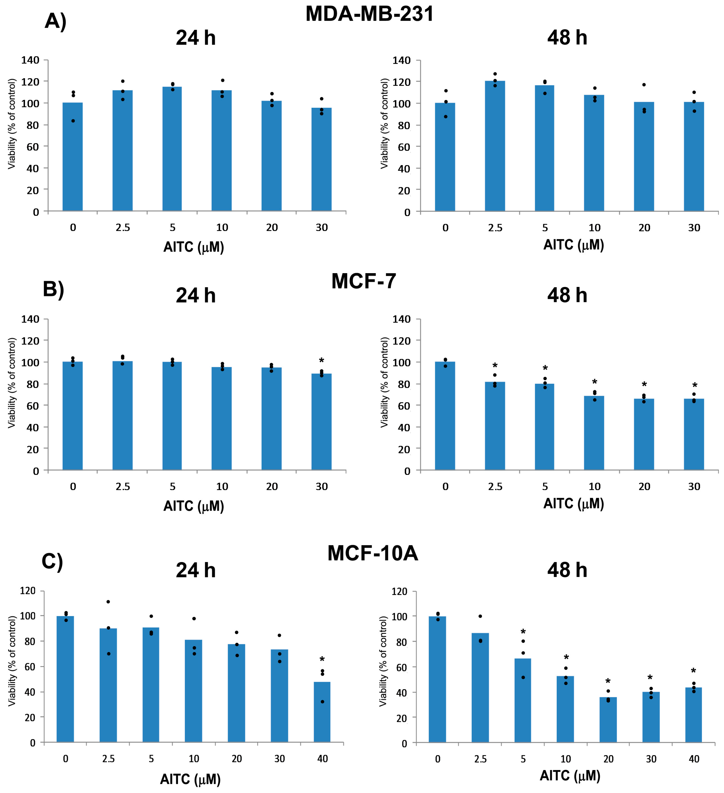
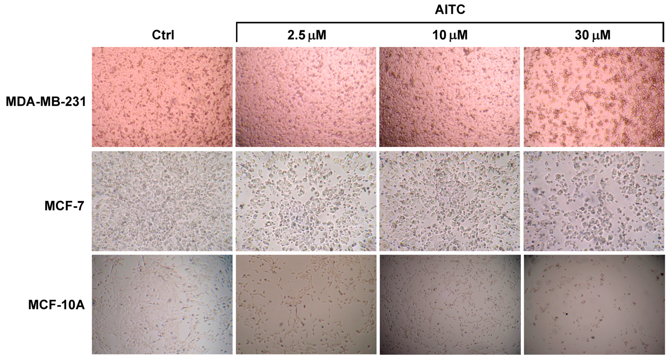
2.1. AITC Did Not Inhibit MDA-MB-231 Cell Proliferation While Affected MCF-7 and MCF-10A Cells

We planned the experiment to investigate whether AITC can inhibit proliferation of MDA-MB-231 breast cancer cells. For our study, we selected 2.5, 5, 10, 20, and 30 µM concentrations based on previous reports [16,26]. Cells were treated with various concentrations of AITC for 24 and 48 h. AITC did not inhibit, rather slightly increased, the proliferation of these cells (Figure 1 and Figure 2A). In contrast, AITC inhibited proliferation of MCF-7 cells in a dose and time-dependent manner (Figure 1 and Figure 2B). We also investigated the effect of AITC on cell viability of MCF-10A non-tumorigenic breast cells. MCF-10A cells were treated with AITC at 0, 2.5, 5, 10, 20, 30, and 40 µM for 24 and 48 h. Our results indicate that AITC shows toxic effects on this non-tumorigenic breast cell line (Figure 1 and Figure 2C). The IC50 values of AITC were 527.8 µM (at 24 h) and not calculable (at 48 h) for MDA-MB-231, 188.1 (at 24 h) and 126.0 µM (at 48 h) for MCF-7, 53.72 (at 24 h), and 14.23 µM (at 48 h) for MCF-10A.

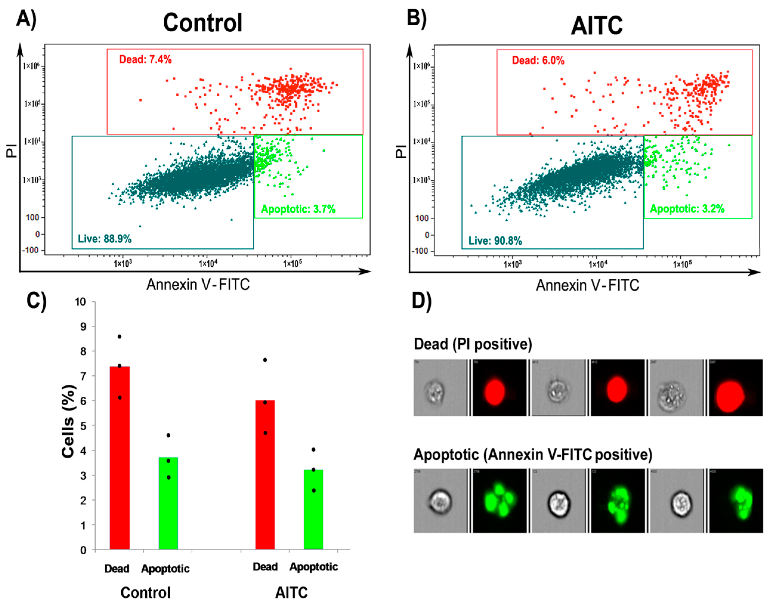
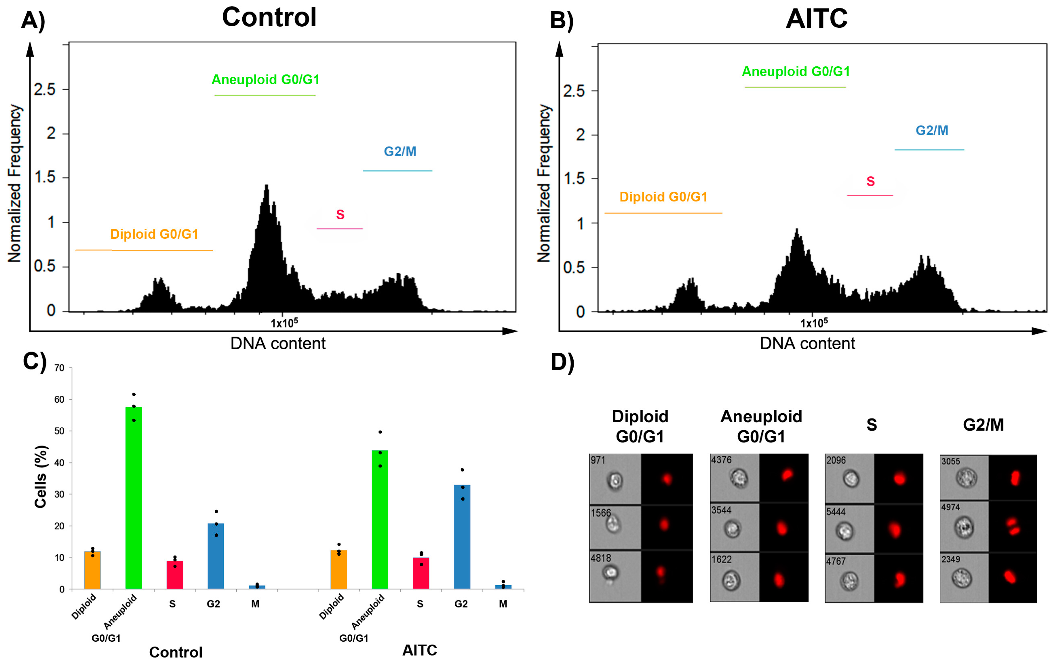
2.2. AITC Did Not Induce Apoptosis and Cell Cycle Arrest

Apoptosis was analyzed by flow cytometer in MDA-MB-231 cells after treatment with 10 µM AITC for 24 h. Approximately 3.2% and 6.0% of the AITC-treated cells were positive for Annexin V-FITC (Annexin V conjugated to green-fluorescent fluorescein isothiocyanate dye) and PI (propidium iodide) after 24 h, respectively (Figure 3B–D). In comparison, 3.7% and 7.4% of the control cells were positive for Annexin V-FITC and PI, respectively (Figure 3A,C,D). Our results indicate that AITC did not induce, rather slightly decreased, apoptosis in these cells.
Cell cycle control is important in cancer progression. Hence, we studied the effects of AITC on cell cycle progression in MDA-MB-231 cells. Cytofluorimetric analysis indicated that AITC did not induce the arrest of phases of the cell cycle significantly. Approximately 12.2%, 43.8%, 9.8%, 32.9%, and 1.2% of AITC-treated cells were noted in G0/G1 (diploid), G0/G1 (aneuploid), S, G2, and M phases, respectively (Figure 4B–D). By contrast, approximately 11.8%, 57.5%, 8.9%, 20.7%, and 1.1% of control cells were noted in G0/G1 (diploid), G0/G1 (aneuploid), S, G2, and M phases, respectively (Figure 4A,C,D). These results suggest that AITC has no ability to induce the cell cycle arrest in MDA-MB-231 cells.

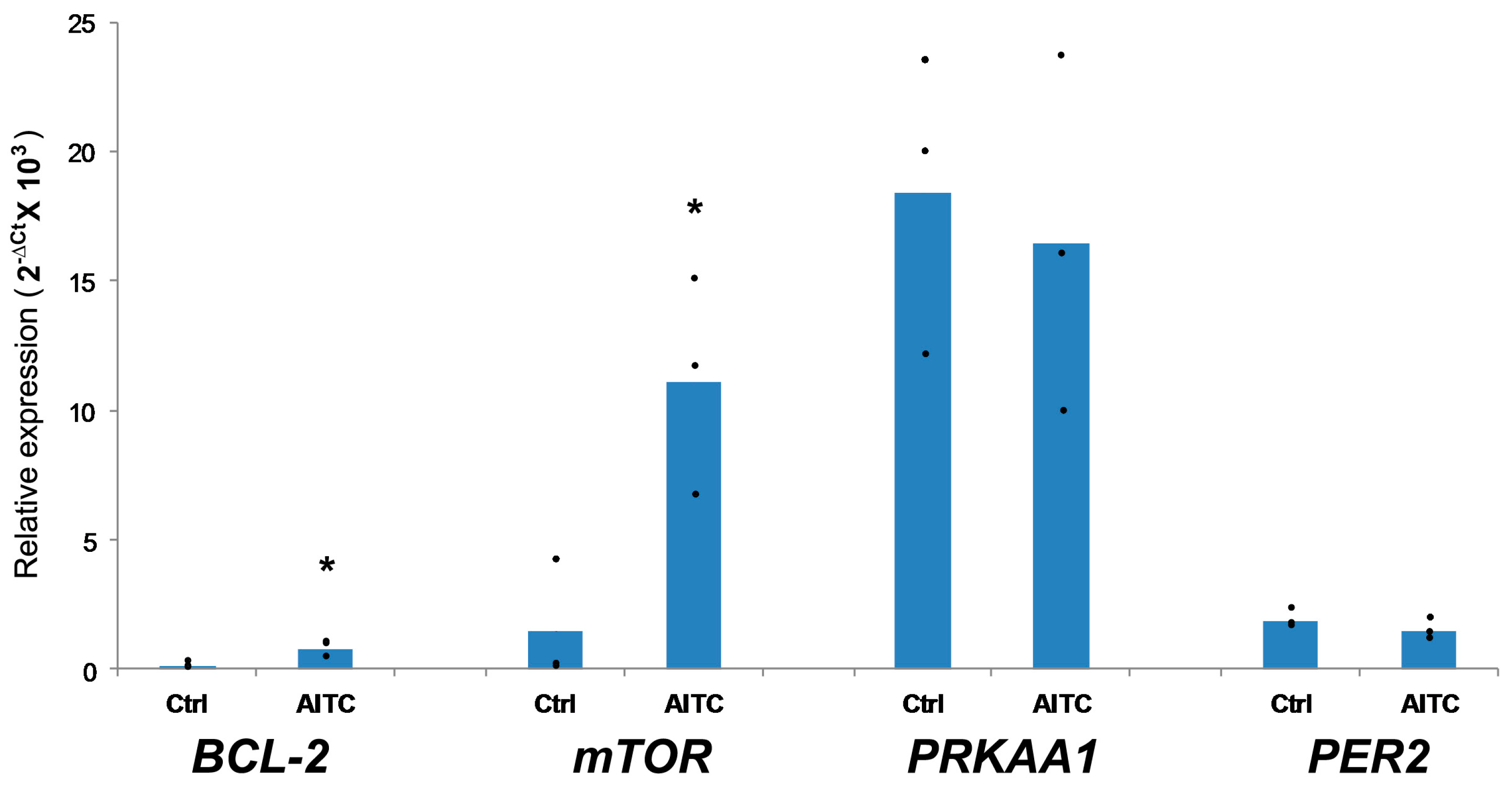
2.3. AITC UpregulatedBCL-2 and mTOR Expression, While Induced No Changes in PRKAA1 and PER2 Expression

In this study, we also measured the expression levels of selected BCL-2, mTOR, PRKAA1, and PER2 genes in MDA-MB-231 cells after treatment with AITC (10 µM) for 48 h. We observed that AITC significantly increased the expression of BCL-2 and mTOR (Figure 5). In contrast, AITC did not affect the expression of PRKAA1 and PER2 (Figure 5). These results suggest that AITC may not inhibit, rather may promote, breast cancer cell growth.

2.4. AITC Did Not Affect Caspase-8, Caspase-9, PARP, p-mTOR, and NF-κB p65 Proteins Level, While Downregulated Beclin-1 Protein Level

Next, we performed experiments to study the expression levels of some selected Caspase-8, Caspase-9, PARP, Beclin-1, and p-mTOR proteins (Figure 6). These genes are aberrantly expressed in breast cancer. Several previous studies have reported that phytochemicals can modulate the expressions of these molecules in various cancer cells including breast cancer [1,43,44,45,46,47]. Hence, we analyzed the expression levels of these proteins by western blot technique. Cells were treated with 10 µM AITC for 48 h and we found that AITC did not affect the level of Caspase-8, Caspase-9, PARP, and p-mTOR, and NF-κB p65 significantly. In contrast, AITC decreased the level of Beclin-1 significantly. These results indicate that AITC shows no growth inhibitory effect on MDA-MB-231 cells.

3. Discussion

Many previous studies have shown that natural compounds can inhibit cancer initiation, development, and progression [48]. They have been proposed as potential cancer preventive agents, although some natural compounds such as isothiocyanates show carcinogenesis promoting effects [49].
In this study, we demonstrate that AITC cannot inhibit, rather can promote, the proliferation of MDA-MB-231 breast cancer cells. In contrast, AITC can inhibit the proliferation of MCF-7 breast cancer cells. Importantly, we observed that AITC significantly affected MCF-10A non-tumorigenic breast cells. We observed that AITC did not induce apoptosis and cell cycle arrest in MDA-MB-231 cells, as demonstrated by flow cytometry. We also found that AITC failed to alter the expression of Caspase-8, Caspase-9, PARP, and p-mTOR proteins, but reduced the expression of Beclin-1 proteins. In addition, AITC increased the expression of BCL-2 and mTOR genes, and did not affect PRKAA1 and PER2 genes. We did not use the MCF-7 cells for our further study since our MTT results confirmed the previous report [17].
Apoptosis is an important biological process by which cell death occurs and is generally characterized by DNA and nuclear fragmentation, chromatin condensation, cytoplasmic membrane blebbing, and messenger RNA degradation. Apoptosis is regulated by various factors such as Fas receptors, Caspases, and BCL-2 family members [27,50,51]. This process is defective in various diseases including cancer. We found that AITC at 10 µM did not induce apoptosis in MDA-MB-231 breast cancer cells, as demonstrated by flow cytometry. Earlier studies have shown that AITC increased Caspase-8 in MDA-MB-231 cells [17] and HL60 human leukemia cells [24]; increased Caspase-9 in MDA-MB-231 [17] and MDA-MB-468 breast [16], UM-UC-3 bladder [19] cancer cells, and GBM 8401 brain malignant glioma cells [20]; decreased BCL-2 in MDA-MB-231 [17] and MDA-MB-468 [16] breast cancer cells, and PC-3 prostate cancer cells in vitro [22] and in vivo [23]; and increased PARP in MDA-MB-231 breast cancer [17] and SW620 colorectal adenocarcinoma cells [52]. We further studied the expression of these molecules in AITC-treated cells. Unexpectedly, we found that AITC (10 µM) did not affect the expression of Caspase-8, Caspase-9, and PARP proteins, as well as increased the expression of BCL-2 gene. Our results suggest that AITC could not induce apoptosis in MDA-MB-231 cells.
Previous studies reported that AITC induced S phase cell cycle arrest in bladder cancer [53] and lung cancer [26] cells, and G2/M phase cell cycle arrest in bladder cancer [53], lung cancer [26], MDA-MB-468 breast cancer [16], brain malignant glioma [20], prostate cancer [22], and leukemia [24] cells. In this study, we observed that AITC did not induce cell cycle arrest significantly. These results suggest that AITC may not inhibit the growth of MDA-MB-231 cells.
Beclin-1 is aberrantly expressed in breast cancer and has been suggested as a therapeutic target [54]. We found that AITC decreased Beclin-1 level. Study reported that overexpressed Beclin-1 may promote autophagy in autophagy-defective breast cancer cells [55]. Thus, we suggest that AITC may promote disruption of autophagy in MDA-MB-231 cells.
The abnormal expression of mTOR is illustrated in breast cancer cells [56,57]. Previous studies reported that isothiocyanates can suppress mTOR in cancer cells [58,59,60,61]. In our study, we found that AITC upregulated mTOR expression, indicating AITC may promote breast cancer progression. Although mTOR was upregulated in AITC-treated cells, p-mTOR protein level was not significantly upregulated. We are currently unable to provide a definitive explanation for this paradox, but it is likely that this is not due to a simple difference in sensitivity between the two assays. Indeed, mTOR signaling pathway in MDA-MB-231 cells can be activated mainly via mTOR overexpression rather than phosphorylation of this molecule. A previous study confirms our assumption. This study reported that untreated MCF-7 cells show high p-mTOR levels and relatively low mTOR levels while MDA-MB-231 cells show an opposite profile [62].
NF-κB is overexpressed in breast cancer [63]. Various natural products including AITC have been shown to inhibit this oncogenic factor in breast cancer [46,64]. In this study, we observed that AITC did not affect NF-κB p65 significantly, which suggests that AITC has no ability to suppress this molecule in breast cancer.
It has been shown that activation of AMPKα1 and AMPKα suppressed the growth of colon cancer [65] and breast cancer [66] cells, respectively. Our result revealed that AITC did not affect the expression of PRKAA1 in MDA-MB-231 cells, indicating AITC may not inhibit MDA-MB-231 breast cancer cell growth.
PER2 is aberrantly expressed in breast cancer [67,68]. PER2 may act as tumor suppressor gene [69,70]. Studies have shown that PER2 can regulate cell cycle-related molecules Cyclin D and Cyclin E [71], and cell adhesion-related molecule β-catenin [72]. We found that AITC did not affect PER2 expression, suggesting AITC may not modulate this molecule in MDA-MB-231 breast cancer cells.
Our results do not confirm previously published results about AITC on MDA-MB-231 cells [17], however changes in mTOR suggest a metabolic activation that can be addressed to the different medium used in the two studies. We used DMEM with high glucose, sodium pyruvate and l-glutamine, while the previous study used RPMI-1640 with l-glutamine [17]. DMEM high glucose is a medium with more nutrients compared to RPMI-1640 hence it can stimulate more the growth of MDA-MB231 cells. The increased metabolism might be responsible of the results obtained in this study.
The different response of AITC on proliferation of MDA-MB-231 compared to MCF-7 was further investigated. Since MDA-MB-231 cells do not have estrogen receptor (ER) and MCF-7 cells have, we hypothesized that the different responses of the two cell lines to AITC may be related to an interaction with ER. To test this hypothesis, we treated MCF-7 cells with ER inhibitor fulvestrant and AITC (Figure S1). Fulvestrant showed a synergic action with AITC at 24 h and an additive action at 48 h, suggesting that AITC may have an interaction with ER or estrogen signaling pathway.
In conclusion, the results of our present study indicate that AITC does not inhibit the proliferation of MDA-MB-231 breast cancer cells. AITC may not suppress, rather may promote, the aberrant expression of molecules related to proliferation, apoptosis, and autophagy in these breast cancer cells. Moreover, AITC can display cytotoxic effect on healthy breast cells. Although AITC has been suggested as a potential anticancer agent, this phytochemical may not have potential inhibitory activity in triple negative breast cancer cells. The results of this in vitro study warrant more preclinical and clinical studies on the beneficial and harmful effects of AITC in healthy and cancer cells.

4. Materials and Methods

4.1. Chemicals and Reagents

AITC, 3-(4,5-dimethylthiazol-2-yl)-2,5-diphenyltetrazolium bromide (MTT), propidium iodide (PI), ethanol, and isopropanol were purchased from Sigma-Aldrich (St. Louis, MO, USA). AITC was dissolved in ethanol at a concentration of 10 mM and stored at 4 °C. Dulbecco’s Modified Eagle Medium (DMEM), Dulbecco’s Modified Eagle Medium: Nutrient Mixture F-12 (DMEM/F12), and fetal bovine serum (FBS) were purchased from Euroclone (Pero, Italy). Annexin V-FITC apoptosis detection kit was purchased from eBioscience Inc. (San Diego, CA, USA), RIPA buffer was purchased from Thermo Fisher Scientific (Waltham, MA, USA). BCL-2, mTOR, PRKAA1, and PER2 gene primers were purchased from Integrated DNA Technologies (Coralville, IA, USA). Primary antibodies against Caspase-8, Caspase-9, PARP, Beclin-1, p-mTOR, NF-κB p65, and β-actin, and secondary antibodies were purchased from Cell Signaling Technology (Danvers, MA, USA).

4.2. Cell Line and Culture Conditions

MDA-MB-231, MCF-7 human breast cancer cell lines and MCF-10A human breast epithelial cell line were purchased from Istituto Zooprofilattico Sperimentale della Lombardia e dell’Emilia Romagna (IZSLER, Brescia, Italy). MDA-MB-231 and MCF-7 cells were grown in DMEM with high glucose (4500 mg/L), sodium pyruvate (110 mg/L) and l-glutamine (584 mg/L) supplemented with 10%FBS and 1% penicillin–streptomycin. MCF-10A cells were grown in DMEM/F12 supplemented with 5% horse serum, 20 ng/mL EGF, 0.5 µg/mL hydrocortisone, 100 ng/mL cholera toxin, 10 µg/mL insulin and 1% penicillin–streptomycin. They were maintained at 37 °C and 5% CO2 in an incubator. The growth medium was changed after 48 or 72 h, and cells were trypsinized and subcultured when they reached 80–90% confluence.

4.3. Cell Viability Analysis

To analyze cell viability, we used MTT assay. Cells (2 × 10⁴/well) were seeded in 96-well plate. After 24 h, cells were treated with AITC at 0, 2.5, 5, 10, 20, 30, and 40 µM. After 24 h or 48 h of treatment, 10 µL of MTT (5 mg/mL in PBS) was added to each well and the plate was incubated again for 3 h at 37 °C in incubator. Next, medium was removed, 200 µL of isopropanol was added, and the absorbance was read at 595 nm [73]. Cell viability rate was calculated following the formula: cell viability (%) = (average OD value of AITC-treated cells/average OD value of control cells) × 100.

4.4. Apoptosis Analysis

For apoptosis analysis, MDA-MB-231 cells (1 × 10⁵/well) were cultured in 6-well for 24 h and treated with AITC at 10 µM. After 24 h of treatment, floating and adherent cells were collected and stained with Annexin V-FITC and PI following the manufacturer’s instructions. Stained cells were then analyzed using an imaging flow cytometer named Flow Sight (Amnis Corporation, Seattle, WA, USA) and IDEAS Software version 6.2 (EMD Millipore, Burlington, MA, USA).

4.5. Cell Cycle Analysis

MDA-MB-231 cells (3.3 × 10⁵) were seeded in T25 flasks for 24 h and treated with AITC at 10 µM for 24 h. After that both floating and adherent cells were collected and fixed in 70% ethanol (cold) for 24 h. Ethanol was removed, cells were then washed twice with PBS, and resuspended in 1 mL of PBS containing 0.02 mg/mL PI, 0.2 mg/mL DNase-free RNase A, and 0.1% Triton X-100, and incubated for 15 min at 37 °C in incubator. After incubation, cells were analyzed using a flow cytometer [74].

4.6. Western Blotting

MDA-MB-231 cells (3.3 × 10⁵) were seeded in T25 flasks. After 24 h, cells were treated with 10 µM AITC for 48 h, and collected for protein expression analysis. Protein extraction and quantification as well as western blotting were performed according to protocol described elsewhere [75,76,77].

4.7. RNA Extraction and Real-Time PCR

MDA-MB-231 cells (3.3 × 10⁵) were seeded in T25 flasks. After 24 h, cells were treated with 10 µM AITC for 48 h. After incubation, the cells were collected for RNA extraction. Total RNA was isolated using the RNeasy Mini Kit (QIAGEN, Hilden, Germany) according to the manufacturer’s protocol. RNA quality and quantification were evaluated with a Nanodrop 1000 spectrophotometer (Thermo Scientific, Wilmington, DE, USA). cDNA was synthesized using the High-Capacity cDNA Reverse Transcription Kit (Applied Biosystems, Foster City, CA, USA) according to the manufacturer’s protocol. Gene expression levels were assayed using the TaqMan system (Applied Biosystems, Foster City, CA, USA). GAPDH was used as a housekeeping gene. The Ct (threshold cycle) value of each gene of interest was normalized to Ct value of GAPDH and the relative gene expression data were analyzed using the equation: 2−ΔCt.

4.8. Statistical Analysis

Data were collected from three independent experiments. The average of intra-assay replications is presented as individual dots on top of bar graphs whose height represent the average of all the experiments. One-way ANOVA with post-hoc Tukey HSD (in the case of multiple independent groups) and Student’s t-Test (in the case of two independent groups) with Bonferroni’s correction (in the case of multiple testing) were used to evaluate the statistical significance and statistical analyses were performed using SPSS 19.0 (Statistical Package for the Social Sciences Inc., Armonk, New York, NY, USA) software. In figures, asterisk indicates the significant differences of data in comparison with the control. Data were considered statistically significant at p < 0.05. IC50 values were calculated using GraphPad Prism 7.03 (GraphPad Software, San Diego, CA, USA).

Supplementary Materials

Supplementary materials can be found at www.mdpi.com/1422-0067/19/1/145/s1.

Acknowledgments

Md. Abu Sayeed was a recipient of fellowship from the Polytechnic University of Marche, Ancona, Italy. The National Institute on Health and Science on Ageing, Ancona, Italy, gave facility to use the flow cytometer.

Author Contributions

Md. Abu Sayeed conceived and designed the study, performed experiments, and wrote the manuscript; Massimo Bracci conceived and designed the study, analyzed the data, and wrote the manuscript; Veronica Ciarapica, Ernesta Pieragostini, Simona Gaetani and Federica Monaco assisted in performing the experiments; Marco Malavolta and Mauro Provinciali analyzed the cytofluorometric data; Guendalina Lucarini and Venerando Rapisarda revised the manuscript; Roberto Di Primio approved the final manuscript; and Lory Santarelli supervised the project and approved the final manuscript.

Conflicts of Interest

The authors declare no conflict of interest.

References

  1. Sayeed, M.A.; Bracci, M.; Lazzarini, R.; Tomasetti, M.; Amati, M.; Lucarini, G.; Di Primio, R.; Santarelli, L. Use of potential dietary phytochemicals to target miRNA: Promising option for breast cancer prevention and treatment? J. Funct. Foods 2017, 28, 177–193. [Google Scholar] [CrossRef]
  2. Dandawate, P.R.; Subramaniam, D.; Jensen, R.A.; Anant, S. Targeting cancer stem cells and signaling pathways by phytochemicals: Novel approach for breast cancer therapy. Semin. Cancer Biol. 2016, 40–41, 192–208. [Google Scholar] [CrossRef] [PubMed]
  3. Kushad, M.M.; Brown, A.F.; Kurilich, A.C.; Juvik, J.A.; Klein, B.P.; Wallig, M.A.; Jeffery, E.H. Variation of glucosinolates in vegetable crops of Brassica oleracea. J. Agric. Food Chem. 1999, 47, 1541–1548. [Google Scholar] [CrossRef] [PubMed]
  4. Hao, G.; Wang, Q.; Liu, B.; Liu, J. Phytochemical profiling of five medicinally active constituents across 14 Eutrema species. Fitoterapia 2016, 110, 83–88. [Google Scholar] [CrossRef] [PubMed]
  5. Loebers, A.; Muller-Uri, F.; Kreis, W. A young root-specific gene (ArMY2) from horseradish encoding a MYR II myrosinase with kinetic preference for the root-specific glucosinolategluconasturtiin. Phytochemistry 2014, 99, 26–35. [Google Scholar] [CrossRef] [PubMed]
  6. Zhang, Y. Allyl isothiocyanate as a cancer chemopreventive phytochemical. Mol. Nutr. Food Res. 2010, 54, 127–135. [Google Scholar] [CrossRef] [PubMed]
  7. Kim, Y.J.; Lee, D.H.; Ahn, J.; Chung, W.J.; Jang, Y.J.; Seong, K.S.; Moon, J.H.; Ha, T.Y.; Jung, C.H. Pharmacokinetics, Tissue Distribution, and Anti-Lipogenic/Adipogenic Effects of Allyl-Isothiocyanate Metabolites. PLoS ONE 2015, 10, e0132151. [Google Scholar] [CrossRef] [PubMed]
  8. Encinas-Basurto, D.; Ibarra, J.; Juarez, J.; Burboa, M.G.; Barbosa, S.; Taboada, P.; Troncoso-Rojas, R.; Valdez, M.A. Poly(lactic-co-glycolic acid) nanoparticles for sustained release of allyl isothiocyanate: Characterization, in vitro release and biological activity. J. Microencapsul. 2017, 34, 231–242. [Google Scholar] [CrossRef] [PubMed]
  9. Lashkari, E.; Wang, H.; Liu, L.; Li, J.; Yam, K. Innovative application of metal-organic frameworks for encapsulation and controlled release of allyl isothiocyanate. Food Chem. 2017, 221, 926–935. [Google Scholar] [CrossRef] [PubMed]
  10. Ahn, M.; Kim, J.; Bang, H.; Moon, J.; Kim, G.O.; Shin, T. Hepatoprotective effects of allyl isothiocyanate against carbon tetrachloride-induced hepatotoxicity in rat. Chem. Biol. Interact. 2016, 254, 102–108. [Google Scholar] [CrossRef] [PubMed]
  11. Subedi, L.; Venkatesan, R.; Kim, S.Y. Neuroprotective and Anti-Inflammatory Activities of Allyl Isothiocyanate through Attenuation of JNK/NF-κB/TNF-α Signaling. Int. J. Mol. Sci. 2017, 18, 1423. [Google Scholar] [CrossRef] [PubMed]
  12. Wagner, A.E.; Boesch-Saadatmandi, C.; Dose, J.; Schultheiss, G.; Rimbach, G. Anti-inflammatory potential of allyl-isothiocyanate—Role of Nrf2, NF-κB and microRNA-155. J. Cell. Mol. Med. 2012, 16, 836–843. [Google Scholar] [CrossRef] [PubMed]
  13. Matsuda, H.; Ochi, M.; Nagatomo, A.; Yoshikawa, M. Effects of allyl isothiocyanate from horseradish on several experimental gastric lesions in rats. Eur. J. Pharmacol. 2007, 561, 172–181. [Google Scholar] [CrossRef] [PubMed]
  14. Nowicki, D.; Rodzik, O.; Herman-Antosiewicz, A.; Szalewska-Palasz, A. Isothiocyanates as effective agents against enterohemorrhagic Escherichia coli: Insight to the mode of action. Sci. Rep. 2016, 6, 22263. [Google Scholar] [CrossRef] [PubMed]
  15. Manyes, L.; Luciano, F.B.; Manes, J.; Meca, G. In vitro antifungal activity of allyl isothiocyanate (AITC) against Aspergillus parasiticus and Penicillium expansum and evaluation of the AITC estimated daily intake. Food Chem. Toxicol. 2015, 83, 293–299. [Google Scholar] [CrossRef] [PubMed]
  16. Tsai, S.C.; Huang, W.W.; Huang, W.C.; Lu, C.C.; Chiang, J.H.; Peng, S.F.; Chung, J.G.; Lin, Y.H.; Hsu, Y.M.; Amagaya, S.; et al. ERK-modulated intrinsic signaling and G(2)/M phase arrest contribute to the induction of apoptotic death by allyl isothiocyanate in MDA-MB-468 human breast adenocarcinoma cells. Int. J. Oncol. 2012, 41, 2065–2072. [Google Scholar] [CrossRef] [PubMed]
  17. Bo, P.; Lien, J.C.; Chen, Y.Y.; Yu, F.S.; Lu, H.F.; Yu, C.S.; Chou, Y.C.; Yu, C.C.; Chung, J.G. Allyl Isothiocyanate Induces Cell Toxicity by Multiple Pathways in Human Breast Cancer Cells. Am. J. Chin. Med. 2016, 44, 415–437. [Google Scholar] [CrossRef] [PubMed]
  18. Savio, A.L.; da Silva, G.N.; Salvadori, D.M. Inhibition of bladder cancer cell proliferation by allyl isothiocyanate (mustard essential oil). Mutat. Res. 2015, 771, 29–35. [Google Scholar] [CrossRef] [PubMed]
  19. Geng, F.; Tang, L.; Li, Y.; Yang, L.; Choi, K.S.; Kazim, A.L.; Zhang, Y. Allyl isothiocyanate arrests cancer cells in mitosis, and mitotic arrest in turn leads to apoptosis via Bcl-2 protein phosphorylation. J. Biol. Chem. 2011, 286, 32259–32267. [Google Scholar] [CrossRef] [PubMed]
  20. Chen, N.G.; Chen, K.T.; Lu, C.C.; Lan, Y.H.; Lai, C.H.; Chung, Y.T.; Yang, J.S.; Lin, Y.C. Allyl isothiocyanate triggers G2/M phase arrest and apoptosis in human brain malignant glioma GBM 8401 cells through a mitochondria-dependent pathway. Oncol. Rep. 2010, 24, 449–455. [Google Scholar] [PubMed]
  21. Lai, K.C.; Lu, C.C.; Tang, Y.J.; Chiang, J.H.; Kuo, D.H.; Chen, F.A.; Chen, I.L.; Yang, J.S. Allyl isothiocyanate inhibits cell metastasis through suppression of the MAPK pathways in epidermal growth factorstimulated HT29 human colorectal adenocarcinoma cells. Oncol. Rep. 2014, 31, 189–196. [Google Scholar] [CrossRef] [PubMed]
  22. Xiao, D.; Srivastava, S.K.; Lew, K.L.; Zeng, Y.; Hershberger, P.; Johnson, C.S.; Trump, D.L.; Singh, S.V. Allyl isothiocyanate, a constituent of cruciferous vegetables, inhibits proliferation of human prostate cancer cells by causing G2/M arrest and inducing apoptosis. Carcinogenesis 2003, 24, 891–897. [Google Scholar] [CrossRef] [PubMed]
  23. Srivastava, S.K.; Xiao, D.; Lew, K.L.; Hershberger, P.; Kokkinakis, D.M.; Johnson, C.S.; Trump, D.L.; Singh, S.V. Allyl isothiocyanate, a constituent of cruciferous vegetables, inhibits growth of PC-3 human prostate cancer xenografts in vivo. Carcinogenesis 2003, 24, 1665–1670. [Google Scholar] [CrossRef] [PubMed]
  24. Xu, K.; Thornalley, P.J. Studies on the mechanism of the inhibition of human leukaemia cell growth by dietary isothiocyanates and their cysteine adducts in vitro. Biochem. Pharmacol. 2000, 60, 221–231. [Google Scholar] [CrossRef]
  25. Ling, X.; Westover, D.; Cao, F.; Cao, S.; He, X.; Kim, H.R.; Zhang, Y.; Chan, D.C.; Li, F. Synergistic effect of allyl isothiocyanate (AITC) on cisplatin efficacy in vitro and in vivo. Am. J. Cancer Res. 2015, 5, 2516–2530. [Google Scholar] [PubMed]
  26. Tripathi, K.; Hussein, U.K.; Anupalli, R.; Barnett, R.; Bachaboina, L.; Scalici, J.; Rocconi, R.P.; Owen, L.B.; Piazza, G.A.; Palle, K. Allyl isothiocyanate induces replication-associated DNA damage response in NSCLC cells and sensitizes to ionizing radiation. Oncotarget 2015, 6, 5237–5252. [Google Scholar] [CrossRef] [PubMed]
  27. Hengartner, M.O. The biochemistry of apoptosis. Nature 2000, 407, 770–776. [Google Scholar] [CrossRef] [PubMed]
  28. Stennicke, H.R.; Jurgensmeier, J.M.; Shin, H.; Deveraux, Q.; Wolf, B.B.; Yang, X.; Zhou, Q.; Ellerby, H.M.; Ellerby, L.M.; Bredesen, D.; et al. Pro-caspase-3 is a major physiologic target of caspase-8. J. Biol. Chem. 1998, 273, 27084–27090. [Google Scholar] [CrossRef] [PubMed]
  29. Li, H.; Zhu, H.; Xu, C.J.; Yuan, J. Cleavage of BID by caspase 8 mediates the mitochondrial damage in the Fas pathway of apoptosis. Cell 1998, 94, 491–501. [Google Scholar] [CrossRef]
  30. Oliver, F.J.; de la Rubia, G.; Rolli, V.; Ruiz-Ruiz, M.C.; de Murcia, G.; Murcia, J.M. Importance of poly(ADP-ribose) polymerase and its cleavage in apoptosis. Lesson from an uncleavable mutant. J. Biol. Chem. 1998, 273, 33533–33539. [Google Scholar] [CrossRef] [PubMed]
  31. Satoh, M.S.; Lindahl, T. Role of poly(ADP-ribose) formation in DNA repair. Nature 1992, 356, 356–358. [Google Scholar] [CrossRef] [PubMed]
  32. Nicholson, D.W.; Ali, A.; Thornberry, N.A.; Vaillancourt, J.P.; Ding, C.K.; Gallant, M.; Gareau, Y.; Griffin, P.R.; Labelle, M.; Lazebnik, Y.A.; et al. Identification and inhibition of the ICE/CED-3 protease necessary for mammalian apoptosis. Nature 1995, 376, 37–43. [Google Scholar] [CrossRef] [PubMed]
  33. Zhong, Y.; Wang, Q.J.; Li, X.; Yan, Y.; Backer, J.M.; Chait, B.T.; Heintz, N.; Yue, Z. Distinct regulation of autophagic activity by Atg14L and Rubicon associated with Beclin 1-phosphatidylinositol-3-kinase complex. Nat. Cell Biol. 2009, 11, 468–476. [Google Scholar] [CrossRef] [PubMed]
  34. Erlich, S.; Mizrachy, L.; Segev, O.; Lindenboim, L.; Zmira, O.; Adi-Harel, S.; Hirsch, J.A.; Stein, R.; Pinkas-Kramarski, R. Differential interactions between Beclin 1 and Bcl-2 family members. Autophagy 2007, 3, 561–568. [Google Scholar] [CrossRef] [PubMed]
  35. Baeuerle, P.A.; Baltimore, D. NF-κB: Ten years after. Cell 1996, 87, 13–20. [Google Scholar] [CrossRef]
  36. Gupta, S. Molecular signaling in death receptor and mitochondrial pathways of apoptosis (Review). Int. J. Oncol. 2003, 22, 15–20. [Google Scholar] [CrossRef] [PubMed]
  37. Laplante, M.; Sabatini, D.M. mTOR signaling in growth control and disease. Cell 2012, 149, 274–293. [Google Scholar] [CrossRef] [PubMed]
  38. Xu, Y.; Toh, K.L.; Jones, C.R.; Shin, J.Y.; Fu, Y.H.; Ptacek, L.J. Modeling of a human circadian mutation yields insights into clock regulation by PER2. Cell 2007, 128, 59–70. [Google Scholar] [CrossRef] [PubMed]
  39. Gwinn, D.M.; Shackelford, D.B.; Egan, D.F.; Mihaylova, M.M.; Mery, A.; Vasquez, D.S.; Turk, B.E.; Shaw, R.J. AMPK phosphorylation of raptor mediates a metabolic checkpoint. Mol. Cell 2008, 30, 214–226. [Google Scholar] [CrossRef] [PubMed]
  40. DeBerardinis, R.J.; Thompson, C.B. Cellular metabolism and disease: What do metabolic outliers teach us? Cell 2012, 148, 1132–1144. [Google Scholar] [CrossRef] [PubMed]
  41. Xu, H.; Zhou, Y.; Coughlan, K.A.; Ding, Y.; Wang, S.; Wu, Y.; Song, P.; Zou, M.H. AMPKalpha1 deficiency promotes cellular proliferation and DNA damage via p21 reduction in mouse embryonic fibroblasts. Biochim. Biophys. Acta 2015, 1853, 65–73. [Google Scholar] [CrossRef] [PubMed]
  42. Obba, S.; Hizir, Z.; Boyer, L.; Selimoglu-Buet, D.; Pfeifer, A.; Michel, G.; Hamouda, M.A.; Goncalves, D.; Cerezo, M.; Marchetti, S.; et al. The PRKAA1/AMPKalpha1 pathway triggers autophagy during CSF1-induced human monocyte differentiation and is a potential target in CMML. Autophagy 2015, 11, 1114–1129. [Google Scholar] [CrossRef] [PubMed]
  43. Singh, B.N.; Singh, H.B.; Singh, A.; Naqvi, A.H.; Singh, B.R. Dietary phytochemicals alter epigenetic events and signaling pathways for inhibition of metastasis cascade: Phytoblockers of metastasis cascade. Cancer Metastasis Rev. 2014, 33, 41–85. [Google Scholar] [CrossRef] [PubMed]
  44. Sayeed, M.A.; Bracci, M.; Lucarini, G.; Lazzarini, R.; Di Primio, R.; Santarelli, L. Regulation of microRNA using promising dietary phytochemicals: Possible preventive and treatment option of malignant mesothelioma. Biomed. Pharmacother. 2017, 94, 1197–1224. [Google Scholar] [CrossRef] [PubMed]
  45. Gupta, S.C.; Kim, J.H.; Prasad, S.; Aggarwal, B.B. Regulation of survival, proliferation, invasion, angiogenesis, and metastasis of tumor cells through modulation of inflammatory pathways by nutraceuticals. Cancer Metastasis Rev. 2010, 29, 405–434. [Google Scholar] [CrossRef] [PubMed]
  46. Hasanpourghadi, M.; Pandurangan, A.K.; Mustafa, M.R. Modulation of oncogenic transcription factors by bioactive natural products in breast cancer. Pharmacol. Res. 2017. [Google Scholar] [CrossRef] [PubMed]
  47. Li, Y.; Li, S.; Meng, X.; Gan, R.Y.; Zhang, J.J.; Li, H.B. Dietary Natural Products for Prevention and Treatment of Breast Cancer. Nutrients 2017, 9. [Google Scholar] [CrossRef] [PubMed]
  48. Pandey, M.K.; Gupta, S.C.; Nabavizadeh, A.; Aggarwal, B.B. Regulation of cell signaling pathways by dietary agents for cancer prevention and treatment. Semin. Cancer Biol. 2017, 46, 158–181. [Google Scholar] [CrossRef] [PubMed]
  49. Hirose, M.; Yamaguchi, T.; Kimoto, N.; Ogawa, K.; Futakuchi, M.; Sano, M.; Shirai, T. Strong promoting activity of phenylethyl isothiocyanate and benzyl isothiocyanate on urinary bladder carcinogenesis in F344 male rats. Int. J. Cancer 1998, 77, 773–777. [Google Scholar] [CrossRef]
  50. Green, D.R. Apoptotic pathways: Paper wraps stone blunts scissors. Cell 2000, 102, 1–4. [Google Scholar] [CrossRef]
  51. Ashkenazi, A.; Dixit, V.M. Death receptors: Signaling and modulation. Science 1998, 281, 1305–1308. [Google Scholar] [CrossRef] [PubMed]
  52. Lau, W.S.; Chen, T.; Wong, Y.S. Allyl isothiocyanate induces G2/M arrest in human colorectal adenocarcinoma SW620 cells through down-regulation of Cdc25B and Cdc25C. Mol. Med. Rep. 2010, 3, 1023–1030. [Google Scholar] [PubMed]
  53. Savio, A.L.; da Silva, G.N.; de Camargo, E.A.; Salvadori, D.M. Cell cycle kinetics, apoptosis rates, DNA damage and TP53 gene expression in bladder cancer cells treated with allyl isothiocyanate (mustard essential oil). Mutat. Res. 2014, 762, 40–46. [Google Scholar] [CrossRef] [PubMed]
  54. Jung, Y.Y.; Lee, Y.K.; Koo, J.S. The potential of Beclin 1 as a therapeutic target for the treatment of breast cancer. Expert Opin. Ther. Targets 2016, 20, 167–178. [Google Scholar] [CrossRef] [PubMed]
  55. Liang, X.H.; Jackson, S.; Seaman, M.; Brown, K.; Kempkes, B.; Hibshoosh, H.; Levine, B. Induction of autophagy and inhibition of tumorigenesis by beclin 1. Nature 1999, 402, 672–676. [Google Scholar] [CrossRef] [PubMed]
  56. Liu, J.; Li, H.Q.; Zhou, F.X.; Yu, J.W.; Sun, L.; Han, Z.H. Targeting the mTOR pathway in breast cancer. Tumor Biol. 2017, 39. [Google Scholar] [CrossRef] [PubMed]
  57. Davis, N.M.; Sokolosky, M.; Stadelman, K.; Abrams, S.L.; Libra, M.; Candido, S.; Nicoletti, F.; Polesel, J.; Maestro, R.; D’Assoro, A.; et al. Deregulation of the EGFR/PI3K/PTEN/Akt/mTORC1 pathway in breast cancer: Possibilities for therapeutic intervention. Oncotarget 2014, 5, 4603–4650. [Google Scholar] [CrossRef] [PubMed]
  58. Bommareddy, A.; Hahm, E.R.; Xiao, D.; Powolny, A.A.; Fisher, A.L.; Jiang, Y.; Singh, S.V. Atg5 regulates phenethyl isothiocyanate-induced autophagic and apoptotic cell death in human prostate cancer cells. Cancer Res. 2009, 69, 3704–3712. [Google Scholar] [CrossRef] [PubMed]
  59. Boreddy, S.R.; Pramanik, K.C.; Srivastava, S.K. Pancreatic tumor suppression by benzyl isothiocyanate is associated with inhibition of PI3K/AKT/FOXO pathway. Clin. Cancer Res. 2011, 17, 1784–1795. [Google Scholar] [CrossRef] [PubMed]
  60. Shao, W.Y.; Yang, Y.L.; Yan, H.; Huang, Q.; Liu, K.J.; Zhang, S. Phenethyl isothiocyanate suppresses the metastasis of ovarian cancer associated with the inhibition of CRM1-mediated nuclear export and mTOR-STAT3 pathway. Cancer Biol. Ther. 2017, 18, 26–35. [Google Scholar] [CrossRef] [PubMed]
  61. Wiczk, A.; Hofman, D.; Konopa, G.; Herman-Antosiewicz, A. Sulforaphane, a cruciferous vegetable-derived isothiocyanate, inhibits protein synthesis in human prostate cancer cells. Biochim. Biophys. Acta 2012, 1823, 1295–1305. [Google Scholar] [CrossRef] [PubMed]
  62. Pan, Y.; Shi, J.; Ni, W.; Liu, Y.; Wang, S.; Wang, X.; Wei, Z.; Wang, A.; Chen, W.; Lu, Y. Cryptotanshinone inhibition of mammalian target of rapamycin pathway is dependent on oestrogen receptor α in breast cancer. J. Cell. Mol. Med. 2017, 21, 2129–2139. [Google Scholar] [CrossRef] [PubMed]
  63. Suman, S.; Sharma, P.K.; Rai, G.; Mishra, S.; Arora, D.; Gupta, P.; Shukla, Y. Current perspectives of molecular pathways involved in chronic inflammation-mediated breast cancer. Biochem. Biophys. Res. Commun. 2016, 472, 401–409. [Google Scholar] [CrossRef] [PubMed]
  64. Rajakumar, T.; Pugalendhi, P.; Jayaganesh, R.; Ananthakrishnan, D.; Gunasekaran, K. Effect of allyl isothiocyanate on NF-κB signaling in 7,12-dimethylbenz(a)anthracene and N-methyl-N-nitrosourea-induced mammary carcinogenesis. Breast Cancer 2018, 25, 50–59. [Google Scholar] [CrossRef] [PubMed]
  65. Lee, Y.K.; Park, S.Y.; Kim, Y.M.; Kim, D.C.; Lee, W.S.; Surh, Y.J.; Park, O.J. Suppression of mTOR via Akt-dependent and -independent mechanisms in selenium-treated colon cancer cells: Involvement of AMPKalpha1. Carcinogenesis 2010, 31, 1092–1099. [Google Scholar] [CrossRef] [PubMed]
  66. Yoon, J.H.; Ahn, S.G.; Lee, B.H.; Jung, S.H.; Oh, S.H. Role of autophagy in chemoresistance: Regulation of the ATM-mediated DNA-damage signaling pathway through activation of DNA-PKcs and PARP-1. Biochem. Pharmacol. 2012, 83, 747–757. [Google Scholar] [CrossRef] [PubMed]
  67. Chen, S.T.; Choo, K.B.; Hou, M.F.; Yeh, K.T.; Kuo, S.J.; Chang, J.G. Deregulated expression of the PER1, PER2 and PER3 genes in breast cancers. Carcinogenesis 2005, 26, 1241–1246. [Google Scholar] [CrossRef] [PubMed]
  68. Winter, S.L.; Bosnoyan-Collins, L.; Pinnaduwage, D.; Andrulis, I.L. Expression of the circadian clock genes Per1 and Per2 in sporadic and familial breast tumors. Neoplasia 2007, 9, 797–800. [Google Scholar] [CrossRef] [PubMed]
  69. Fu, L.; Lee, C.C. The circadian clock: Pacemaker and tumour suppressor. Nat. Rev. Cancer 2003, 3, 350–361. [Google Scholar] [CrossRef] [PubMed]
  70. Fu, L.; Pelicano, H.; Liu, J.; Huang, P.; Lee, C. The circadian gene Period2 plays an important role in tumor suppression and DNA damage response in vivo. Cell 2002, 111, 41–50. [Google Scholar] [CrossRef]
  71. Yang, X.; Wood, P.A.; Oh, E.Y.; Du-Quiton, J.; Ansell, C.M.; Hrushesky, W.J. Down regulation of circadian clock gene Period 2 accelerates breast cancer growth by altering its daily growth rhythm. Breast Cancer Res. Treat. 2009, 117, 423–431. [Google Scholar] [CrossRef] [PubMed]
  72. Wood, P.A.; Yang, X.; Taber, A.; Oh, E.Y.; Ansell, C.; Ayers, S.E.; Al-Assaad, Z.; Carnevale, K.; Berger, F.G.; Pena, M.M.; et al. Period 2 mutation accelerates ApcMin/+ tumorigenesis. Mol. Cancer Res. 2008, 6, 1786–1793. [Google Scholar] [CrossRef] [PubMed]
  73. Staffolani, S.; Manzella, N.; Strafella, E.; Nocchi, L.; Bracci, M.; Ciarapica, V.; Amati, M.; Rubini, C.; Re, M.; Pugnaloni, A.; et al. Wood dust exposure induces cell transformation through EGFR-mediated OGG1 inhibition. Mutagenesis 2015, 30, 487–497. [Google Scholar] [CrossRef] [PubMed]
  74. Darzynkiewicz, Z.; Juan, G.; Bedner, E. Determining cell cycle stages by flow cytometry. In Current Protocols in Cell Biology; John Wiley & Sons, Inc.: Hoboken, NJ, USA, 2001; Chapter 8. [Google Scholar]
  75. Manzella, N.; Bracci, M.; Strafella, E.; Staffolani, S.; Ciarapica, V.; Copertaro, A.; Rapisarda, V.; Ledda, C.; Amati, M.; Valentino, M.; et al. Circadian Modulation of 8-Oxoguanine DNA Damage Repair. Sci. Rep. 2015, 4, 13752. [Google Scholar] [CrossRef] [PubMed]
  76. Tomasetti, M.; Monaco, F.; Manzella, N.; Rohlena, J.; Rohlenova, K.; Staffolani, S.; Gaetani, S.; Ciarapica, V.; Amati, M.; Bracci, M.; et al. MicroRNA-126 induces autophagy by altering cell metabolism in malignant mesothelioma. Oncotarget 2016, 7, 36338–36352. [Google Scholar] [CrossRef] [PubMed]
  77. Islam, M.S.; Giampieri, F.; Janjusevic, M.; Gasparrini, M.; Forbes-Hernandez, T.Y.; Mazzoni, L.; Greco, S.; Giannubilo, S.R.; Ciavattini, A.; Mezzetti, B.; et al. An anthocyanin rich strawberry extract induces apoptosis and ROS while decreases glycolysis and fibrosis in human uterine leiomyoma cells. Oncotarget 2017, 8, 23575–23587. [Google Scholar] [PubMed]
Figure 1. Representative photographs captured with 25× magnification of MDA-MB-231, MCF-7, and MCF-10A cells (control and after treatment with AITC for 48 h).
Ijms 19 00145 g001
Figure 2. Effects of AITC on proliferation in MDA-MB-231, MCF-7, and MCF-10A cells. MDA-MB-231 (A); MCF-7 (B); and MCF-10A (C) cells were treated with various concentrations of AITC for 24 and 48 h, and then cell viability was determined by the MTT (methylthiazolyldiphenyl-tetrazolium bromide) assay. Values are presented as individual dots, and symbol asterisk indicates significant (p < 0.05) difference as compared to the control cells.
Ijms 19 00145 g002
Figure 3. AITC did not induce apoptosis in MDA-MB-231 cells: (A,B) flow cytometric analysis of cell apoptosis; (C) histogram showing dead and apoptotic rates of control and AITC-treated cells; and (D) representative flow cytometric images of propidium iodide (PI; red fluorescence) and Annexin V-FITC (green fluorescence) positive cells.
Ijms 19 00145 g003
Figure 4. AITC did not induce cell cycle arrest in MDA-MB-231 cells: (A,B) flow cytometric analysis of cell cycle; (C) histogram showing rate of control and AITC-treated cells of different cell cycle phases; and (D) representative flow cytometric images of cell cycle phases.
Ijms 19 00145 g004
Figure 5. Relative gene expression levels of BCL-2, mTOR, PRKAA1, and PER2 in MDA-MB-231 cells after treatment with 10 µM AITC for 48 h. * p < 0.05.
Ijms 19 00145 g005
Figure 6. Protein levels of Beclin-1, Caspase-8, Caspase-9, PARP, p-mTOR, and NF-κB p65 in MDA-MB-231 cells after treatment with 10 µM AITC for 48 h. β-actin was used as a loading control. * p < 0.05.
Ijms 19 00145 g006

