Variation on Molecular Structure, Crystallinity, and Optical Properties of Dentin Due to Nd:YAG Laser and Fluoride Aimed at Tooth Erosion Prevention

 , , and
Abstract
:
1. Introduction
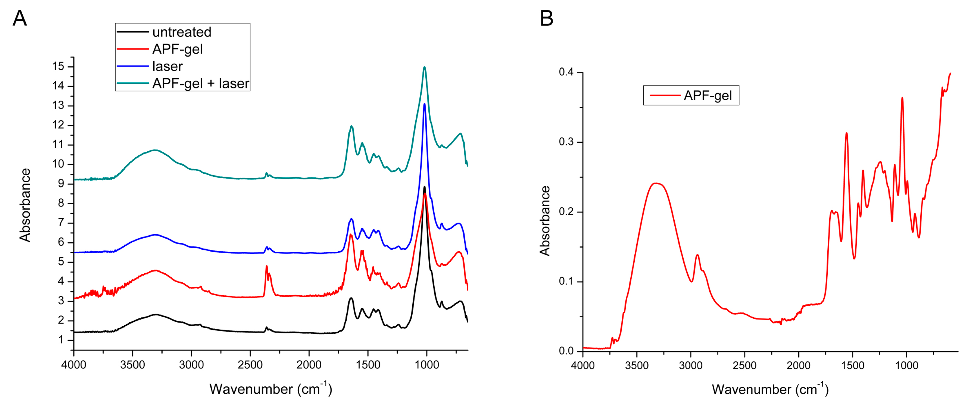
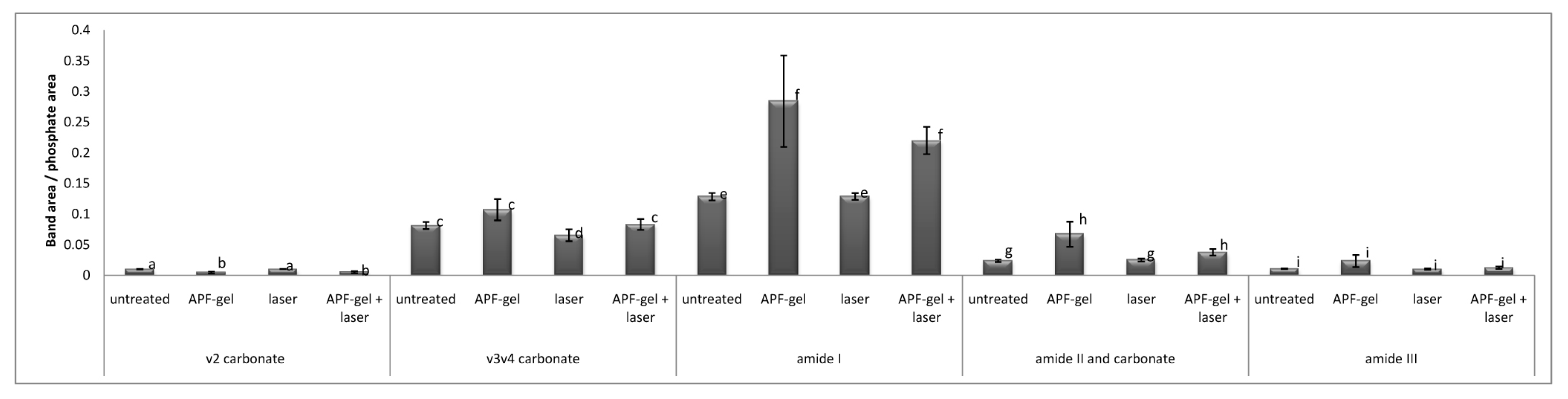
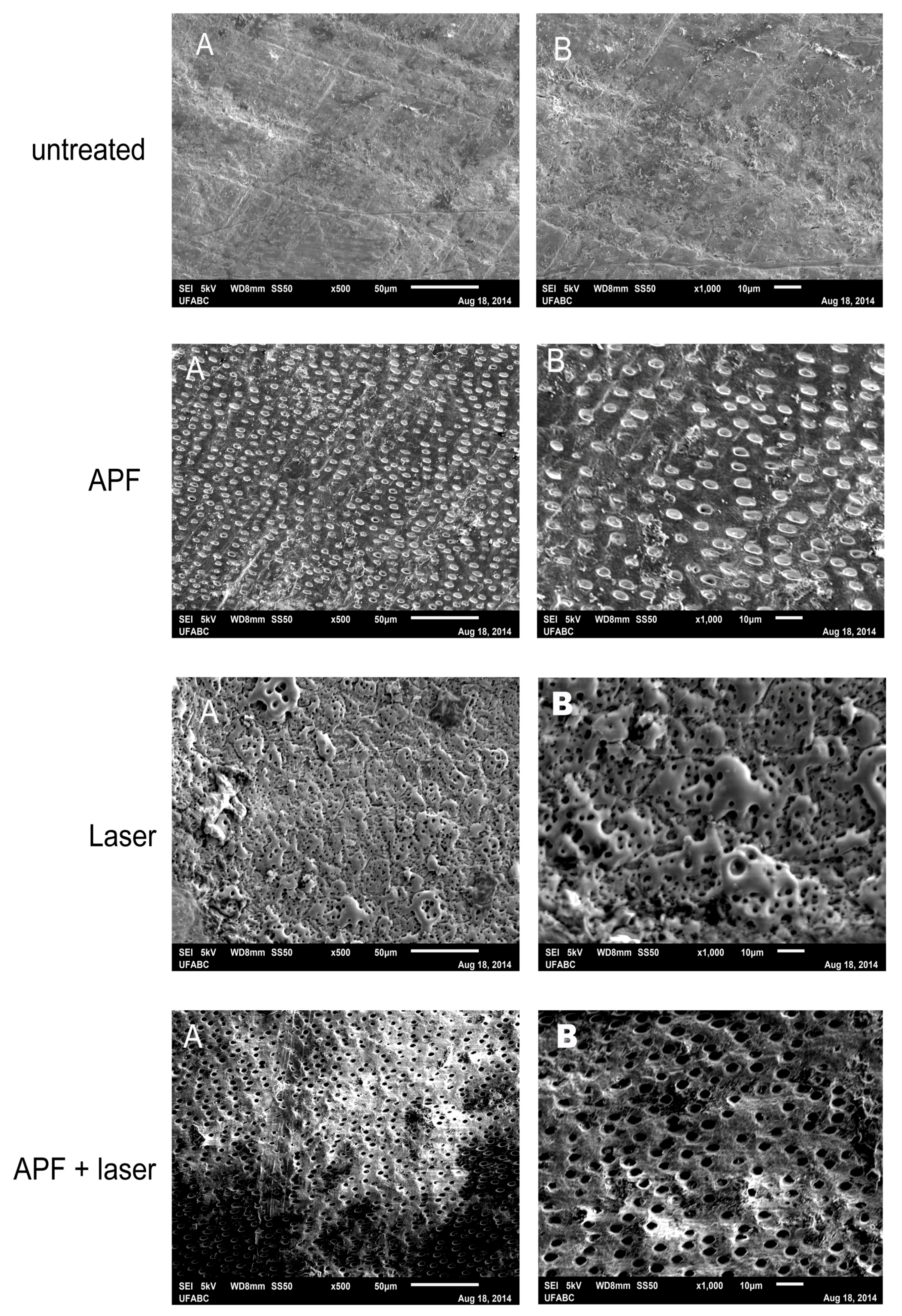
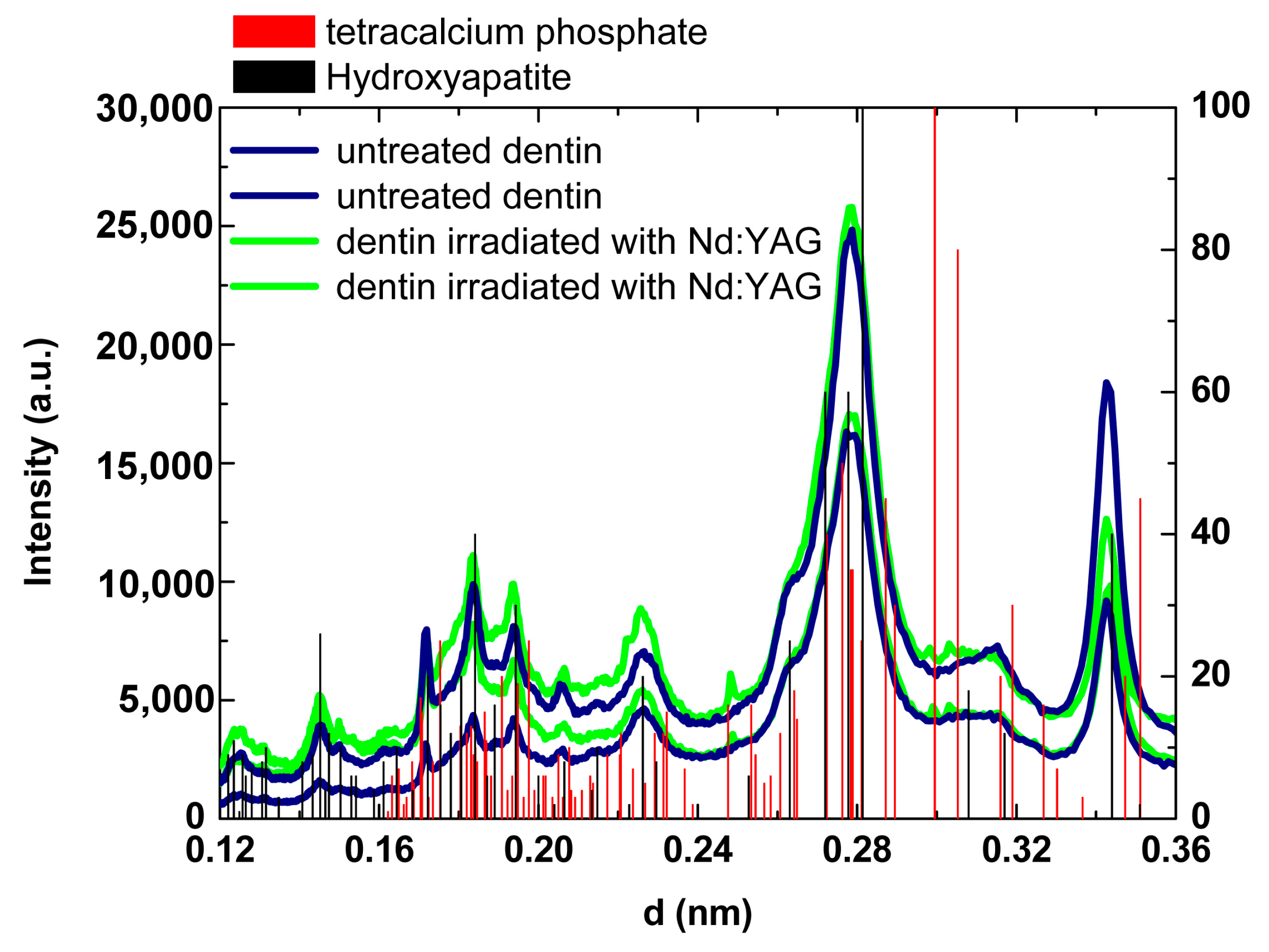
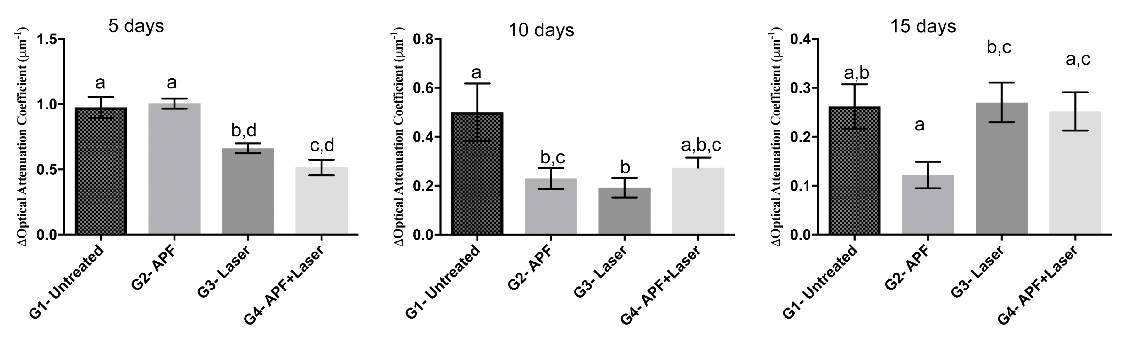
2. Results
3. Discussion
4. Materials and Methods
4.1. Experimental Design
4.2. Preparation of the Specimens and Treatments
4.3. Compositional Analysis
4.4. Crystallographic Analysis
4.5. Morphological Analysis
4.6. Erosive/Abrasive Cycling
4.7. Wear Depth and Area Assessment
4.8. Statistical Analysis
Acknowledgments
Author Contributions
Conflicts of Interest
References
- Jaeggi, T.; Lussi, A. Prevalence, incidence and distribution of erosion. Monogr. Oral Sci. 2006, 20, 44–65. [Google Scholar] [CrossRef] [PubMed]
- Lussi, A.; Schlueter, N.; Rakhmatullina, E.; Ganss, C. Dental erosion—An overview with emphasis on chemical and histopathological aspects. Caries Res. 2011, 45, S2–S12. [Google Scholar] [CrossRef] [PubMed]
- Ganss, C.; Lussi, A.; Schlueter, N. Dental erosion as oral disease. Insights in ethiological factors and pathomecanisms, and current strategies for prevention and therapy. Am. J. Dent. 2012, 25, 351–364. [Google Scholar] [PubMed]
- Kimura, Y.; Wilder-Smith, P.; Arrastia-Jitosho, A.M.A.; Matsumoto, K.; Berns, M.W. Effects of nanosecond pulsed Nd:YAG laser irradiation on dentin resistance to artificial caries-like lesions. Lasers Surg. Med. 1997, 20, 15–21. [Google Scholar] [CrossRef]
- Zezell, D.M.; Boari, H.G.D.; Ana, P.A.; Eduardo, C.P.; Powell, G.L. Nd:YAG laser in caries prevention: A clinical trial. Lasers Surg. Med. 2009, 41, 31–35. [Google Scholar] [CrossRef] [PubMed]
- Ana, P.A.; Tabchoury, P.M.; Cury, J.A.; Zezell, D.M. Effect of Er,Cr:YSGG laser and professional fluoride application on enamel demineralization and on fluoride retention. Caries Res. 2012, 46, 441–451. [Google Scholar] [CrossRef] [PubMed]
- Featherstone, J.D.B. Caries detection and prevention with laser energy. Dent. Clin. N. Am. 2000, 44, 955–969. [Google Scholar] [PubMed]
- Ana, P.A.; Bachmann, L.; Zezell, D.M. Laser Effects on enamel for caries prevention. Laser Phys. 2006, 16, 865–875. [Google Scholar] [CrossRef]
- Featherstone, J.D.B.; Fried, D.; Bitten, E. Mechanism of laser induced solubility reduction of dental enamel. Proc. SPIE 1997, 2973, 112–116. [Google Scholar] [CrossRef]
- Chen, C.C.; Huang, S.T. The effects of laser and fluoride on the acid resistance of decalcified human enamel. Photomed. Laser Surg. 2009, 27, 447–452. [Google Scholar] [CrossRef] [PubMed]
- Wen, X.; Zhang, L.; Liu, R.; Deng, M.; Wang, Y.; Liu, L.; Nie, X. Effects of pulsed Nd:YAG laser on tensile bond strength and caries resistance of human enamel. Oper. Dent. 2014, 39, 273–282. [Google Scholar] [CrossRef] [PubMed]
- Correa-Afonso, A.M.; Pécora, J.D.; Palma-Dibb, R.G. Influence of laser irradiation on pits and fissures: An in vitro study. Photomed. Laser Surg. 2013, 31, 82–89. [Google Scholar] [CrossRef] [PubMed]
- Azevedo, D.T.; Faraoni-Romano, J.J.; Derceli, J.R.; Palma-Dibb, R.G. Effect of Nd:YAG laser combined with fluoride on the prevention of primary tooth enamel demineralization. Braz. Dent. J. 2012, 23, 104–109. [Google Scholar] [CrossRef] [PubMed] [Green Version]
- Seino, P.Y.; Freitas, P.M.; Marques, M.M.; de Souza Almeida, F.C.; Botta, S.B.; Moreira, M.S. Influence of CO2 (10.6 µm) and Nd:YAG laser irradiation on the prevention of enamel caries around orthodontic brackets. Lasers Med. Sci. 2015, 30, 611–616. [Google Scholar] [CrossRef] [PubMed]
- Raucci-Neto, W.; Castro-Raucci, L.M.; Lepri, C.P.; Faraoni-Romano, J.J.; Gomes da Silva, J.M.; Palma-Dibb, R.G. Nd:YAG laser in occlusal caries prevention of primary teeth: A randomizedclinicaltrial. Lasers Med. Sci. 2015, 30, 761–768. [Google Scholar] [CrossRef] [PubMed]
- Magalhães, A.C.; Levy, F.M.; Rizzante, F.A.; Rios, D.; Buzalaf, M.A. Effect of NaF and TiF(4) varnish and solution on bovine dentin erosion plus abrasion in vitro. Acta Odontol. Scand. 2012, 70, 160–164. [Google Scholar] [CrossRef] [PubMed]
- Magalhães, A.C.; Rios, D.; Machado, M.A.A.M.; da Silva, S.M.B.; Lizarelli, R.F.Z.; Bagnato, V.S.; Buzalaf, M.A. Effect of Nd:YAG irradiation and fluoride application on dentine resistance to erosion in vitro. Photomed. Laser Surg. 2008, 26, 559–563. [Google Scholar] [CrossRef] [PubMed]
- Naylor, F.V.; Aranha, A.C.C.; Eduardo, C.P.; Arana-Chavez, V.E.; Sobral, M.A.P. Micromorphological analysis of dentinal structure after irradiation with Nd:YAG laser and immersion in acidic beverages. Photomed. Laser Surg. 2006, 24, 745–753. [Google Scholar] [CrossRef] [PubMed]
- João-Souza, S.H.; Scaramucci, T.; Hara, A.T.; Aranha, A.C.C. Effect of Nd:YAG laser irradiation and fluoride application in the progression of dentin erosion in vitro. Lasers Med. Sci. 2015, 30, 2273–2279. [Google Scholar] [CrossRef] [PubMed]
- Chiga, S.; Toro, C.V.T.; Lepri, T.P.; Turssi, C.P.; Corona, S.A.M. Combined effect of fluoride varnish to Er:YAG or Nd:YAG laser on permeability of eroded root dentine. Arch. Oral Biol. 2016, 64, 24–27. [Google Scholar] [CrossRef] [PubMed]
- Moraes, M.C.D.; Freitas, A.Z.; Aranha, A.C.C. Progression of erosive lesions after Nd:YAG laser and fluoride using optical coherence tomography. Lasers Med. Sci. 2017, 32, 1–8. [Google Scholar] [CrossRef] [PubMed]
- Passos, V.F.; Melo, M.A.; Silva, F.F.; Rodrigues, L.K.; Santiago, S.L. Effects of diode laser therapy and stannous fluoride on dentin resistance under different erosive acid attacks. Photomed. Laser Surg. 2014, 32, 146–151. [Google Scholar] [CrossRef] [PubMed]
- Wegehaupt, F.J.; Sener, B.; Attin, T.; Schmidlin, P.R. Anti-erosive potential of amine fluoride, cerium chloride and laser irradiation application on dentine. Arch. Oral Biol. 2011, 56, 1541–1547. [Google Scholar] [CrossRef] [PubMed] [Green Version]
- De-Melo, M.A.; Passos, V.F.; Alves, J.J.; Barros, E.B.; Santiago, S.L.; Rodrigues, L.K. The effect of diode laser irradiation on dentin as a preventive measure against dental erosion: An in vitro study. Lasers Med. Sci. 2011, 26, 615–621. [Google Scholar] [CrossRef] [PubMed]
- Steiner-Oliveira, C.; Nobre-dos-Santos, M.; Zero, D.T.; Eckert, G.; Hara, A.T. Effect of a pulsed CO2 laser and fluoride on the prevention of enamel and dentine erosion. Arch. Oral Biol. 2010, 55, 127–133. [Google Scholar] [CrossRef] [PubMed]
- Wiegand, A.; Magalhães, A.C.; Navarro, R.S.; Schmidlin, P.R.; Rios, D.; Buzalaf, M.A.; Attin, T. Effect of titanium tetrafluoride and amine fluoride treatment combined with carbon dioxide laser irradiation on enamel and dentin erosion. Photomed. Laser Surg. 2010, 28, 219–226. [Google Scholar] [CrossRef] [PubMed] [Green Version]
- Boari, H.G.D.; Ana, P.A.; Eduardo, C.P.; Powell, G.L.; Zezell, D.M. Absorption and thermal study of dental enamel when irradiated with Nd:YAG laser with the aim of caries prevention. Laser Phys. 2009, 19, 1–7. [Google Scholar] [CrossRef]
- Robinson, C.; Shore, R.C.; Brookes, S.J.; Strafford, S.; Wood, S.R.; Kirkham, J. The chemistry of enamel caries. Crit. Rev. Oral Biol. Med. 2000, 11, 481–495. [Google Scholar] [CrossRef] [PubMed]
- Ana, P.A.; Kauffmann, C.M.F.; Bachmann, L.; Soares, L.E.S.; Martin, A.A.; Gomes, A.S.L.; Zezell, D.M. FT-Raman spectroscopic analysis of Nd:YAG and Er,Cr:YSGG laser irradiated enamel for preventive purposes. Laser Phys. 2014, 24, 035603. [Google Scholar] [CrossRef]
- Fowler, B.O.; Kuroda, S. Changes in heated and in laser-irradiated human tooth enamel and their probable effects on solubility. Calcif. Tissue Int. 1986, 38, 197–208. [Google Scholar] [CrossRef] [PubMed]
- Oho, T.; Morioka, T. A possible mechanism of acquired acid resistance of human dental enamel by laser irradiation. Caries Res. 1990, 24, 86–92. [Google Scholar] [CrossRef] [PubMed]
- Nelson, D.G.; Jongebloed, W.L.; Arends, J. Morphology of enamel surfaces treated with topical fluoride agents: SEM considerations. J. Dent. Res. 1983, 62, 1201–1208. [Google Scholar] [CrossRef] [PubMed]
- Pandurangappa, C.; Lakshminarasappa, B.N.; Nagabhushana, B.M. Synthesis and characterization of CaF2 nanocrystals. J. Alloys Compd. 2010, 489, 592–595. [Google Scholar] [CrossRef]
- Bachmann, L.; Craievich, A.F.; Zezell, D.M. Crystalline structure of dental enamel after Ho:YLF laser irradiation. Arch. Oral Biol. 2004, 49, 923–929. [Google Scholar] [CrossRef] [PubMed]
- Bachmann, L.; Rosa, K.; Ana, P.A.; Zezell, D.M.; Craievich, A.F.; Kellermann, G. Crystalline structure of human enamel irradiated with Er,Cr:YSGG laser. Laser Phys. Lett. 2008, 6, 159–162. [Google Scholar] [CrossRef]
- Zezell, D.M.; Ana, P.A.; Benetti, C.; Goulart, V.P.; Bachmann, L.; Tabchoury, C.P.M.; Cury, J.A. Compositional and crystallographic changes on enamel when irradiated by Nd:YAG or Er,Cr:YSGG lasers and its resistance to demineralization when associated with fluoride. Proc. SPIE 2010, 7549, 75490G1–75490G12. [Google Scholar] [CrossRef]
- He, H.; Yu, J.; Song, Y.; Lu, S.; Liu, H.; Liu, L. Thermal and morphological effects of the pulsed Nd:YAG laser on root canal surfaces. Photomed. Laser Surg. 2009, 27, 235–240. [Google Scholar] [CrossRef] [PubMed]
- Sato, K. Relation between acid dissolution and histological alteration of heated tooth enamel. Caries Res. 1983, 17, 490–495. [Google Scholar] [CrossRef] [PubMed]
- Tenuta, L.M.A.; Cerezetti, R.V.; Del Bel Cury, A.A.; Tabchoury, C.P.M.; Cury, J.A. Fluoride release from CaF2 and enamel demineralization. J. Dent. Res. 2008, 87, 1032–1036. [Google Scholar] [CrossRef] [PubMed]
- Huang, G.F.; Lan, W.H.; Guo, M.K.; Chiang, C.P. Synergistic effect of Nd:YAG laser combined with fluoride varnish on inhibition of caries formation in dental pits and fissures in vitro. J. Formos. Med. Assoc. 2001, 100, 181–185. [Google Scholar] [PubMed]
- Delbem, A.C.; Cury, J.A. Effect of application time of APF and NaF gels on microhardness and fluoride uptake of in vitro enamel caries. Am. J. Dent. 2002, 15, 169–172. [Google Scholar] [PubMed]
- Tagomori, S.; Morioka, T. Combined effects of laser and fluoride on acid resistance of human dental enamel. Caries Res. 1989, 23, 225–251. [Google Scholar] [CrossRef] [PubMed]
- Lee, R.; Chan, K.H.; Jew, J.; Simon, J.C.; Fried, D. Synergistic effect of fluoride and laser irradiation for the inhibition of the demineralization of dental enamel. Proc. SPIE 2017, 10044. [Google Scholar] [CrossRef]
- Zamataro, C.B.; Ana, P.A.; Benetti, C.; Zezell, D.M. Influence of Er,Cr:YSGG laser on CaF2-like products formation because of professional acidulated fluoride or to domestic dentifrice application. Microsc. Res. Tech. 2013, 76, 704–713. [Google Scholar] [CrossRef] [PubMed]
- Popescu, D.P.; Sowa, M.G.; Hewko, M.D.; Choo-Smith, L.P. Assessment of early demineralization in teeth using the signal attenuation in optical coherence tomography images. J. Biomed. Opt. 2008, 13, 054053. [Google Scholar] [CrossRef] [PubMed]
- Cara, A.C.B.; Zezell, D.M.; Ana, P.A.; Maldonado, E.P.; Freitas, A.Z. Evaluation of two quantitative analysis methods of optical coherence tomography for detection of enamel demineralization and comparison with microhardness. Lasers Surg. Med. 2014, 46, 666–671. [Google Scholar] [CrossRef] [PubMed]
- Maia, A.M.A.; de Freitas, A.Z.; Campello, S.D.; Gomes, A.S.L.; Karlsson, L. Evaluation of dental enamel caries assessment using Quantitative Light Induced Fluorescence and Optical Coherence Tomography. J. Biophotonics 2016, 9, 596–602. [Google Scholar] [CrossRef] [PubMed]
- Mujat, C.; Van Der Veen, M.H.; Ruben, J.L.; Ten Bosch, J.J.; Dogariu, A. Optical path-length spectroscopy of incipient caries lesions in relation to quantitative light induced fluorescence and lesions characteristics. Appl. Opt. 2003, 42, 2079–2086. [Google Scholar] [CrossRef]
- Cury, J.A.; Oliveira, M.J.; Martins, C.C.; Tenuta, L.M.; Paiva, S.M. Available fluoride in toothpastes used by Brazilian children. Braz. Dent. J. 2010, 21, 396–400. [Google Scholar] [CrossRef] [PubMed]
- Queiroz, C.S. Modelos de Estudos in vitro Para Avaliar o Efeito do Fluoreto na Desmineralização e Remineralização do Esmalte e Dentina. Ph.D. Thesis, UNICAMP, Campinas, Brazil, 2004. [Google Scholar]
- Takahashi, R.; Jin, J.; Nikaido, T.; Tagami, J.; Hickel, R.; Kunzelmann, K.H. Surface characterization of current composites after toothbrush abrasion. Dent. Mater. J. 2013, 32, 75–82. [Google Scholar] [CrossRef] [PubMed]
- Voronets, J.; Jaeggi, T.; Buergin, W.; Lussi, A. Controlled toothbrush abrasion of softened human enamel. Caries Res. 2008, 42, 286–290. [Google Scholar] [CrossRef] [PubMed]


| Groups | 5 Days (n = 15) | 10 Days (n = 15) | 15 Days (n = 15) |
|---|---|---|---|
| Untreated (n = 45) | 26,344.8 a ± 4263.1 | 28,197.1 a ± 6470.0 | 34,111.5 a ± 9269.9 |
| APF (n = 45) | 25,882.2 a ± 3621.1 | 27,958.3 a ± 5318.8 | 35,187.2 a,b ± 8762.8 |
| Laser (n = 45) | 12,960.1 b ± 5162.7 | 15,007.1 b ± 8831.0 | 14,760.2 c ± 11,515.5 |
| APF + Laser (n = 45) | 16,171.8 b ± 5445.1 | 21,705.0 b ± 4446.6 | 24,580.1 b,c ± 9505.8 |
| Groups | 5 Days (n = 15) | 10 Days (n = 15) | 15 Days (n = 15) |
|---|---|---|---|
| G1—Untreated (n = 45) | 9.78 a ± 2.9 | 10.66 a ± 3.6 | 12.80 a ± 5.0 |
| G2—APF (n = 45) | 9.25 a ± 2.5 | 10.09 a ± 1.9 | 14.80 a ± 3.9 |
| G3—Laser (n = 45) | 5.99 b ± 2.7 | 7.23 b ± 2.9 | 6.97 b ± 4.0 |
| G4—APF + Laser (n = 45) | 6.05 b ± 3.2 | 7.57 b ± 2.8 | 8.13 b ± 4.2 |
© 2018 by the authors. Licensee MDPI, Basel, Switzerland. This article is an open access article distributed under the terms and conditions of the Creative Commons Attribution (CC BY) license (http://creativecommons.org/licenses/by/4.0/).
Share and Cite
Pereira, D.L.; Freitas, A.Z.; Bachmann, L.; Benetti, C.; Zezell, D.M.; Ana, P.A. Variation on Molecular Structure, Crystallinity, and Optical Properties of Dentin Due to Nd:YAG Laser and Fluoride Aimed at Tooth Erosion Prevention. Int. J. Mol. Sci. 2018, 19, 433. https://doi.org/10.3390/ijms19020433
Pereira DL, Freitas AZ, Bachmann L, Benetti C, Zezell DM, Ana PA. Variation on Molecular Structure, Crystallinity, and Optical Properties of Dentin Due to Nd:YAG Laser and Fluoride Aimed at Tooth Erosion Prevention. International Journal of Molecular Sciences. 2018; 19(2):433. https://doi.org/10.3390/ijms19020433
Chicago/Turabian StylePereira, Daísa L., Anderson Z. Freitas, Luciano Bachmann, Carolina Benetti, Denise M. Zezell, and Patricia A. Ana. 2018. "Variation on Molecular Structure, Crystallinity, and Optical Properties of Dentin Due to Nd:YAG Laser and Fluoride Aimed at Tooth Erosion Prevention" International Journal of Molecular Sciences 19, no. 2: 433. https://doi.org/10.3390/ijms19020433