Article

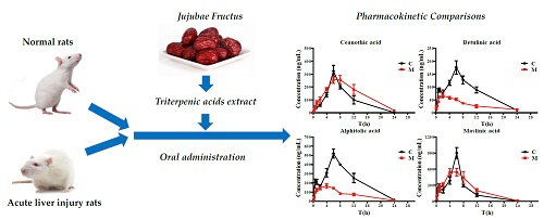
Pharmacokinetic Comparisons of Multiple Triterpenic Acids from Jujubae Fructus Extract Following Oral Delivery in Normal and Acute Liver Injury Rats

1
Jiangsu Collaborative Innovation Center of Chinese Medicinal Resources Industrialization/State Key Laboratory Cultivation Base for Traditional Chinese Medicine Quality and Efficacy, Nanjing University of Chinese Medicine, Nanjing 210023, China
2
Institute of Botany, Jiangsu Province and Chinese Academy of Science, Nanjing 210014, China
3
Shaanxi Collaborative Innovation Center of Chinese Medicinal Resources Industrialization, Shaanxi University of Chinese Medicine, Xianyang 712046, China
*
Authors to whom correspondence should be addressed.
Int. J. Mol. Sci. 2018, 19(7), 2047; https://doi.org/10.3390/ijms19072047
Submission received: 2 July 2018 / Revised: 10 July 2018 / Accepted: 11 July 2018 / Published: 13 July 2018
(This article belongs to the Special Issue Plant Natural Products for Human Health)

Abstract

:
Jujubae Fructus, the dried fruit of Ziziphus jujuba, has been used as Chinese medicine and food for centuries. Triterpenic acids have been found to be the major bioactive constituents in Jujubae Fructus responsible for their hepatoprotective activity in previous phytochemical and biological studies, while few pharmacokinetic studies have been conducted. To reveal the kinetics of the triterpenic acids under the pathological liver injury state, an established ultra-performance liquid chromatography coupled with a mass spectrometry method was applied for the simultaneous quantitation of seven triterpenic acids (ceanothic acid, epiceanothic acid, pomonic acid, alphitolic acid, maslinic acid, betulinic acid, and betulonic acid) in plasma samples of normal and acute liver injury rats induced by CCl4. The results showed that there were significant differences (p < 0.05) in the pharmacokinetic parameters of seven triterpenic acids between model and normal groups. The AUC0–t and AUC0–∞ of epiceanothic acid (5227 ± 334 μg⋅h/L vs. 1478 ± 255 μg⋅h/L and 6127 ± 423 μg⋅h/L vs. 1482 ± 255 μg⋅h/L, respectively) and pomonic acid (4654 ± 349 μg⋅h/L vs. 1834 ± 225 μg⋅h/L and 4776 ± 322 μg⋅h/L vs. 1859 ± 230 μg⋅h/L, respectively) in model rats were significantly higher than those in normal rats, and the CLz/F of them were significantly decreased (0.28 ± 0.02 L/h/kg vs. 1.36 ± 0.18 L/h/kg and 19.96 ± 1.30 L/h/kg vs. 53.15 ± 5.60 L/h/kg, respectively). In contrast, the above parameters for alphitolic acid, betulinic acid and betulonic acid exhibited the quite different trend. This pharmacokinetic research might provide useful information for the clinical usage of triterpenic acids from Jujubae Fructus.

Graphical Abstract

1. Introduction

Jujubae Fructus, the fruit of Ziziphus jujuba Mill., has been used as herb medicine and food for centuries in China [1,2]. According to traditional Chinese medicine theory, Jujubae Fructus could reinforce spleen and stomach, and is commonly used for the treatment of anorexia, fatigue and loose stools related to deficiency syndromes of the spleen and hysteria in women [3]. The controlled clinical trials also showed that Jujubae Fructus extract is an effective treatment for chronic constipation [4] and type 2 diabetes [5]. In addition, in northern China, the decoction of Jujubae Fructus are claimed as useful remedies for the management and/or control of hepatitis in folk [6].
In support of its traditional efficacy, modern researches have revealed that Jujubae Fructus has pharmacological properties including hepatoprotective [6], gastrointestinal protective [7], anti-inflammatory [8], immunomodulating [9] and hematopoiesis effects [10]. Among them, ethanolic extract of Jujubae Fructus with a dose of 200 mg/kg could significantly decrease ALT and AST, and attenuate histopathology of hepatic injury induced by carbon tetrachloride (CCl4), and the results indicated that hepatic protective effects of Jujubae Fructus were relevant to modulate the oxidative stress in hepatic injury [6,11]. These results further confirmed the traditional efficacy of Jujubae Fructus on the hepatoprotective effect. Phytochemical and biological studies showed that these multiple bioactivities of Jujubae Fructus could be attributed to its various constituents, such as triterpenic acids [12], polysaccharides [2], phenolic acids [13], flavonoids [14], nucleoside and amino acids [15,16]. Among these components, triterpenic acids, such as betulinic acid, alphitolic acid, maslinic acid, etc., have been reported to possess biological effects of hepatoprotective, anti-inflammatory, antimicrobial and antioxidant activities [6,8,17,18], which have attracted great attention from researchers.
The liver is a crucial organ for metabolism and detoxification in the human body, and liver disease has nowadays become one of the most common causes of death [19]. There are reports that acute liver injury is the main initiating factor and pathological basis for liver fibrosis, hepatitis, cirrhosis and even liver cancer, which could result in terminal liver failure [20,21]. The previous studies have reported the hepatoprotective effect of Jujubae Fructus, and triterpenic acids have been considered as the main bioactive compounds for the above activity [6,11]. However, there were few reports on the pharmacokinetic studies of triterpenic acids from Jujubae Fructus in liver injury model animals, and relevant pharmacokinetic studies of Jujubae Fructus in humans were also rare. It is well known that under the pathological condition of liver injury, pharmacokinetic and metabolic behaviors of drugs are often altered [20,22,23]. Thus, based on the ultra-performance liquid chromatography coupled with mass spectrometry (UHPLC-MS/MS) method established in the previous study [24], the pharmacokinetics of triterpenic acids from Jujubae Fructus in normal and acute liver injury rats were compared in this paper for the purpose of providing clinical reference for Jujubae Fructus.

2. Results and Discussion

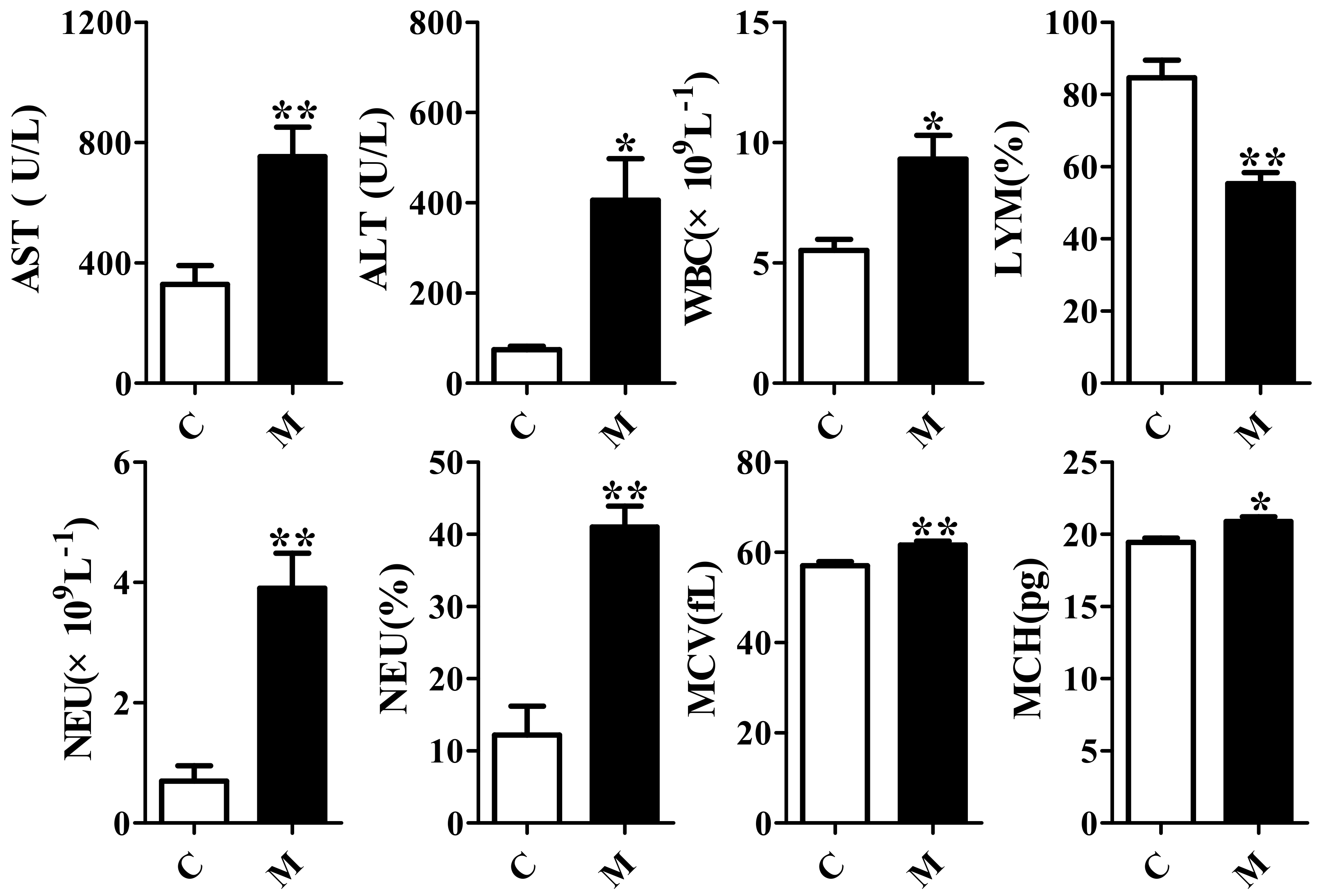
2.1. Validation of the Acute Liver Injury Rats Model

To verify whether the rat model of acute liver injury was successful, peripheral blood routine, levels of alanine aminotransferase (ALT), aspartate aminotransferase (AST) and histopathological characteristics were analyzed. All the results are presented in Figure 1 and Figure 2. It was shown that the liver injury score and the levels of white blood cell count (WBC), neutrophil (NEU) count and ratio, erythrocyte mean corpuscular volume (MCV), mean corpuscular hemoglobin (MCH), AST and ALT of the model group after being injected intraperitoneally with CCl4 increased significantly (p < 0.05) compared to control group. Lymphocyte (LYM) ratios of the model group decreased significantly. It was known that the levels of AST and ALT were all related to liver function. Besides, it has reported that liver injury induced by CCl4 could cause inflammation [19,25], which could be presented with the increases of WBC and NEU. Thus, the above results indicated that the acute liver injury rat model was successful, and could be used for the following experiment.

2.2. Method Validation
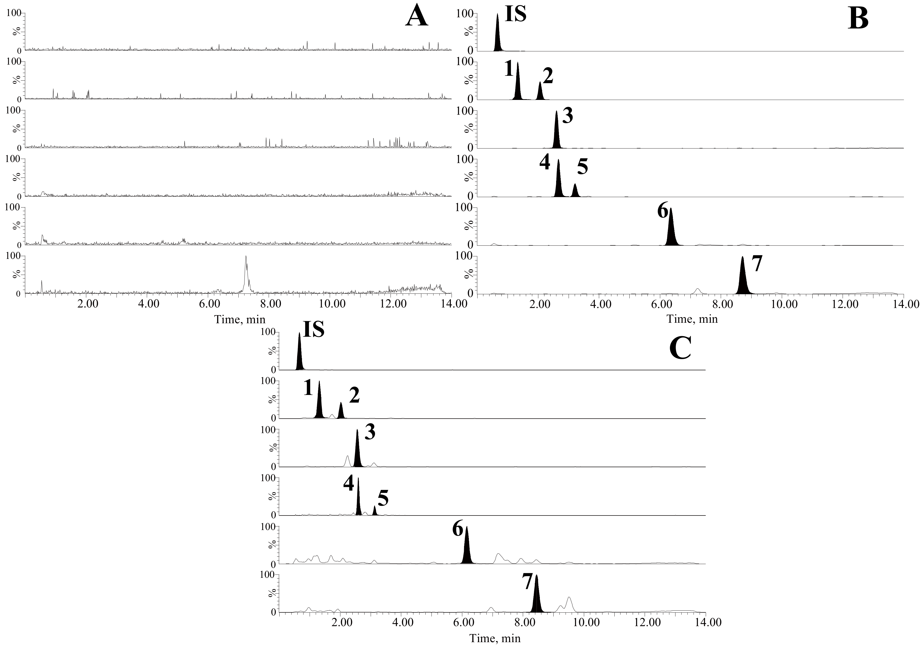
The method for separation and detection of analytes was performed in the established UHPLC-MS/MS method previously [24] with appropriate adjustments.

2.2.1. Selectivity

Figure 3 showed the chromatograms obtained from the blank plasma of a rat, blank plasma spiked with the standards of seven mixed triterpenic acids and internal standard (IS), and rat plasma acquired at 6 h after oral administration of triterpenic acids extract (TAE). No significant endogenous interference or metabolites were found in the blank plasma at the retention times of standards and IS, which revealed that the selectivity of the method was acceptable.

2.2.2. Linearity and Lower Limit of Quantification (LLOQ)

The linearity of the proposed method was evaluated by means of representative calibration curves, correlation coefficients and a linear range of the seven standards, and LLOQs were used for determining the sensitivity of the method. As shown in Table 1, all the correlation coefficients (R2) are ≥0.9930, which indicated the good linearity of all analytes and the LLOQs of the seven triterpenic acids in plasma were suitable for quantitative detection.

2.2.3. Precision and Accuracy

As shown in Table 2, the deviation in intra- and inter-day precision of all the analytes in QC samples were ≤10.11% and ≤14.29%, respectively, and the accuracies (RE) of those analytes ranged from −2.03% to 14.87%. The results indicated that the method was accurate, precise and was acceptable for analysis of biological samples due to the values being within the acceptable criteria.

2.2.4. Extraction Recovery and Matrix Effect

The results of extraction recovery and the matrix effect are shown in Table 3. It was shown that the extraction recoveries ranged from 78.98% to 103.8%, and the matrix effects were between 75.28% and 109.3% with the RSD values less than 15.0% for the seven analytes at three QC concentrations. As for IS, the extraction recoveries and matrix effects were 87.37–98.45% and 75.52–80.31%, respectively. All the results suggested the reliable extraction recoveries of these analytes and no significant matrix effect in this experiment.

2.2.5. Stability

The QC samples with different conditions (three freeze-thaw cycles; 12 h at room temperature; 24 h at 4 °C; 20 days at −20 °C) were used to investigate the stability of the seven triterpenic acids, and the results (Table 4) showed that the RSD values were all less than 13.59%, which indicated that all analytes were stable throughout the whole test.

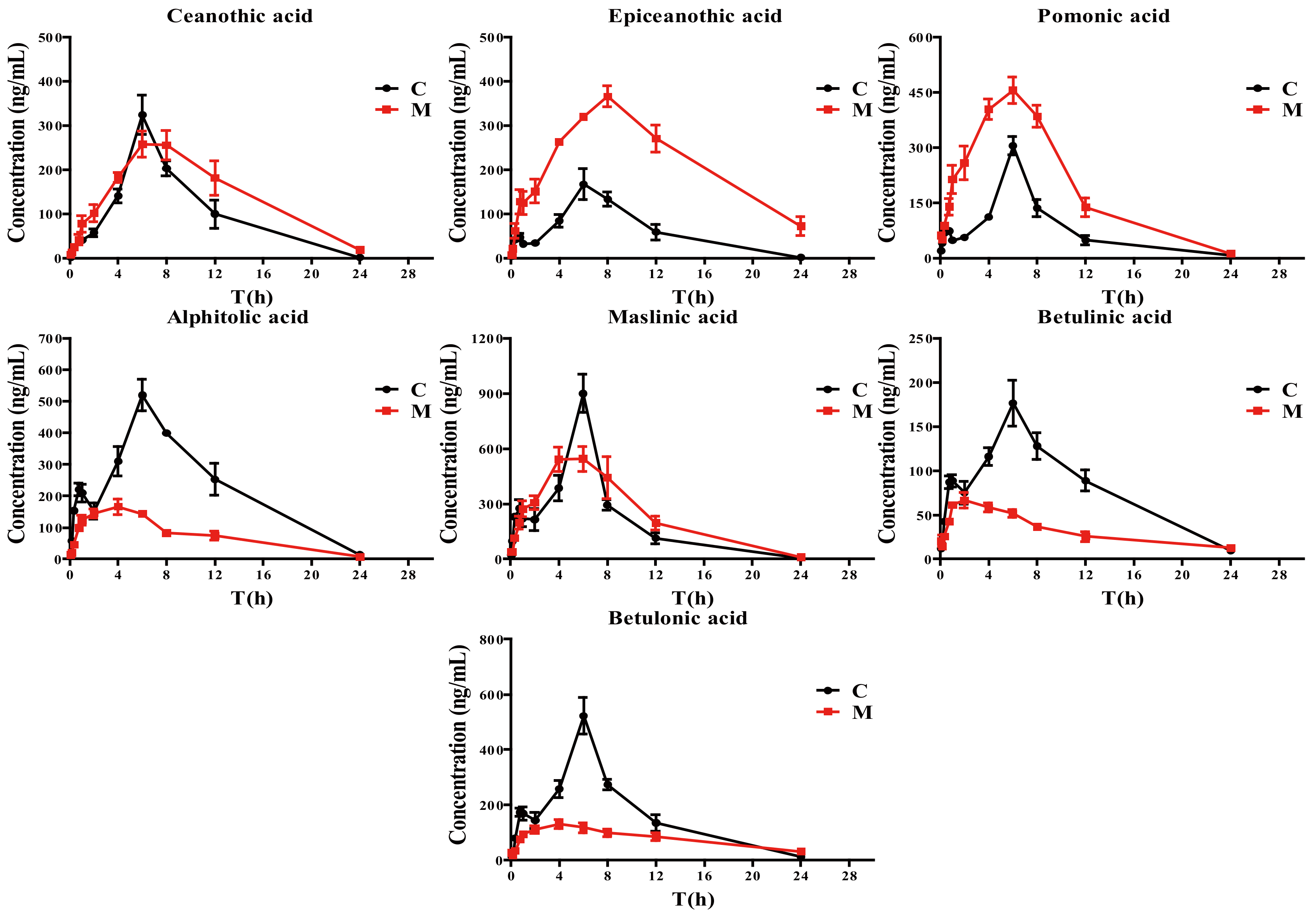
2.3. Pharmacokinetic Study

The pharmacokinetics of seven triterpenic acids in plasma after a single oral administration of TAE in normal and acute liver injury rats were analyzed by the validated UHPLC-MS/MS method. The pharmacokinetic parameters obtained with the non-compartment module of Drug and Statistic (DAS) 3.2.8 pharmacokinetic software are listed in Table 5. The mean concentration-time profiles are presented in Figure 4.
As shown in Figure 4, the consistent plasma concentration-time profiles in normal rats were found for these seven analytes, which may be attributed to their similar chemical structures. However, this phenomenon was not found in the acute liver injury model, which could be ascribed to the pathological changes of the liver. Moreover, certain pharmacokinetic parameters for these triterpenic acids in acute liver injury rats showed significant differences from those in normal rats, especially for the area under the time curve (AUC0–t and AUC0–∞) and the apparent plasma clearance (CLz/F). The AUC0–t and AUC0–∞ of epiceanothic acid and pomonic acid achieved from the drug concentration-time in acute liver injury rats after oral administration of TAE were significantly higher than those in normal rats, and the CLz/F of them were significantly decreased. These indicated that the acute liver injury could increase the bioavailiability of epiceanothic acid and pomonic acid, and decrease their elimination. In contrast, the mean AUC0–t, AUC0–∞ and the mean peak concentration (Cmax) of alphitolic acid, betulinic acid and betulonic acid in acute liver injury rats were achieved with relatively lower values compared to those in normal rats. The obvious higher CLz/Fs of alphitolic acid, betulinic acid and betulonic acid compared to normal rats were also found. The above results suggested that the systemic exposure of alphitolic acid, betulinic acid and betulonic acid were weakened and the elimination increased under the liver injury pathological condition. Additionally, the Tmax of betulinic acid and betulonic acid of the model group were lower than those of the normal group (p < 0.05), and the AUC0–t of ceanothic acid and the Cmax of epiceanothic acid were significantly higher in acute liver injury rats. The other pharmacokinetic parameters in the model rats were found to be different but not significant compared with the normal rats.
It is well known that the liver plays important roles in drug biotransformation, metabolism, detoxification and so on [19]. It contains various enzymes involved in drug metabolism, including cytochrome P450 which converts the drug to active metabolites and directly affects the rate of metabolism [26]. Liver injury might lead to liver cell degeneration, necrosis and changes in cytochrome P450 isoenzyme contents, which could alter the disposition of drugs in the body [22,27]. There have been reports on changes in biotransformation, clearance and pharmacokinetics of drugs in liver injury [28].
Besides, the influence of intestinal drug transport and its microbiota might be an important factor in the absorption and bioavailability of the orally administered medicines. It has been reported that liver injury often causes the increase in intestinal permeability and endotoxin, and the disorder of the intestinal microbiota [29,30,31]. The increased endotoxin could further cause liver injury more severely and might be a vicious cycle [30]. All the above reasons might synthetically result in differences in pharmacokinetic behavior between acute liver injury and normal rats after oral administration of TAE. However, the hypotheses are still undefined and need further validation.
It is worth noting that the plasma concentration–time profiles of epiceanothic acid and pomonic acid were markedly different from alphitolic acid, betulinic acid and betulonic acid in acute liver injury rats administered TAE from Jujubae Fructus. The AUC0–t and AUC0–∞ of epiceanothic acid and pomonic acid achieved from the drug concentration-time in acute liver injury rats were significantly higher than those in normal rats, and the CLz/F of them were significantly decreased. In contrast, the mean AUC0–t, AUC0–∞ and CLz/F of alphitolic acid, betulinic acid and betulonic acid in acute liver injury rats showed quite a different trend. This phenomenon might be attributed to their subtle difference in chemical structures which could lead to different metabolic pathways. Therefore, further studies for the investigation of triterpenic acids metabolism and distribution in vivo are warranted.
At present, there are some clinical reports about the liver protection of Jujubae Fructus, but there is little relevant pharmacokinetic study of Jujubae Fructus in humans. Thus, the depth clinical studies to validate the proposed hypothesis need to be conducted in the future.

3. Materials and Methods

3.1. Chemicals and Reagents

Acetonitrile and methanol were purchased from Merck KGaA (Darmstadt, Germany). Chloramphenicol used as the internal standard (IS) was obtained from Aladdin reagent Co., Ltd. (Shanghai, China). Ammonium acetate and CCl4 were purchased from Sinopharm Chemical Reagent Co., Ltd. (Shanghai, China). Deionized water was prepared by a Milli-Q system (Millipore, Bedford, MA, USA). Jujubae Fructus was gathered at Liuling, Shanxi Province, China. The standards (>98% purity) including ceanothic acid, epiceanothic acid, pomonic acid, alphitolic acid, maslinic acid, betulinic acid and betulonic acid were isolated from Z. jujuba fruits in our laboratory, and their structures were identified by NMR, HPLC and MS. Other reagents used were of analytical grade.
TAE of Jujubae Fructus prepared in our previous experiment [24] was used in this experiment, which contains ceanothic acid, epiceanothic acid, pomonic acid, alphitolic acid, maslinic acid, betulinic acid, and betulonic acid with the contents of 0.78, 0.44, 24.08, 4.97, 17.31, 16.79 and 14.29 mg/g, respectively.

3.2. Instrumentation and Chromatographic Conditions

A Waters Acquity™ UPLC system (Waters Corp., Milford, MA, USA) equipped with a Waters Xevo™ TQ/MS (Waters Corp.) was used. Separation and detection of analytes was performed with the established method described previously [24]. Data acquired was analyzed by MassLynx V4.1 workstation (Waters Corp.).

3.3. Animals and Induction of Acute Liver Injury

Male Sprague-Dawley rats (SPF, 220-240 g) were bought from Experimental Animal Center of Zhejiang Province and the permit number was SCXK (zhe) 2014-0001 (project identification code: No1703300021, 31/03/2017). Animals were housed in Drug Safety Evaluation Center of Nanjing University of Chinese Medicine, Nanjing, China. Animal welfare and all experimental protocols were performed in accordance with the Regulations of Experimental Animal Administration (State Committee of Science and Technology of the People’s Republic of China) and approved by the Animal Ethics Committee of Nanjing University of Chinese Medicine. These rats were housed under standard environment with food and water provided ad libitum. After adaptation for 7 days, the 12 rats were randomly divided into 2 groups: control group (C) and acute liver injury group (model group, M). Rats of M group were injected intraperitoneally with 50% CCl4 once at a dose of 2 mL/kg. CCl4 used for injection was dissolved in peanut oil. To verify whether the model was successful, whole blood and serum samples were collected from the retro-orbital plexus of rats after modeling for measuring the peripheral blood routine parameters using ADVIA120 fully automatic blood analyzer (Bayer, Germany) and the levels of ALT and AST with Dimension Xpand automatic biochemical analyzer (Bayer, Germany). Furthermore, after the final collection of blood, the rats were anesthetized and sacrificed with 10% choral hydrate (350 mg/kg ip) and the liver samples were taken and fixed in formaldehyde solution for histopathological analysis.

3.4. Sample Preparation

After being thawed at room temperature (18–25 °C), each plasma sample (100 μL) was precipitated with 300 μL acetonitrile and 20 μL IS. The mixture was vortexed for 1 min and centrifuged at 15,000× g for 15 min. Then, 2 μL of supernatant was injected for UHPLC-MS/MS analysis.

3.5. Preparation of Standard Solutions, Calibration Standards and Quality Control (QC) Samples

A mixed stock solution containing 126.9 μg/mL of ceanothic acid, 129.4 μg/mL of epiceanothic acid, 125.6 μg/mL of pomonic acid, 124.4 μg/mL of alphitolic acid, 126.3 μg/mL of maslinic acid, 130.0 μg/mL of betulinic acid and 129.4 μg/mL of betulonic acid was prepared with methanol as a solvent. A series of working standard solutions were prepared from the mixed stock solution by sequential dilution with methanol. Calibration solutions were prepared by spiking 10 μL of working solution into 100 μL blank plasma to obtain a mixed final dilution of 2.31–2951 ng/mL ceanothic acid, 2.35–3009 ng/mL epiceanothic acid, 2.28–2922 ng/mL pomonic acid, 2.26–2892 ng/mL alphitolic acid, 2.29–2936 ng/mL maslinic acid, 2.36–3023 ng/mL betulinic acid, and 2.35–3009 ng/mL betulonic acid. The quality control (QC) samples were prepared at low, medium, and high concentrations (23.05, 368.8, and 2951 ng/mL ceanothic acid, 23.51, 376.1, and 3009 ng/mL epiceanothic acid, 22.82, 365.2, and 2922 ng/mL pomonic acid, 22.60, 361.6, and 2892 ng/mL alphitolic acid, 22.94, 367.0, and 2936 ng/mL maslinic acid, 23.62, 377.9, and 3023 ng/mL betulinic acid, and 23.51, 376.1, and 3009 ng/mL betulonic acid) in the same way as calibration solutions. The stock solution of IS (9.76 μg/mL chloramphenicol) was also prepared in methanol.

3.6. Method Validation

This proposed method was validated according to US-FDA Bioanalytical Method Validation Guidance [32], and selectivity, linearity, precision, extraction recovery, matrix effect and stability were assessed.

3.6.1. Selectivity

The chromatograms of the rat blank plasma, blank plasma spiked with the seven standards and IS, and rat plasma acquired at 6 h after gavage of TAE, were analyzed and compared to investigate the selectivity of the method [33].

3.6.2. Linearity and LLOQ

The peak area ratio (y) of the analyte to the IS vs. the nominal concentration (x, ng/mL) was used to plot the calibration curve and determine the linearity with weighted (1/x2) least square linear regression [34]. LLOQ of the method was determined based on the signal to noise ratio of 10:1 with the acceptable precision in six replicates of blank plasma (RSD ≤ 20%) [35].

3.6.3. Accuracy and Precision

The intra-day and inter-day accuracy and precision were assessed by determining the concentration of six replicates of QC samples (as described in section ‘3.5’) at three concentration levels (low, medium and high) on the same day and on three consecutive days, respectively. The accuracy was described as relative error (RE, %) and precision was expressed as relative standard deviation (RSD, %) [36]. The acceptability criteria for accuracy and precision were required within ±15% according to the guidelines of FDA.

3.6.4. Recovery and Matrix Effect

Extraction recovery and matrix effect were evaluated with six replicates of QC samples at three concentrations. Extraction recovery of the seven triterpenic acids were performed by comparing the peak area of every analyte extracted from plasma samples with that of post-extraction spiked plasma blank [24]. For evaluation of the matrix effect, the peak areas of the analytes in post-extraction standard plasma samples (B) were compared with those of pure methanol containing an equivalent amount of standards at QC levels (A) [37].

3.6.5. Stability

The stability of analytes in rat plasma were assessed by analyzing six replicates of QC samples at three concentration levels. The QC samples in different storage conditions including three freeze-thaw cycles (from −80 °C to room temperature), 12 h at room temperature, and 20 days at −20 °C, were used to evaluate the freeze-thaw, short-term, and long-term stability, respectively. In addition, the autosampler stability was also evaluated after samples were stored in the autosampler at 4 °C for 24 h [38].

3.7. Pharmacokinetic Study in Rat and Statistical Analysis

After fasting for 12 h with free access to water, rats in both C and M groups were intragastrically administered TAE (dissolved in water with 10% tween-80) at a dose of 4.0 g/kg. The administered volume was 10 mL/kg for each rat, each time. Serial blood samples (about 500 μL for each) were collected into heparinized tubes from the retro-orbital plexus at 0, 5, 10, 20, 45, 60, 120, 240, 360, 480, 720 and 1440 min after oral administration. All blood samples were centrifuged at 3500 rpm at 4 °C for 10 min, then the supernatants were separated and stored at −80 °C until analysis. The pharmacokinetic parameters were calculated by Drug and Statistic (DAS) 3.2.8 pharmacokinetic software in a non-compartment model. The experimental data were expressed as mean ± SEM. Independent Samples t-test via the software SPSS 22.0 (IBM SPSS, Chicago, IL, USA) was used for evaluation of statistical significance.

4. Conclusions

A rapid, sensitive, and simple UHPLC-MS/MS method was used for the determination of seven triterpenic acids in the plasma of acute liver injury and normal rats after oral administration of TAE. The results demonstrated that acute liver injury induced by CCl4 could alter the pharmacokinetic parameters of seven triterpenic acids, such as AUC0–t, AUC0–∞, CLz/F and Cmax. And the differences might be due to the changes in liver function, intestinal permeability and intestinal microbiome in the acute liver injury pathological state. These pharmacokinetic results in the pathological state of acute liver injury might provide more useful information for the application of Jujubae Fructus in treating liver disease.

Author Contributions

S.G. and J.-A.D. designed the study. Y.L., S.G. and Q.R. performed the laboratory work. Y.L., S.G. and D.W. analyzed the data and wrote the paper. M.Z., S.S. revised the manuscript. J.-A.D., Z.T. and Q.R. contributed reagents/materials/analysis tools.

Funding

This research was funded by National Natural Science Foundation of China (No. 81473538), Six Talent Peaks Project in Jiangsu Province (No. YY-026) and Henry Fok Education Foundation (No. 141040).

Conflicts of Interest

The authors declare no conflict of interest.

References

  1. Lam, C.T.W.; Gong, A.G.W.; Lam, K.Y.C.; Zhang, L.M.; Chen, J.-P.; Dong, T.T.X.; Lin, H.-Q.; Tsim, K.W.K. Jujube-containing herbal decoctions induce neuronal differentiation and the expression of anti-oxidant enzymes in cultured PC12 cells. J. Ethnopharmacol. 2016, 188, 275–283. [Google Scholar] [CrossRef] [PubMed]
  2. Ji, X.; Peng, Q.; Yuan, Y.; Shen, J.; Xie, X.; Wang, M. Isolation, structures and bioactivities of the polysaccharides from jujube fruit (Ziziphus jujuba Mill.): A review. Food Chem. 2017, 227, 349–357. [Google Scholar] [CrossRef] [PubMed]
  3. Gao, Q.-H.; Wu, C.-S.; Wang, M. The Jujube (Ziziphus Jujuba Mill.) Fruit: A review of current knowledge of Fruit composition and health benefits. J. Agric. Food Chem. 2013, 61, 3351–3363. [Google Scholar] [CrossRef] [PubMed]
  4. Naftali, T.; Feingelernt, H.; Lesin, Y.; Rauchwarger, A.; Konikoff, F.M. Ziziphus jujuba extract for the treatment of chronic idiopathic constipation: a controlled clinical trial. Digestion 2008, 78, 224–228. [Google Scholar] [CrossRef] [PubMed]
  5. Yazdanpanah, Z.; Ghadiri-Anari, A.; Mehrjardi, A.V.; Dehghani, A.; Zardini, H.Z.; Nadjarzadeh, A. Effect of Ziziphus jujube fruit infusion on lipid profiles, glycaemic index and antioxidant status in type 2 diabetic patients: a randomized controlled clinical trial. Phytother. Res. 2017, 31, 755–762. [Google Scholar] [CrossRef] [PubMed]
  6. Shen, X.; Tang, Y.; Yang, R.; Yu, L.; Fang, T.; Duan, J.A. The protective effect of Zizyphus jujube fruit on carbon tetrachloride-induced hepatic injury in mice by anti-oxidative activities. J. Ethnopharmacol. 2009, 122, 555–560. [Google Scholar] [CrossRef] [PubMed]
  7. Huang, Y.-L.; Yen, G.-C.; Sheu, F.; Chau, C.F. Effects of water-soluble carbohydrate concentrate from Chinese jujube on different intestinal and fecal indices. J. Agric. Food Chem. 2008, 56, 1734–1739. [Google Scholar] [CrossRef] [PubMed]
  8. Yu, L.; Jiang, B.P.; Luo, D.; Shen, X.C.; Guo, S.; Duan, J.A.; Tang, Y. P. Bioactive components in the fruits of Ziziphus jujuba Mill. against the inflammatory irritant action of Euphorbia plants. Phytomedicine 2012, 19, 239–244. [Google Scholar] [CrossRef] [PubMed]
  9. Chen, J.; Du, C.Y.; Lam, K.Y.; Zhang, W.L.; Lam, C.T.; Yan, A.L.; Yao, P.; Lau, D.T.; Dong, T.T.; Tsim, K.W. The Standardized extract of Ziziphus jujuba Fruit (Jujube) regulates pro-inflammatory cytokine expression in cultured murine macrophages: suppression of lipopolysaccharide-stimulated NF-κB Activity. Phytother. Res. 2014, 28, 1527–1532. [Google Scholar] [CrossRef] [PubMed]
  10. Chen, J.; Lam, C.T.; Kong, A.Y.; Zhang, W.L.; Zhan, J.Y.; Bi, C.W.; Chan, G.K.; Lam, K.Y.; Yao, P.; Dong, T.T.; et al. The Extract of Ziziphus jujuba fruit (Jujube) induces expression of erythropoietin via hypoxia-inducible factor-1 alpha in cultured Hep3B Cells. Planta Med. 2014, 80, 1622–1627. [Google Scholar] [PubMed]
  11. Rajopadhye, A.; Upadhye, A.S. Estimation of bioactive compound, maslinic acid by HPTLC, and evaluation of hepatoprotective activity on fruit pulp of Ziziphus jujuba Mill. cultivars in India. eCAM 2016, 2016, 4758734. [Google Scholar] [PubMed]
  12. Lee, S.M.; Min, B.S.; Lee, C.G.; Kim, K.S.; Kho, Y.H. Cytotoxic triterpenoids from the fruits of Zizyphus jujuba. Planta Med. 2003, 69, 1051–1054. [Google Scholar] [PubMed]
  13. Wang, B.N.; Liu, H.F.; Zheng, J.B.; Fan, M.T.; Cao, W. Distribution of phenolic acids in different tissues of jujube and their antioxidant activity. J. Agric. Food Chem. 2011, 59, 1288–1292. [Google Scholar] [CrossRef] [PubMed]
  14. Chen, J.P.; Li, Z.G.; Maiwulanjiang, M.; Zhang, W.L.; Zhan, J.Y.; Lam, C.T.; Zhu, K.Y.; Yao, P.; Choi, R.C.; Lau, D.T.; et al. Chemical and biological assessment of Ziziphus jujuba fruits from China: different geographical sources and developmental stages. J. Agric. Food Chem. 2013, 61, 7315–7324. [Google Scholar] [CrossRef] [PubMed]
  15. Guo, S.; Duan, J.A.; Qian, D.; Tang, Y.; Wu, D.; Su, S.; Wang, H.; Zhao, Y. Content variations of triterpenic acid, nucleoside, nucleobase, and sugar in jujube (Ziziphus jujuba) fruit during ripening. Food Chem. 2015, 167, 468–474. [Google Scholar] [CrossRef] [PubMed]
  16. Guo, S.; Duan, J.A.; Qian, D.; Tang, Y.; Qian, Y.; Wu, D.; Su, S.; Shang, E. Rapid determination of amino acids in fruits of Ziziphus jujuba by hydrophilic interaction ultra-high-performance liquid chromatography coupled with triple-quadrupole mass spectrometry. J. Agric. Food Chem. 2013, 61, 2709–2719. [Google Scholar] [CrossRef] [PubMed]
  17. Fujiwara, Y.; Hayashida, A.; Tsurushima, K.; Nagai, R.; Yoshitomi, M.; Daiguji, N.; Sakashita, N.; Takeya, M.; Tsukamoto, S.; Ikeda, T. Triterpenoids isolated from Zizyphus jujuba inhibit foam cell formation in macrophages. J. Agric. Food Chem. 2011, 59, 4544–4552. [Google Scholar] [CrossRef] [PubMed]
  18. Guo, S.; Duan, J.A.; Tang, Y.P.; Zhu, Z.H.; Qian, Y.F.; Yang, N.Y.; Shang, E.X.; Qian, D.W. Characterization of nucleosides and nucleobases in fruits of Ziziphus jujuba by UPLC-DAD-MS. J. Agric. Food Chem. 2010, 58, 10774–10780. [Google Scholar] [CrossRef] [PubMed]
  19. Chiu, Y.J.; Chou, S.C.; Chiu, C.S.; Kao, C.P.; Wu, K.C.; Chen, C.J.; Tsai, J.C.; Peng, W.H. Hepatoprotective effect of the ethanol extract of Polygonum orientale on carbon tetrachloride-induced acute liver injury in mice. J. Food Drug Anal. 2018, 26, 369–379. [Google Scholar] [CrossRef] [PubMed]
  20. Zhang, Y.; Li, H.; Hu, T.; Li, H.; Jin, G.; Zhang, Y. Metabonomic profiling in study hepatoprotective effect of polysaccharides from Flammulina velutipes on carbon tetrachloride-induced acute liver injury rats using GC-MS. Int. J. Biol. Macromol. 2018, 110, 285–293. [Google Scholar] [CrossRef] [PubMed]
  21. Xie, J.; Wang, W.; Dong, C.; Huang, L.; Wang, H.; Li, C.; Nie, S.; Xie, M. Protective effect of flavonoids from Cyclocarya paliurus leaves against carbon tetrachloride-induced acute liver injury in mice. Food Chem. Toxicol. 2018. [Google Scholar] [CrossRef] [PubMed]
  22. Li, P.; Lu, Q.; Jiang, W.; Pei, X.; Sun, Y.; Hao, H.; Hao, K. Pharmacokinetics and pharmacodynamics of rhubarb anthraquinones extract in normal and disease rats. Biomed. Pharmacother. 2017, 91, 425–435. [Google Scholar] [CrossRef] [PubMed]
  23. Kropeit, D.; McCormick, D.; Erb-Zohar, K.; Moiseev, V.S.; Kobalava, Z.D.; Stobernack, H.P.; Zimmermann, H.; Rubsamen-Schaeff, H. Pharmacokinetics and safety of the anti-human cytomegalovirus drug letermovir in subjects with hepatic impairment. Br. J. Clin. Pharmacol. 2017, 83, 2678–2686. [Google Scholar] [CrossRef] [PubMed]
  24. Li, Y.; Guo, S.; Hua, T.T.; Wang, Y.Y.; Wei, D.D.; Zhao, M.; Su, S.L.; Duan, J.A. Comparative pharmacokinetics of triterpenic acids in normal and immunosuppressed rats after oral administration of Jujubae Fructus extract by UPLC-MS/MS. J. Chromatogr. B 2018, 1077, 13–21. [Google Scholar] [CrossRef] [PubMed]
  25. Geier, A.; Kim, S.K.; Gerloff, T.; Dietrich, C.G.; Lammert, F.; Karpen, S.J.; Stieger, B.; Meier, P.J.; Matern, S.; Gartung, C. Hepatobiliary organic anion transporters are differentially regulated in acute toxic liver injury induced by carbon tetrachloride. J. Hepatol. 2002, 37, 198–205. [Google Scholar] [CrossRef]
  26. Wang, W.; Wang, S.; Liu, J.; Cai, E.; Zhu, H.; He, Z.; Gao, Y.; Li, P.; Zhao, Y. Sesquiterpenoids from the root of Panax Ginseng protect CCl4-induced acute liver injury by anti-inflammatory and anti-oxidative capabilities in mice. Biomed. Pharmacother. 2018, 102, 412–419. [Google Scholar] [CrossRef] [PubMed]
  27. Xie, Y.; Hao, H.P.; Wang, H.; Guo, C.; Kang, A.; Wang, G.J. Reversing effects of lignans on CCl4-induced hepatic CYP450 down regulation by attenuating oxidative stress. J. Ethnopharmacol. 2014, 155, 213–221. [Google Scholar] [CrossRef] [PubMed]
  28. Schrieber, S.J.; Wen, Z.M.; Vourvahis, M.; Smith, P.C.; Fried, M.W.; Kashuba, A.D.M.; Hawke, R.L. Pharmacokinetics of silymarin is altered in patients with hepatitis C virus and nonalcoholic fatty liver disease and correlates with plasma caspase-3/7 activity. Drug Metab. Dispos. 2008, 36, 1909–1916. [Google Scholar] [CrossRef] [PubMed]
  29. Fouts, D.E.; Torralba, M.; Nelson, K.E.; Brenner, D.A.; Schnabl, B. Bacterial translocation and changes in the intestinal microbiome in mouse models of liver disease. J. Hepatol. 2012, 56, 1283–1292. [Google Scholar] [CrossRef] [PubMed] [Green Version]
  30. Jiang, F.J.; Zhao, Y.L.; Wang, J.B.; Wei, S.S.; Wei, Z.M.; Li, R.S.; Zhu, Y.; Sun, Z.Y.; Xiao, X.H. Comparative pharmacokinetic study of paeoniflorin and albiflorin after oral administration of Radix Paeoniae Rubra in normal rats and the acute cholestasis hepatitis rats. Fitoterapia 2012, 83, 415–421. [Google Scholar] [CrossRef] [PubMed]
  31. Li, Y.T.; Wang, L.; Chen, Y.; Chen, Y.B.; Wang, H.Y.; Wu, Z.W.; Li, L.J. Effects of gut microflora on hepatic damage after acute liver injury in rats. J. Trauma 2010, 68, 76–83. [Google Scholar] [CrossRef] [PubMed]
  32. US Department of Health and Human Services; Food and Drug Administration&Center for Drug Evaluation and Research. Guidance for industry: Bioanalytical method validation. Fed. Regist. 2001, 66, 206–207. [Google Scholar]
  33. Liu, Y.; Pu, Y.; Zhang, T.; Ding, Y.; Wang, B.; Cai, Z. Rapid and sensitive determination of timosaponin AIII in rat plasma by LC-MS/MS and its pharmacokinetic application. Int. J. Mol. Sci. 2013, 14, 3656–3670. [Google Scholar] [CrossRef] [PubMed]
  34. Zhao, M.; Qian, D.; Shang, E.X.; Jiang, S.; Guo, J.; Liu, P.; Su, S.L.; Duan, J.A.; Du, L.; Tao, J. Comparative pharmacokinetics of the main compounds of Shanzhuyu extract after oral administration in normal and chronic kidney disease rats. J. Ethnopharmacol. 2015, 173, 280–286. [Google Scholar] [CrossRef] [PubMed]
  35. Liu, J.H.; Cheng, Y.Y.; Hsieh, C.H.; Tsai, T.H. The Herb-drug pharmacokinetic interaction of 5-fluorouracil and its metabolite 5-fluoro-5,6-dihydrouracil with a traditional Chinese medicine in rats. Int. J. Mol. Sci. 2018, 19, 25. [Google Scholar] [CrossRef] [PubMed]
  36. Du, P.; Lei, M.; Liu, Y.; Yang, S. Simultaneous determination and pharmacokinetic study of six components in rat plasma by HPLC-MS/MS after oral administration of Acanthopanax sessiliflorus fruit extract. Int. J. Mol. Sci. 2017, 18, 45. [Google Scholar] [CrossRef] [PubMed]
  37. Zhao, M.; Tao, J.H.; Qian, D.W.; Liu, P.; Shang, E.X.; Jiang, S.; Guo, J.M.; Su, S.L.; Duan, J.A.; Du, L.Y. Simultaneous determination of loganin, morroniside, catalpol and acteoside in normal and chronic kidney disease rat plasma by UPLC-MS for investigating the pharmacokinetics of Rehmannia glutinosa and Cornus officinalis Sieb drug pair extract. J. Chromatogr. B 2016, 1009, 122–129. [Google Scholar] [CrossRef] [PubMed]
  38. Pang, H.Q.; Tang, Y.P.; Cao, Y.J.; Tan, Y.J.; Jin, Y.; Shi, X.Q.; Huang, S.L.; Sun, D.Z.; Sun, J.; Tang, Z.S.; et al. Comparatively evaluating the pharmacokinetic of fifteen constituents in normal and blood deficiency rats after oral administration of Xin-Sheng-Hua Granule by UPLC-MS/MS. J. Chromatogr. B 2017, 1061, 372–381. [Google Scholar] [CrossRef] [PubMed]
Figure 1. Changes in alanine aminotransferase (ALT), aspartate aminotransferase (AST), and peripheral blood routine between control group (C) and model group of acute liver injury (M). WBC: white blood cell count, LYM: lymphocyte ratio, NEU: neutrophil count/ratio, MCV: erythrocyte mean corpuscular volume, MCH: mean corpuscular hemoglobin (means ± SEM, n = 6, * p < 0.05, ** p < 0.01 vs. control group).
Ijms 19 02047 g001
Figure 2. Pathological sections of liver, means ± SEM, n = 6, ** p < 0.01 vs. control group. (A) blank group: Local hepatocellular necrosis, nucleus fragmentation dissolves or pyknosis, and eosinophilic cytoplasm can be seen as shown by black arrows; degeneration of hepatocytes, swelling of cell bodies, irregular vacuoles and eosinophilic particles in the cytoplasm were shown by yellow arrows; (B) acute liver injury group: hepatocyte necrosis, nucleus fragmentation or dissolution, and eosinophilic cytoplasmic enhancement were shown by the black arrow; inflammatory cell infiltration was indicated by the yellow arrow; hepatic cell vesicle steatosis was shown by the green arrow; some hepatocytes in the vicinity of the necrotic lesions are degenerated, as indicated by the red arrow; pathological mitoses can be seen, as indicated by the blue arrows; and (C) Column chart of liver injury score.
Ijms 19 02047 g002
Figure 3. Typical UHPLC-MS/MS chromatograms of (A) blank plasma, (B) blank plasma spiked with the analytes and IS, and (C) plasma sample from a normal rat at 6 h after oral administration of TAE, which were detected with multiple reaction monitoring mode.; ceanothic acid (1), epiceanothic acid (2), pomonic acid (3), alphitolic acid (4), maslinic acid (5), betulinic acid (6), betulonic acid (7).
Ijms 19 02047 g003
Figure 4. Mean plasma concentration–time curves of seven triterpenic acids after oral administration of TAE for control (C) and acute liver injury model groups (M) (means ± SEM, n = 6).
Ijms 19 02047 g004
Table 1. Regression equation and LLOQ of seven compounds.
CompoundLinear Regression EquationR2Range (ng/mL)LLOQ (ng/mL)
Ceanothic acidy = 2.347 × 10⁻³ x + 8.488 × 10−20.99824.61–29512.93
Epiceanothic acidy = 1.832 × 10⁻³ x − 3.799 × 10−20.99872.35–30090.92
Pomonic acidy = 2.989 × 10⁻⁴ x − 1.377 × 10−30.999722.82–29226.47
Alphitolic acidy = 4.411 × 10⁻³ x + 4.185 × 10−10.993022.60–28927.34
Maslinic acidy = 1.657 × 10⁻³ x + 1.111 × 10−10.996822.94–293615.02
Betulinic acidy = 6.084 × 10⁻³ x + 9.910 × 10−20.999623.62–302317.23
Betulonic acidy = 3.058 × 10⁻³ x + 1.718 × 10−20.999923.51–300922.68
Table 2. Precision and accuracy for the determination of the seven compounds.
CompoundConcentration (ng/mL)Intra-DayInter-Day
Accuracy (RE, %)Precision (RSD, %)Accuracy (RE, %)Precision (RSD, %)
Ceanothic acid23.0514.391.989.856.29
368.814.121.688.647.18
29515.628.494.778.03
Epiceanothic acid23.5110.403.945.6611.33
376.19.971.335.955.43
30095.047.302.746.52
Pomonic acid22.82−2.0310.119.029.31
365.213.736.6311.3312.443
29229.429.4911.099.75
Alphitolic acid22.6012.421.3810.9513.39
361.612.182.399.765.14
28925.877.691.195.90
Maslinic acid22.9412.034.8814.6114.29
367.014.871.1712.999.18
293612.446.588.215.77
Betulinic acid23.6212.867.0511.938.80
377.913.933.9710.597.69
30234.425.632.434.96
Betulonic acid23.5113.507.8210.3112.88
376.112.882.1410.528.78
30098.418.575.528.75
Table 3. Recoveries and matrix effects of the seven compounds in rat plasma.
CompoundConcentration (ng/mL)Recovery (%, Mean ± S.D.)Matrix Effect (%, Mean ± S.D.)
Ceanothic acid23.0587.77 ± 3.8894.42 ± 4.37
368.889.99 ± 6.0891.57 ± 12.23
295183.88 ± 2.3082.64 ± 1.67
Epiceanothic acid23.5191.06 ± 13.0691.71 ± 8.25
376.190.87 ± 6.21108.4 ± 15.8
300982.01 ± 1.28105.9 ± 1.6
Pomonic acid22.8278.98 ± 2.9375.28 ± 9.39
365.289.32 ± 5.7394.82 ± 12.17
292283.31 ± 3.72109.3 ± 3.9
Alphitolic acid22.6090.09 ± 3.6285.95 ± 6.71
361.693.48 ± 5.9382.60 ± 7.93
289285.33 ± 0.8077.22 ± 0.67
Maslinic acid22.9494.67 ± 8.4989.02 ± 8.68
367.093.23 ± 5.6693.72 ± 8.65
293683.58 ± 2.2489.37 ± 1.33
Betulinic acid23.62103.8 ± 11.389.83 ± 10.73
377.9100.5 ± 8.9101.8 ± 14.8
302389.34 ± 1.8191.10 ± 0.28
Betulonic acid23.51101.0 ± 8.192.62 ± 7.51
376.1101.7 ± 7.2100.4 ± 13.5
300985.79 ± 2.10102.0 ± 2.0
Table 4. Stabilities of the seven compounds in rat plasma.
CompoundConcentration (ng/mL)Three Freeze-Thaw Cycles (RSD%)12 h at Room Temperature (RSD%)24 h at 4 °C (RSD%)20 Days at −20 °C (RSD%)
Ceanothic acid23.059.1012.5910.458.57
368.82.5610.125.912.04
29513.4111.688.147.15
Epiceanothic acid23.519.2213.339.926.33
376.13.059.676.301.65
30095.4612.278.076.30
Pomonic acid22.8213.4213.5912.5011.71
365.25.3310.547.157.01
29224.1712.678.427.72
Alphitolic acid22.609.7811.3211.991.82
361.63.068.646.142.88
28924.2811.399.524.90
Maslinic acid22.9410.219.4713.6110.65
367.03.107.765.341.17
29365.5411.549.726.31
Betulinic acid23.6210.4911.329.315.82
377.93.616.965.463.35
30235.3211.958.624.50
Betulonic acid23.5111.1810.504.537.47
376.13.446.994.082.48
30096.4711.466.138.51
Table 5. Pharmacokinetic parameters of seven compounds after an oral administration in normal and model rats (means ± SEM, n = 6).
CompoundGroupCmaxCLz/FTmaxT1/2zAUC0–tAUC0–∞
(μg/L)(L/h/kg)(h)(h)(μg⋅h/L)(μg⋅h/L)
Ceanothic acidC326.9 ± 67.41.25 ± 0.086.67 ± 0.472.06 ± 0.262474 ± 1682479 ± 171
M286.5 ± 21.10.87 ± 0.068.67 ± 1.254.11 ± 0.483431 ± 171 *3567 ± 232
Epiceanothic acidC169.7 ± 34.41.36 ± 0.187.33 ± 0.472.45 ± 0.031478 ± 2551482 ± 255
M371.9 ± 19.9 *0.28 ± 0.02 *7.33 ± 0.477.00 ± 1.335227 ± 334 **6127 ± 423 **
Pomonic acidC304.9 ± 53.853.15 ± 5.606.00 ± 0.002.88 ± 0.541834 ± 2251859 ± 230
M495.2 ± 60.919.96 ± 1.30 *6.00 ± 0.823.87 ± 0.584654 ± 349 **4776 ± 322 **
Alphitolic acidC526.7 ± 45.63.53 ± 0.276.67 ± 0.473.72 ± 0.465446 ± 3465580 ± 379
M171.0 ± 21.9 **10.25 ± 0.59 **4.67 ± 0.474.18 ± 0.651855 ± 126 **1912 ± 112 **
Maslinic acidC899.5 ± 144.413.50 ± 0.676.00 ± 0.002.17 ± 0.365026 ± 2455040 ± 239
M578.7 ± 78.611.96 ± 1.246.00 ± 0.823.06 ± 0.135879 ± 7025931 ± 715
Betulinic acidC189.5 ± 20.533.90 ± 3.038.00 ± 1.413.58 ± 0.571951 ± 1801995 ± 192
M69.8 ± 7.8 *70.09 ± 4.33 **1.33 ± 0.24 *9.00 ± 2.20774 ± 42 *945 ± 54 *
Betulonic acidC522.7 ± 65.613.65 ± 0.616.00 ± 0.003.74 ± 0.923939 ± 984107 ± 190
M133.2 ± 14.8 *22.80 ± 0.78 **3.33 ± 0.47 *10.49 ± 1.561928 ± 205 **2450 ± 88 **
* p < 0.05, ** p < 0.01 vs. control group.

