Article

Meiotic and Mitotic Phenotypes Conferred by the blm1-1 Mutation in Saccharomyces cerevisiae and MSH4 Suppression of the Bleomycin Hypersusceptibility

by
Georgia Anyatonwu
,
Ediberto Garcia
,
Ajay Pramanik
,
Marie Powell
and
Carol Wood Moore
*
City University of New York Medical School and Sophie Davis School of Biomedical Education, Department of Microbiology and Immunology, Convent Ave at 138th St, New York, NY 10031, USA
*
Author to whom correspondence should be addressed.
Int. J. Mol. Sci. 2003, 4(1), 1-12; https://doi.org/10.3390/i4010001
Submission received: 7 June 2002 / Accepted: 12 November 2002 / Published: 30 January 2003

Abstract

:
Oxidative damage can lead to a number of diseases, and can be fatal. The blm1-1 mutation of Saccharomyces cerevisiae confers hypersusceptibility to lethal effects of the oxidative, anticancer and antifungal agent, bleomycin. For the current report, additional defects conferred by the mutation in meiosis and mitosis were investigated. The viability of spores produced during meiosis by homozygous normal BLM1/BLM1, heterozygous BLM1/blm1-1, and homozygous mutant blm1-1/blm1-1 diploid strains was studied and compared. Approximately 88% of the tetrads derived from homozygous blm1-1/blm1-1 mutant diploid cells only produced one or two viable spores. In contrast, just one tetrad among all BLM1/BLM1 and BLM1/blm1-1 tetrads only produced one or two viable spores. Rather, 94% of BLM1/BLM1 tetrads and 100% of BLM1/blm1-1 tetrads produced asci with four or three viable spores. Thus, at least one copy of the BLM1 gene is essential for the production of four viable spores after meiosis. During mitotic growth, mutant blm1-1 strains grew at reduced rates and produced cells with high frequencies of unusual morphologies compared to wild-type strains. These results indicated BLM1 is also essential for normal mitotic growth. We also investigated the suppression by the MSH4 gene, a meiosis-specific MutS homolog, of the bleomycin hypersusceptibility of blm1-1 mutant cells, and the relationship of MSH4 to BLM1. We screened a genomic library, and isolated the MSH4 gene on the basis of its ability to suppress lethal effects of bleomycin in blm1-1 cells. However, genetic mapping studies indicated that BLM1 and MSH4 are not the same gene. The possibility that chromosomal nondisjunction could be the basis for the inability of blm1-1/ blm1-1 mutant cells to produce four viable spores after meiosis is discussed.

Introduction

The structures of bleomycins and structurally-related phleomycin are shown in Figure 1. These low molecular weight compounds cause single- and double-stranded breaks in DNA [1] and damage to fungal cell walls [2]. They bind to DNA and generate free radicals in an oxygen-dependent, metal-dependent reaction [3,4,5]. Bleomycins and phleomycins are radiomimetic in that they produce effects similar to those observed after exposure of DNA to ionizing radiation [6,7,8,9]. As a result, the drugs are employed as tools in studies of oxidative damage to cells [10]. To gain knowledge about how cells respond to oxidative damage, we isolated a series of mutants in Saccharomyces cerevisiae that confer hypersensitivity to killing by the bleomycins and phleomycins. One of these mutants contained the recessive blm1-1 mutation [11,12].
Additional defects conferred by blm1-1 were investigated for the current report. We also describe the cloning and isolation of the MSH4 gene, essential for wild-type spore viability in S. cerevisiae [13]. The Msh4 protein is a homolog of the MutS DNA mismatch repair protein in bacteria [14]. Tetrad analyses indicated that the BLM1 gene is essential for the production of four viable spores after meiosis, and mitotic analyses indicated BLM1 is essential for normal mitotic growth.
Figure 1. Structures of bleomycins and phleomycins. Figure taken from Moore, 1999.
Ijms 04 00001 g001

Materials and Methods

Strains and Plasmids

The yeast and bacterial strains used in this work are listed in Table 1.
Table 1. Strains and plasmids used.
Strain or PlasmidGenotype or Selectable MarkersSource
Saccharomyces cerevisiae
CM1457-79AMATα ade2-1 and/or ade2-40 trp1-1 and/or trp5bThis laboratory
CM1457-79BMATa ade2-1 and/or ade2-40 ilv1-92 trp1-1 and/or trp5bThis laboratory
CM1457-79CMATa ade2-1 and/or ade2-40 blm1-1trp1-1 and/or trp5b leu2-3 and/or leu2-112 ura3-1This laboratory
CM1457-79DMATα ade2-1 and/or ade2-40 blm1-1 ilv1-92 leu2-3 and/or leu2-112 trp1-1 and/or trp5b ura3-1This laboratory
PS593/6HMATα his3Δ200 leu2-3 or leu2-112 msh4::LEU2 trp1-289 ura3-52Dr. Nancy Hollingsworth; Dr. Shirleen Roeder [15]
Plasmids
YCp50amp tet ARS1 CEN4 URA3Johnston and Davis, 1984 [16]
pPM118tet ARS1 CEN4 URA3This laboratory
pPM118-4tet ARS1 CEN4 URA3This laboratory

Media and Growth Conditions

Non-synthetic complete solid medium (YPAD), containing 2% glucose, 2% Bacto-peptone, (Difco Laboratories, Detroit, Michigan), 1% Bacto-yeast extract (Difco), 2% Bacto-agar (Difco), and 0.16 mg/ml adenine sulfate were used for the nonselective growth of all yeast strains. If liquid YPAD medium was required, agar was not included in the preparation. Solid synthetic medium (SD) was used for the selective growth of desired strains. It contained 0.2% Bacto-yeast nitrogen base (Difco), 0.5% ammonium sulfate, 2% glucose, and 2.5% Bacto-agar. For strain selection or for the growth of individual strains during screening procedures, the specific amino acid requirements of each strain were added during the preparation of SD medium.

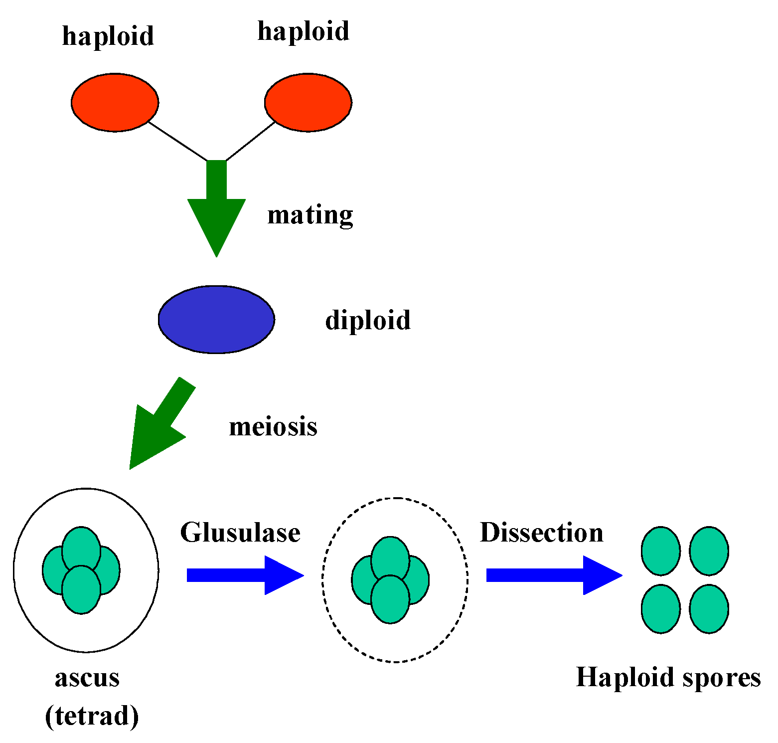
Sporulation and Dissection of Tetrads

The steps involved in mating of haploid strains to produce diploid cells, sporulation of diploids, and analysis of tetrads after meiosis are outlined in Figure 2. The yeast haploids of opposite mating types were crossed, and the resulting diploids were selected on supplemented SD media. A colony from the selected diploids was inoculated into sterile liquid YPAD medium and incubated with aeration at 30°C overnight. Cells were grown to stationary phase, then inoculated at a cell density of 1000 cells/ml into presporulation media containing 0.8% Bacto-yeast extract (Difco), 0.3% Bacto-peptone (Difco), 10%
Figure 2. Flow chart outlining the steps involved in meiosis and the production of ascospores in S. cerevisiae. Haploid strains of opposite mating types were mated to produce diploid cells. Diploid cells were then sporulated, and ascal cell walls were treated with the lytic enzyme, glusulase. The treated asci or tetrads were microdissected to release and analyze the four spore products of meiosis.
Ijms 04 00001 g002
glucose, and 0.16 mg/ml adenine sulfate. The culture was incubated at 23°C until the cells grew to a density of 1 x 10⁷ to 5 x 10⁷ cells/ml (mid-exponential growth). Cells were pelleted, washed twice, and resuspended into sporulation media containing 1% potassium acetate and the amino acids required for sporulation of the diploids. The tetrads obtained after sporulation were dissected using a micromanipulator (Singer Instruments Co. Ltd., Somerset, England) and incubated on solid YPAD at 30°C.

Zeocin Treatments

Zeocin is a 20-fold dilution of phleomycin, and was purchased from Invitrogen (Carlsbad, CA). Aliquots from the stock solution of Zeocin were added to the desired concentration in SD or YPAD media which had been sterilized and cooled to approximately 45°C. Zeocin medium was always prepared within 24 hours prior to use. Yeast strains listed in Table 1 were replica-plated on Zeocin media and grown for 3 days at 30°C to determine their susceptibility to killing by the drug.

E. coli and Yeast Transformations

E. coli transformation was carried out according the CaCl2 method described by Sambrook et al. [17]. Transformation of yeast cells was carried out using lithium acetate in the method of Ito and co-workers [18] and as modified by Rose [19] and Rose and Broach [20].

Results

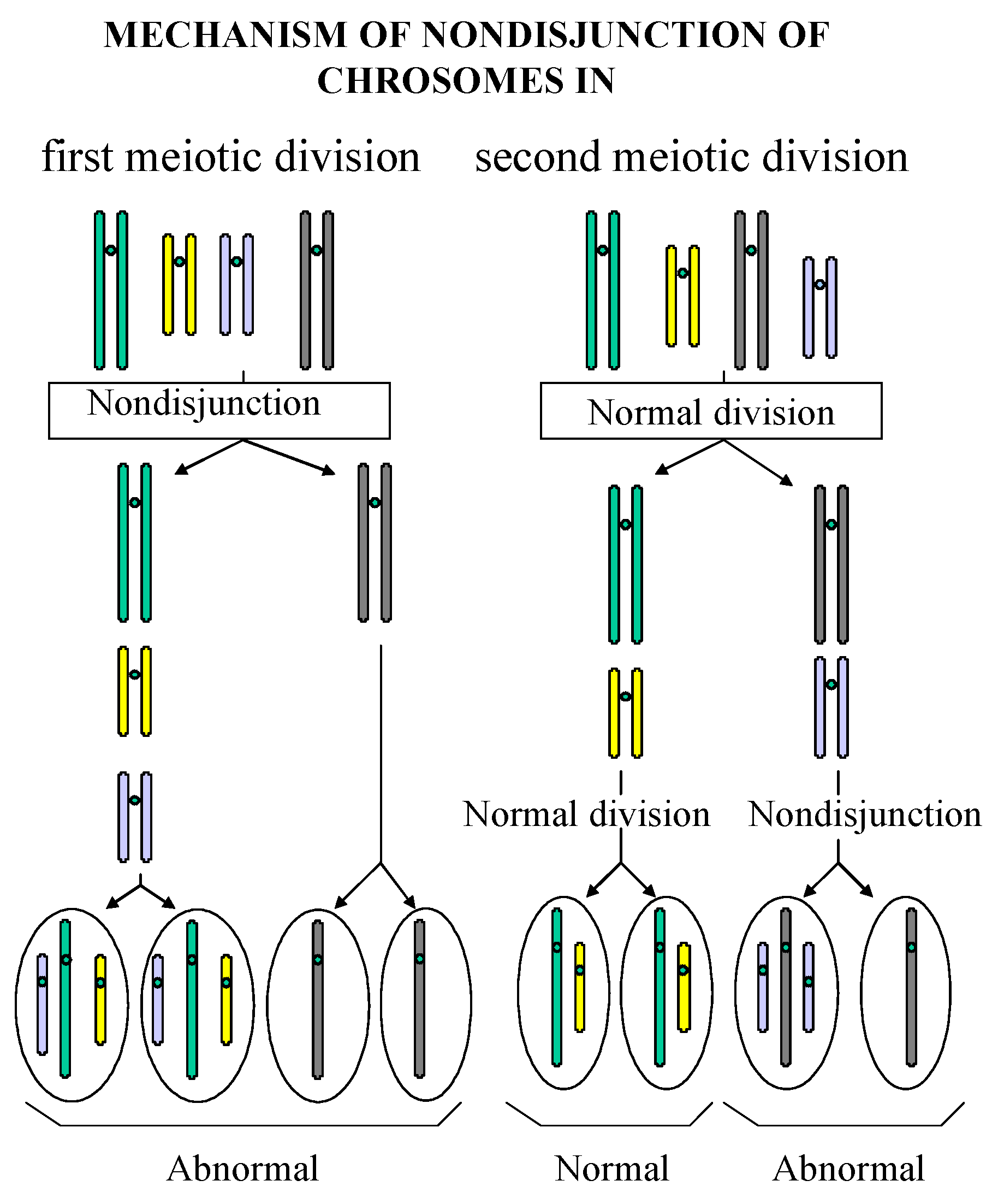
Reduced Viability of Spores from Homozygous Mutant Strains

The viability of spores from homozygous blm1-1/blm1-1 mutant strains was compared to that of spores from BLM1/BLM1 homozygous normal strains and heterozygous BLM1/blm1-1 strains. The homozygous normal diploid was created by crossing two wild-type haploid segregants, CM1457-79A and CM1457-79B (Table 1). The heterozygous diploid was created by crossing a wild-type haploid, CM1457-79A, to a mutant haploid, CM1457-79C. The homozygous mutant diploid was created by crossing two mutant haploids, CM1457-79C and CM1457-79D. Each diploid was sporulated, and the tetrads obtained from each sporulated diploid were dissected (Figure 2).
The viability of the spores in each tetrad is tabulated in Table 2. Ninety-four percent of the tetrads from the BLM1/BLM1 diploid and 100% of the tetrads from the BLM1/blm1-1 diploids produced four or three viable spores. In contrast, no tetrads from blm1-1/blm1-1 cells produced asci with four viable spores, and only 13% of the tetrads produced three viable spores. One- or two-spored viability was predominant. Thus, in the presence of one or two copies of the BLM1 gene (as in homozygous or heterozygous diploids), tetrads appear capable of completing meiosis and producing four viable spores. However, in the absence of the BLM1 gene, cells appear unable to complete meiosis and produce four viable spores. These results indicate that the BLM1 gene is essential for the viability of all four spores after meiosis, and that the blm1-1 mutation confers a deficiency in meiosis since the homozygous mutant cells are unable to produce four viable spores.
Table 2a. Viability of spores from BLM1/BLM1, BLM1/blm1-1, and blm1-1/blm1-1 diploid strains
Diploid TypeTotal number of tetrads dissectedNo viable spores1 viable spore2 viable spores3 viable spores4 viable spores
Homozygous normal1601069
Heterozygous1400059
Homozygous mutant31072040
Table 2b. Percentage of viable spores produced by each genotype
Diploid TypeTotal number of tetrads dissectedSporulation (%)1 viable spore (%)2 viable spores (%)3 viable spores (%)4 viable spores (%)
Homozygous normal1670±5.81603856
Heterozygous1466.3±4.51003664
Homozygous mutant3152.1±4.112365130
1Standard errors of the means were calculated from three to five replicates.

Mitotic Phenotype of blm1-1 Mutant Strains: Reduced Growth Rate and Elevated Frequencies of Irregularly-shaped Cells

Mutant blm1-1 strains were found to grow at reduced rates compared to the wild-type strains. At stationary phase, the titers of blm1-1 haploid strains were approximately 25% less than those of BLM1 cells (Table 3). During growth, the blm1-1 mutant cells also assumed peculiar shapes at high frequencies. When quantitated, approximately five percent of blm1-1 cells in stationary-phase populations were abnormally shaped. In addition, twice as many cells appeared swollen in blm1-1 populations than in BLM1 populations.
Table 3. Comparisons of Growth and Abnormally-shaped Cells in BLM1 and blm1-1 Strains 1
StrainsFinal Titers (cells/ml)Number and Percent of Irregularly- shaped Cells 2Normal-sized Single Cells and Budded Cells 3Number and Percent of Enlarged Cells 4Total Number of Cells
Means of experiments one and two
BLM1 (CM1457-79A) 5.00 x 1080%(462) 84.6%(84) 15.4%546 (100%)
Experiments three and four: means of both strains
blm1-1 (CM1457-79C and CM1457-79D)3.60 x 10⁸(28.5) 5.23%(350) 64.1%(167) 30.6%546 (100%)
Experiments five and six: means of both strains
blm1-1 (CM1457-79C and CM1457-79D)3.78 x 10⁸(29) 5.31%(347) 63.6%(170) 31.1%546 (100%)
1Each cell was classified based on the following criteria and relative sizes.
2Irregularly-shaped cells: Ijms 04 00001 i001.
3 Normal-sized single cells and budded cells: Ijms 04 00001 i002.
4Enlarged cells: Ijms 04 00001 i003.

Isolation of Plasmids, Restriction Analyses, and DNA Sequence Analyses

In addition to the meiotic defect, the blm1-1 mutation confers hypersensitivity to lethal effects of bleomycin [21]. Thus, this phenotype was used to isolate plasmids that relieved the drug hypersensitivity. Isolation of the pPM118 and pPM118-4 plasmids from a S. cerevisiae (S288C) genomic library was previously described [22]. A 32P-labelled 3.8 kb BamHI-ClaI fragment from pPM118 was used as a probe to screen the genomic library and isolate pPM118-4.
To identify the complete sequences of the inserts in pPM118 and pPM118-4, approximately 200 bases at each end of the inserts were sequenced by two custom-made primers originating from the vector sequences. These four end sequences were then used to search the nucleotide sequence databases through the National Center for Biological Information (NCBI) using the BLAST program [23]. These sequences were also used to search the SGD (Saccharomyces Genomic Database). The search results indicated the cloned sequence on pPM118 contains a region from chromosome VI between 133748 and 140147 bases, including the full MSH4 gene (Figure 3). Restriction maps, chromosomal origin and the overlapping region of pPM118 and pPM118-4 are shown in Figure 3, along with the identification and location of the open reading frame of the MSH4 gene.
Figure 3. Restriction maps of the inserts of the plasmids pPM118 and pPM118-4, and the location of the MSH4 gene. Restriction maps of the two chromosomal inserts are shown with their corresponding overlapping region. B: BamHI, C: ClaI, E: EcoRI, P: Pst I. Inserts originated from chromosome VI and the coordinates for the entire region cloned on pPM118 and pPM118-4 extends from 133748 to 141542 bases. Map of chromosomal features from SGD: VTC2 (brown), ARS605 (yellow), Msh4 (red), tRNA-Asn (green), Ty1LTR (pink), Ty2LTR (orange), Ty2 (purple), TyAGag (aqua), TyB (aqua).
Ijms 04 00001 g003

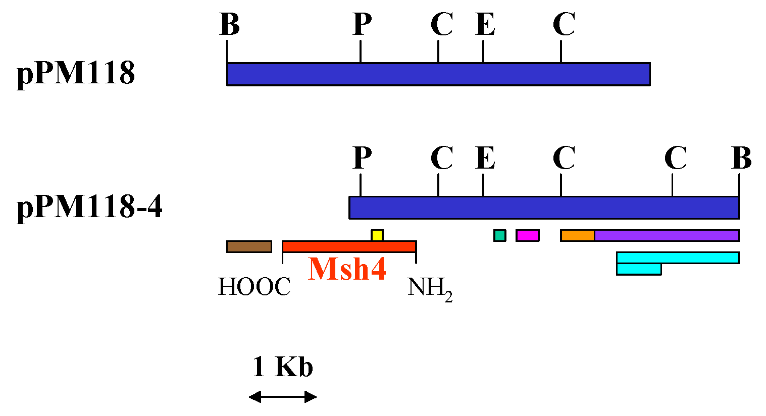
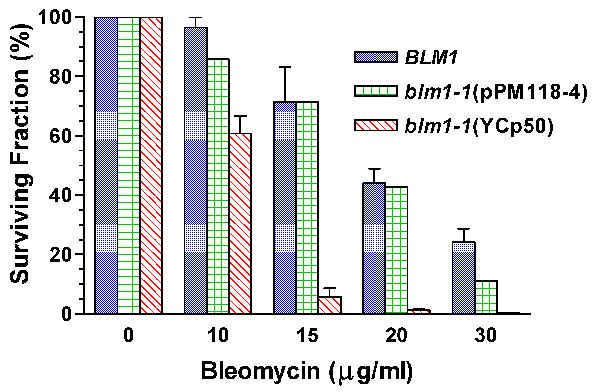
Relief of Drug Hypersensitivity

The relief of the hypersensitivity to killing by bleomycin conferred by the pPM118-4 plasmid is illustrated in Figure 4. For comparison, the figure also illustrates the resistance conferred by the wild-type or normal BLM1 gene and the hypersensitivity conferred by the mutant blm1-1 gene (mutant blm1-1 strains transformed with the YCp50 vector alone). The drug resistance conferred by pPM118-4 is comparable to the resistance of wild-type cells at all drug concentrations with the exception of the
Figure 4. A representative comparison of the resistance of normal (BLM1, CM1401-5B) and transformed blm1-1 mutant strains to lethal effects of bleomycin. The blm1-1 mutant strain, CM1401-5C, was transformed with the plasmid (pPM118-4) or with the YCp50 vector alone. The normal segregant and the transformed mutant strains were plated on supplemented SD medium containing various concentrations of bleomycin. Plates were incubated at 30°C for 3 days. The survival value of the last cell is 0.3±0.2%.
Ijms 04 00001 g004
very high concentration of 30 μg/ml bleomycin. The resistance provided by pPM118-4 is also high at 30 μg/ml bleomycin, though somewhat less than wild-type cells. There are several possible reasons for the somewhat decreased resistance relative to wild-type cells at the highest drug dose. For example, if MSH4 is a suppressor of the blm1-1 phenotype, rather than the same gene as BLM1, it may not be able to fully relieve the drug hypersusceptibility of mutant cells at high drug concentrations. Second, whether MSH4 and BLM1 are or are not the same gene, the full gene may be required on the plasmid to obtain full complementation. Third, the encoded protein may not be produced as efficiently from the plasmid as it is from the chromosome. Fourth, the region of the gene on pPM118 may contain an alteration.

Genetic Mapping Showed that MSH4 is not the BLM1 Structural Gene

The MHS4 gene has one function that is similar to that of the BLM1 gene — both genes are required in meiosis for wild-type levels of spore viability in S. cerevisiae. Thus, to determine if BLM1and MSH4 are the same gene, linkage analysis was carried out by genetic mapping to determine if MSH4 is linked to (and thus could be) the BLM1 gene.
A mutant strain, PS593/6H, with the LEU2 marker replacing nucleotides 168-2290 of the MSH4 coding region [15], was kindly provided by Dr. Nancy Hollingsworth (Department of Biochemistry and Cell Biology, State University of New York at Stony Brook, Stony Brook, NY). This strain was crossed to a leu2/blm1-1 strain, CM1457-79C. The resulting diploid was genotypically MSH4/msh4::LEU2, BLM1/blm1-1. The LEU2 gene was used to track the msh4 gene in linkage analysis, and Zeocin hypersensitivity was used to track the blm1-1 mutation. If the two genes are the same, the genotypes in the tetrad segregants would be one or the other of the parental ditypes (PD). That is, there will be 2:2 segregation of LEU2 (leucine prototrophy) with BLM1 (wild-type or normal drug resistance), and leu2 (leucine auxotrophy) with the blm1-1 mutation (drug hypersensitivity).
Segregation of BLM1, blm1-1, MSH4 and msh4::LEU2 in meiosis is shown in Table 4. The segregation pattern from the tetrad analysis of the cross PS593/6H x CM1457-79C revealed that 35.7% of the tetrads were PD, 42.9% of the tetrads were non-parental ditypes (NPD; the genotypes of all four spores are different from the genotypes of the parental haploids), and 21.4% were tetratypes (TT; two spores are parental genotypes and two spores are not). For two genes to be linked, the ratio of PD: NPD: TT is >1: <1:0 [24]. The ratio obtained from this assay is <1:1<1. Actually, the ratio of each genotype among the total spores is close to 1:1:1:1, consistent with random assortment of each gene on its respective chromosome. Thus, the linkage analysis indicates that MSH4 and BLM1 are different genes.
A master plate containing solid YPAD medium and inoculated with parental strains, all the spores from dissected tetrads, and positive controls were created. Cells from this plate were replica-plated on SD+7 media lacking leucine and on complete media containing Zeocin. The MSH4 gene was tracked by the wild-type LEU+ phenotype from PS593/6H, and the BLM1 phenotype was introduced into the cross by PS593/6H.
Table 4. Analysis of tetrad segregation of CM1457-79C (blm1-1,leu2) x PS593/6H (msh4::LEU2, BLM1). (The number of spores of each genotype is tabulated. Only results from four-spored asci are tabulated)
Classes of tetradsNumber of tetradsNumber of sporesblm1-1, leu2BLM1, LEU2BLM1, leu2blm1-1, LEU2
Parental Ditype (PD)520101000
Non-Parental Ditype (NPD)624001212
Tetratype (TT)3123333
Total spores145613131515

Discussion

Analyses of the meiotic and mitotic phenotypes conferred by the blm1-1 mutation indicate that in addition to the bleomycin and phleomycin hypersusceptibility, the blm1-1 mutation confers defects in meiosis and mitosis. These results have not previously been reported. It is not known at the present time what mechanism accounts for the low spore viability after meiosis in homozygous blm1-1/blm1-1 mutant diploid cells. Compared to the homozygous normal BLM1/BLM1 and heterozygous BLM1/blm1-1 diploid strains, sporulation was not dramatically lower in the homozygous mutant cells (Table 2). But the fact that only one or two spores from 88% of the tetrads survived indicates some step in meiosis is likely to be defective. One possibility for the defect is that nondisjunction of one or more chromosomes occurs at high frequency during meiosis in the mutant diploids. A failure of chromosomes to properly separate could occur at either the first or second meiotic division, as shown in Figure 5. If the nondisjunction were limited to a specific chromosome, some spores would receive an extra chromosome and some spores would not receive that chromosome (Figure 5). If the nondisjunction were not limited to a single chromosomal pair, different homologous chromosomes would fail to properly separate during meiosis. Either situation could lead to defective meiotic products and an inability to produce four viable spores. Nondisjunction events during mitosis might account for the blm1-1/blm1-1 mitotic phenotypes of reduced growth rates and high frequencies of abnormal cell morphologies. The decreased growth rates may actually reflect the inviability of some of the cells during mitotic growth.
Figure 5. An illustration of the fates of two chromosomal pairs during meiosis. One pair of homologous chromosome behaves normally as it goes through meiosis I and meiosis II. The other pair does not disjoin in either the first or the second meiotic division.
Ijms 04 00001 g005
Disturbance of the structural components of the synaptonemal complex in meiosis can lead to nondisjunction of homologous chromosomes [25]. Although the relationship between the Blm1p and nondisjunction is unknown, it is known that Msh4p functions in the formation of the synaptonemal complex [15]. The MSH4 gene in S. cerevisiae encodes a protein involved in crossing over between chromosomes in meiosis, and the gene is not transcribed in mitosis [25]. Recently, Msh4p was shown to function in chromosomal synapsis in meiosis, and regulate the distribution of meiotic crossovers along chromosomes [15]. The protein localizes to discrete sites in meiotic chromosomes when synapsis initiates, and promotes formation of the synaptonemal complex [15].

Acknowledgements

We thank Dr. Nancy Hollingsworth for graciously providing strains for these studies, Bristol-Myers Squibb Company for providing Blenoxane for these experiments, and Mr. Ronald Edwards for his assistance.
This work was supported by The National Science Foundation, Grants #9874878 and #HRD-9628726, Aaron Diamond Foundation, National Institutes of Health (including the RCMI AIDS Infrastructure [Grant #G12RR03060], Support of Continuing Research Excellence [Grant #2 SO6 GM08168-19], and Minority Biomedical Research Programs), City University of New York Medical School/Sophie Davis School of Biomedical Education, and the College Research Scholarship Program of City College of the City University of New York.

References

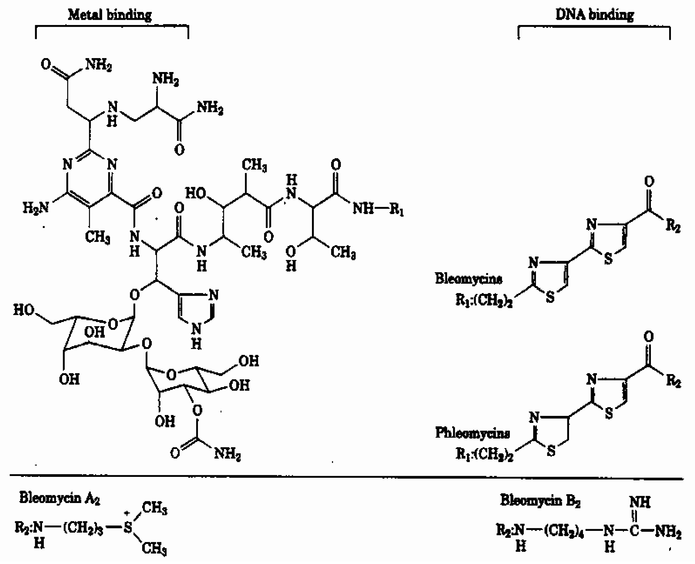
  1. Moore, C. W. Internucleosomal cleavage and chromosomal degradation by bleomycin and phleomycin in yeast. Cancer Res. 1988, 48, 6837–6843. [Google Scholar] [PubMed]
  2. Beaudouin, R.; Lim, S. T.; Steide, J. A.; Powell, M.; McKoy, J.; Pramanik, A. J.; Johnson, E.; Moore, C. W.; Lipke, P. N. Bleomycin affects cell wall anchorage of mannoproteins in Saccharomyces cerevisiae. Antimicrob. Agents Chemother. 1993, 37, 1264–1269. [Google Scholar] [CrossRef] [PubMed]
  3. Burger, R. M. Cleavage of nucleic acids by bleomycin. Chemical Reviews 1998, 98, 1153–1170. [Google Scholar] [CrossRef] [PubMed]
  4. Petering, D. H.; Byrnes, R. W.; Antholine, W. E. The role of redox-active metals in the mechanism of action of bleomycin. Chem. Biol. Interact. 1990, 73, 133–182. [Google Scholar] [CrossRef] [PubMed]
  5. Steighner, R. J.; Povirk, L. F. Bleomycin-induced DNA lesions at mutational hot spots: implications for the mechanism of double-strand cleavage. Proc. Natl. Acad. Sci. U. S. A. 1990, 87, 8350–8354. [Google Scholar] [CrossRef] [PubMed]
  6. Moore, C. W. Responses of radiation-sensitive mutants of Saccharomyces cerevisiae to lethal effects of bleomycin. Mutat. Res. 1978, 51, 165–180. [Google Scholar] [CrossRef] [PubMed]
  7. Moore, C. W. Control of in vivo (cellular) phleomycin sensitivity by nuclear genotype, growth phase, and metal ions. Cancer Res. 1982, 42, 929–933. [Google Scholar] [PubMed]
  8. Moore, C. W. Ligase-deficient yeast cells exhibit defective DNA rejoining and enhanced gamma ray sensitivity. J. Bacteriol. 1982, 150, 1227–1233. [Google Scholar] [PubMed]
  9. Moore, C. W. Degradation of DNA and structure-activity relationship between bleomycins A2 and B2 in the absence of DNA repair. Biochemistry 1990, 29, 1342–1347. [Google Scholar] [CrossRef] [PubMed]
  10. Moore, C. W. Bleomycin. In Encyclopedia of Molecular Biology; Creighton, T., Ed.; John Wiley and Sons, Inc.: New York, 1999; pp. 292–297. [Google Scholar]
  11. Moore, C. W. Isolation and partial characterization of mutants of Saccharomyces cerevisiae altered in sensitivities to lethal effects of bleomycins. J. Antibiot. (Tokyo) 1980, 33, 1369–1375. [Google Scholar] [CrossRef] [Green Version]
  12. Moore, C. W. Further characterizations of bleomycin-sensitive (blm) mutants of Saccharomyces cerevisiae with implications for a radiomimetic model. J. Bacteriol. 1991, 173, 3605–3608. [Google Scholar] [PubMed]
  13. Ross-Macdonald, P.; Roeder, G. S. Mutation of a meiosis-specific MutS homolog decreases crossing over but not mismatch correction. Cell 1994, 79, 1069–1080. [Google Scholar]
  14. Eisen, J. A. A phylogenomic study of the MutS family of proteins. Nucleic Acids Res. 1998, 26, 4291–4300. [Google Scholar] [CrossRef] [PubMed]
  15. Novak, J. E.; Ross-Macdonald, P. B.; Roeder, G. S. The budding yeast Msh4 protein functions in chromosome synapsis and the regulation of crossover distribution. Genetics 2001, 158, 1013–1025. [Google Scholar] [PubMed]
  16. Johnston, M.; Davis, R. W. Sequences that regulate the divergent GAL1-GAL10 promoter in Saccharomyces cerevisiae. Mol. Cell Biol. 1984, 4, 1440–1448. [Google Scholar] [PubMed]
  17. Sambrook, J.; Fritsch, E. F.; Maniatis, T. Oligotide-Mediatede Mutagenesis by Selection Against Template Strands That Contain Uracil (Kunkel Method). In Molecular Cloning: a Laboratory Manual; Ford, N., Nolan, C., Ferguson, M., Eds.; Cold Spring Harbor Laboratory Press: Cold Spring Harbor, NY, 1989; pp. 15.74–15.79. [Google Scholar]
  18. Ito, H.; Fukuda, Y.; Murata, K.; Kimura, A. Transformation of intact yeast cells treated with alkali cations. J. Bacteriol. 1983, 153, 163–168. [Google Scholar] [PubMed]
  19. Rose, A. H. Isolations of Genes by Complementation in Yeast. In Methods in Enzymology; Academic Press: New York, 2002; pp. 481–504. [Google Scholar]
  20. Rose, M. D.; Broach, J. R. Cloning Genes by Complementation in Yeast. In Methods in Enzymology; Academic Press: New York, 1991; pp. 195–230. [Google Scholar]
  21. Moore, C. W. Further characterizations of bleomycin-sensitive (blm) mutants of Saccharomyces cerevisiae with implications for a radiomimetic model. J. Bacteriol. 1991, 173, 3605–3608. [Google Scholar] [PubMed]
  22. Pramanik, A.; Martinez, M.; McKoy, J. F.; Ospina, R.; Daly, J.; Moore, C. W. A mutant approach and molecular strategy to study fungal cell walls. Cell. Mol. Biol. (Noisy. -le-grand) 1995, 41 Suppl 1, S73–S81. [Google Scholar]
  23. Altschul, S. F.; Gish, W.; Miller, W.; Myers, E. W.; Lipman, D. J. Basic local alignment search tool. J. Mol. Biol. 1990, 215, 403–410. [Google Scholar] [CrossRef] [PubMed]
  24. Kaiser, C.; Michaelis, S.; Mitchell, A. Meiotic Mapping. In Methods in Yeast Genetics: a Cold Spring Harbor Laboratory Course Manual; Cold Spring Harbor Laboratory Press, 1994; pp. 25–35. [Google Scholar]
  25. Roeder, G. S. Meiotic chromosomes: it takes two to tango. Genes Dev. 1997, 11, 2600–2621. [Google Scholar]

