Article

Expression of Myosin Light Chain Kinase in Kidney of Streptozotocin-Induced Diabetic Rats

1
Institute of Clinical Pharmacology, Anhui Medical University, Hefei 230032, Anhui, P.R.China
2
Laboratory of Molecular Biology and Department of Biochemistry, Anhui Medical University, Hefei 230032, Anhui, P.R.China
3
Department of Respiratory Disease, Anhui Medical University, Hefei 230032, Anhui, P.R.China
4
Anhui Province Key Laboratory of Genomic Research and the Key Laboratory of Gene Resource Utilization for Severe Disease (Anhui Medical University ), Ministry of Education P.R.China, Hefei 230032, Anhui, P.R.China
*
Author to whom correspondence should be addressed.
Int. J. Mol. Sci. 2006, 7(11), 510-518; https://doi.org/10.3390/i7110510
Submission received: 2 October 2006 / Accepted: 13 November 2006 / Published: 21 November 2006
(This article belongs to the Special Issue Interaction of Biological Molecules)

Abstract

:
Nephropathy is one of the most common complications of diabetes mellitus which remains incompletely understood. We reported the expression of myosin light chain kinase (MLCK) in the kidney of diabetic rats and investigated the correlation between MLCK and diabetic nephropathy by observing the expression of MLCK. The diabetic model rats were induced by an intraperitoneal injection of streptozotocin (STZ) and the insulin-treated rats were subcutaneously injected with protamine zine insulin 3u/d. The kidneys were excised and immersed in 4% polyoxymethylene after 12 weeks later. The expression of MLCK was analyzed by immunohistochemical staining and Western blot. Immunohistochemical analysis and Western blot assay indicated that the MLCK expression was higher in kidney of diabetic rats than that in control and it was decreased in kidney of insulin-treated rats. Our results suggested that the over expression of MLCK may be related with the development of diabetic nephropathy.

1. Introduction

Diabetes is a complex disorder resulting in large and small vessel disease and impaired organ function, which is characterized by hyperglycemia, a relative or absolute lack of insulin, an inclination to vascular disease and nephropathy. Diabetic nephropathy is characterized by thickening of basement membranes and mesangial expansion with progression into glomerulosclerosis, tubular atrophy and interstitial fibrosis, ultimately resulting in renal failure [1,2,3,4]. A wide variety of mechanisms in the pathogenesis of diabetes have been proposed, including accumulation of nonenzymatic glycated end products in the kidney, oxidation of renal glycoproteins by reactive oxygen species, intracellular accumulation of sorbitol generated by the reduction of glucose by aldose reductase, involvement of mitogen-activated protein kinase and growth factors [5,6,7,8]. The diverse mechanisms indicates that a vast number of molecules and different signal transduction pathways are involved in its pathogenesis [9,10]. But, the mechanism of diabetic nephropathy remains incompletely understood.
Studies have demonstrated altered papillary muscle mechanics and changes in contractile proteins in the alloxan-induced diabetic rabbit model. Diminished velocity of shortening, an increased duration of isometric contraction–relaxation and prominent abnormalities of contractile proteins could be reversed by insulin [11]. Studies also have documented that myocardial performance is impaired in the renal of chronically diabetic rats and rabbits. Abnormalities of the contractile and regulatory proteins could be responsible for the mechanical defects in streptozotocin (STZ)-diabetic renal. The major research on contractile proteins in the diabetic state has focused on myosin ATPase and its isoenzymes [1,12]. The calmodulin-dependent myosin light chain kinase (MLCK), which transfers the γ-phosphate from ATP to myosin is essential for contraction [13,14,15]. Some studies have shown the relationship between MLCK and vessel diseases [16], while few reports concerned to the association between MLCK and diabetic nephropathy. Here, we report the relationship between MLCK and diabetes nephropathy and investigate the variation of MLCK in the development of diabetes.

2. Materials and Methods

2.1 Animal and experiment design

Male Sprague-Dawley rats (180–200g body weight) were obtained from Nanjing Animal Centre (Nanjing, China) and kept in accordance with our Institutional Animal Care Committee guidelines. Diabetes mellitus model was induced by an intraperitoneal injection of STZ at a dose of 65 mg/kg body weight in a citrate buffer (pH4.5). Rats in the control group were injected with saline vehicle. Tail vein blood glucose levels were measured in 48h after injection. The diabetic model was believed to been established according to its blood glucose which was greater than 16.7mmol/L and kept for at least 5 days. The diabetic rats were randomly divided into diabetic group and insulin-treated group. The insulin-treated rats were subcutaneously injected with protamine zine insulin 3u/d for 12 weeks. Throughout the study the rats were maintained on standard laboratory diet with free access to water. All rats were sacrificed after 12 weeks.

2.2 Tissue sample and isolation.

After collecting blood samples from common carotid artery to determinate glucose level, fresh kidney tissue was isolated and washed with cold Hanks solution (137mM NaCl, 5.0mM KCl, 1.3mM CaCl2, 0.8mM MgSO4●7H2O, 0.6mM Na2HPO4, 0.4mM KH2PO4, 3.0mM NaHCO3, 5.6mM glucose, pH7.4) and immersed in 4% polyoxymethylene. The fixed kidneys were embedded in paraffin for immunohistochemical and H&E staining. The rest tissues were cut into small slices and homogenized or stored at –80°C for Western blot. Blood Glucose (Glu), Urine Protein (urine pro), Urea Nitrogen (Bun) and creatinine (Cr) levels were measured enzymatically using an autoanalyser.

2.3 Immunohistochemistry

The kidney tissue were sectioned at an average thickness of 6 μm and stored at -20 ºC until use. The sections were blocked with 5% BSA (Sigma), 5% normal rabbit serum (Vector Laboratories, Burlingame, CA, USA) and incubated with the anti-MLCK monoclonal antibodies overnight, followed by incubation with 1:200 diluted HRP-conjugated goat antibody against rabbit IgG and treated with a metal-enhanced 3, 3’-diaminobenzidine (DAB; Pierce, Rockford, IL, USA) for 3 min. After a thorough washing, the sections were mounted on slide glasses with Vectashield (Vector Laboratories, Burlingame, CA, USA). Section and its integral absorbance of MLCK expression were examined with microscope.

2.4 Western blot analysis

The kidney tissue were washed three times in PBS, and then lysed in RIPA buffer (1% Nonidet P-40, 1% sodium deoxycholate, 0.1% SDS, 150mM NaCl, 10mM sodium phosphate buffer pH 7.2, 2mM EDTA, 10mg/ml aprotinin, 10mg/ml leupeptin, 2 mM PMSF, 2 mM sodium orthovanadate, 10 mM sodium pyrophosphate, and 20 mM sodium fluoride). The lysates were centrifuged at 15000 ×g for 30 min at 4°C, the supernatants were used for western blot. The total protein concentration of each sample was measured by using MicroBCA Protein Assay Reagent Kit (Pierce, Rockford, IL, USA). The same amount of lysate from each line in SDS sample buffer was with 10% SDS-polyacrylamide gel electrophoresis and electroblotted onto a PVDF membrane, which was then blocked with 5% fat-free milk in PBST (PBS, 0·1% Tween 20) for 1h at room temperature. Anti-MLCK monoclonal antibody (1:1000 dilution) was incubated overnight at 4°C, followed by incubation with 1:20 diluted HRP-conjugated goat antibody against rabbit IgG, and stained with enhanced chemiluminescence reagent (Pierce, Rockford, IL, USA). Densitometric scanning of the exposed X-ray film was used for semi-quantitative measurement of the protein bands. Three independent experiments were performed, and the results were reproducible.

2.5 Statistical analysis

Statistical analyses were carried out using unpaired Students t-test with SPSS 8.0. All data is expressed as mean ± standard error of the mean (S.E.M.). Statistical significance was accepted at a level of p<0.05.

3. Results

3.1 Experimental animal model

The diabetic rat model was established by an intraperitoneal injection of STZ. The blood glucose of the rats was higher than 16.7 mM, whereas untreated rats had normal levels. The elevated level of blood glucose in STZ-injected rats was maintained during the entire experimental period.

3.2 The general character

Blood glucose of STZ-treated rats was 27.79±9.28 mM and higher than control rats (6.49±0.79 mM) (P<0.01). Body weight of the STZ-treated rats (156.50±10.13g) was lower than control rats (367.33±26.73 g). After being treated with insulin, blood glucose decreased to 8.75±2.56 mM and 3.0-fold less than that in diabetic rats (P<0.01). Body weight of insulin treated was 354.67±31.94g and was no difference compared with control group. The diabetic rats had a decrease of kidney weight to 1.10±0.12g compared with 1.40±0.32g in control (P<0.01). Urine Pro of STZ-treated rats was 21.8±2.76 (mg/24h) and higher than control rats (6.87±0.23mg/24h) (P<0.01). Similar results of Cr and BUN were obtained (81.68±12.43µM and 19.27±2.39mM tended to 61.56±13.42µM and 9.31±1.64mM). However, after treated with insulin , the levels of Pro, Cr, BUN decreased to 12.1±1.32mg/24h, 74.36±11.32µM and 13.26±2.71 mM respectively (Table 1).
Table 1. The effects of streptozotocin-diabetes and insulin treatment on the blood glucose, Body weight , Pro, Cr , BUN and Kidney weight levels in the rats( mean ± S.D)*P<0.05 **P<0.01 significance relative to controls and ▲ P<0.01 significance relative to diabetic group.
BloodControlDiabeticDiabetic + Insulin
Glucose (mM)6.49±0.7927.7±9.28**8.75±2.56* ▲
Body weight (g)367.33±26.73156.50±10.13**354.67±31.94 ▲
kidney weight (g)1.40±0.321.10±0.12** 1.27±0. 25*
Urine pro (mg/24h)6.87±0.2321.8±2.76**12.1±1.32 ▲
Cr (µM)61.56±13.4281.68±12.43**74.36±11.32* ▲
BUN (mM)9.31±1.6419.27±2.39**13.26±2.71* ▲

3.3 Tissue staining

With the development of diabetes mellitus, evident morphological changes were observed and diabetic glomerulosclerosis became worse. After treated with insulin, the volume of glomeruli returned to normal level (Figure 1).
Figure 1. H&E staining was performed in glomerulus and renal tubule from control, streptozotocin-diabetes and insulin-treated rats. A: control group ;B: diabetic group C: diabetic and insulin-treated group.
Ijms 07 00510 g001

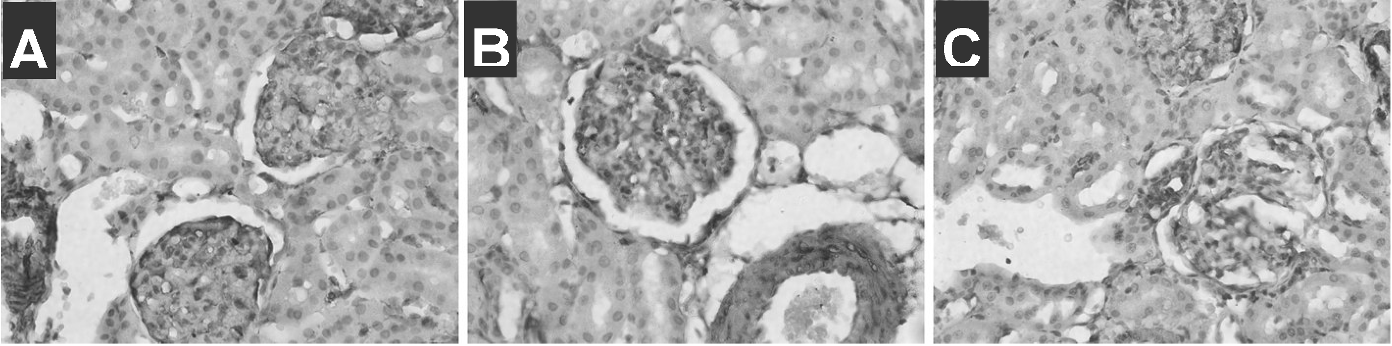
3.4 Immunohistochemistry examination

MLCK was expressed in glomerulus, renal tubule and glomerular arteriolar in control by immunohisochemistry. The expression of MLCK in glomerulus of STZ-treated rats was higher than that in control. After treated with insulin, the expression of MLCK in glomerulus was a little lower than that in control. The change of integral absorbance of MLCK expression in gomenulus also indicated that results (Figure 2, Table 2).
Figure 2. Immunohistochemistry analysis of MLCK was performed in glomerulus and renal tubule from control, streptozotocin-diabetes and insulin-treated rats. Immunohistochemistry analysis were prepared using a anti-MLCK monoclonal antibody as described in METHODS. A: control group; B: diabetic group C: diabetic and insulin-treated group
Ijms 07 00510 g002
Table 2. The effects of streptozotocin-diabetes and insulin treatment on the integral absorbance in gomenulus of kidney ( mean ± S.D)**P<0.01 significance relative to controls and ▲ P<0.01 significance relative to diabetic group.
GroupnMLCK(absorbance)
Negative control613±7
Control683±7 **
Diabetic6120±28 **
Diabetic+ Insulin658±9 ** ▲

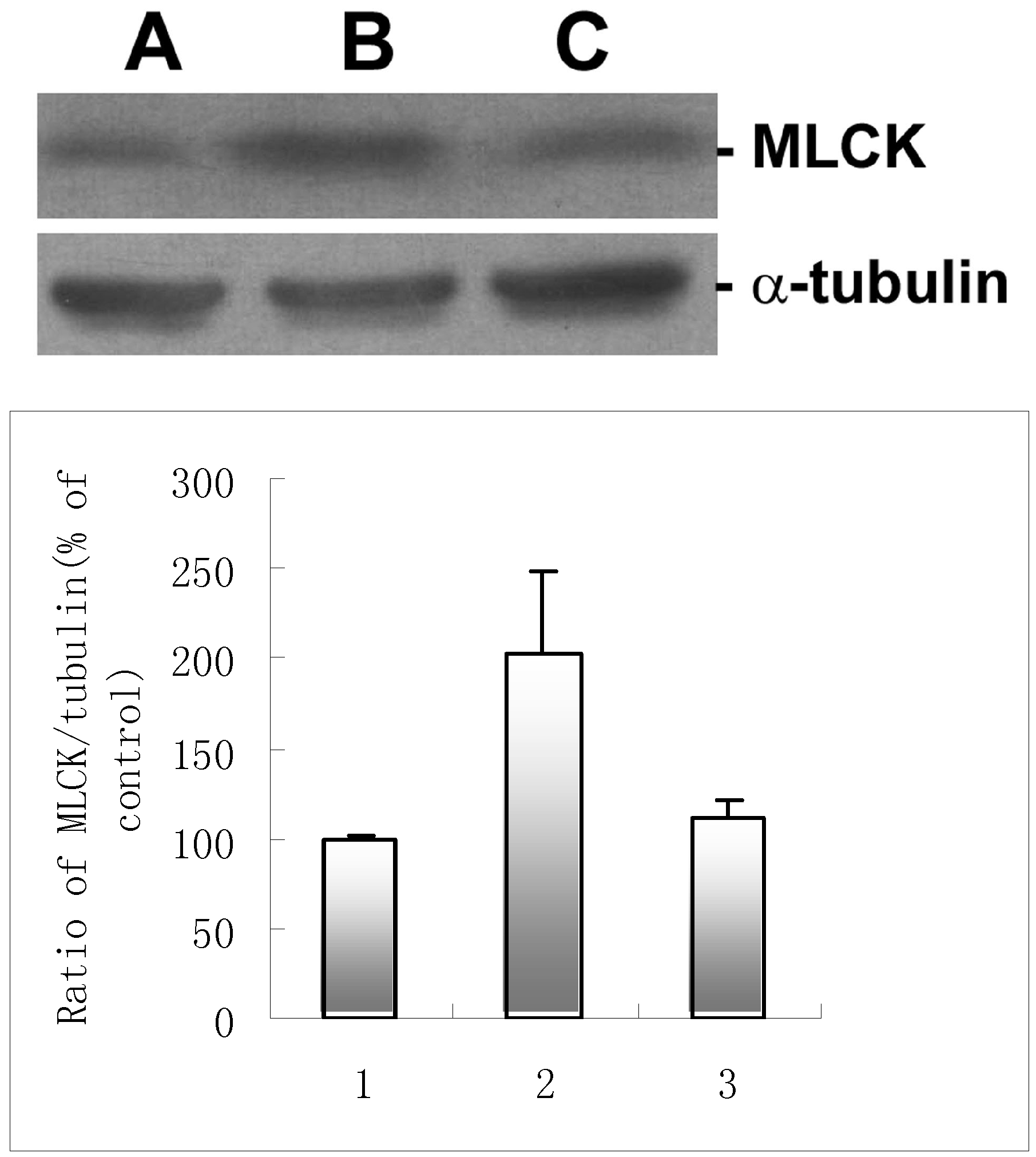
3.5 Western blot examination

The expression of MLCK in kidney of STZ-treated rats (203±45) was higher than that in control (100). After treated with insulin, the expression of MLCK in kidney (112±9) was a little higher than that in control. Quantitative analysis of this data by densitometric scanning normalized to the internal α-tubulin control is shown (Figure 3).
Figure 3. Western blot analysis of MLCK was performed in kidney of from control, streptozotocin-diabetes and insulin-treated rats. Kidney homogenates were prepared as described in METHODS. Western blot analysis was performed using a anti-MLCK and α-tubulin monoclonal antibody . Laser scanning densitometry was conducted to quantitata the differences. Densitometric units of the control group(MLCK/ α-tubulin) were set to 100. the internal α-tubulin control is also shown. A: control group ;B: diabetic group C: diabetic and insulin-treated group
Ijms 07 00510 g003

4. Discussion

MLCK is a Ca2+ /calmodulin activated enzyme in the kinase family which catalyses the phosphorylation of the 20-kD myosin light chain (MLC-20) [17,18]. In skeletal muscle, phosphorylation of the MLC-20 correlates with potentiated twitch tension after repetitive stimulation [19]. In smooth muscle cells, this phosphorylation leads to an increase in actomyosin ATPase activity and contraction which appears to be required for initiation of contraction [20]. Phosphorylation of MLC-20 by smooth muscle MLCK is a key event initiate to smooth muscle contraction. Although the roles of MLCK in non-muscle cells are not well defined, a variety of morphological changes such as cellular motility and organelle movement occur concurrently with increasing in cytoplasmic Ca2+ levels, light chain phosphorylation and activation of MLCK [21,22].
Myosin, one of the major contractile proteins in muscle cells as well as nonmuscle cells, consists of two heavy chains and two light chains. Myosin heavy chain (MHC) isoforms (SM1, SM2 and SMemb) are not only determination of the contractile properties of muscles, such as velocity of shortening or the economy of force production. But they are also molecular markers for muscle development and disease. The previous study showed that strong mesangial staining of SMemb was observed in cases with diffuse glomerulosclerosis [23]. The myosin isoenzymes demonstrate marked shifts in rodent hearts during pathologic states such as hypertensive hypertrophy, diabetes, myocardial infarction and increasing age. Myosin ATPase activity is one of the mechanisms in the contractile protein system that might be a controlling factor. Among other potential contractile proteins in diabetes, we are interested in the role of MLCK in the pathogenesis of kidney disease. In this study, we focus on the expression of MLCK with regard to its relevance to diabetic nephropathy.
In present study, diabetic model was established by an intraperitoneal injection of STZ, commonly used to induce diabetic. It has been shown that injection of STZ is associated with the development of diabetes as previously reported [24,25]. Following the development of Diabetes, evident morphology was changed. This study also showed that the expression of MLCK was positive in glomerulus, renal tubule and glomerular arteriolar. In glomerulus of STZ-treated rats the expression of MLCK was higher than that in control. The results indicated that the expression of MLCK is correlated with diabetic. From those results we proposed that the changes of mesangial expansion, glomerulosclerosis, tubular atrophy, and interstitial fibrosis may be associated with the increase of the expression of MLCK. MLCK catalyses the phosphorylation of MLC,which is associated with the gap formations and hyperpermeability of renal cells. Breakdown of this cell, which is to serve as a barrier to fluid and solute flux across the blood vessel wall, leads to increased permeability and the development of diabetic. In the study, it is found that MLCK expression increased more and more when diabetic was initiated and provided a circumstance of diabetic formation. It appears that among the molecular events the expression of MLCK may play a crucial role in the protection against diabetes. Treatment of diabetic rats with insulin, which has been reported to reverse the decreased myofibrillar and myosin ATPase activities and heart dysfunction, was observed to reverse the increased level of MLCK. This can be seen to provide evidence that increased MLCK may represent one of the mechanisms associated with mesangial expansion and glomerulosclerosis in diabetic nephropathy.
Since the importance of mitogen-activated protein kinase (MAPK) was reported in many studies. MLCK, which can be directly activated by MAPK, appears involved [26,27,28]. MLCK activation was associated with increase in MLC phosphorylation which is related with diabetic nephropathy. Among the molecular events it is plausible that activation of MAPK indirectly increase MLCK expression may play a crucial role.

5. Conclusion

In summary, the expression of MLCK may be involved in the development of diabetic nephropathy. The expression was higher in kidney of diabetic rats than that in control and it was decreased in kidney of insulin-treated rats. It should be noted that the exact mechanism to induce MLCK expression was not known which remains to be elucidated in our future study.

Acknowledgements

This study was Supported by National Natural Science Foundation of China (No. 03570750, 39870324), Grant for Excellent Young Teachers of Ministry of education of China and Natural Science Foundation of anhui provincial education department (No. 2004KJ211).

References and Notes

  1. Malhotra, A.; Sanghi, V. Regulation of contractile proteins in diabetic heart. Cardiovasc Res. 1997, 34, 34–40. [Google Scholar] [CrossRef]
  2. Tsao, T.S.; Stenbit, A.E.; Factor, SM.; Chen, W.; Rossetti, L.; Charron, M.J. Prevention of insulin resistance and diabetes in mice heterozygous for GLUT4 ablation by transgenic complementation of GLUT4 in skeletal muscle. Diabetes. 1999, 48, 775–782. [Google Scholar] [CrossRef]
  3. Voziyan, P.A.; Metz, T.O.; Baynes, J.W.; Hudson, B.G. A post-Amadori inhibitor pyridoxamine also inhibits chemical modification of proteins by scavenging carbonyl intermediates of carbohydrate and lipid degradation. J Biol Chem. 2002, 277, 3397–3403. [Google Scholar] [CrossRef]
  4. Feliers, D.; Duraisamy, S.; Faulkner, J.L.; Duch, J.; Lee, A.V.; Abboud, H.E. Activation of renal signaling pathways in db/db mice with type 2 diabetes. Kidney Int. 2001, 60, 495–504. [Google Scholar] [CrossRef]
  5. Gooch, J.L.; Tang, Y.; Ricono, J.M.; Abboud, H.E. Insulin-like growth factor-I induces renal cell hypertrophy via a calcineurin-dependent mechanism. J Biol Chem. 2001, 276, 42492–42500. [Google Scholar] [CrossRef]
  6. Reddy, M.A.; Thimmalapura, P.R.; Lanting, L.; Nadler, J.L.; Fatima, S.; Natarajan, R. The oxidized lipid and lipoxygenase product 12(S)-hydroxyeicosatetraenoic acid induces hypertrophy and fibronectin transcription in vascular smooth muscle cells via p38 MAPK and cAMP response element-binding protein activation. Mediation of angiotensin II effects. J Biol Chem. 2002, 277, 9920–9928. [Google Scholar] [CrossRef]
  7. Natarajan, R.; Gerrity, R.G.; Gu, J.L.; Lanting, L.; Thomas, L.; Nadler, J.L. Role of 12-lipoxygenase and oxidant stress in hyperglycaemia-induced acceleration of atherosclerosis in a diabetic pig model. Diabetologia. 2002, 45, 125–133. [Google Scholar] [CrossRef]
  8. Chen, S.; Cohen, M.P.; Lautenslager, G.T.; Shearman, C.W.; Ziyadeh, F.N. Glycated albumin stimulates TGF-beta 1 production and protein kinase C activity in glomerular endothelial cells. Kidney Int. 2001, 59, 673–681. [Google Scholar] [CrossRef]
  9. Sun, L.; Pan, X.; Wada, J.; Haas, C.S.; Wuthrich, R.P.; Danesh, F.R. Isolation and functional analysis of mouse UbA52 gene and its relevance to diabetic nephropathy. J Biol Chem. 2002, 277, 29953–29962. [Google Scholar]
  10. Kondo, T.; Kahn, C.R. Altered insulin signaling in retinal tissue in diabetic states. J Biol Chem. 2004, 279, 37997–38006. [Google Scholar] [CrossRef]
  11. Yu, P.K.; Yu, D.Y.; Cringle, S.J. Endothelial F-actin cytoskeleton in the retinal vasculature of normal and diabetic rats. Curr Eye Res. 2005, 30, 279–290. [Google Scholar] [CrossRef]
  12. Nair, K.S. Aging muscle. Am J Clin Nutr. 2005, 81, 953–963. [Google Scholar]
  13. Isotani, E.; Zhi, G.; Lau, K.S.; Huang, J.; Mizuno, Y.; Persechini, A. Real-time evaluation of myosin light chain kinase activation in smooth muscle tissues from a transgenic calmodulin-biosensor mouse. Proc Natl Acad Sci. 2004, 101, 6279–6284. [Google Scholar] [CrossRef]
  14. Smith, L.; Su, X.; Lin, P.; Zhi, G.; Stull, J.T. Identification of a novel actin binding motif in smooth muscle myosin light chain kinase. J Biol Chem. 1999, 274, 29433–29438. [Google Scholar] [CrossRef]
  15. Klingenberg, D.; Gunduz, D.; Hartel, F.; Bindewald, K.; Schafer, M.; Piper, H.M. MEK/MAPK as a signaling element in ATP control of endothelial myosin light chain. Am J Physiol Cell Physiol. 2004, 286, 807–812. [Google Scholar] [CrossRef]
  16. Tran, Q.K.; Watanabe, H.; Le, H.Y.; Pan, L.; Seto, M.; Takeuchi, K. Myosin light chain kinase regulates capacitative ca(2+) entry in human monocytes/macrophages. Arterioscler. Thromb Vasc Biol. 2001, 21, 509–515. [Google Scholar] [CrossRef]
  17. Kim, Y.; Chang, S. Modulation of actomyosin contractility by myosin light chain phosphorylation/dephosphorylation through Rho GTPases signaling specifies axon formation in neurons. Biochem Biophys Res Commun. 2004, 318, 579–587. [Google Scholar] [CrossRef]
  18. Deng, J.T.; Lierop, J.E.; Sutherland, C.; Walsh, M.P. Ca2+-independent smooth muscle contraction. a novel function for integrin-linked kinase. J Biol Chem. 2001, 276, 16365–16373. [Google Scholar] [CrossRef]
  19. Tubman, L.A.; MacIntosh, B.R.; Maki, W.A. Myosin light chain phosphorylation and posttetanic potentiation in fatigued skeletal muscle. Pflugers Arch. 1996, 431, 882–887. [Google Scholar]
  20. Zhou, H.; Murthy, K.S. Distinctive G protein-dependent signaling in smooth muscle by sphingosine 1-phosphate receptors S1P1 and S1P2. Am J Physiol Cell Physiol. 2004, 286, 1130–1138. [Google Scholar] [CrossRef]
  21. Wadgaonkar, R.; Nurmukhambetova, S.; Zaiman, A.L.; Garciam, J.G. Mutation analysis of the non-muscle myosin light chain kinase (MLCK) deletion constructs on CV1 fibroblast contractile activity and proliferation. J Cell Biochem. 2003, 88, 623–634. [Google Scholar] [CrossRef]
  22. Goeckeler, Z.M.; Masaracchia, R.A.; Zeng, Q.; Chew, T.L.; Gallagher, P.; Wysolmerski, R.B. Phosphorylation of myosin light chain kinase by p21-activated kinase PAK2. J Biol Chem. 2000, 275, 18366–18374. [Google Scholar] [CrossRef]
  23. Makino, H.; Kashihara, N.; Sugiyama, H.; Kanao, K.; Sekikawa, T.; Okamoto, K.; Maeshima, Y.; Ota, Z.; Nagai, R. Phenotypic modulation of the mesangium reflected by contractile proteins in diabetes. Diabetes 1996, 45, 488–495. [Google Scholar] [CrossRef]
  24. Ichinose, K.; Maeshima, Y.; Yamamoto, Y.; Kitayama, H.; Takazawa, Y.; Hirokoshi, K. Antiangiogenic endostatin Peptide ameliorates renal alterations in the early stage of a type 1 diabetic nephropathy model. Diabetes. 2005, 54, 2891–2903. [Google Scholar] [CrossRef]
  25. Adhikary, L.; Chow, F.; Nikolic-Paterson, D.J.; Stambe, C.; Dowling, J.; Atkins, R.C. Abnormal p38 mitogen-activated protein kinase signalling in human and experimental diabetic nephropathy. Diabetologia. 2004, 47, 1210–1222. [Google Scholar]
  26. Huang, C.; Jacobson, K.; Schaller, M.D. MAP kinases and cell migration. J Cell Sci. 2004, 117, 4619–4628. [Google Scholar] [CrossRef]
  27. Adachi, T.; Stafford, S.; Kayaba, H.; Chihara, J.; Alam, R. Myosin light chain kinase mediates eosinophil chemotaxis in a mitogen-activated protein kinase-dependent manner. J Allergy Clin Immunol. 2003, 11, 13–116. [Google Scholar]
  28. Deng, M.; Williams, C.J.; Schultz, R.M. Role of MAP kinase and myosin light chain kinase in chromosome-induced development of mouse egg polarity. Dev Biol. 2005, 278, 358–366. [Google Scholar] [CrossRef] [Green Version]

