Article

Enzyme Chemiluminescence Immuno Assay of Free hCGβ in Serum Based on Superparamagnetic Polymer Microbeads

1
State Key Bioelectronics Laboratory, Southeast University, Sipailou2#, Nanjing, 210096, P.R. China
2
Institute of Chemistry & Hydrometallurgy, Jinchuan Group Co., Ltd., Lanzhou Road, Jinchang,737100, P.R.China
3
Laboratory of Separation Science and Engineering, State Key Laboratory of Biochemical Engineering, Institute of Process Engineering,Chinese Academy of Sciences, Beijing, 100080, P.R. China
4
Technical Institute of Physics and Chemistry, Chinese Academy of Science, Datun Street First 3#, Beijing, 100101, P.R. China
*
Author to whom correspondence should be addressed.
Int. J. Mol. Sci. 2006, 7(8), 274-288; https://doi.org/10.3390/i7080274
Submission received: 26 May 2006 / Accepted: 3 August 2006 / Published: 24 August 2006

Abstract

:
Superparamagnetic polymers (SPMP) microbead was an excellent form for nanosized magnetic particles less than 10nm to realize their potential applications in many fields. We developed a novel modified suspension polymerization method for the production of superparamagnetic poly (methacrylate divinylbenzene) (PMA-DVB) microbeads, with amino groups on their surface after simple modifications. We applied these SPMP microbeads to establish a sandwich enzyme chemiluminence immuno (ECLIA) procedure of detecting the free hCGβ in serum. It was proved to a better method compared to the ELISA, since it need half of the sample volume, simpler protocols, and the time it need was shortened from 2 hours to 1 hour. The detection limit was 0.22 mIU•m-1, one order lower than the ELISA assay. Its linear range was between 0.45—185.2 mIU•m-1. And in the detections of clinical serum samples, the related coefficient between the two methods’ results was 0.955.

1. Introduction

The prospects of superparamagnetic nanoparticles are more and more extensively thought highly of nowadays. The most unique feature of the magnetic particles is their response to magnetic force, and this feature has been utilized in many fields, such as giant magnetoresistance (GMR) biosensors [1], diagnosis [2], and hyperthermia therapy of cancers [3], drug targeting [4], cell sorting [5], isolation of rare hematopoietic progenitor cells [6], sensitive clinical immunoassays [7], etc. Different synthesis approaches of the applicable magnetic particles or beads were developed for different purposes [8]. However, as a result of anistropic dipolar attraction, nanometer-sized particles intend to aggregate into larger clusters, losing the particular properties associated with their nanometer dimensions. A protection shell of coating materials was prepared to cover the superparamagnetic nanoparticles to prevent the core agglomeration. Li et al [9] introduced the initiator 10-carboxydecanyl 2-bromo-2-methylthiopropanoate onto the surface of maghemite, utilized surface-initiated atom transfer radical polymerization (ATRP) to synthesize perfect polystyrene/maghemite core/shell polymeric nanobeads. Kim et al [10] synthesized composite nanostructures from magnetic nanoparticles and cross-linked amphiphilic block copolymers with magnetomicelles. Qiu et al [11] succeeded in preparing polypyrrole-Fe3O4 magnetic nanocomposite by ultrasonic irradiation, and magnetic nanobeads were endowed with electric conductivity. All these magnetic nanobeads had only one or two single original nanoparticles as their magnetic cores, which would result in weak magnetophoresis ability [6], and in turn the magnetic separation effect would be weakened.
Recently, we developed a modified suspension polymerization method to prepare super­paramagnetic polymers (SPMP) microbeads with nanosized magnetic particles (less than 10nm in diameter) within their polymeric framework [12]. Not like the normal polymeric beads of several hundred micrometers, these SPMP microbeads were much smaller, around 3 microns in their diameter. Because there were many 8-nm magnetite nanoparticles distributed and immobilized in the inner polymeric frameworks, our SPMP microbeads had much higher magnetite contents and higher magnetophoresis ability than those reported core-shell nanobeads [6,7(b),9,10,11] with only one or two single magnetic original particles as the cores. In this paper, we coupled antibody with the synthesized SPMP microbeads, and set up an enzyme chemiluminescent immuno assay (ECLIA).
The detection of tumor markers in clinical assays was more and more important in the diagnosis of many cancers. Recently, it was brought forward that free hCGβ may replace hCG and hCGβ as the tumor marker of procreatic neoplasms, renal cell carcinoma (RCC) [13], and Down syndrome [14]. The level of free hCGβ is more valuable to diagnose cancers in the reproductive system [15], since there is cross reaction between Human Chorionic Gonadotropin (hCG) and luteinizing hormone (LH), thyroidstimulating hormone (TSH), which were used as markers of tumor assays. In clinical assays, free hCGβ was conventionally detected by radioimmuno assay (RIA), electrochemical immunoassay (EIA). Methods that had been reported were in-house ELISA [16], RIA [17], enhanced chemiluminescence immunoassay [18],amperometric immunosensors based on a conducting immunocomposite [19],flowing immunoassay [20]. By far no mature ELICA had been established for the detection for free hCGβ. We applied these SPMP microbeads to establish an enzyme chemiluminescence immuno assay (ECLIA) of free hCGβ in serum, which was an easy-performing, low cost, rapid, and quantitative method.

2. Results and Discussion

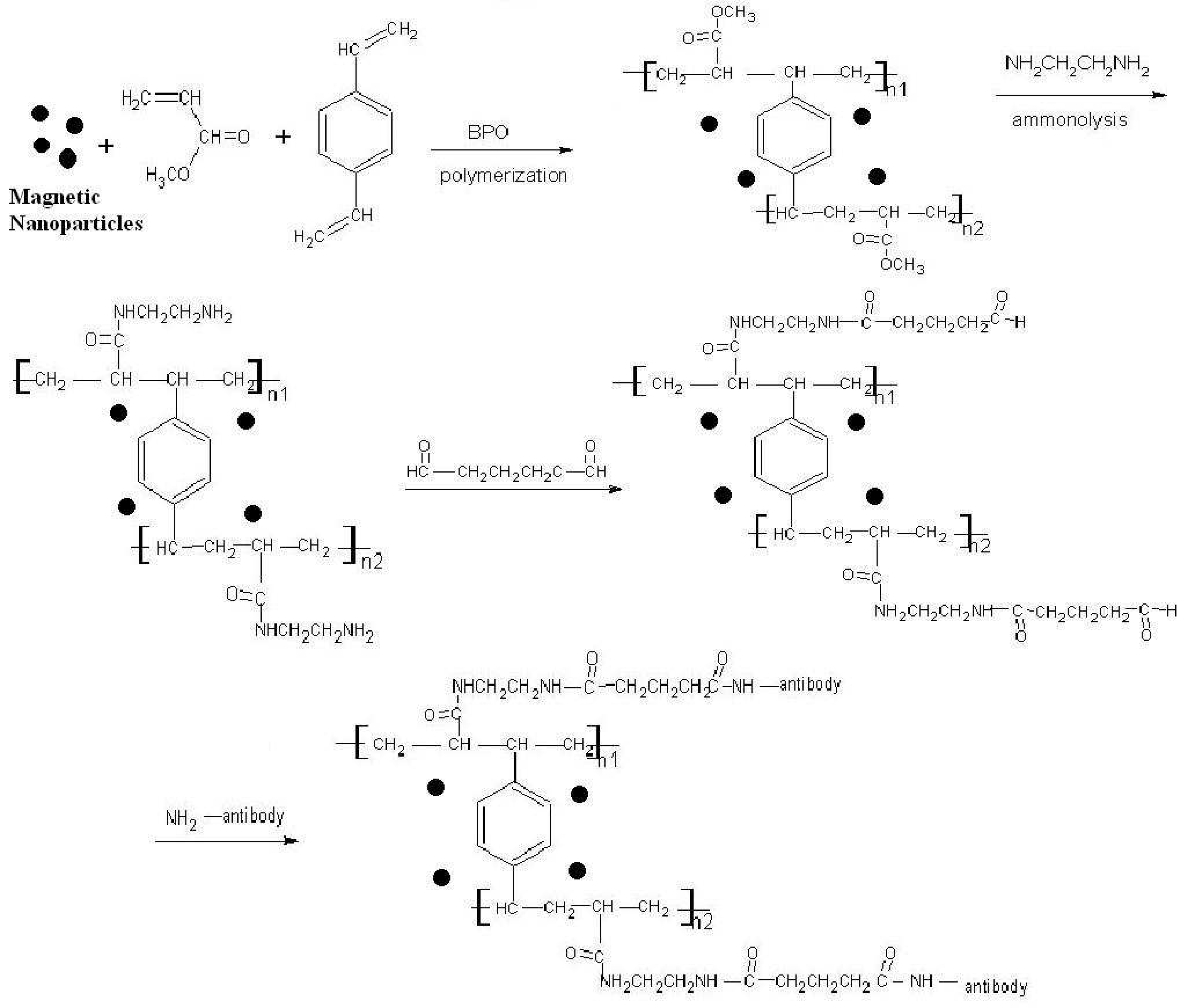
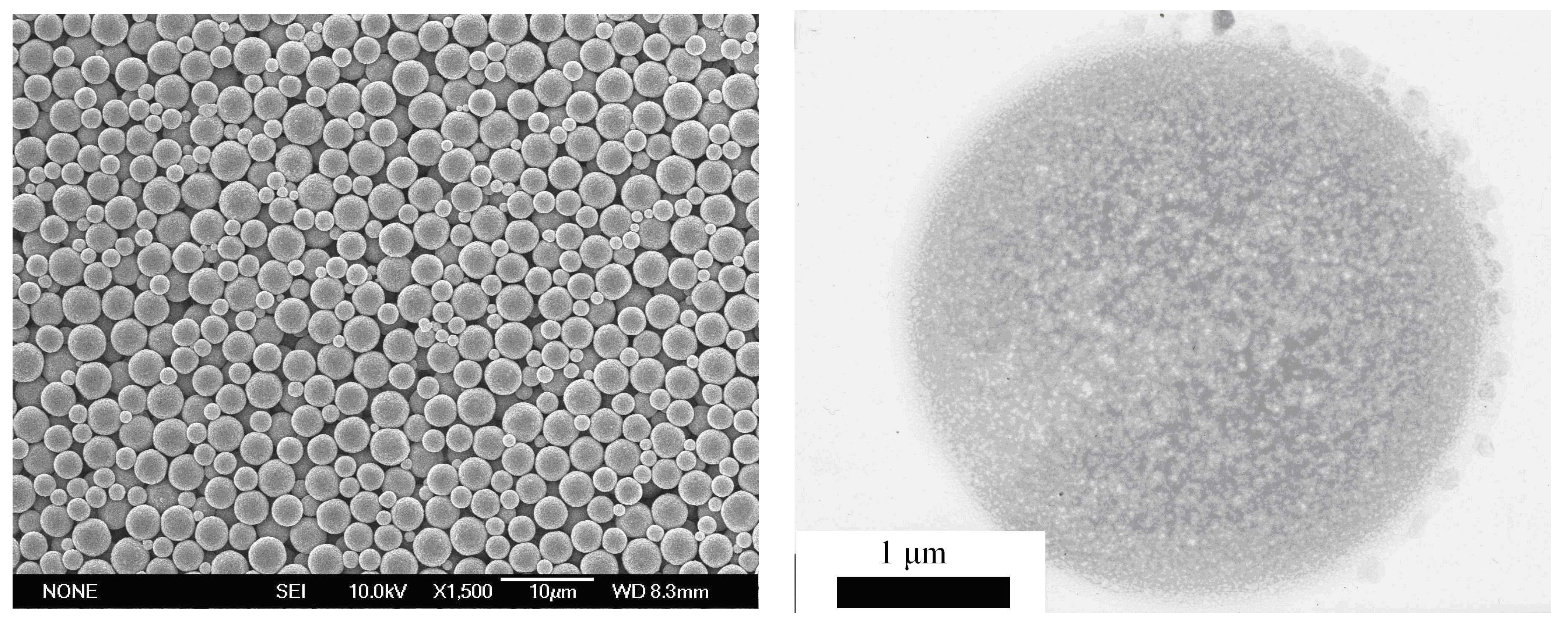
2.1. Preparation and surface modification of the SPMP microbeads

The magnetic polymer microspheres obtained from conventional suspension polymerization methods were in the size of several hundred micrometers. We developed a novel modified suspension polymerization method for the production of superparamagnetic polymer microspheres with micron size, which have narrow size distribution and high magnetite contents [12]. As seen in Scheme 1, magnetic fluid was prepared by a conventional coprecipitation method,then oleic acid was added dropwise into the suspension while heating. After the excess oleic acid was removed, the magnetic precipitate was redispersed in hexane to form magnetic fluid. The obtained Fe3O4 fluid and benzoyl peroxide (BPO) were dispersed in a mixture of methacrylate (MA) and divinyl benzene (DVB), and agitated until Fe3O4 was dissolved completely. The mixture was then transferred into a beaker containing polyvinyl alcohol (PVA). With rapid agitation, the mixture temperature was increased to reaction temperature under the protection of nitrogen and kept. The resulting magnetic microspheres were thoroughly washed to remove the excess monomer. After being agitated in methylformamide (DMF) and ethylenediamine (EDA), the ester groups on the poly(MA-DVB) microspheres were converted into amino groups, and the resulting microspheres were denoted as poly(MA-DVB)-NH2. To facilitate the covalent attachment of antibody, the amino groups on the surface of magnetic poly(MMA-DVB)-NH2 were transferred to aldehyde groups by the glutaraldehyde method.
The adding of the DVB as crosslinker made the polymer structure change from linear type to three-dimensional crosslink type, just as what we could see from the Scheme 1, which increased greatly the mechanic strength and the stability of SPMP. The detailed numbers of MMA and DVB in the polymer macromolecules relied on their molecular ratio. Wang [21] thought that this kind of cross-linking was necessary. Because a simple noncovalent linkage between the iron oxide cores and the polymeric chain were not sufficient to achieve a permanent linkage. Instead, a dynamic exchange between the polymeric chains and other competing molecules possessing a -COOH group such as peptides and amino acids in the biological systems could lead to the dissociation of the polymeric chains from the core surfaces. The loss of the coating material and its terminal functionalities could demolish the biospecificity of these materials. Kim [10] also found that this cross-linkage was particularly well suited to protect and functionalize materials such as Fe3O4, γ-Fe2O3 for which strongly binding surface ligands were not readily available, for it could fix the nanostructures topologically and these structures were stable to further synthetic transformations of surface functional groups.
Poly(MA-DVB), the SPMP microbeads synthesized by the modified suspension polymerization method, possessed the perfect sphere configuration, shown in Scheme 2(a). Most of these spheres were about 3 μm in diameters, but there still were a few below 2.5 μm due to the unhomogeneousity of the microemulsion diameter variating in 0.5-3 μm. Scheme 2(b) was the TEM picture of a single microbead. From the picture, we could see that the surface of the magnetic Poly(MA-DVB) microbeads was not so smooth, and there were full of small protuberances, and some tiny block in the outside of the sphere. These protuberances and blocks would increase the surface area to be ammonolysis by EDA, and provide amino groups and aldehyde groups at high density [22]. Such a procedure to synthesize the SPMP microbeads often bring several advantages to these magnetic beads. First, the integration and fix of many tiny magnetic cores guarantee the superparamagnetism and magnetophoresis ability due to the large amount of magnetic material percentage. Second, the organic surface of these polymers was more biocompitable than inorganic surface. The SPMP obtained by our method were small, inexpensive, and chemically and physically stable, and can be easily coupled to biomolecules at high density. Simply reacted with EDA and Glutaraldehyde, these SPMP could carry aldehyde groups at high density, which could chemically combined directly with biomolecules through many chemical groups, for example H2N-, HO-, HS-. Third, the polymers could protect the magnetic cores from influencing of the environmental factors.
Scheme 1. Synthesis, modification and coupling routines of SPMP poly(MA-DVB) microbeads.
Ijms 07 00274 g001
Note: Before polymerization of the MA and DVB, the 8 nm primitive Fe3O4 particles were first prepared by coprecipitation of Fe2+ and Fe3+ under the protection of N2, and followed by the hydrophobic treatment of the oleic acid coating. When the polymerization took place, this oleic acid-coated magnetite suspension was distributed in the microemusion with the monomers MA and DVB. The detailed numbers of MA and DVB in the polymer macromolecules relied on their molecular ratio, and the Scheme 1 just showed the principle scheme. Nevertheless the adding of the DVB as colinker made the polymer structure change from linear type to three-dimensional crosslink type, which increased greatly the mechanic strength and the stability of SPMP.
The magnetic properties of SPMP microspheres were recorded by VSM at room temperature. Scheme 3 showed their magnetization curves. No hysteresis loop was observed at this temperature, suggesting that the magnetic microspheres were superparamagnetic, which indicated that there would no magnetic interactions among magnetic microspheres in a zero magnetic field environment. This feature would result in the easy dispersion of the magnetic microspheres. The saturation magnetization of SPMP microspheres was 13.8 emu/g, which was higher than those reported by other similar works [23]. With such high saturation magnetization, they could be easily and quickly separated from a suspension. This could be used to the magnetic separation of proteins on a large scale.

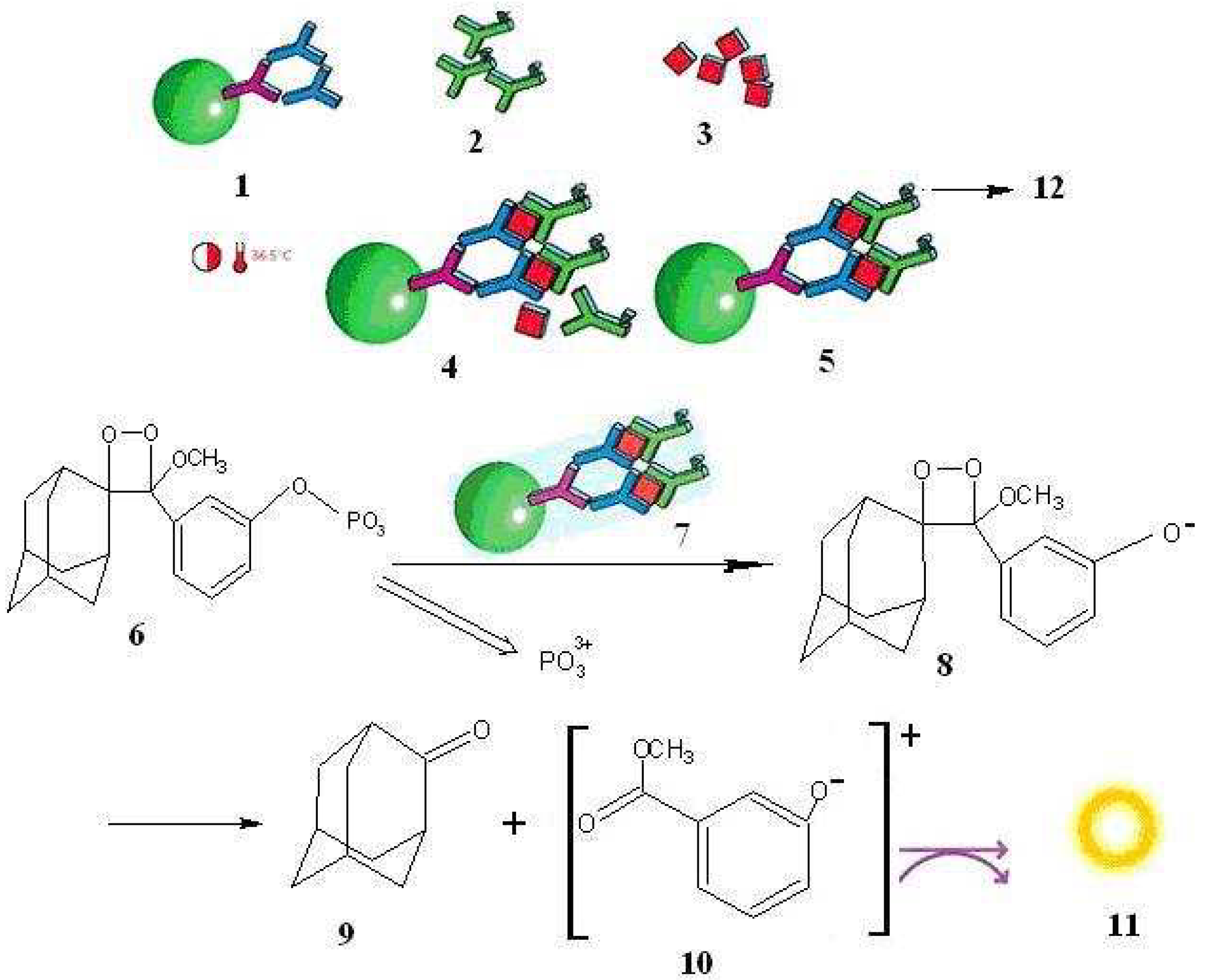
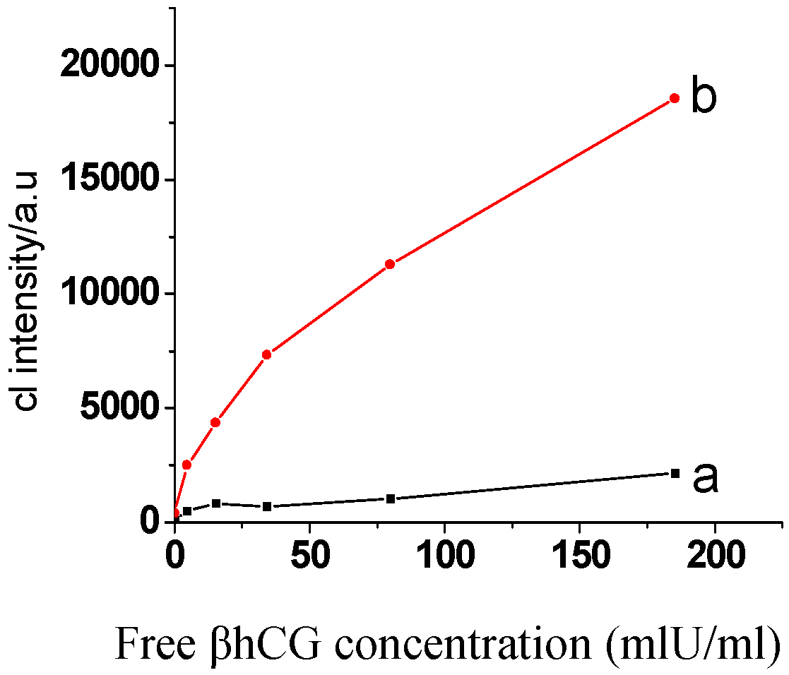
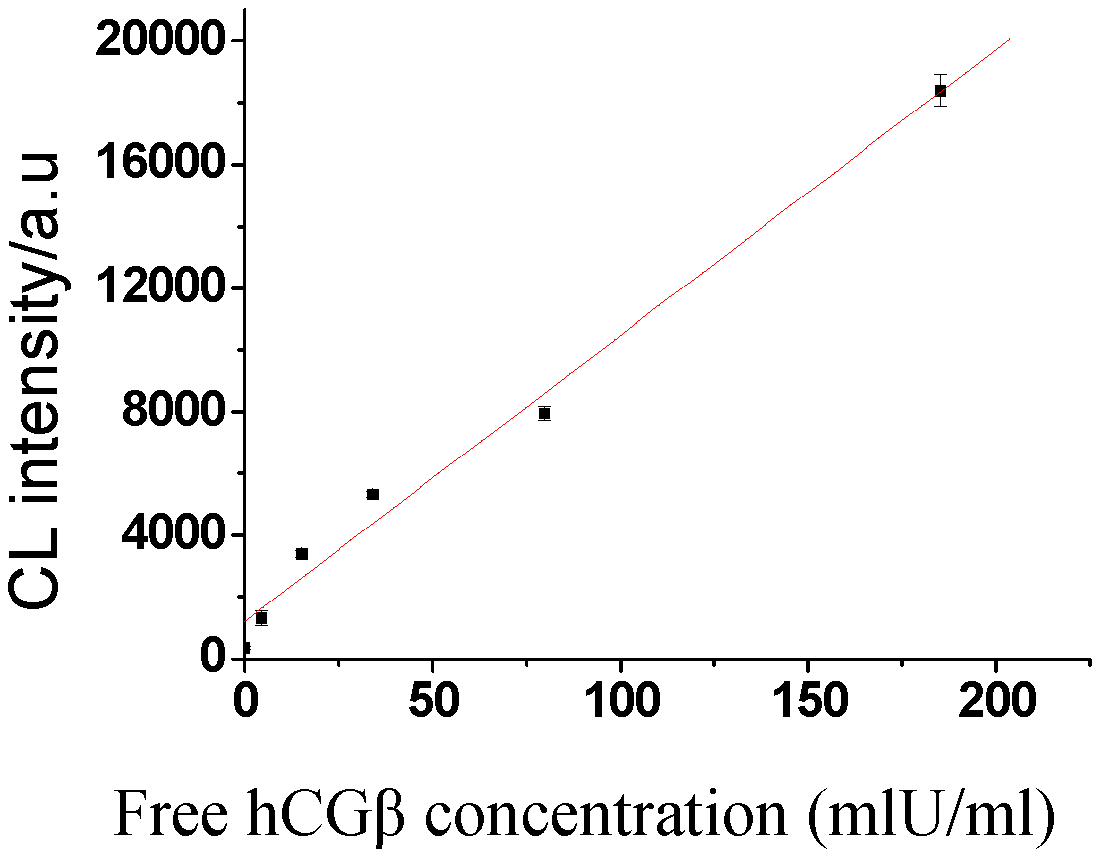
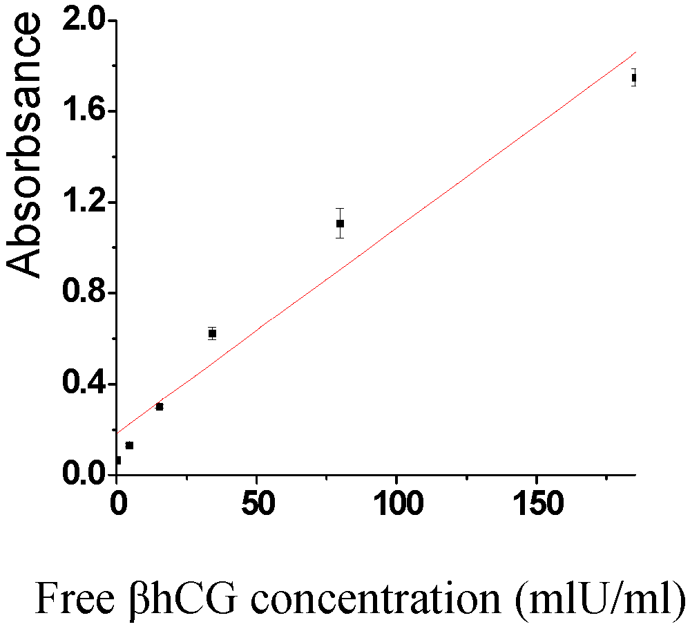
2.2. Application in the detection of free hCGβ

Magnetic beads came into use in enzyme chemiluminescence immunoassay (ECLIA) just in recent years. In this method magnetic separation, immunological protocols and chemiluminecence detection technique were combined ingeniously, which left out the complicated package of antibodies in microcells, thus the time and procedures were cut down. Santandreu et al. [19] thought that the high selectivity displayed between the antibodies and their antigens had motivated the development of immunoassay analytical techniques, while magnetic beads provided immunosensing systems with renewable surfaces for the sake of regeneration.
Scheme 2. SEM and TEM pictures of the SPMP microbeads synthesized by the modified suspension microemusion polymerization method (a. left picture; b. right picture).
Ijms 07 00274 g002
In our point, the advantage of superparamagenetic microbeads in ECLIA was mainly in their superparamagnetism. Namely, their magnetism was proportional to the strength of outside eternal magnetic field without any remnant magnetism. When the field was removed, the good dispersion of these SPMP microbeads could be ensured after repeated uses of magnetic field. Otherwise, the specific biomolecules bound to these beads would not has much possibility to bump and react with their corresponding biomolecules in liquid, or nonspecifically reacted molecules could not be diluted effectively due to the aggregation among particles. But superparamagnetism was usually demonstrated when the size of the particles was less than 10nm. Thus some obstacles prevented the very tiny superparamagnetic nanoparticles to realize their application. First, some biological molecules are larger than 10nm, some (E.G., cells) are even several tens microns. Significant reduction in particle size must be achieved to provide the surface area required, but too small a particle may not carry enough magnetite and, in practice, would cease to be magnetic [12(b)] due to their little magnetophoresis ability [6]. Second, as a result of anisotropic dipolar attraction, pristine nanoparticles tend to aggregate into large clusters and thus lose the specific properties associated with single-domain, magnetic nanostructures [24]. Immobilizing these very tiny pristine nanoparticles in the framework of polymers successfully solved both of the two obstacles.
Scheme 3. the magnetic hysteris loop by vibrating samples magneticity
Ijms 07 00274 g003
Whatever advantages of magnetic beads in assays people focused on, the magnetic ECLIA has by and by come into use in the detection of other tumor markers such as carcino-embryonic antigen (CEA), alpha-fetoprotein (AFP), carbohydrate antigen (CA) 125、CA199, and Zhao et al [25] detected the hCG in saliva samples in this method.
Scheme 4 demonstrated the proposed mechanism of the newly developed assay. In the assay, the molecules for determination were successively reacted with the two kinds of monoclonal antibodies, which were separately labeled by alkaline phosphatase (ALP) and fluoreszeinthiocyanat (FITC) in their ferrocenemonocarboxylic acid (Fc) fragments, to form a sandwich double antibodies’ complex, ALP-labeled-antibody-antigen-FITC-labeled-antibody. The SPMP microbeads, which were coupled with anti-FITC antibody, then specifically reacted with the sandwich complex, and deposited to the bottom of the microcells in the attraction of the eternal magnetic field. The unreacted molecules were removed by repeated cleansing and magnetic separation. The new chemiluminescence substrate, 3(2´spiroadamantane)-4- methoxy-4(3´´ phosphoryloxy)phenyl-1,2-dioxetane (AMPPD), was used to produce stable photo signals with the aid of alkaline phosphatase(ALP)without any background. We added the AMPPD working solution to the complex, and the ALP labeled on it would catalyze the AMPPD to decompose and release moderately stable dephosphorylated anion, which would rapidly break into an adamantanone and an excited methyl meta-oxybenzoate (AMP-D), then the stable photonic signal in 477nm [26] was emitted in 30 minutes and keep unchangeable in 7days [25]. The photonics number counted was direct proportional to the amount of ALP. Therefore, the quantitative determination of the samples could be obtained by detection of the strength of photonic signals. A similar procedure was utilized by Zhao [25] to determine the concentration of intact hCG in saliva samples. The chemiluminescence dectector we used herein was BPCL (-1-KGC), which can detect ultrafaint signals, with the advantages of easy manipulation, low price and cost.
Scheme 4. diagram of ECLIA based on superparamagnetic polymer microbeads.
Ijms 07 00274 g004
Note:1. microbeads coupled with anti-FITC, and anti-FITC antibody specifically reacted with the FITC which was labeled on the Fc fragment of anti-free hCGβ antibody; 2. antibody which was labeled with alkaline phosphate (ALP) enzyme; 3. free hCGβ for determination; 4. incubation when the sandwhich complex informed; 5. washing to remove unreacted components; 6. AMPPD; 7. the cleansed complex on which ALP would catalyze the AMPPD to decompose and emit photonics; 8. dephorylated anion,a moderately table middle product of AMPPD; 9. adamantanone; 10. methyl meta-oxybenzoate anion, AMP-D; 11. photonic signal at 477nm; 12. alkaline phosphate enzyme.

2.3. Optimization of the operation parameters of the sandwich magnetic ECLIA of free hCGβ

The optimized assay conditions were as the following: Amount of the substrate AMPPD was 10μl, after adding 10μl AMPPD working solution to the tube, detected the photonic signals in 30 minutes. There was better linearity between the CL intensity and the concentration of free hCGβ when 5μl sample were used. The increase of the samples would increase the expenditure, and too strong signals were prone to attain the saturated maximum or overflow the detection limit of the instrument. The detection voltage was an important parameter that could influence the results. Comparatively, the signal in 920V had better linearity. When the voltage increased to 950V, the signal strength was attained to saturated state, and began to fluctuate, sometimes even appeared sharp fall.
In order to investigate the unspecific absorption of the tubes used in the assays without antibodies complex to determine the antigens. We added SPMP beads solution in one group, and added blank solution without SPMP microbeads after adding 5μl sample. Then incubated in 37°C to make the specific reaction take place, and after separation and 10μl AMPPD was added to the both tubes. After 30 minutes the signals of the two tubes were detected in 920V to compare their chemiluminecent intensities. The intensity of the assays without SPMP beads meant the extend of unspecific absorption , since no complex was left in the tube except for the absorbed free hCGβ and antibody labeled ALP during the course of separation and washing, which would finally result in the chenmiluminencence. Shown as Scheme 5, the intensity due to the tube’s unspecific absorption was not as much more enormous rise with the increasing of the free hCGβ concentration. All the assays’ signal intensity was very weak and the photonic numbers did not exceed 2000 even in 180 IU•l-1, which was the maximum concentration of the kits from Bio-Ekon Biotechnology Co. Ltd. Compared with them, the intensity of assays using SPMP beads which were combined with anti-FITC antibodies was much stronger. Under the same conditions, the signals with SPMP beads were at least six times stronger than those without even at low concentration, and much higher with the increasing of concentration of free hCGβ. We explained the reason for this phenomenon as following: the outside eternal magnetic plate improved greatly the separation efficiency of the mixture after the sandwich immuno reaction, and decrease the unspecific absorption of unreacted molecules, and also reduced the loss of the immuno complex during the course of repeated separations and cleansing. Therefore the application of superparamagnetic beads in enzyme immuno assays (EIA) would remarkably enhance the intensity of the CLA signals, and raise the sensitivity of these assays in the same reaction conditions, such as pH, temperature, concentration, and so on.
Scheme 5. the unspecific absorption of the CL intensity
Ijms 07 00274 g005
(a) without SPMP microbeads
(b) with SPMP microbeads
Scheme 6. the calibration of ECLIA
Ijms 07 00274 g006
Scheme 7. the calibration of ELISA
Ijms 07 00274 g007

2.4. Sensitivity and recovery rate

Pipette the standard serum in different concentration gradients and repeated the assays according to the protocols of ECLA based on SPMP microbeads and the magnetic ELISA method separately and detected their corresponding photonic signals, and drawn their calibration curves. From the Scheme 6 we could see that the detection limit of ECLIA we developed was 0.22 IU•l-1, and the linear range was 0.45-185.2 IU•l-1, which was confined by the kit’s maximum concentration, and not the theatrical upper limit. The fitting linear equation was Y=99.50X+1223.42, R=0.995. Seen from the Scheme 7, the corresponding detection limit of the spectrometry assays was 2.28 IU•l-1. The fitting linear equation was Y=0.0094X+0.2055, and the linear range was 4.56-185.2, R=0.972. Obviously the ECLIA was more sensitive assays, for its lower limit was much lower than that of the spectrometry assays, and the linear range was wider.
Added a certain amount of standard concentration serum to a sample, detected the result, and compared it to the theatrical concentration value, the zero-concentration recovery could be obtained. Two standard samples, the concentrations of which were 15.22 IU•l-1 and 79.80 IU•l-1, were both diluted at the ratios of 1:2, 1:4, and 1:8, the diluted concentration values were detected, and the non-zero concentration recovery would be obtained compared to their predicated concentration values. Also we calculated the CV value among batches was less than 8.0%, and that among batches was less than 15%, by carrying out repeated detection for at least five times, to a same zero-concentration recovery experiment sample.

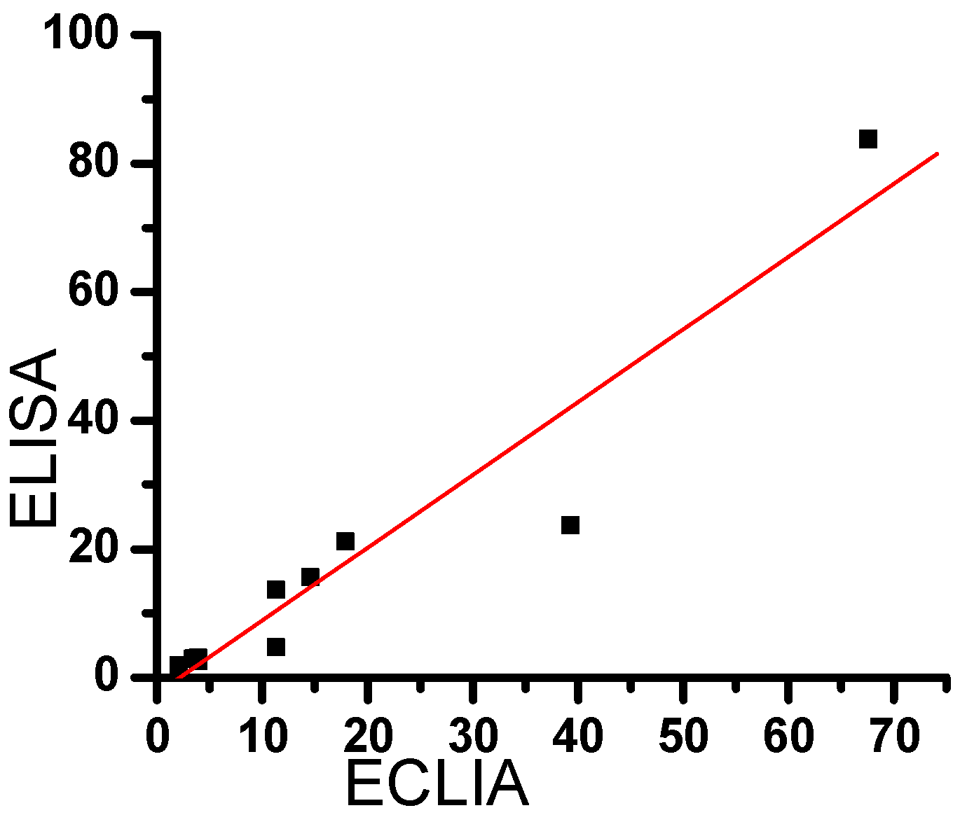
2.5. Comparison between the results of clinical samples, by ECLIA and ELISA.

10 blood samples were collected in vacutainers from volunteers, tops were removed and bloods centrifuged for 10 min. A pair of aliquots (5μl) was taken from each serum sample. We detected these clinical samples in the both of spectrometry assay and SPMPECLIA, and compared their results, we could calculated the related coefficient between the two methods was 0.955(Scheme 8). Therefore we could replace the ELISA methods by the ECLIA in the assays of free hCGβ in serum.
Scheme 8. correlation line of clinical serum samples’ results by ECLIA and ELISA
Ijms 07 00274 g008
Pipette the standard serum in different concentration gradients and repeated the assays according to the protocols of ECLIA based on SPMP microbeads and the ELISA method separately and detected their corresponding photonic signals, and drawn their calibration curves. We could see that the detection limit of MECLIA we developed was 0.22 IU•l-1, and the linear range was 0.45-185.2 IU•l-1, which was confined by the kit’s maximum concentration, and not the theatrical upper limit. The fitting linear equation was Y=99.50X+1223.42, R=0.995. Seen from the Scheme 7, the corresponding detection limit of the ELISA was 2.28 IU•l-1. The fitting linear equation was Y=0.0094X+0.2055, and the linear range was 4.56-185.2, R=0.972. Obviously the ECLIA was more sensitive assays, for its lower limit was much lower than that of the spectrometry assays, and the linear range was wider. Also we calculated the CV value among batches was less than 8.0%, and that among batches was less than 15%, by carrying out repeated detection for at least five times, to a same zero-concentration recovery experiment sample.
10 blood samples were collected in vacutainers from volunteers, tops were removed and bloods centrifuged for 10 min. A pair of aliquots (5μl) was taken from each serum sample. We detected these clinical samples in the both of spectrometry assay and ECLIA based on SPMP, and compared their results in Scheme 8, we could calculate the related coefficient between the two methods was 0.955.

3. Conclusions

A kind of superparamagnetic microspheres with higher magnetic content were synthesized in a modified suspension polymerization routine, and a sandwich magnetic enzyme chemiluminecent assay of free hCGβ in serum was built based on them. The ALP-AMPPD was used as the chemiliuminencent system. The detection limit of MECLIA we developed was 0.22 IU•l-1, and the linear range was 0.45-185.2 IU•l-1, which was confined by the kit’s maximum concentration, and not the theatrical upper limit. The fitting linear equation was Y=99.50X+1223.42, R=0.995. Compared with the original ELISA kit, its low limit was much lower, and the linear range was wider, their related coefficient between the two methods in detecting clinical samples was 0.955. Moreover the magnetic ECLIA need half of the sample volume, simpler protocols. And the time it need was shortened from 2 hours to 1 hour. Therefore we could replace the ELISA methods by the magnetic ECLIA in the assays of free hCGβ in serum .

4. Experimental Section

4.1. General

BPCL(-1-KGC) Ultra-weak Luminescence Analyzer, Developed by Technical Institute of Physics in Chinese Academy of Science; adjustable pipette in 2-20μl and 50-200μl, Shanghai Dalong medical instrument Co., Ltd.; oscillator, Shanghai Ester transformer Co., Ltd; magnetic separator; Beijing Bio-Ekon Biotechnology Co. Ltd.; digital thermostat bath HH-4, Changzhou Guohua electrical appliance corporation; tubes specially for Serozyme diagnostic system, Bio-Ekon Biotechnology Co. Ltd.; DYNEX MRX-HD, DYNEX Technologies Co. Ltd.; 8X12 cells plate (Shenzhen Jingmei Biological Engineering Co., Ltd.). Scanning electronic microscope (SEM), JSM-6700F, JEOL, Japan. Transmitting Electronic microscope( TEM), JEM-200 CX, JEOL Co., Ltd.
Methyl methacrylate, analytical pure, washed with 5% sodium hydroxide aqueous solution; benzoyl peroxide (BPO), divinyl benzene (DVB), sodium hydroxide, polyvinyl alcohol (PVA) 1788, methylic alcohol, aqueous ammonia, FeCl2•4H2O, FeCl3•6H2O, all of the above were analytical pure and from Beijing Shuanghuan reagent Co., Ltd.. ethylenediamine (EDA), purchased from Beijing Chemical Reagents Company (Beijing, China).
Bovine pancreatic alkaline phosphatase (ALP, 1U/ml), Free hCGβ standard solution and quality control samples, Free hCGβ, ALP labeled anti-free hCGβ, anti-free hCGβ antibody labeled by 0.6% Fluoreszeinthiocyanat (FITC), magnetic nanobeads coupled with anti-FITC antibody, Serozyme substrate,serozyme block reagent, and detergent (pH=7.4) made of 0.1mol/L Tris-HCl and 0.5% Tween-20, all of the above were from Beijing Bio-Ekon Biotechnology Co. Ltd.. Na2CO3-NaHCO3 buffer, by the first Nanjing Chemical Factory. 3(2´spiroadamantane)-4- methoxy- 4(3´´ phosphoryloxy)phenyl-1,2-dioxetane (AMPPD) was the gift from Beijing Yongning Zhiben independent Laboratory. All water used here was Millipore ultrapure and deionized water.

4.2. Preparation of Oleic Acid-coating Fe3O4 Magnetic Fluid (1)

Magnetic fluid was prepared by a conventional coprecipitation method with some modifications. FeCl3·6H2O (11.6 g) and FeCl2·4H2O (4.3 g) were dissolved in 400 ml of deionized water under nitrogen gas protection with vigorous stirring at 85°C. NH3·H2O (25%; 15 ml) was added first to the solution, and then 9 ml of oleic acid was added dropwise into the suspension within 20 min. After several minutes, the magnetic precipitate was isolated from the solvent by magnetic decantation. The precipitate was washed with deionized water several times to remove the excess oleic acid. The magnetic precipitate was redispersed in an organic carrier liquid such as hexane to form the magnetic fluid.

4.3. Synthesis of Poly(MA-DVB) SPMP microspheres (2)

The Poly(MA-DVB) microspheres were prepared by a modified suspension polymerization method. In a typical experiment, 30g of the magnetic Fe3O4 gel and 3g of BPO were dispersed in a mixture of 95ml MA and 5ml DVB (crosslinker) to form the organic phase, and agitated until Fe3O4 was dissolved completely. The mixture was then transferred into a 2-liter beaker containing 25g PVA and 30g NaCl dissolved in 1000 ml H2O, which was equipped with four vertical stainless steel baffleplates, a nitrogen inlet, and a 4-paddle mechanical stirrer. With agitation at 1000rpm, the mixture temperature was increased evenly from 45°C to 60°C within 1 hr, then the temperature was maintained at 60°C for 2 hr until finally the temperature was increased to 70°C and kept for 2 hr. The reaction mixture was cooled to room temperature, and the resulting magnetic microspheres were thoroughly washed with deionized water and ethanol to remove the excess monomer.

4.4. Surface Modification of Poly(MA-DVB) SPMP microspheres with EDA(3)

In order to prepare the EDA-modified Poly(MA-DVB) microspheres, the following procedure was applied. 3g sample of Poly(MA-DVB) microspheres was washed with DMF two times and put in a solution of 100ml DMF plus 100ml EDA. The mixture was agitated gently at 110°C for 12 h. After being cooled to room temperature, the Poly(MA-DVB) microspheres were separated by magnetic decantation and washed with water and ethanol to remove the residual DMF. After modification, ester groups on the Poly(MA-DVB ) were converted into amino groups. The resulting microspheres were denoted as Poly(MA-DVB)-NH2.

4.5. Surface functionalization and activation (4)

To facilitate the covalent attachment of antibody, the amino groups on the surface of magnetic Poly(MA-DVB)-NH2 were transferred to aldehyde groups by the glutaraldehyde method. After agitating at 30 °C overnight, the glutaraldehyde-activated magnetic supports were washed with deionized water three times and stored for future use. The detailed principle routine of the above steps was shown in Scheme 1.

4.6. Combination of anti-FITC (5)

The anti-FITC antibodies was incubated with the glutaraldehyde-activated magnetic poly(MA–DVB) microspheres in PBS (pH7.4) for 4 hours, and followed by repeated magnetic separations and washing with PBS.

4.7. Protocols of the whole assay of ECLIA (6)

5μl standard serum, quality controlling serum, and sample serum into different tubes, which were then respectively added two kinds of 10μl monoclonal anti-free hCGβ antibodies labeled by correspondingly FITC and ALP. Enough mixing were obtained by shaking of the oscillator, and incubated in 37°C for 12 minutes. 10μl nanosized magnetic beads solution was added and followed by oscillation for 2 minutes, and incubation in 37°C for 5 minutes. Eternal magnetic plate was used to deposit the magnetic complex for 2 minutes. Decanted the liquid of all tubes (which were all attached to the magnetic plate), and pat the upside-down tubes on clean and dry paper for at least five times. Rinse the remnant in the tubes with 300μl detergent each time for at least 3 to 5 times, pat the remained solution on paper. Then 10μl AMPPD solution was added to each tube, the strength of photonic signals were detected by BPCL after 30 minutes.

4.8. Collection of the clinical serum samples (7)

All the clinical samples were obtained from the clinical laboratories of Jiangsu Provincial Institute of Tumor Research and Jiangsu Provincial Center of Women and Children’s Health-Care. The venous blood was exsanguinated by the experienced nurse from the hospitalized patients who suffered from the cervical cancer or other reproductive organs’ cancers. And the separated serum was used for determination.

Acknowledgements

We thank for the sponsor of the National Scientific Funds of China, NO. 60223002 and 60121101. Thanks also go to Hong Jianguang for his effort in TEM operation,and Dr. Chen Tingfeng and Dr. Zhang Jiaming for their clinical samples.

References and Notes

  1. (a) Baselt, D.R.; Lee, G.L.; Natesan, M.; Metzger, S.W.; Sheehan, P.E.; Colton, R. J. A Biosensor Based on Magnetoresistance Technology. Biosens.Bioelectron. 1998, 13, 731-739; (b) Li, G.; Sun, S.; Wilson, R.J.; White, R. L.; Pourmand, N.; Wang, S. X. Spin Valve Sensors for Ultrasensitive Detection of Superparamagnetic Nanoparticles for Biological Applications. Sensors and Actuators A, 2006, 126, 98-106.
  2. (a) Kircher, M. F.; Mahmood, U.; King, R. S.; Weissleder, R.; Josephson, L. A Multimodal Nanoparticle for Preoperative Magnetic Resonance Imaging and Intraoperative Optical Brain Tumor Delineation. Cancer Research 2003, 63, 8122-8125; (b) Funovics M.; Montet, X.; Reynolds, F.; Weissleder, R.; Josephson, L. Nanoparticles for the Optical Imaging of Tumor E-selectin. Neoplasia 2005, 7, 904-911.
  3. Jordan, A.; Scholz, R.; Scholz, R.; Maier-Hauff, K.; Johannsen, M.; Wust, P.; Nadobny, J. Presentation of a New Magnetic Field Therapy System for the Treatment of Human Solid Tumors with Magnetic Fluid Hyperthermia. Journal of Magnetism and Magnetic Materials 2001, 225, 118–126. [Google Scholar] [CrossRef]
  4. Ito, A.; Shinkai, M.; Honda, H.; Kobayashi, T. Medical Application of Functionalized Magnetic Nanoparticles. Journal of Bioscience and Bioengineering 2005, 100, 1–11. [Google Scholar] [CrossRef]
  5. Sun, L.; Zborowski, M.; Chalmers, J.J. Continuous, Flow-through Immunomagnetic Cell Sorting in a Quadrupole Field. Cytometry 1998, 33, 469–475. [Google Scholar] [CrossRef]
  6. McCloskey, K.E.; Chalmers, J.J.; Zborowski, M. Magnetic Cell Separation: Characterization of Magnetophoretic Mobility. Anal. Chem. 2003, 75, 6868–6874. [Google Scholar] [CrossRef]
  7. (a) Luxton, R.; Badesha, J.; Kiely, J.; Hawkins, P. Use of External Magnetic Fields To Reduce Reaction Times in an Immunoassay Using Micrometer-Sized Paramagnetic Particles as Labels (Magnetoimmunoassay). Anal. Chem. 2004, 76, 1715-1719; (b) Chou, P.; Chen, S.; Liao, H.; Lin, P.; Her, G.; Lai, A. C.; Chen, J.; Lin, C.; Chen, Y. Nanoprobe-Based Affinity Mass Spectrometry for Selected Protein Profiling in Human Plasma. Anal. Chem. 2005, 77, 5990-5997; (c) Matsunaga, T.; Kawasaki, M.; Yu, X.; Tsujimura, N; Nakamura, N. Chemiluminescence Enzyme Immunoassay Using Bacterial Magnetic Particles. Anal. Chem. 1996, 68, 3551-3554; (d) Tanaka, T.; Matsunaga, T. Fully Automated Chemiluminescence Immunoassay of Insulin Using Antibody-Protein A-Bacterial Magnetic Particle Complexes. Anal. Chem. 2000, 72, 3518-3522.
  8. Osaka, T.; Matsunaga, T.; Nakanishi, T.; Arakaki, A.; Niwa, D.; Iida, H. Synthesis of Magnetic Nanoparticles and Their Application to Bioassays. Anal. Bioanal. Chem. 2006, 384, 593–600. [Google Scholar] [CrossRef]
  9. Li, G.; Fan, J.; Gao, Y. Cross-linking the Linear Polymeric Chains in the ATRP Synthesis of Iron Oxide/Polystyrene Core/Shell Nanoparticles. Chem. Mater. 2004, 16, 1835–1837. [Google Scholar] [CrossRef]
  10. Kim, B.-S.; Qiu, J.-M.; Wang, J.-P.; Taton, T.A. Magnetomicelles: Composite Nanostructures from Magnetic Nanoparticles and Cross-Linked Amphiphilic Block Copolymers. Nano Lett. 2005, 5, 1987–1991. [Google Scholar] [CrossRef]
  11. Qiu, G.; Wang, Q.; Nie, M. Polypyrrole-Fe3O4 Magnetic Nanocomposite Prepared by Ultrasonic Irradiation. Macromol. Mater. Eng. 2006, 291, 68–74. [Google Scholar] [CrossRef]
  12. (a) Liu, X.; Guan, Y.; Ma, Z.; Liu, H. Suface Modification and Characterization of Magnetic Polymer Nanospheres Prepared by Miniemulsion Polymerization. Langmuir 2004, 20, 10278-10283; (b) Ma, Z.-Y.; Guan, Y.-P.; Liu, X.-Q.; Liu, H.-Z. Preparation and characterization of micron-sized non-porous magnetic polymer microspheres with immobilized metal affinity ligands by modified suspension polymerization. Journal of Appl. Polym. Sci. 2005, 96, 2174-2180; (c) Liu, X.; Guan, Y.; Shen, R.; Liu, H. Immobilization of Lipase onto Micron-size Magnetic Beads. Journal of Chromatogr. B 2005, 822, 91-97.
  13. Hotakainen, K.; Ljungberg, B.; Haglund, C.; Nordling, S.; Paju, A.; Stenman, U.H. Expression of the Free Beta-subunit of Human Chorionic Gonadotropin in Renal Cell Carcinoma: Prognostic Study on Tissue and Serum. Int. J. Cancer 2003, 104, 631–635. [Google Scholar] [CrossRef]
  14. (a) Spencer, K. The Influence of Fetal Sex in Screening for Down Syndrome in the Second Trimester Using AFP and Free β-hCG. Prenat. Diagn. 2000, 20, 648-651; (b) Knight, G.J. ; Palomaki, G.E. ; Neveux, L.M., et al. HCG and the Free β-subunit as Screening Tests for Down Syndrome. Prenat. Diagn. 1998, 18, 235-245; (C) Hedstro, M.J.; Grenman, R.; Ramsay, H. Concentration of Free HCGβ Subunit in Serum as a Prognostic Marker for Squamous-cell Carcinoma of the Oral Cavity and Oropharynx. Int. J. Cancer (Pred. Oncol.) 1999, 84, 525-528 ; (d) Hallahan, T.; Krantz, D.; Orlandi, F.; Rossi, C.; Curcio, P.; Macri, S.; Larsen, J.; Buchanan, P.; Macri, J. First Trimester Biochemical Screening for Down Syndrome: Free Beta HCG versus Intact HCG. Prenat. Diagn. 2000, 20, 785-789.
  15. (a) Wenstrom, K.D.; Owen, J.; Chu, L. B. Free β-hCG Subunit Versus Intact hCG in Down Syndrome Screening. Obstetrics & Gynecology 1997, 90, 370-374; (b) Caroppo, E.; Niederberger, C.; Iacovazzi, P.A.; Correale, M.; Palagiano, A.; Amato, G.D. Human Chorionic Gonadotropin Free β-subunit in the Human Seminal Plasma: a New Marker for Spermatogenesis? European Journal of Obstetrics & Gynecology and Reproductive Biology 2003, 106, 165-169. (c) Nishimura, R. ; Koizumi, T. ; Yokotani, T. ; Taniguchi, R. ; Morisue, K. ; Yoshimura, M. ; Hiranmoy, D. ; Yamaguchi, S. ; Nakagawa, T. ; Hasegewa, K. ; Yasui, H. Molecular Heterogeneity of hCGβ-related Glycoproteins and the Clinical Relevance in Trophoblastic and Non-trophoblastic Tumors. International Journal of Gynecology & Obstetrics 1998, 60 suppl., S29-S32.
  16. Hsu, J.-J.; Spencer, K.; Aitken, D.A.; Crossley, J.; Choi, T.; Ozaki, M.; Tazawa, H. Urinary Free Beta HCG, Beta Core Fragment and Total Oestriol as Markers of Down Syndrome in the Second Trimester of Pregnancy. Prenat. Diagn. 1999, 19, 146–158. [Google Scholar] [CrossRef]
  17. Luppa, P.; Spöttl, G.; Saller, B.; Neumeier, D. Cross – Reactivity of a Commercial Chemiluminescence Immunoassay for Human Chorionic Gonadotropin with the Free Beta – subunit . Journal of Biolumin. Chemilumin. 1992, 7, 195–201. [Google Scholar] [CrossRef]
  18. (a) Cole, L.A. ; Kellner, L.H. ; Isozaki, T. Comparison of 12 Assays for Detecting hCG and Related Molecules in Urine Samples from Down Syndrome Pregnancies. Prenat. Diagn. 1997, 17, 607-614 ; (b) Lin, J.; Ju, H. Electrochemical and Chemiluminescent Immunosensors for Tumor markers. Biosensors and Bioelectronics 2005, 20, 1461-1470.
  19. Santandreu, M.; Alegret, S.; Fàbregas, E. Determination of β-HCG Using Amperometric Immunosensors Based on a Conducting Immunocomposite. Analytica Chimica Acta 1999, 396, 181–188. [Google Scholar] [CrossRef]
  20. (a) Nakamural, N.; Lim, T.-K.; Jeong, J.-M.; Matsunaga, T. Flow Immunoassay for Detection of Human Chorionic Gonadotrophin Using a Cation Exchange Resin Packed Capillary Column. Analytica Chimica Acta 2001, 439, 125-130; (b) Lim, T.-K.; Matsunaga, T. Construction of Electrochemical Flow Immunoassay System Using Capillary Columns and Ferrocene Conjugated Immunoglobulin G for Detection of Human Chorionic Gonadotrophin. Biosensors & Bioelectronics 2001, 16, 1063-1069; (c) Nakamura1,N.; Lim T.-K.; Jeong J.-M.; Matsunaga, T. Flow Immunoassay for Detection of Human Chorionic Gonadotrophin Using a Cation Exchange Resin Packed Capillary Column. Analytica Chimica Acta 2001, 439, 125-130; (d) Lin, J.-M.; Tsuji, A.; Maeda, M. Chemiluminescent Flow Injection Determination of Alkaline Phosphatase and Its Applications to Enzyme Immunoassays. Analytical Chimica Acta 1997, 339, 139-146.
  21. Wang, Y.; Zhu, R.; He, W. Magnetic Properties of Polystyrene-b-poly(2-hydroxylethylmethacrylate)/metal Hybrids. J Appl. Polym. Sci. 2006, 99, 2314–2319. [Google Scholar] [CrossRef]
  22. (a) Ma W.-L. ; Zheng W.-L. Molecular Oncology, Chin. Sci. press, 2003, September, Beijing; (b) Yang, C.; Guan, Y.; Xing, J.; Liu, J.; An, Z.; Liu, H. Preparation and Surface Modification of Magnetic PMMA Microspheres. Science in China (Ser.B Chemistry) 2004, 34, 265-269; (c) Liu, X. Q.; Guan, Y. P.; Xing, J. M.; Ma, Z. Y.; Liu, H. Z. Synthesis and Properties of Micro-sized Magnetic Polymer Spheres with Epoxy Groups. Chinese J. Chem. Eng. 2003, 11, 731-735.
  23. (a) Lee, J.; Senna, M. Preparation of Monodispersed Polystyrene Microspheres Uniformly Coated by Magnetite via Heterogeneous Polymerization. Colloid Polym. Sci. 1995, 273, 76-82; (b) Liu, X. Y.; Ding, X. B.; Zheng, Z. H. Synthesis of Novel Magnetic Polymer Microspheres with Amphiphilic Structure. J Appl. Polym. Sci. 2003, 90, 1879-1884.
  24. Lu, Y.; Yin, Y.; Mayers, B.T.; Xia, Y. Modifying the Surface Properties of Superparamagnetic Iron Oxide Nanoparticles through a Sol-Gel Approach. Nano Lett. 2002, 2, 183–186. [Google Scholar] [CrossRef]
  25. Zhao, L.-X.; Lin, J.-M.; Qu, F. Micro-plate Magnetic Chemiluminescence Enzyme Immunoassay for Rapid, Sensitive Determination of Human Chorionic Gonadotropin (hCG). Acta Chimca Sinica 2004, 64, 71–77. [Google Scholar]
  26. Gillevet, P.M. Chemiluminescent Multiplex DNA Sequencing. Nature 1990, 348, 657–658. [Google Scholar] [CrossRef]
  • Sample Availability: Available from the authors.

