Article

UVB-Protective Effects of Isoflavone Extracts from Soybean Cake in Human Keratinocytes

by
Han-Sun Chiang
1,
Wen-Bin Wu
1,
Jia-You Fang
2,
Bing-Huei Chen
3,
Tsai-Hua Kao
3,
Ying-Ting Chen
1,
Chieh-Chen Huang
4 and
Chi-Feng Hung
1,*
1
School of Medicine, Fu-Jen Catholic University, Taipei Hsien, Taiwan
2
Pharmaceutics Laboratory, Graduate Institute of Natural Products, Chang Gung University, Kweishan, Taoyuan, Taiwan
3
Department of Nutrition and Food Sciences, Fu-Jen Catholic University, Taipei Hsien, Taiwan
4
Department of Dermatology, Shin Kong Wu Ho-Su Memorial Hospital, Taipei, Taiwan
*
Author to whom correspondence should be addressed.
Int. J. Mol. Sci. 2007, 8(7), 651-661; https://doi.org/10.3390/i8070651
Submission received: 14 May 2007 / Revised: 20 June 2007 / Accepted: 28 July 2007 / Published: 11 July 2007
(This article belongs to the Special Issue Phenolics and Polyphenolics)

Abstract

:
It has been shown by chromatography that aglycone, glucoside, acetylglucoside and malonylglucoside isoflavone extracts prepared from soybean cake showed better antioxidant activities than isoflavone standards. Consequently, the aim of this study was to evaluate the protective effects of these isoflavone extracts against ultraviolet B (UVB)-induced keratinocyte damage. Our results demonstrated that these soybean cake isoflavone extracts could inhibit UVB-induced keratinocyte death. Moreover, they could inhibit UVB-induced intracellular release of hydrogen peroxide (H2O2) Furthermore, these isoflavone extracts differentially inhibited UVB-induced MAPK phosphorylation. The ERK1/2 and p38 phosphorylation was not inhibited by all tested isoflavone extracts, whereas JNK phosphorylation was inhibited by group I to group III isoflavone extracts. Since these isoflavone extracts are relative stable and easily obtained than the isoflavone standards, we suggest that soybean cake may be a useful potential source for developing effective skin care agents in against photoaging.

1. Introduction

Diets rich in soybean products are associated with reduced incidence of cardiovascular disease, osteoporosis and certain human cancers [13]. Animal studies have shown that soy diets block oxidative degradation of lipoproteins, prevent atherosclerosis of blood vessels and inhibit radiation-and carcinogen-induced tumors of various tissues. Soybeans contain ingredients with potential biological effects and isoflavone is one of the most extensively studied ingredients. Twelve isoflavones are found in soybean and are present in four chemical forms: malonylglucosides (malonyldaidzin, malonylgenistin, and malonylglycitin), acetylglucosides (acetyldaidzin, acetylgenistin, and acetylglycitin), glucosides (daidzin, genistin, and glycitin) and aglycones (daidzein, genistein, and glycitein). Among these, the glucoside is the predominate form in soybean [45]. In vitro and in vivo studies have shown that isoflavone aglycones such as genistein inhibit ultraviolet light-induced damage [68]. However, it has also been shown that isoflavone glycosides such as genistin, daizin and glycitin have biological activities on melanoma cells and myoblasts [7,9]. Therefore, it is possible that aglycones and glycosides can show some physiological relevance.
Soybean cake is a by-product during processing of soybean oil. Defatted soybean contains a high amount (2121.9 μg/g) of isoflavones [4]. Kao and coworkers have isolated four isoflavone extracts, namely malonylglucoside, acetylglucoside, glucoside and aglycone, from soybean cake by chromatography [5,10]. They found these isoflavone extracts possess differential antioxidant activities. For example, the acetylglucoside extract exhibits the highest efficiency in 1,1-diphenyl-2-picrylhydrazyl (DPPH) scavenging assay and the glucoside extract is the most efficient for chelating metal ions [5]. Overall, the isoflavone extracts show better antioxidant activities than isoflavone standards [5]. Therefore, the purpose of this study was to determine the protective effects of these four isoflavone extracts on Ultraviolet B (UVB)-induced keratinocyte damage, including their effects on UVB-induced hydrogen peroxide (H2O2) generation and mitogen-activated protein kinase (MAPK) signaling [extracellular-regulated kinase (ERK1/2), p38 and c-jun N-terminal kinase (JNK)] in keratinocytes.

2. Materials and Methods

2.1. Materials

3-(4,5-Dimethylthiazol-2-yl)-2,5-diphenyltetrazolium bromide (MTT), aprotinin, leupeptin, phenylmethylsulfonyl fluoride (PMSF), sodium fluoride (NaF) and sodium orthovanadate were purchased from Sigma Chemical Co. (St Louis, MO). Antibodies raised against p38 and p-JNK were from Cell Signaling Technology (Beverly, MA). Antibodies raised against JNK, ERK1/2 and p-p38 were from R&D system, Inc. (Minneapolis, MN). Antibody raised against p-ERK1/2 was from Santa Cruz Biotechnology (Santa Cruz, CA)

2.2. Preparation of Isoflavone extracts

Isoflavone extracts (group I~IV) were prepared by a method as previously described by Kao et al. [5]. A sample (50 g) was mixed with deionized water-ethanol (150 ml, 1:1, v/v) and the mixture was shaken for 2 h. After centrifuging at 6000 rpm for 20 min (25 °C), the supernatant was collected and filtered through a glass filter paper. Then soybean cake extract (80 ml) was poured onto the top of a glass column (375 × 45 mm I.D.) containing Diaion HP-20 adsorbent (200 g) which was prewetted with ethanol (1 l) and deionized water (1 l). The water-soluble impurities were removed with deionized water (400 ml), followed by water-ethanol (900 ml, 85:15, v/v) to elute malonylglucosides and water-ethanol (3300 ml, 73:27, v/v) to elute glucosides. The residual isoflavones (acetylglucosides and aglycones) were eluted with water-ethanol (200 ml, 30:70, v/v) and water-ethanol (400 ml, 5:95, v/v), respectively. The two fractions were mixed, then evaporated to dryness under vacuum and dissolved in isopropanol. The aglycone and acetylglucoside fractions were separated respectively by injection of isopropanol solution (20 ml) into a Yamazen Hi-FlashTM silica gel column, with the mobile phase changed to n-hexane-isopropanol-ethanol (8:9:1, v/v/v) and flow rate adjusted to 20 ml/min. Each fraction was injected onto a High Pressure Liquid Chromatography (HPLC) system to monitor the composition and concentration of isoflavone. Contents in aglycone, glucoside, acetylglucoside and malonylglucoside isoflavone extracts are shown in Table 1.

2.3. Cell Culture

Human immortalized keratinocytes (HaCaT cells) were maintained in Dulbecco’s modified Eagle’s medium (DMEM) with 10 % fetal calf serum (GibcoBRL, Invitrogen Life Technologies, Carlsbad, CA), 100 units/ml penicillin, and 100 μg/ml streptomycin (Sigma Chemical Co., St. Louis, MO). The cells were cultured in a humidified incubator at 37 °C and 5l % CO2. For most experiments, cells reaching a 90 % – 95 % of confluency were starved in DMEM at 37 °C for 24 h.

2.4. Drug treatment and UVB irradiation

Keratinocytes cultured on 1.5-cm or 3-cm culture dish (Costar, Cambridge, MA) were pretreated with isoflavone extracts for the indicated time. After two washes with phosphate-buffered saline (PBS) cells were incubated with DMEM (1 ml). Then, cells were irradiated by UVB in a Bio-Sun system illuminator from VL (Vilber Lourmat, France) with a UV peak at 312 nm. Ultraviolet B was supplied by a closely spaced array of two UVB lamps, which delivered uniform irradiation at a distance of 10 cm. UVB irradiation dose was 40 mJ/cm2. After UVB exposure, cells were fed with fresh DMEM containing isoflavones, incubated for the indicated time, and collected for further analysis.

2.5. Cell viability assay (MTT assay)

The viability of cells was determined by the MTT assay as previously described [11] with a minor modification. Briefly, PBS-or isoflavone-pretreated cells were exposed to UVB and incubated for an additional 24 h. After a brief wash with medium, MTT (0.5 mg/mL in DMEM) was used for the quantification of living metabolically active cells [12]. Mitochondrial dehydrogenases metabolized MTT to a purple formazan dye, which was measured photometrically at 550 nm. Cell viability is proportional to the absorbance measured [13].

2.6. Flow cytometric analysis of intracellular H2O2

Intracellular production of H2O2 was assayed as previously described [14] with a minor modification. Briefly, confluent keratinocytes starved with DMEM were preteated with various concentrations of isoflavones for 12 h. Cells were washed with PBS and DMEM, and then treated with dihydrorhodamine 123 (10 μg/mL) in DMEM for 30 mins. After a brief wash, cells were irradiated by UVB and then were collected by scraping and centrifugation. The cell pellets were resuspended in 1 mL PBS and then analyzed immediately by flow cytometer (Partech GmBH, Munster, Germany) at excitation and emission wavelengths of 488 and 525 nm, respectively. Fluorescence signals of 10,000 cells were collected to calculate mean fluorescence intensity of a single cell.

2.7. Cell lysate preparation and Western blot analysis of JNK, ERK1/2 and p38

Keratinocytes treated with or without UVB were washed with PBS twice. Cells were lysed in lysis buffer [17 mM Tris–HCl, pH 7.4, 50 mM NaCl, 5 mM EDTA. 1 mM sodium fluoride, 1% Triton X-100, 1% sodium deoxycholate, 0.1% SDS, 1 mM sodium orthovanadate, 1 mM PMSF, and 1 μg/mL aprotinin and leupeptin (freshly prepared)]. After sonication, the lysate was centrifuged (14,000 g for 10 min at 4°C), and supernatant was removed. The protein content was quantified by Pierce protein assay kit (Pierce, Rockford, IL). Total protein was separated by electrophoresis on 10% SDS–polyacrylamide gels and the proteins were electroblotted onto PVDF membranes and then probed using the indicated specific antibodies. Immunoblots were detected by enhanced chemiluminescence (Chemiluminescence Reagent Plus from NEN, Boston, MA).

2.8. Statistical analysis

Otherwise where indicated, data were expressed as mean ± standard errors (SE). Comparison of means of two groups of data was made by using the unpaired, two-tailed Student t-test. All data are analyzed by SigmaPlot 2002 for Windows Version 8.0.

3. Results and Discussion

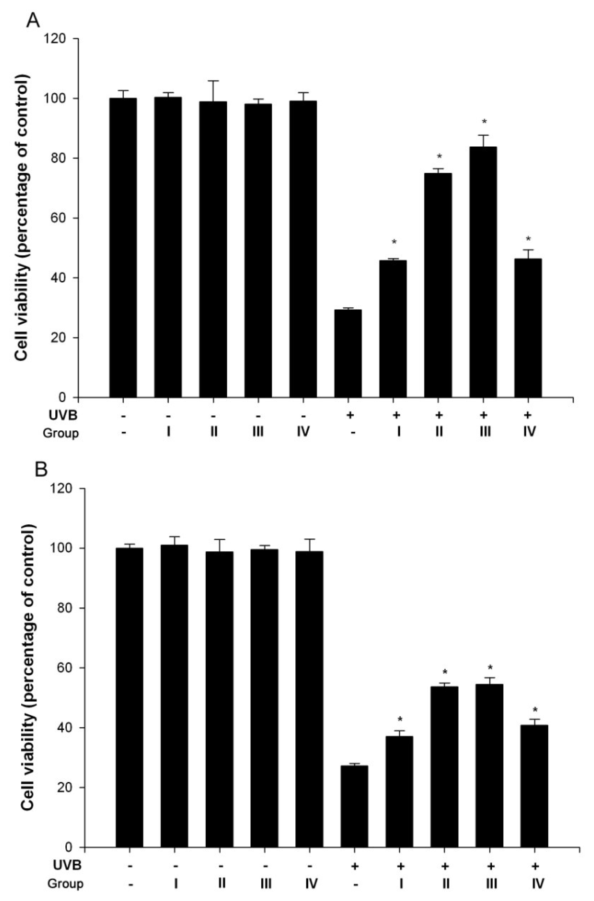
3.1. Isoflavone extracts inhibited UVB-induced cell death

To determine protective effects of isoflavone extracts obtained from soybeans (group I~IV, Table 1) on UVB-irradiated keratinocytes, cell viability was determined by the MTT assay. Figure 1 shows that isoflavone extracts alone didn’t affect keratinocyte viability but keratinocytes viability was decreased after UVB exposure.
However, UVB-induced cell death was inhibited by the treatment of group I~IV isoflavone extracts at concentrations in 0.5 and 1 % (Figure 1). 0.5 % of isoflavone extracts was sufficient to exert maximum protective effect in UVB-irradiated keratinocytes. These results indicate that aglycone, glucoside, acetylglucoside and malonylglucoside isoflavone extracts are able to prevent UVB-induced human skin damage.

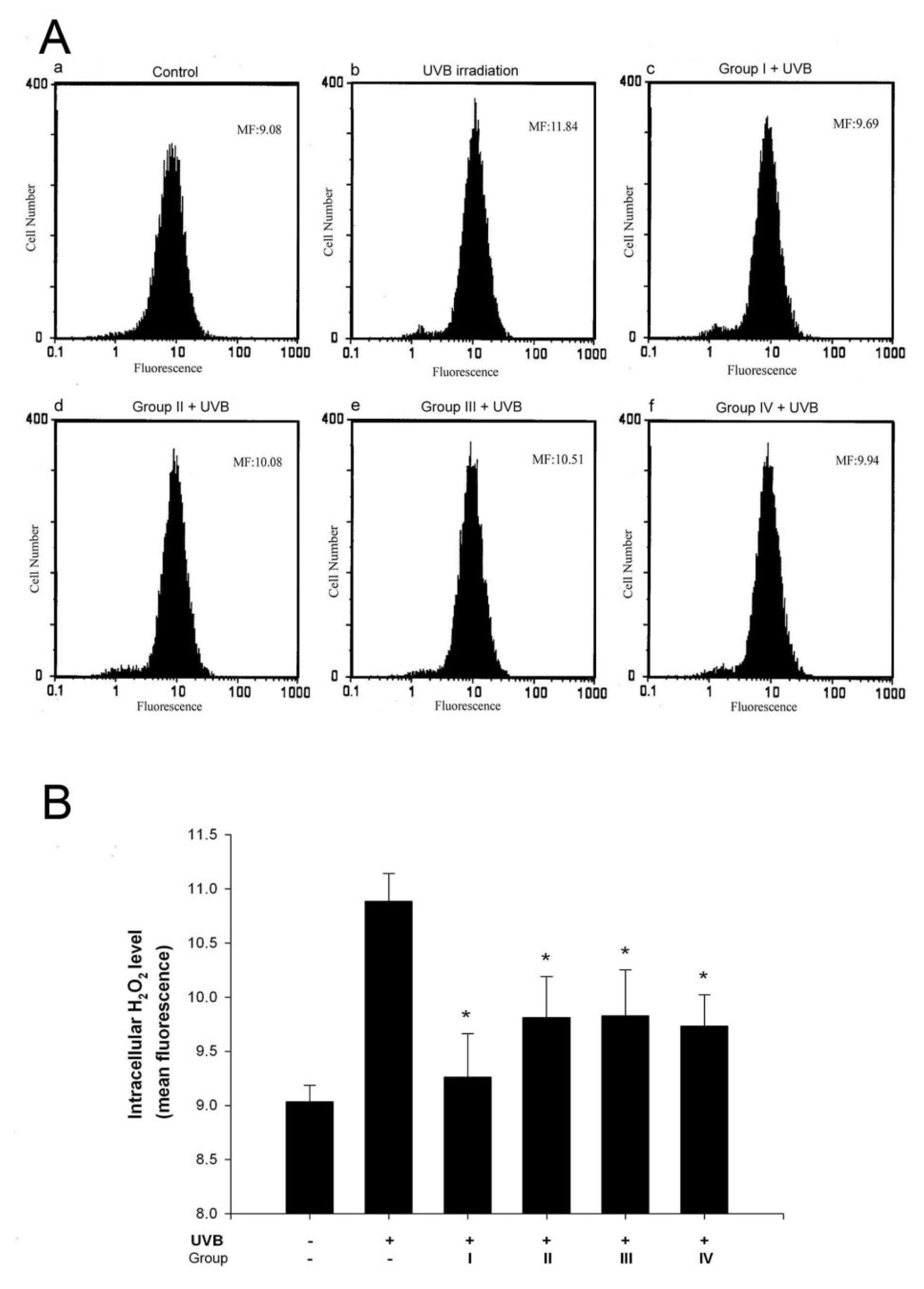
3.2. Isoflavone extracts inhibit UVB-induced H2O2 generation in keratinocytes

It has been shown that H2O2 is generated in cultured human skin cells during UVB irradiation [15,16]. In addition, group I~IV isoflavone extracts have been shown to possess antioxidant activity [5]. Therfore, we determined whether these extracts affect UVB-induced intracellular H2O2 production. Intracellular H2O2 in keratinocytes exposed to UVB was measured by dihydrorhodamine 123 (DHR 123), which has been shown to react with H2O2 in the presence of peroxidase and is extensively used as a probe for the detection of intracellular H2O2 [16;17].
As shown in Figure 2, flowcytometric analysis showed that mean fluorescence, i.e. H2O2 production, was increased in UVB-treated cells (Figure 2A, panels a and b). The increase of intracellular H2O2 by UVB irradiation was decreased by the treatment of these isoflavone extracts (Figure 2), while the basal level of intracellular H2O2 was not affected (data not shown). The result directly demonstrated that all isoflavone extracts have potent scavenging activity which can prevent UV induced intracellular H2O2 production.

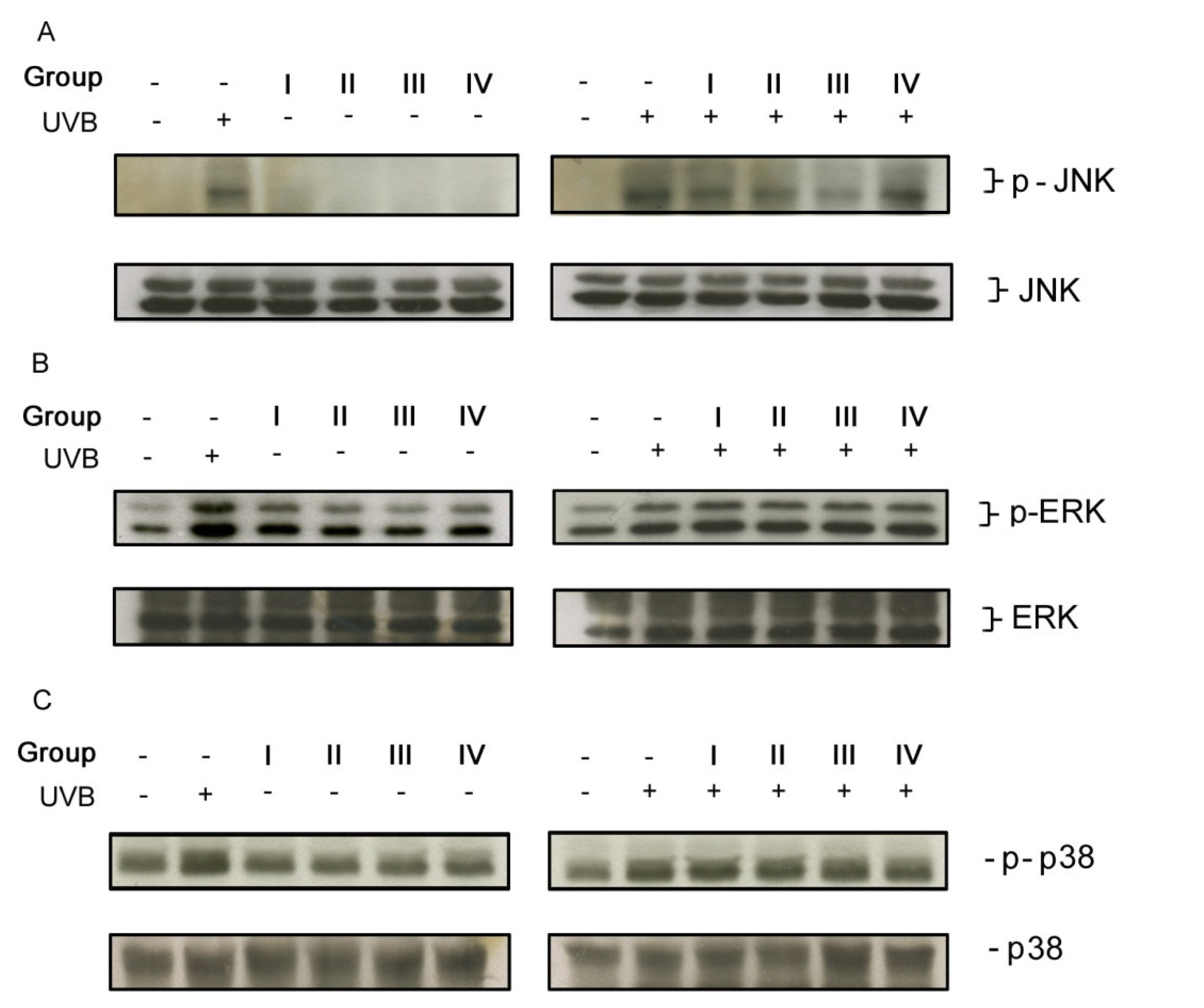
3.3. Isoflavone extracts differentially inhibited UVB-induced MAP kinase signaling pathway

Ultraviolet B irradiation has been shown to activate ERK1/2, JNK, and p38 kinase [14;16;18], which may lead to skin cell damage. Thus, we examined if these isoflavone extracts could affect UVB-induced MAPK activation. We observed that ERK1/2, JNK, and p38 phosphorylation were apparently increased in UVB-irradiated keratinocytes.
The basal level of JNK phosphorylation was not affected by isoflavone extracts, but UVB-induced JNK activation was inhibited by group I~III but not by group IV isoflavone extracts. Group III isoflavone extract elicited more inhibitory effect than group I and II (Figure 3A, upper panels). However, UVB-induced ERK1/2 and p38 activation in keratinocytes was not significantly affected by group I~IV isoflavone extracts (Figure 3B and 3C, upper panels), suggesting that aglycone, glucoside, acetylglucoside and malonylglucoside isoflavones differentially affect MAPK activation. The inhibition by these isoflavones was not due to uneven loading because reprobing of the immunoblots with antibodies raised against total JNK, ERK1/2, and p38 showed equal loading of each sample (Figure 3A, B, C, lower panels).

Discussion

In the present study we examined the protective effects in four groups of isoflavone extracts from soybean cake on UVB-induced keratinocyte damage. Treatment with isoflavone extracts (groups I~IV) significantly increased cell viability in UVB-irradiated keratinocytes. Both glucoside (group II) and acetylglucoside (group III) extracts exhibited higher efficacy in preventing UVB-induced cell death than malonylglucoside and aglycone extracts (Figure 1). This is consistent with the results reported by Kao and coworkers [5] stating that among these four isoflavone extracts, the glucoside and acetylglucoside extracts are relatively stronger antioxidants in scavenging DPPH free radical and chelating metal ions [5]. This suggests that UVB protective effect of these isoflvones is related with their antioxidant activities. However, group I~IV isoflavone extracts exerted similar inhibitory effects on UVB-induced H2O2 production (Figure 2). Since H2O2 and other reactive oxygen species play an important role in causing UVB-induced skin cell damage [19], the scavenging effects on free radicals other than H2O2 by these isoflavone extracts may also contribute to their protective effect on UVB-induced cell death.
UV irradiation has been reported to upregulate expression of transcription factors [20;21], which is mediated by the sequential activation of cytoplasmic protein kinases. To date, three structurally related but biochemically and functionally distinct MAPK signal pathways have been identified, including ERK1/2, JNK, and p38 [22]. Peus et al.[17,23] have demonstrated that ERK1/2, JNK, and p38 are activated in human epidermal keratinocytes following exposure to UVB irradiation. Several lines of evidence have also supported that the activation of these kinases correlates with skin cell death in response to UVB [24]. Assefa et al. [25] have shown that UVB induce keratinocyte apoptosis and produce a sustained p38 activation, which in turn leads to cytochrome c release and procaspase-3 activation. Gene disruption studies have shown that JNK mediates UV-stimulated apoptosis via the mitochondrial pathway by a Bax/Bak-dependent mechanism [26]. These studies indicate that at least p38 and/or JNK are involved in UVB irradiation-induced keratinocyte death. In the present study, we found that these four isoflavones differentially affected UVB irradiation-induced MAPK activation. Groups I~III exhibited a pronounced inhibitory effect on UVB irradiation-induced JNK phosphorylation, whereas group IV did not. Moreover, these isoflavone extracts did not have inhibitory effects on UVB irradiation-induced p38 and ERK1/2 activation (Figure 3). However, all these isoflavone extracts reversed UVB irradiation-induced keratinocyte death (Figure 1), suggesting group IV may act through affecting other signaling pathways.
Although group I~IV isoflavone extracts can reverse UVB irradiation-induced keratinocyte death, it was found they have differential reversal effects (Figure 1). It has been reported that UVB irradiation dose at 40 mJ/cm2 induces not only H2O2 but also many DNA damages such as cyclobutane pyrimidine dimers and 6–4 photoproducts in keratinocytes [27,28]. Therefore, an agent that can activate the DNA repair system may also have an anti-cell death ability. Based on this hypothesis and the mentions discussed above, group II and III isoflavone extracts possibly have relatively stronger antioxidant activity on ROS, inhibitory effect on JNK activation, and inductory effect on DNA repairing. However, whether they can directly activate the DNA repair system in human keratinocytes needs to be further determined.
In conclusion, our study demonstrated that isoflavone extracts from soybean cake can inhibit UVB-induced intracellular H2O2 production, JNK activation, and consequently protect human keratinocytes against UVB-induced death. Since these isoflavone extracts are relative stable and obtained easily than isoflavone standards, soybean cake may be the source for developing effective agents in skin care against photoaging.
Figure 1. Group I~IV isoflavone extracts inhibited UVB-induced keratinocyte death. Human keratinocytes pretreated with various concentrations of isoflavone extracts (A: 0.5 %; B: 1 %) were exposed to UVB irradiation (40 mJ/cm2) and then incubated for an additional 24 h in the absence or presence of isoflavones. *P < 0.05 vs. UVB-exposed cells without isoflavone treatment.
Ijms 08 00651f1
Figure 2. Group I~IV isoflavone extracts inhibited UVB irradiation-induced intracellular H2O2 production in human keratinocytes. (A) Intracellular H2O2 production [denoted by mean fluorescence (MF)] was expressed as histogram and (B) the results from four independent experiments was analyzed. *P < 0.05 vs. UVB-exposed cells without isoflavone treatment.
Ijms 08 00651f2
Figure 3. Effect of isoflavone extracts on UVB-induced JNK, ERK and p38 phosphorylation. The results are the representatives of four to six experiments.
Ijms 08 00651f3
Table 1. Contents in group I~IV isoflavone extracts from soybean.
ExtractsIsoflavone nameConcentration (μg/ml)
aglycone extract (Group I)daidzein47.3 ± 0.5
genistein36.4 ± 0.1
glycitein16.3 ± 0.2
glucoside extract (Group II)daidzin25.3 ± 0.1
genistin45.9 ± 1.6
glycitin29.2 ± 0.2
acetylglucoside extract (Group III)acetyldaidzin19.7 ± 0.9
acetylgenistin69.0 ± 1.6
acetylglycitin12.8 ± 0.7
malonylglucoside extract (Group IV)malonyldaidzin195.2 ± 1.2
malonylgenistin140.4 ± 4.8
malonylglycitin64.0 ± 0.8

Acknowledgements

This work was supported by the research grants from the National Science Council and from Fu-Jen Catholic University, Taipei, Taiwan.

References

  1. Armstrong, B.; Doll, R. Environmental factors and cancer incidence and mortality in different countries, with special reference to dietary practices. Int. J. Cancer 1975, 15, 617–631. [Google Scholar]
  2. Correa, P. Epidemiological correlations between diet and cancer frequency. Cancer Res 1981, 41, 3685–3690. [Google Scholar]
  3. Messina, M.; Barnes, S. The role of soy products in reducing risk of cancer. J. Natl. Cancer Inst 1991, 83, 541–546. [Google Scholar]
  4. Kao, T.H.; Chen, B.H. An improved method for determination of isoflavones in soybean powder by liquid chromatography. Chromatographia 2002, 56, 423–430. [Google Scholar]
  5. Kao, T.H.; Chen, B.H. Functional components in soybean cake and their effects on antioxidant activity. J. Agric. Food Chem 2006, 54, 7544–7555. [Google Scholar]
  6. Wei, H.; Saladi, R.; Lu, Y.; Wang, Y.; Palep, S.R.; Moore, J.; Phelps, R.; Shyong, E.; Lebwohl, M.G. Isoflavone genistein: photoprotection and clinical implications in dermatology. J. Nutr 2003, 133, 3811S–3819S. [Google Scholar]
  7. Russo, A.; Cardile, V.; Lombardo, L.; Vanella, L.; Acquaviva, R. Genistin inhibits UV light-induced plasmid DNA damage and cell growth in human melanoma cells. J. Nutr. Biochem 2006, 17, 103–108. [Google Scholar]
  8. Wei, H.; Spencer, J.; elfand, J.; Phelps, R.; Lebwohl, M. The isoflavone genistein: A new agent in dermatology? Cosmet. Dermatol 2001, 14, 13–19. [Google Scholar]
  9. Ji, S; Willis, GM; Frank, GR; Cornelius, SG; Spurlock, ME. Soybean isoflavones, genistein and genistin, inhibit rat myoblast proliferation, fusion and myotube protein synthesis. J. Nutr. 1999, 129, 1291–1297. [Google Scholar]
  10. Kao, T.H.; Lu, Y.F.; Chen, B.H. Preparative column chromatography of four groups of isoflavones from soybean cake. Eur. Food Res. Technol 2005, 221, 459–465. [Google Scholar]
  11. Huang, C.C.; Fang, J.Y.; Wu, W.B.; Chiang, H.S.; Wei, Y.J.; Hung, C.F. Protective effects of (−)- epicatechin-3-gallate on UVA-induced damage in HaCaT keratinocytes. Arch. Dermatol. Res. 2005, 296, 473–481. [Google Scholar]
  12. Mosmann, T. Rapid colorimetric assay for cellular growth and survival: application to proliferation and cytotoxicity assays. J. Immunol. Methods 1983, 65, 55–63. [Google Scholar]
  13. Green, L.M.; Reade, J.L.; Ware, C.F. Rapid colorimetric assay for cell viability: application to the quantitation of cytotoxic and growth inhibitory lymphokines. J. Immunol. Methods 1984, 70, 257–268. [Google Scholar]
  14. Wu, W.B.; Chiang, H.S.; Fang, J.Y.; Chen, S.K.; Huang, C.C.; Hung, C.F. (+)-Catechin prevents ultraviolet B-induced human keratinocyte death via inhibition of JNK phosphorylation. Life Sci. 2006, 79, 801–807. [Google Scholar]
  15. Peus, D.; Meves, A.; Vasa, R.A.; Beyerle, A.; O’Brien, T.; Pittelkow, M.R. H2O2 is required for UVB-induced EGF receptor and downstream signaling pathway activation. Free Radic. Biol. Med 1999, 27, 1197–1202. [Google Scholar]
  16. Katiyar, S.K.; Afaq, F.; Azizuddin, K.; Mukhtar, H. Inhibition of UVB-induced oxidative stress-mediated phosphorylation of mitogen-activated protein kinase signaling pathways in cultured human epidermal keratinocytes by green tea polyphenol (-)-epigallocatechin-3-gallate. Toxicol. Appl. Pharmacol 2001, 176, 110–117. [Google Scholar]
  17. Peus, D.; Vasa, R.A.; Beyerle, A.; Meves, A.; Krautmacher, C.; Pittelkow, M.R. UVB activates ERK1/2 and p38 signaling pathways via reactive oxygen species in cultured keratinocytes. J. Invest. Dermatol 1999, 112, 751–756. [Google Scholar]
  18. Afaq, F.; Malik, A.; Syed, D.; Maes, D.; Matsui, M.S.; Mukhtar, H. Pomegranate fruit extract modulates UV-B-mediated phosphorylation of mitogen-activated protein kinases and activation of nuclear factor kappa B in normal human epidermal keratinocytes paragraph sign. Photochem. Photobiol 2005, 81, 38–45. [Google Scholar]
  19. Takahashi, H.; Suzuki, Y.; Miyauchi, Y.; Hashimoto, Y.; Ishida-Yamamoto, A.; Iizuka, H. Roxithromycin decreases ultraviolet B irradiation-induced reactive oxygen intermediates production and apoptosis of keratinocytes. J. Dermatol. Sci 2004, 34, 25–33. [Google Scholar]
  20. Devary, Y.; Gottlieb, R.A.; Smeal, T.; Karin, M. The mammalian ultraviolet response is triggered by activation of Src tyrosine kinases. Cell 1992, 71, 1081–1091. [Google Scholar]
  21. Bender, K.; Blattner, C.; Knebel, A.; Iordanov, M.; Herrlich, P.; Rahmsdorf, H.J. UV-induced signal transduction. J. Photochem. Photobiol. B 1997, 37, 1–17. [Google Scholar]
  22. Su, B.; Karin, M. Mitogen-activated protein kinase cascades and regulation of gene expression. Curr. Opin. Immunol 1996, 8, 402–411. [Google Scholar]
  23. Peus, D.; Vasa, R.A.; Meves, A.; Beyerle, A.; Pittelkow, M.R. UVB-induced epidermal growth factor receptor phosphorylation is critical for downstream signaling and keratinocyte survival. Photochem. Photobiol 2000, 72, 135–140. [Google Scholar]
  24. Assefa, Z.; Van Laethem, A.; Garmyn, M.; Agostinis, P. Ultraviolet radiation-induced apoptosis in keratinocytes: on the role of cytosolic factors. Biochim. Biophys. Acta 2005, 1755, 90–106. [Google Scholar]
  25. Assefa, Z.; Vantieghem, A.; Garmyn, M.; Declercq, W.; Vandenabeele, P.; Vandenheede, J.R.; Bouillon, R.; Merlevede, W.; Agostinis, P. p38 mitogen-activated protein kinase regulates a novel, caspase-independent pathway for the mitochondrial cytochrome c release in ultraviolet B radiation-induced apoptosis. J. Biol. Chem 2000, 275, 21416–21421. [Google Scholar]
  26. Lei, K.; Davis, R.J. JNK phosphorylation of Bim-related members of the Bcl2 family induces Bax-dependent apoptosis. Proc. Natl. Acad. Sci. U S A 2003, 100, 2432–2437. [Google Scholar]
  27. Takasawa, R.; Nakamura, H.; Mori, T.; Tanuma, S. Differential apoptotic pathways in human keratinocyte HaCaT cells exposed to UVB and UVC. Apoptosis 2005, 10, 1121–30. [Google Scholar]
  28. Courdavault, S.; Baudouin, C.; Charveron, M.; Canguilhem, B.; Favier, A.; Cadet, J.; Douki, T. Repair of the three main types of bipyrimidine DNA photoproducts in human keratinocytes exposed to UVB and UVA radiations. DNA Repair (Amsterdam) 2005, 4, 836–44. [Google Scholar]

