Nanomaterials-Based Optical Techniques for the Detection of Acetylcholinesterase and Pesticides

Abstract
: The large amount of pesticide residues in the environment is a threat to global health by inhibition of acetylcholinesterase (AChE). Biosensors for inhibition of AChE have been thus developed for the detection of pesticides. In line with the rapid development of nanotechnology, nanomaterials have attracted great attention and have been intensively studied in biological analysis due to their unique chemical, physical and size properties. The aim of this review is to provide insight into nanomaterial-based optical techniques for the determination of AChE and pesticides, including colorimetric and fluorescent assays and surface plasmon resonance.1. Introduction
Acetylcholinesterase (AChE) catalyzes the hydrolysis of the neurotransmitter acetylcholine (ACh) to inactive choline. Inhibition of AChE will lead to the accumulation of acetylcholine in the synaptic cleft, resulting in impeded neurotransmission. AChE has regained great attention recently due to its association with Alzheimer's disease (AD) and other neurodegenerative diseases characterized by low ACh levels owing to catabolism by AChE [1,2]. Moreover, AChE is the primary target of inhibition by organophosphorus compounds such as nerve agents and pesticides (Figure 1); thus, public concern about the development of detection devices for effectively monitoring pesticides has also grown steadily [3–8].
The combination of enzymatic reactions with the various methods of monitoring enzymatic products has allowed the development of enzyme-based devices for sensitive and rapid determination of acetylcholine, AChE and its inhibitors [9–14]. In line with the rapid development of nanotechnology, nanomaterials have attracted great attention and have been intensively studied in biological analysis and detection due to their unique chemical, physical and size properties [15,16]. Commonly, nanomaterials can be employed for development of AChE-based sensing devices in the following three ways: (1) nanomaterials are used as enzyme carriers for loading a large amount of AChE to enhance the detection signal [17,18], especially in the electrochemical detection protocols, (2) nanomaterials act as peroxidase- or oxidase-like activities catalysts to catalyze the oxidation of various substrates including 2,2′-azino-bis(3-ethylbenzo-thiazoline-6-sulfonic acid) diammonium salt and 3,3,5,5-tetramethylbenzidine (TMB) by enzyme-generated hydrogen peroxide (H2O2) for colorimetric or fluorescence detection of acetylcholine and AChE inhibitors [19,20], and (3) nanomaterials are employed as the direct signal sources [21–23]. Electrochemical techniques based on the inhibition of AChE are attractive for the detection of acetylcholine and AChE inhibitors. In a typical configuration, the enzyme is deposited onto the surface of the working electrode. The activity of AChE is then monitored by adding the substrate to the solution and follow-up measuring the redox current of the enzymatic products. To improve the performance characteristics of the AChE-based electrochemical methods, electrode materials including nanomaterials have allowed for large quantities of enzyme to be immobilized, provide a favorable microenvironment to maintain the enzyme activity, and facilitate the oxidation of the enzymatic products. Nanomaterials used for the fabrication of AChE-based electrochemical biosensors have been summarized in recent review papers [10,24–26]. In this work, we highlighted the progress in development of nanomaterials-based optical techniques for the determination of AChE and pesticides.
2. Nanomaterials-Based Optical Techniques for Detection of Acetylcholine and AChE Inhibitors
The AChE-based sensing systems include the use of AChE alone or combination with choline oxidase (ChO). The AChE inhibition in the single and bienzyme systems is monitored by determining the generated enzyme production. AChE can hydrolyze acetylcholine or acetylthiocholine (a synthesized analogues of acetylcholine) to produce choline or thiocholine (a thiol compound), respectively. In the single enzyme system, AChE hydrolyzes acetylthiocholine to produce the thiocholine (Equation (1)). In the bienzyme system, AChE catalyzes the hydrolysis of acetylcholine into acetate and choline (Equation (2)). The choline is subsequently converted by ChO, producing hydrogen peroxide in the presence of oxygen (Equation (3)). Traditional optical methods to measure the levels of AChE and its inhibitors include the spectrophotometric thiol assay by using Ellman's reagent and the colorimetric detection of H2O2 produced by oxidation of the AChE-induced choline by using horseradish peroxidase (HRP) [27,28]. However, the methods lack sufficient sensitivity and require time-consuming sample-handling procedures. In order to enhance the detection sensitivity, advanced techniques based on metallic/magnetic nanoparticles and quantum dots have been developed recently, including colorimetric and fluorescent assays and surface plasmon resonance. Their preparation, modification and detection principle are presented herein.
2.1. Colorimetric Assays
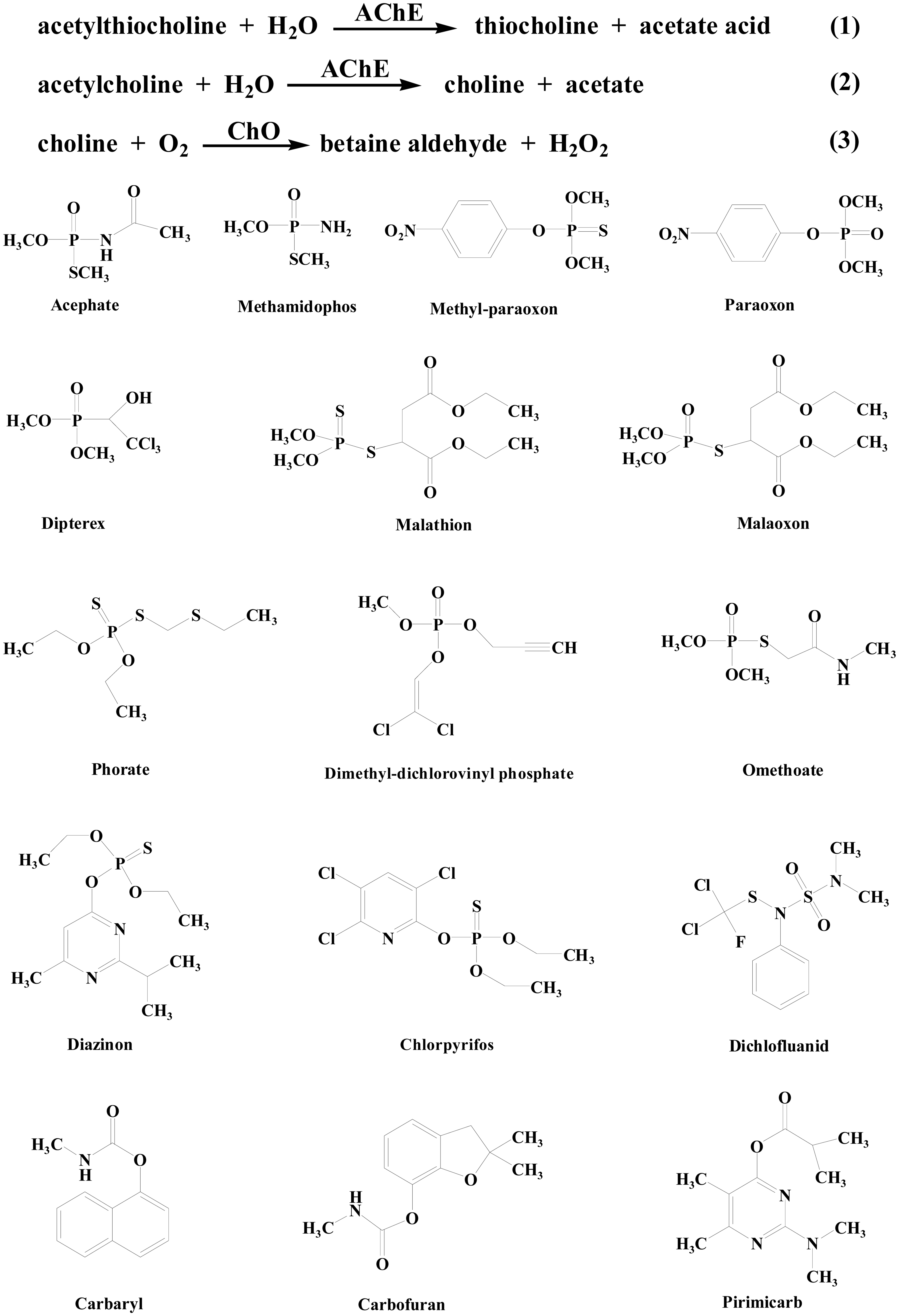
Because of the high extinction coefficients and the unique size-dependent optical properties of gold nanoparticles (AuNPs), AuNPs-based colorimetric assays have recently become useful for many types of analytes without the need for advanced instruments, including screening the enzyme activity and measuring the concentrations of nucleic acid, proteins, metal ions and other small molecules [29–33]. In this process, molecular events can be transformed into color changes. Usually, the color changes are highly sensitive to the size, shape, capping agents, and medium refractive index, as well as the aggregation states of AuNPs, which can be confirmed by the significant absorption band shift in the visible region of the electromagnetic spectrum. Based on the unique physical properties of AuNPs, Pavlov et al., presented the first colorimetric detection of AChE inhibitors based on the color change of AuNPs [21]. In the work, AChE mediated hydrolysis of acetylthiocholine to yield a reducing reagent thiocholine that modulated the growth of AuNPs seeds in the presence of AuCl4−. The catalytic growth of AuNPs was prevented by inhibition of AChE activity using 1,5-bis(4-allyldimethyl-ammoniumphenyl)-pentane-3-one dibromide or paraoxon, thus enabling a colorimetric assay for AChE inhibitors. However, this method is less sensitive because the enzymatic generation of AuNPs would consume thiocholine produced in the course of enzymatic reactions. Thus, the authors developed another method for the detection of AChE inhibitors based on the modulation of AuNPs growth by the enzymatically generated thiocholine [34]. As shown in Figure 2, the produced thiocholine hindered the deposition of Ag reduced by ascorbic acid from AgNO3 by binding to the surface of the Au seeds. As a result, the formation of Ag-coated AuNPs is blocked (route A). In the presence of AChE inhibitors, hydrolysis of acetylthiocholine by AChE was prevented, which allowed for the deposition of Ag on AuNPs surface (route B).
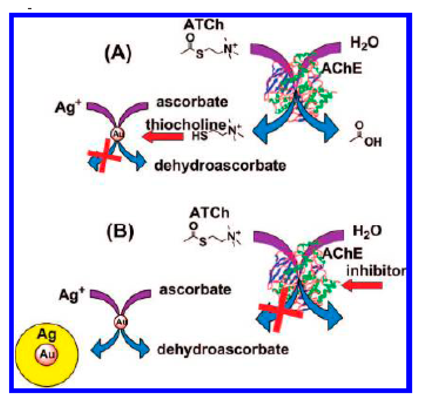
The modulation of aggregation states of AuNPs has also been broadly used as a colorimetric assay for various analytes. With the continual aggregation of AuNPs, the color of the dispersion gradually changes from an initial red to purple, then blue and, finally, yellow. The color change of AuNPs suspension can be confirmed by the significant absorption band shift in the visible region of the electromagnetic spectrum. For this view, Wang et al. demonstrated that the positively charged thiocholine can substitute the citrate on the surface of AuNPs; as a result, the cross-linking/aggregation of interparticles occurs because of the electrostatic interaction between thiocholine and citrate on gold surface, leading to the red-shift of the plasmon absorption of AuNPs suspension [35]. The degree of AuNPs aggregation is strictly dependent on the concentration of the produced thiocholine; thus, AChE activity could be monitored by the AuNPs-based colorimetric assay. With the method, AChE at the concentration as low as 0.6 mU/mL and tacrine (a well-known inhibitor for AChE) below 4 nM can be readily assayed. With the same principle, Sun et al., reported the detection of organophosphate (OP) nerve agents and pesticide using lipoic acid (LA)-capped AuNPs with a pM detection limit [36]. Furthermore, Liu et al., reported the assays of AChE in the cerebrospinal fluid of transgenic mice suffering from Alzheimer's disease and four pesticides (carbaryl, diazinon, malathion and phorate) using rhodamine B (RB)-functionalized AuNPs (RB-AuNPs) as the dual (colorimetric and fluorometric) readouts [37–39]. Specifically, electrostatic absorption of RB onto the surface of AuNPs leads to the quenching of RB's fluorescence (Figure 3). Thiocholine produced from acetylthiocholine by AChE substitutes RB on the surface of AuNPs, resulting in the color change of the solution from red to purple, simultaneously accompanied by the recovery of fluorescence of RB. The detection limit for AChE reaches 0.1 mU/mL and the lowest detectable concentrations for carbaryl, diazinon, malathion, and phorate are 0.1, 0.1, 0.3 and 1 μg/L, respectively.
Besides AuNPs, AgNPs have also been employed by Li et al., for organophosphorus pesticide detection based on the thiocholine-induced aggregation of the citrate-stabilized AgNPs [40]. Similarly, organophosphorus pesticides prevented the production of thiocholine by inhibiting the activity of AChE, thus holding back the aggregation of AgNPs. As a result, a detection limit of 0.18 ng/mL for dipterex was achieved.
Fe3O4 magnetic nanoparticles (MNPs) exhibit peroxidase activity that can catalyze the oxidation of peroxidase substrates in the presence of H2O2 to produce a color reaction [41]. Liang et al. reported a Fe3O4 MNPs-based colorimetric method for the detection of organophosphorus pesticides and nerve agents using AChE and CHO [19]. In this method, AChE and CHO catalyzed the production of H2O2 in the presence of acetylcholine, which then activated MNPs to catalyze the oxidation of colorimetric substrate TMB to produce a color reaction. Inhibition of AChE by organophosphorus pesticides (acephate and methylparaoxon) and the nerve agent Sarin prevented the production of H2O2, resulting in a reduced catalytic oxidation of TMB and a decrease in the color intensity. Acephate, methylparaoxon and Sarin at the concentrations below 1 nM, 10 nM and 5 μM, respectively, can be readily detected.
2.2. Fluorescent Assays
2.2.1. Quantum Dots
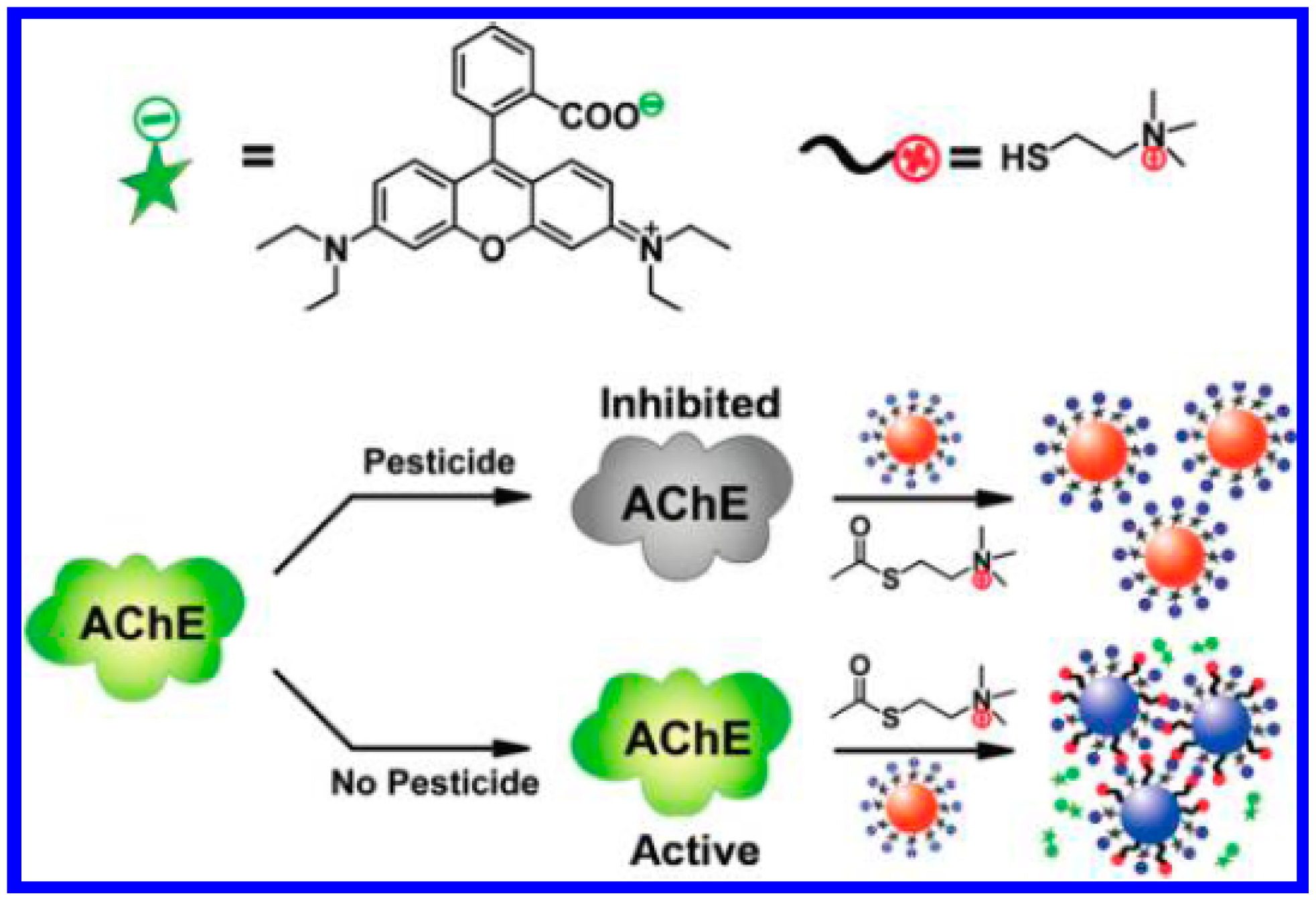
Comparing the chromatography evaluations and electrochemical analysis methods that need either time-consuming operation or complicated labeling and modification procedures, fluorimetric methodologies stand out as rapid, sensitive, and efficient especially in combination with the nanotechnologies and fluorescent nanomaterials. Semiconductor quantum dots (QDs) are the commonly known nanoparticles used in fluorescent sensing. The most important advantages of QDs over organic fluorophores are higher brightness, reduced photobleaching and longer lifetimes. Recently, several groups have reported the QDs-based fluorescence assays for detection of AChE activity and organophosphorus pesticides [22,42–46]. Typically, Pavlov's group demonstrated that thiocholine released from the AChE-catalyzed hydrolysis of acetylthiocholine (ATCh) are able to catalyze the production of fluorescent CdS QDs in the presence of thiosulfate and Cd2+ (Figure 4) [22] and can mediate stabilization of in situ produced CdS quantum dots [46]. As a result, AChE activity and its inhibitors can be determined by the fluorescence intensity of the resulting CdS QDs.
Silicon quantum dots (SiQDs), as inert, nontoxic, abundant, and low-cost nanomaterials, have been demonstrated to be environmentally friendly photoluminescence probes and have attracted much interest. In comparison to other QDs, SiQDs have unique optical and electronic properties, especially favorable biocompatibility. Yi et al. found that the fluorescence of label-free SiQDs could be effectively quenched by enzyme-generated H2O2 [47]. For this view, they further developed a SiQDs-based sensor for pesticides detection based on the fluorescence quenching of SiQDs induced by the enzyme-generated H2O2 [48]. Specifically, AChE hydrolyzed acetylcholine to choline; choline was then enzymatically oxidized by ChOx to produce betaine and H2O2. If the activity of AChE was inhibited by pesticides, the amount of the generated H2O2 would reduce, resulting in an increase in the fluorescence of SiQDs. The method allowed for the detection of carbaryl, parathion, diazinon and phorate at the concentrations below 7.25 ng/L, 32.5 ng/L, 67.6 ng/L and 0.19 mg/L, respectively. Additionally, Shen et al. found that the fluorescence of core-shell silica particles with tetraphenylethylene moieties could be quenched by dabcyl-ACh due to the electrostatic interaction between the silica particles and dabcyl-ACh [49]. After incubation with AChE, dabcyl-ACh was degraded between the residues of dabcyl and ACh, which caused the removal of dabcyl residues from the silica surface and the recovery of fluorescence of silica particles.
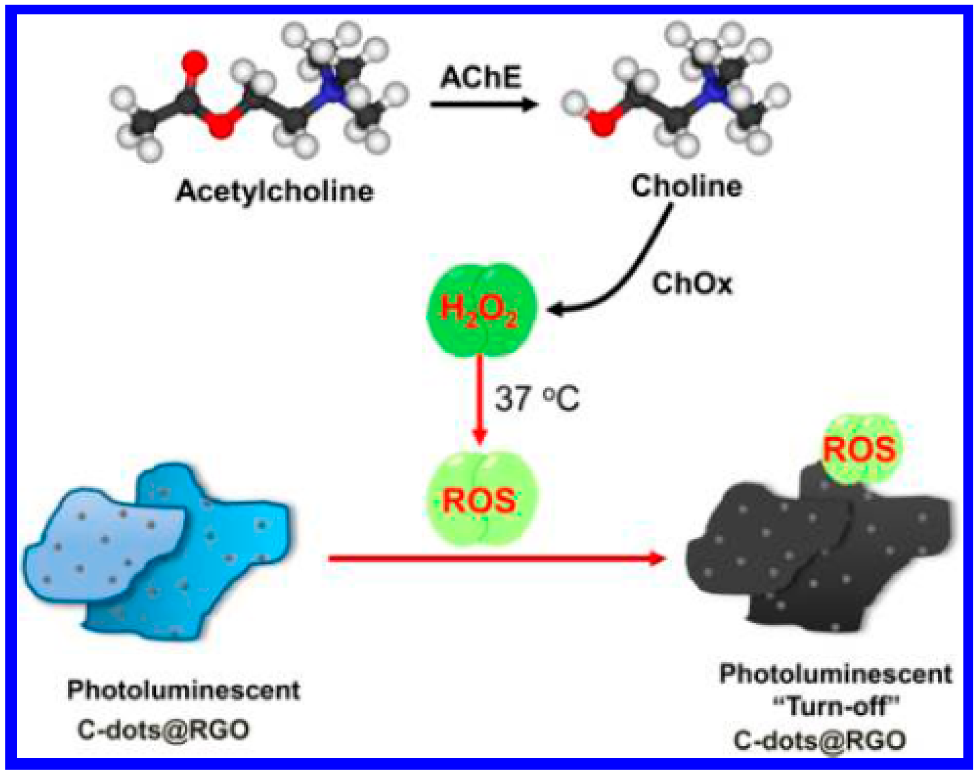
Reduced graphene oxide (RGO) has become a very popular sensing material for the detection of DNA, proteins, and small molecules because of its large planar surface and high photoluminescence quenching efficiency to fluorophores (e.g., organic dyes, quantum dots) [50]. However, as-prepared RGO is usually hydrophobic and nonphotoluminescent, thus limiting its direct use for biological application [51]. Recently, Chang's group reported a strategy for the synthesis of hydrophilic, photoluminescent (PL) carbon dots on RGO (C-dots@RGO) from graphene oxide (GO) through a hydrothermal reduction route using catechin as a reductant [52,53]. Furthermore, they found that the AChE/ChOx-mediated production of H2O2 caused the photoluminescent quenching of the C-dots@RGO via an etching process (Figure 5) [51]. The photoluminescent intensity of the C-dots@RGO is inversely proportional to the acetylcholine concentration in the range of 0.05−10 nM, with a detection limit of 30 pM. By this method, the concentrations of acetylcholine in plasma and blood samples were determined to be 2.6 nM and 6.8 nM, respectively.
2.2.2. Metal Nanoparticles
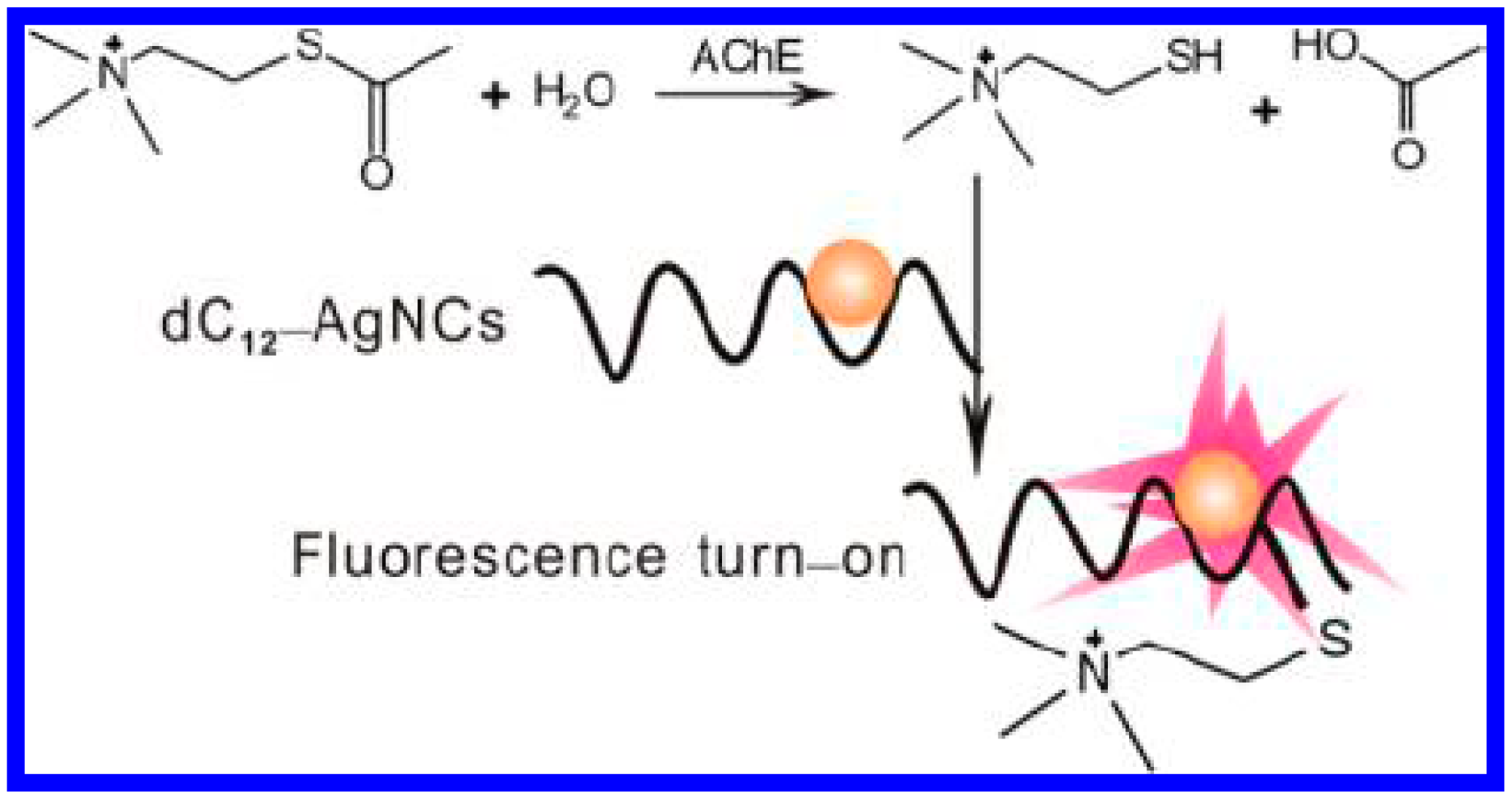
The emerging technology of fluorescent few-atom metal (e.g., Au or Ag) nanoclusters (NCs) offers an attractive compromise between the photostability and brightness of quantum dots and the compact versatility of dye fluorophores [54]. This technology has recently been used in a wide range of chemical or biological detection and cellular imaging applications. For this consideration, Li et al. developed a fluorometric sensor for the detection of AChE activity and its inhibitors [23]. The method was based on the thiocholine-induced fluorescence quenching of DNA-templated copper/silver nanoclusters (DNA-Cu/AgNCs). The AChE activity could be detected as low as 0.05 mU/mL and with a linear range from 0.05 to 2.0 mU/mL. Inversely, Zhang et al., found that the reaction of thiocholine to 12 polycytosine-templated silver nanoclusters (dC12−AgNCs) through the formation of Ag−S bonds lead to the increase of fluorescence of dC12−AgNCs (Figure 6) [55]. The hydrolysis of ATCh chloride was retarded in the presence of the corresponding inhibitor. Thus, AChE activity and its inhibitors could be determined with dC12−AgNCs as the fluorescence probes. This method allowed for the assay of AChE as low as 0.05 mU/mL. Moreover, based on the interaction of thiocholine and Ag(I), Liao et al. reported a “turn-on” fluorescent method for probing of AChE activity and sensing of AChE inhibitors. Specifically, a polyanion (poly(vinyl sulfonate)–PVS) could induce the aggregation and fluorescence quenching of a perylene probe (probe 1). The produced thiocholine interacted with Ag(I) to form a positively charged metal coordination polymer. As a result, the polycation interacted with the polyanion and caused the release of the free probe 1 monomer molecules, and a fluorescence turn on signal was detected [56].
Beside Ag NCs, Au NCs have also been extensively studied because of their intrinsic characteristics such as ease of preparation and chemical stability. Recently, Li et al. synthesized the denatured bovine serum albumin (dBSA)-protected AuNCs and demonstrated their applications in the fluorescent detection of AChE activity in human serum [57]. Specifically, the fluorescence of AuNCs was quenched by the produced thiocholine due to the combination of thiocholine with the dBSA-AuNCs. The method showed a linear range of 0.005–0.15 U/mL for AChE with a detection limit of 0.02 mU/mL. Also, Zhang et al. found that bovine serum albumin (BSA)-stabilized gold nanoclusters (BSA-AuNCs) can be used as the fluorimetric reaction substrate for probing the activity and phosphorylation of AChE and detecting dimethyl-dichlorovinyl phosphate (DDVP) [58]. They suggested that thiocholine released from the AChE-catalyzed hydrolysis of ATC caused the aggregation and fluorescence reduction of BSA-AuNCs. With this method, DDVP could be determined with a detection limit of 13.67 pM.
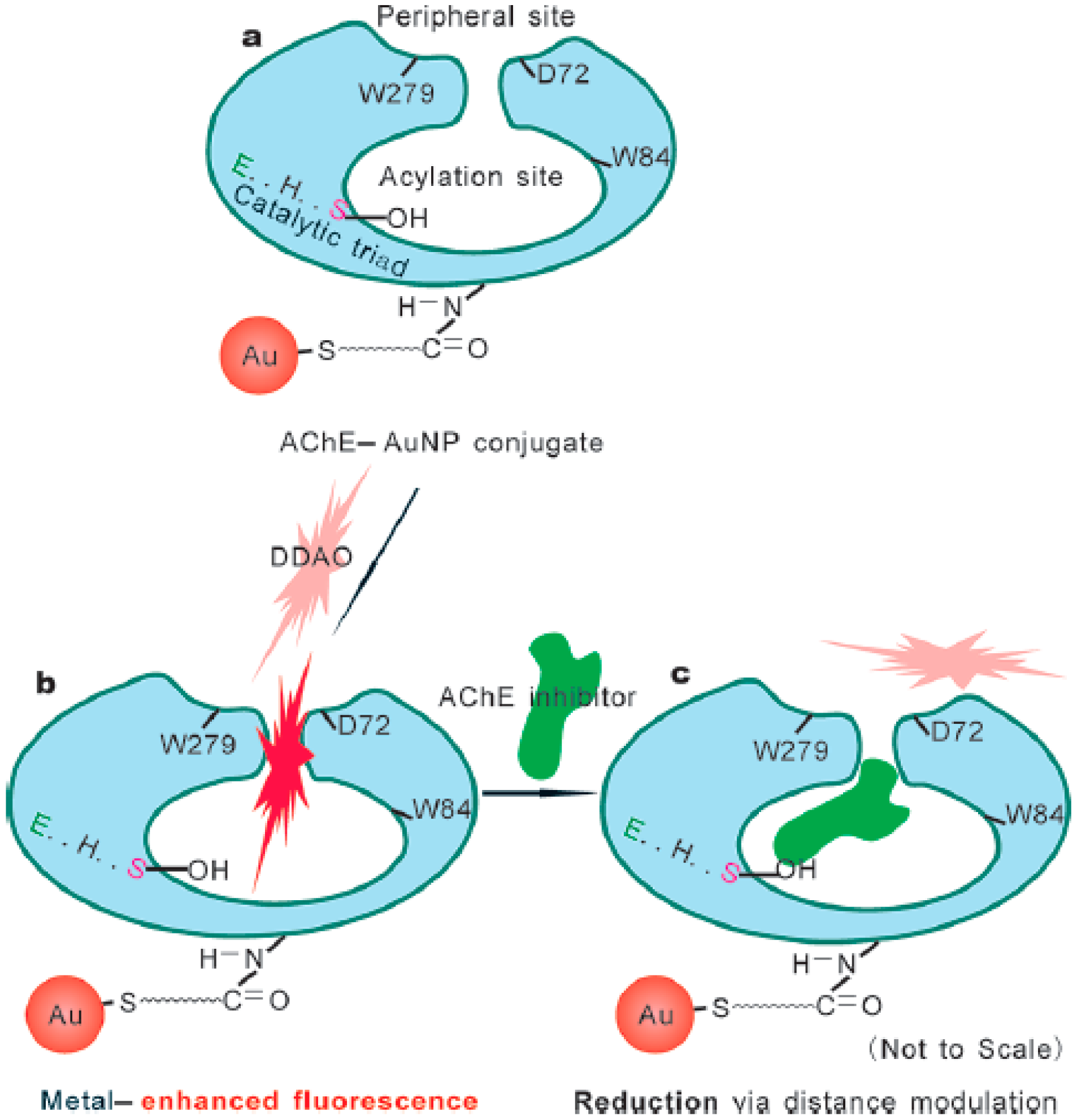
Recently, there has been increasing attention paid to the use of metal NPs-based metal-enhanced fluorescence (MEF) for immunoassays, protein translation and DNA detection [59–62]. Enhancement is attributed primarily to the increased electric field close to the metal NPs induced by incident light. One of factors affecting the enhancement magnitude is the distance between fluorophores and metal nanostructures. Interestingly, Zhang et al. found that AChE could modulate the distance between AuNPs and fluorophore 7-hydroxy-9H-(1,3-dichloro-9,9-dimethylacridin-2-one) (DDAO) (Figure 7) [63]. Binding of DDAO to AChE immobilized onto AuNPs lead to the enhancement of DDAO's fluorescence due to MEF (Figure 7a). Because AChE inhibitors competed with DDAO to bind the peripheral anionic site and penetrate into the active gorge site of AChE, inhibition of AChE activity by AChE inhibitors such as paraoxon and tacrine prevented the interaction of AChE and DDAO, thus resulting in the distance variation between AuNPs and DDAO and reducing the fluorescence signal (Figure 7b). The results demonstrated that the biosensor shows low detection limits for paraoxon (0.4 μM) and tacrine (10 nM).
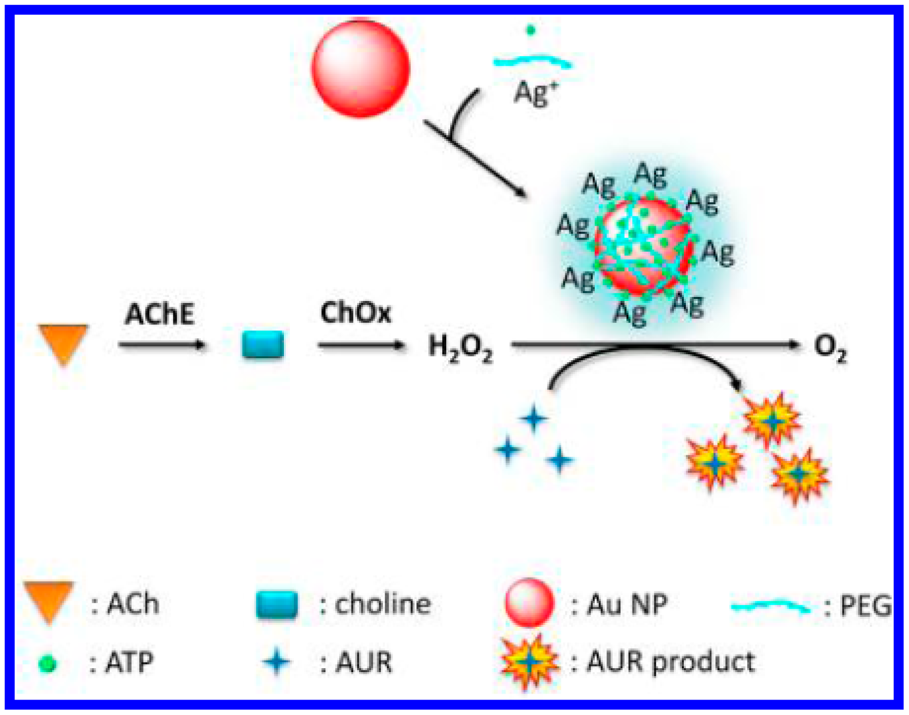
Moreover, nanoparticles (NPs) including Fe3O4, sheet-like FeS, spherical CeO2, single-walled carbon nanotubes, graphene oxide, AgM (M = Au, Pd, Pt) and metallic nanocomposites show peroxidase- or oxidase-like activities [20,41,64,65]. Chang's group suggested that Au/Ag bimetallic nanoparticles promoted the H2O2-mediated oxidation of Amplex UltraRed (AUR) and thus reported the fluorescent detection of acetylcholine (Figure 8) [20]. In this process, AChE catalyzes the hydrolysis of acetylcholine into acetate and choline. The choline is subsequently converted by ChO, producing H2O2 in the presence of oxygen. The as-produced H2O2 was made to react with AUR in the presence of Au/Ag bimetallic NPs. The detection limit of this method for acetylcholine was 0.21 nM.
2.3. Surface Plasmon Resonance
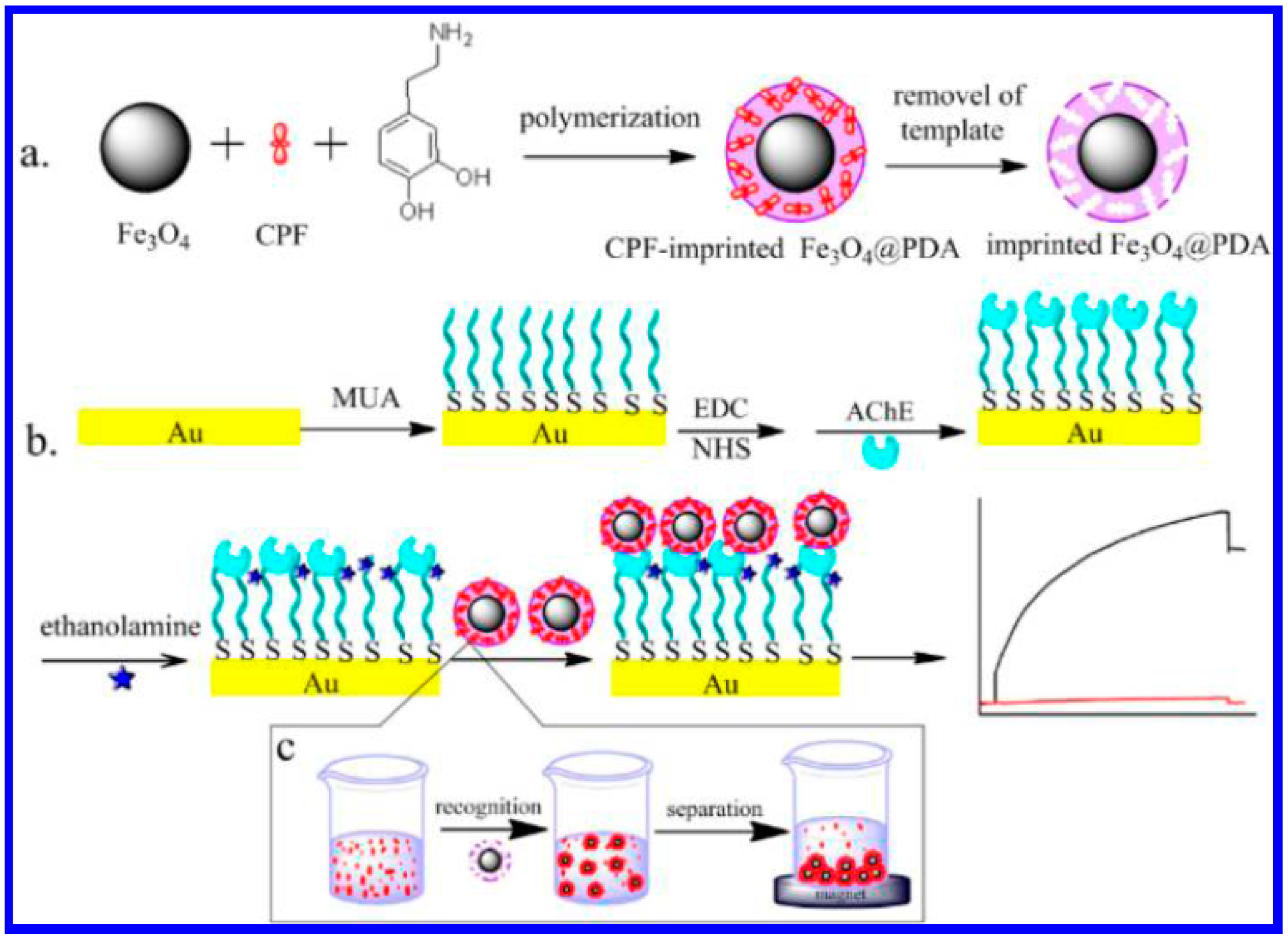
SPR is the collective oscillation of electrons in a solid or liquid stimulated by incident light [66]. The resonance condition is established when the frequency of light photons matches the natural frequency of surface electrons oscillating against the restoring force of positive nuclei. SPR instruments optically monitor changes that occur on certain metal sensor surfaces (typically gold and silver) when sample fluid flows past the surface. Several SPR optical biosensors have been developed for detecting low levels of AChE inhibitors including pesticides residues [67–69]. However, because of the small size of the inhibitors, binding of the targets to the AChE-covered sensing chip dose not bring significant shift in the resonance angle. Thus, the use of the simple SPR method for pesticides detection shows a poor sensitivity. NPs can promote a significant shift in the angle of plasmon resonance. Qiu's group reported the synthesis of magnetic molecular imprinting polymers (MIPs) NPs with high density and accessible recognition sites for chlorpyrifos (CPF) [70]. The magnetic MIPs NPs were synthesized by self-polymerization of dopamine on the surface of Fe3O4 NPs in the presence of template CPF in weak base aqueous solution (Figure 9a). As a result, the target CPF molecules can be rapidly enriched and separated by the imprinted Fe3O4@polydopamine nanoparticles (Fe3O4@PDA NPs) by an external magnetic field. Integrating the CPF-imprinted Fe3O4@PDA NPs to a SPR chip through the specific interactions between the CPF rebound in the recognition cavities in the PDA matrix and the AChE immobilized on sensor chip results in a significant signal amplification due to the high molecular weight of Fe3O4@PDA NPs (Figure 9b). The SPR biosensor showed a low detection limit (0.76 nM) for CPF detection.
3. Conclusions/Outlook
In conclusion, we have reviewed the progress in the optical detection of AChE and pesticides using functional nanoscaffolds made of novel nanomaterials, such as metal and metal oxide nanoparticles, QDs and magnetic beads. Although most of the existing limitations of the AChE-based sensors could be directly related to the selectivity in multicomposite mixtures and complex matrices and the inability of identifying a specific pesticide, the sensitivity of most of the methods is sufficient to detect the minimum levels of total pesticides imposed by regulatory agencies. Moreover, the advances in nanoscience and nanotechnology promise a better future for designing of AChE-based biosensors that would complement or serve as an alternative to the existing expensive and complex chromatographic devices.
Acknowledgments
Partial support of this work by the National Natural Science Foundation of China (Nos. 21205003, 21305004) and the Joint Fund for Fostering Talents of National Natural Science Foundation of China and Henan Province (U1304205) is gratefully acknowledged.
Conflicts of Interest
The authors declare no conflict of interest.
References
- Lane, R.M.; Kivipelto, M.; Greig, N.H. Acetylcholinesterase and its inhibition in Alzheimer disease. Clin. Neuropharmacol. 2004, 27, 141–149. [Google Scholar]
- Pohanka, M. Inhibitors of acetylcholinesterase and butyrylcholinesterase meet immunity. Int. J. Mol. Sci. 2014, 15, 9809–9825. [Google Scholar]
- Lan, W.; Chen, G.; Cui, F.; Tan, F.; Liu, R.; Yushupujiang, M. Development of a novel optical biosensor for detection of organophoshorus pesticides based on methyl parathion hydrolase immobilized by metal-chelate affinity. Sensors 2012, 12, 8477–8490. [Google Scholar]
- Bucur, M.P.; Bucur, B.; Marty, J.L.; Radu, G.L. In vitro investigation of anticholinesterase activity of four biochemical pesticides: Spinosad, pyrethrum, neem bark extract and veratrine. J. Pestic. Sci. 2014, 39, 48–52. [Google Scholar]
- Ben Oujji, N.; Bakas, I.; Istamboulie, G.; Ait-Ichou, I.; Ait-Addi, E.; Rouillon, R.; Noguer, T. Sol-gel immobilization of acetylcholinesterase for the determination of organophosphate pesticides in olive oil with biosensors. Food Control 2014, 30, 657–661. [Google Scholar]
- Alonso, G.A.; Istamboulie, G.; Noguer, T.; Marty, J.L.; Munoz, R. Rapid determination of pesticide mixtures using disposable biosensors based on genetically modified enzymes and artificial neural networks. Sens. Actuators B Chem. 2012, 164, 22–28. [Google Scholar]
- Mishra, R.K.; Dominguez, R.B.; Bhand, S.; Munoz, R.; Marty, J.L. A novel automated flow-based biosensor for the determination of organophosphate pesticides in milk. Biosens. Bioelectron. 2012, 32, 56–61. [Google Scholar]
- Flampouri, K.; Mavrikou, S.; Kintzios, S.; Miliadis, G.; Aplada-Sarlis, P. Development and validation of a cellular biosensor detecting pesticide residues in tomatoes. Talanta 2010, 80, 1799–1804. [Google Scholar]
- Jaffrezic-Renault, N. New trends in biosensors for organophosphorus pesticides. Sensors 2001, 1, 60–74. [Google Scholar]
- Pundir, C.S.; Chauhan, N. Acetylcholinesterase inhibition-based biosensors for pesticide determination: A review. Anal. Biochem. 2012, 429, 19–31. [Google Scholar]
- Andreescu, S.; Marty, J.-L. Twenty years research in cholinesterase biosensors: From basic research to practical applications. Biomol. Eng. 2006, 23, 1–15. [Google Scholar]
- Pohanka, M. Biosensors based on cholinesterases. Chem. Listy 2013, 107, 121–125. [Google Scholar]
- Caetano, J.; Dragunski, D.C.; Pedrosa, V.A.; Machado, S.A.S. Quantification of methomyl levels in cabbage, tomato, and soya milk using a renewable amperometric biosensor. Int. J. Electrochem. Sci. 2013, 8, 7795–7805. [Google Scholar]
- Pedrosa, V.A.; Caetano, J.; Machado, S.A.S.; Bertotti, M. Determination of parathion and carbaryl pesticides in water and food samples using a self assembled monolayer/acetylcholinesterase electrochemical biosensor. Sensors 2008, 8, 4600–4610. [Google Scholar]
- Aragay, G.; Pino, F.; Merkocçi, A. Nanomaterials for sensing and destroying pesticides. Chem. Rev. 2012, 112, 5317–5338. [Google Scholar]
- Valdés, M.G.; González, A.C.V.; Calzón, J.A.G.; Díaz-García, M.E. Analytical nanotechnology for food analysis. Microchim. Acta 2009, 166, 1–19. [Google Scholar]
- Barquero-Quirós, M.; Domínguez-Renedo, O.; Alonso-Lomillo, M.A.; Arcos-Martínez, M.J. Acetylcholinesterase inhibition-based biosensor for Aluminum(III) chronoamperometric determination in aqueous media. Sensors 2014, 14, 8203–8216. [Google Scholar]
- Zhou, H.K.; Gan, N.; Hou, J.G.; Li, T.H.; Cao, Y.T. Enhanced electrochemiluminescence employed for the selective detection of methyl parathion based on a zirconia nanoparticle film modified electrode. Anal. Sci. 2012, 28, 267–273. [Google Scholar]
- Liang, M.; Fan, K.; Pan, Y.; Jiang, H.; Wang, F.; Yang, D.; Lu, D.; Feng, J.; Zhao, J.; Yang, L.; et al. Fe3O4 magnetic nanoparticle peroxidase mimetic-based colorimetric assay for the rapid detection of organophosphorus pesticide and nerve agent. Anal. Chem. 2013, 85, 308–312. [Google Scholar]
- Wang, C.I.; Chen, W.T.; Chang, H.T. Enzyme mimics of Au/Ag nanoparticles for fluorescent detection of acetylcholine. Anal. Chem. 2012, 84, 9706–9712. [Google Scholar]
- Pavlov, V.; Xiao, Y.; Willner, I. Inhibition of the acetycholine esterase-stimulated growth of Au nanoparticles: Nanotechnology-based sensing of nerve gases. Nano Lett. 2005, 5, 649–653. [Google Scholar]
- Saa, L.; Virel, A.; Sanchez-Lopez, J.; Pavlov, V. Analytical applications of enzymatic growth of quantum dots. Chem. Eur. J. 2010, 16, 6187–6192. [Google Scholar]
- Li, W.H.; Li, W.; Hu, Y.; Xia, Y.; Shen, Q.; Nie, Z.; Huang, Y.; Yao, S. A fluorometric assay for acetylcholinesterase activity and inhibitor detection based on DNA-templated copper/silver nanoclusters. Biosens. Bioelectron. 2013, 47, 345–349. [Google Scholar]
- Periasamy, A.P.; Umasankar, Y.; Chen, S.-M. Nanomaterials-acetylcholinesterase enzyme matrices for organophosphorus pesticides electrochemical sensors: A review. Sensors 2009, 9, 4034–4055. [Google Scholar]
- Zhang, W.; Asiri, A.M.; Liu, D.; Du, D.; Lin, Y. Nanomaterial-based biosensors for environmental and biological monitoring of organophosphorus pesticides and nerve agents. Trends Anal. Chem. 2014, 54, 1–10. [Google Scholar]
- Xia, N.; Gao, Y. Carbon nanostructures for development of acetylcholinesterase electrochemical biosensors for determination of pesticides. Int. J. Electrochem. Sci. 2015, 10, 713–724. [Google Scholar]
- Pohanka, M.; Musilek, K.; Kuca, K. Progress of biosensors based on cholinesterase inhibition. Curr. Med. Chem. 2009, 16, 1790–1798. [Google Scholar]
- Rhouati, A.; Istamboulie, G.; Cortina-Puig, M.; Marty, J.L.; Noguer, T. Selective spectrophotometric detection of insecticides using cholinesterases, phosphotriesterase and chemometric analysis. Enzyme Microb. Tech. 2010, 46, 212–216. [Google Scholar]
- Xia, N.; Shi, Y.; Zhang, R.; Zhao, F.; Liu, F.; Liu, L. Simple, rapid and label-free colorimetric assay for arsenic based on unmodified gold nanoparticles and a phytochelatin-like peptide. Anal. Methods 2012, 4, 3937–3941. [Google Scholar]
- Liu, L.; Li, S.; Liu, L.; Deng, D.; Xia, N. Simple, sensitive and selective detection of dopamine using dithiobis(succinimidylpropionate)-modified gold nanoparticles as colorimetric probes. Analyst 2012, 137, 3794–3799. [Google Scholar]
- Nam, J.-M.; Thaxton, C.S.; Mirkin, C.A. Nanoparticle-based bio-bar codes for the ultrasensitive detection of proteins. Science 2003, 301, 1884–1886. [Google Scholar]
- Elghanian, R.; Storhoff, J.J.; Mucic, R.C.; Letsinger, R.L.; Mirkin, C.A. Selective colorimetric detection of polynucleotides based on the distance-dependent optical properties of gold nanoparticles. Science 1997, 277, 1078–1081. [Google Scholar]
- Upadhyayula, V.K.K. Functionalized gold nanoparticle supported sensory mechanisms applied in detection of chemical and biological threat agents: A review. Anal. Chim. Acta 2012, 715, 1–18. [Google Scholar]
- Virel, A.; Saa, L.; Pavlov, V. Modulated growth of nanoparticles. Application for sensing nerve gases. Anal. Chem. 2009, 81, 268–272. [Google Scholar]
- Wang, M.; Gu, X.; Zhang, G.; Zhang, D.; Zhu, D. Continuous colorimetric assay for acetylcholinesterase and inhibitor screening with gold nanoparticle. Langmuir 2009, 25, 2504–2507. [Google Scholar]
- Sun, J.; Guo, L.; Bao, Y.; Xie, J. A simple, label-free AuNPs-based colorimetric ultrasensitive detection of nerve agents and highly toxic organophosphate pesticide. Biosens. Bioelectron. 2011, 28, 152–157. [Google Scholar]
- Liu, D.; Chen, W.; Wei, J.; Li, X.; Wang, Z.; Jiang, X. A highly sensitive, dual-readout assay based on gold nanoparticles for organophosphorus and carbamate pesticides. Anal. Chem. 2012, 84, 4185–4191. [Google Scholar]
- Liu, D.; Chen, W.; Tian, Y.; He, S.; Zheng, W.; Sun, J.; Wang, Z.; Jiang, X. A highly sensitive gold-nanoparticle-based assay for acetylcholinesterase in cerebrospinal fluid of transgenic mice with Alzheimer's Disease. Adv. Healthcare Mater. 2012, 1, 90–95. [Google Scholar]
- Liu, D.; Wang, Z.; Jin, A.; Huang, X.; Sun, X.; Wang, F.; Yan, Q.; Ge, S.; Xia, N.; Niu, G.; et al. Acetylcholinesterase-catalyzed hydrolysis allows ultrasensitive detection of pathogens with the naked eye. Angew. Chem. Int. Ed. 2013, 52, 14065–14069. [Google Scholar]
- Li, Z.; Wang, Y.; Ni, Y.; Kokot, S. Unmodified silver nanoparticles for rapid analysis of the organophosphorus pesticide, dipterex, often found in different waters. Sens. Actuators B Chem. 2014, 193, 205–211. [Google Scholar]
- Gao, L.; Zhuang, J.; Nie, L.; Zhang, J.; Zhang, Y.; Gu, N.; Wang, T.; Feng, J.; Yang, D.; Perrett, S.; et al. Intrinsic peroxidase-like activity of ferromagnetic nanoparticles. Nat. Nanotechnol. 2007, 2, 577–583. [Google Scholar]
- Chen, Z.; Ren, X.; Tang, F. Optical detection of acetylcholine esterase based on CdTe quantum dots. Chin. Sci. Bull. 2013, 58, 2622–2627. [Google Scholar]
- Yu, T.; Ying, T.-Y.; Song, Y.-Y.; Li, Y.-J.; Wu, F.-H.; Dong, X.-Q.; Shen, J.-S. A highly sensitive sensing system based on photoluminescent quantum dots for highly toxic organophosphorus compounds. RSC Adv. 2014, 4, 8321–8327. [Google Scholar]
- Zheng, Z.; Zhou, Y.; Li, X.; Liua, S.; Tang, Z. Highly-sensitive organophosphorous pesticide biosensors based on nanostructured films of acetylcholinesterase and CdTe quantum dots. Biosens. Bioelectron. 2011, 26, 3081–3085. [Google Scholar]
- Buiculescu, R.; Hatzimarinaki, M.; Chaniotakis, N.A. Biosilicated CdSe/ZnS quantum dots as photoluminescent transducers for acetylcholinesterase-based biosensors. Anal. Bioanal. Chem. 2010, 398, 3015–3021. [Google Scholar]
- Garai-Ibabe, G.; Saa, L.; Pavlov, V. Thiocholine mediated stabilization of in situ produced CdS quantum dots: Application for the detection of acetylcholinesterase activity and inhibitors. Analyst 2014, 139, 280–284. [Google Scholar]
- Yi, Y.; Deng, J.; Zhang, Y.; Li, H.; Yao, S. Label-free Si quantum dots as photoluminescence probes for glucose detection. Chem. Commun. 2013, 49, 612–614. [Google Scholar]
- Yi, Y.; Zhu, G.; Liu, C.; Huang, Y.; Zhang, Y.; Li, H.; Zhao, J.; Ya, S. A label-free silicon quantum dots-based photoluminescence sensor for ultrasensitive detection of pesticides. Anal. Chem. 2013, 85, 11464–11470. [Google Scholar]
- Shen, X.; Liang, F.; Zhang, G.; Zhang, D. A new continuous fluorometric assay for acetylcholinesterase activity and inhibitor screening with emissive core-shell silica particles containing tetraphenylethylene fluorophore. Analyst 2012, 137, 2119–2123. [Google Scholar]
- Jiang, G.X.; Susha, A.S.; Lutich, A.A.; Stefani, F.D.; Feldmann, J.; Rogach, A.L. Cascaded FRET in conjugated polymer/quantum dot/dye-labeled DNA complexes for DNA hybridization detection. ACS Nano 2009, 3, 4127–4131. [Google Scholar]
- Wang, C.I.; Periasamy, A.P.; Chang, H.T. Photoluminescent C-dots@RGO probe for sensitive and selective detection of acetylcholine. Anal. Chem. 2013, 85, 3263–3270. [Google Scholar]
- Hsu, P.C.; Shih, Z.Y.; Lee, C.H.; Chang, H.T. Synthesis and analytical applications of photoluminescent carbon nanodots. Green Chem. 2012, 14, 917–920. [Google Scholar]
- Hsu, P.C.; Chang, H.T. Synthesis of high-quality carbon nanodots from hydrophilic compounds: Role of functional groups. Chem. Commun. 2012, 48, 3984–3986. [Google Scholar]
- Guo, W.; Yuan, J.; Dong, Q.; Wang, E. Highly sequence-dependent formation of fluorescent silver nanoclusters in hybridized DNA duplexes for single nucleotide mutation identification. J. Am. Chem. Soc. 2010, 132, 932–934. [Google Scholar]
- Zhang, Y.; Cai, Y.; Qi, Z.; Lu, L.; Qian, Y. DNA-templated silver nanoclusters for fluorescence turn-on assay of acetylcholinesterase activity. Anal. Chem. 2013, 85, 8455–8461. [Google Scholar]
- Liao, D.; Chen, J.; Zhou, H.; Wang, Y.; Li, Y.; Yu, C. In situ formation of metal coordination polymer: A strategy for fluorescence turn-on assay of acetylcholinesterase activity and inhibitor screening. Anal. Chem. 2013, 85, 2667–2672. [Google Scholar]
- Li, H.; Guo, Y.; Xiao, L.; Chen, B. Selective and sensitive detection of acetyl-cholinesterase activity using denatured protein-protected gold nanoclusters as a label-free probe. Analyst 2014, 139, 285–289. [Google Scholar]
- Zhang, N.; Si, Y.; Sun, Z.; Li, S.; Li, S.Y.; Lin, Y.; Wang, H. Lab-on-a-drop: biocompatible fluorescent nanoprobes of gold nanoclusters for label-free evaluation of phosphorylation-induced inhibition of acetylcholinesterase activity towards the ultrasensitive detection of pesticide residues. Analyst 2014, 139, 4620–4628. [Google Scholar]
- Kinkhabwala, A.; Yu, Z.; Fan, S.; Avlasevich, Y.; Müllen, K.; Moerner, W.E. Large single-molecule fluorescence enhancements produced by a bowtie nanoantenna. Nat. Photonics 2009, 3, 654–657. [Google Scholar]
- Anger, P.; Bharadwaj, P.; Novotny, L. Enhancement and quenching of single-molecule fluorescence. Phys. Rev. Lett. 2006. [Google Scholar] [CrossRef]
- Bharill, S.B.; Chen, C.L.; Stevens, B.; Kaur, J.; Smilansky, Z.; Mandecki, W.; Gryczynski, I.; Gryczynski, Z.; Cooperman, B.S.; Goldman, Y.E. Enhancement of single-molecule fluorescence signals by colloidal silver nanoparticles in studies of protein translation. ACS Nano 2011, 5, 399–407. [Google Scholar]
- Li, Y.Q.; Guan, L.Y.; Zhang, H.L.; Chen, J.; Lin, S.; Ma, Z.Y.; Zhao, Y.D. Distance-dependent metal-enhanced quantum dots fluorescence analysis in solution by capillary electrophoresis and its application to DNA detection. Anal. Chem. 2011, 83, 4103–4109. [Google Scholar]
- Zhang, Y.; Hei, T.; Cai, Y.; Gao, Q.; Zhang, Q. Affinity binding-guided fluorescent nanobiosensor for acetylcholinesterase inhibitors via distance modulation between the fluorophore and metallic nanoparticle. Anal. Chem. 2012, 84, 2830–2836. [Google Scholar]
- Asati, A.; Santra, S.; Kaittanis, C.; Nath, S.; Perez, J.M. Oxidase-like activity of polymer-coated cerium oxide nanoparticles. Angew. Chem. Int. Ed. 2009, 48, 2308–2312. [Google Scholar]
- He, W.W.; Wu, X.C.; Liu, J.B.; Hu, X.N.; Zhang, K.; Hou, S.A.; Zhou, W.Y.; Xie, S.S. Design of AgM bimetallic alloy nanostructures (M = Au, Pd, Pt) with tunable morphology and peroxidase-like activity. Chem. Mater. 2010, 22, 2988–2994. [Google Scholar]
- Homola, J. Surface Plasmon Resonance Based Sensors; Springer: Berlin, Germany, 2006. [Google Scholar]
- Mauriz, E.; Calle, A.; Lechuga, L.M.; Quintana, J.; Montoya, A.; Manclus, J. Real-time detection of chlorpyrifos at part per trillion levels in ground, surface and drinking water samples by a portable surface plasmon resonance immunosensor. Anal. Chim. Acta 2006, 561, 40–47. [Google Scholar]
- Lin, T.J.; Huang, K.T.; Liu, C.Y. Determination of organophosphorous pesticides by a novel biosensor based on localized surface plasmon resonance. Biosens. Bioelectron. 2006, 22, 513–518. [Google Scholar]
- Milkani, E.; Lambert, C.R.; McGimpsey, W.G. Direct detection of acetylcholinesterase inhibitor binding with an enzyme-based surface plasmon resonance sensor. Anal. Biochem. 2011, 408, 212–219. [Google Scholar]
- Yao, G.H.; Liang, R.P.; Huang, C.F.; Wang, Y.; Qiu, J.D. Surface plasmon resonance sensor based on magnetic molecularly imprinted polymers amplification for pesticide recognition. Anal. Chem. 2013, 85, 11944–11951. [Google Scholar]


© 2015 by the authors; licensee MDPI, Basel, Switzerland. This article is an open access article distributed under the terms and conditions of the Creative Commons Attribution license ( http://creativecommons.org/licenses/by/4.0/).
Share and Cite
Xia, N.; Wang, Q.; Liu, L. Nanomaterials-Based Optical Techniques for the Detection of Acetylcholinesterase and Pesticides. Sensors 2015, 15, 499-514. https://doi.org/10.3390/s150100499
Xia N, Wang Q, Liu L. Nanomaterials-Based Optical Techniques for the Detection of Acetylcholinesterase and Pesticides. Sensors. 2015; 15(1):499-514. https://doi.org/10.3390/s150100499
Chicago/Turabian StyleXia, Ning, Qinglong Wang, and Lin Liu. 2015. "Nanomaterials-Based Optical Techniques for the Detection of Acetylcholinesterase and Pesticides" Sensors 15, no. 1: 499-514. https://doi.org/10.3390/s150100499