The Utility of the Nitric Oxide Electrochemical Sensor in Biomedical Research

1
Centre for Research in Biomedicine, Faculty of Applied Sciences, University of the West of England, Frenchay Campus, Coldharbour Lane, Bristol, UK
2
Department of Neurochemistry, Institute of Neurology, University College London, Queen Square, London, UK
*
Author to whom correspondence should be addressed.
Sensors 2003, 3(8), 321-329; https://doi.org/10.3390/s30800321
Submission received: 9 January 2003
/
Accepted: 19 January 2003
/
Published: 22 August 2003
(This article belongs to the Special Issue Nitric Oxide Sensors and Their Applications in Biomedical Research)
Abstract
:In recent years World Precision Instruments Inc. (WPI) produced for commercial use a selective and sensitive electrochemical sensor for the detection of the important biological free radical nitric oxide (NO). Though many kinds of NO sensors are now commercially available WPI offers a range of sensors of variable size and applicability for the detection of NO in vivo and in in vitro biomedical samples. This article overviews the working characteristics of the sensors and their utility for biomedical research.
Introduction
The short-lived regulatory gas nitric oxide (NO) plays a very important role in a wide variety of biological and cellular functions including the regulation of blood pressure, the immune response, platelet aggregation and clotting, neurotransmission and possibly respiration. The reliable and specific detection of NO in biological systems (tissues/cells/biological fluids) is paramount to understanding the role of NO further and to designing novel ways of manipulating these regulatory steps for therapeutic biomedical use. Non electrochemical methods for the detection of NO are not well suited for real time measurement and are time consuming to perform, hence in recent years there has been a drive to the development of electrochemical sensors. World Precision Instruments Inc. (WPI) have driven this technological advance and generated the first commercially available, and specific NO sensor for detection of the gas in biological systems without the use of an external reference electrode [1].
WPI offers a range of NO sensors that is constantly being expanded for the sensitive and flexible measurement for a variety of applications in the biomedical field. WPI presently offer replacement shielded electrodes of 2 mm tip diameter as a standard system with micro-sensors designed for greater in vivo access and single cell measurement of 200, 30, and 7 micron tip diameter. Additionally they have recently introduced a 100 nanometer sensor and a microchip based sensor with exceptional sensitivity limits. Furthermore, various accessories for data acquisition, calibration, and a sealed closed chamber which can be utilized with combinations of other selective electrodes offered by WPI (e.g. the oxygen electrode) for measurement of cells in solution are also available. Using these systems NO has been measured in a range of research applications both in vivo and in vitro and studies aimed at understanding the chemistry of NO and those evaluating the effects of NO on biological processes [2,3,4]. NO has been measured from cells in culture [5,6,7], from beating hearts during ischaemia and reperfusion [8,9], in peripheral blood [10], and in human skin [11] and rat kidney tubules [12].
The WPI sensor systems offer distinct advantages over those previously reported and enable quick, easy, and reliable NO measurements. The sensors have developed over the years since the initial launch of the 2 mm standard probe. For simplicity the characteristics, set up and utilization of the standard WPI sensor is described here. For specific details and properties of the full sensor range visit the comprehensive and searchable WPI web site (www.wpiinc.com).
Principles of Detection
The specific detection of NO by the WPI sensors is based on a general principle well used in electrochemistry and extensively reviewed elsewhere [1]. In brief, NO diffuses across a gas-permeable membrane and a thin film of electrolyte covering the probe. The NO gas is oxidized on the sensor which consists of a working and Ag/AgCl reference electrode pair. A potential (approx 900 mV) is applied to the working/measuring electrode relative to a reference electrode and the resulting small redox current due to the oxidation of NO according to the following reaction is measured by an amplifier system and recorder:
NO + 4OH- → NO3- + 2H2O + 3e-
Performance Details
Characteristics of performance of the sensors can be defined in terms of selectivity, sensitivity, stability, and response time. The sensors have an inherently high selectivity by virtue of the potential applied to the electrodes and because the internal electrode is separated from the sample by a gas permeable NO selective membrane. At the potential used the naked sensors will also detect many other species present in biological samples such as nitrite. However, the addition of a multi-layered selective coating has been shown to eliminate most species related to NO research. These include arginine, ascorbic acid, CO, CO2, cysteine, dopamine, ethanol, glucose and other carbohydrates, H2O2, methanol, N-acetyl cysteine, nitrate, nitrite, N2, O2 and proteins. Sensitivity can range from 0.03 pA/nM of NO to 20 pA/nM and is related to the material and the design and size of the sensors.
The NO sensors are sensitive to certain other influences which must be critically controlled, or adjusted for, in order to control stability and make the correct interpretation of the readings obtained. Because the meter detects extremely small currents generated at the electrode probe, various external electrical sources can influence the system and produce extraneous signals. The external noise level depends upon the environment of the laboratory where the electrode is housed. Usually the interference is negligible but if the interference is excessive it may be necessary to ground and shield the instrument and sample. There is a grounding connection on the NO meter itself and the most advisable procedure is to route all electrical equipment in the system, and the sample, to this common ground. This set up ensures all instruments and equipment are associated with one connection to ground - thereby avoiding ground loops. Proper grounding should eliminate most sources of extraneous noise. In rare instances protection against stray electrical fields may be necessary and the best solution is to place all, if not most, of the instruments and the sample into an iron shield Faraday cage.
The sensor range is sensitive to temperature fluctuations. An increase in the background current of the electrode is observed as temperatures change which is equivalent to 100 nM NO/degree C. In order to compensate the instrument is easily adjusted to zero. At physiological temperatures (37°C) however, it may not always be possible to zero the baseline directly on the meter. To eliminate this problem it is advisable to link the electrode to a chart recorder or acquisition system, which infringes no limit to the magnitude of the offset that may be applied to zero the baseline. Because temperature effects the partial pressure of NO in liquid or gas samples, the permeability of the sensor membrane, and the conductivity’s of the meter’s circuit components, the sensitivity of the sensor to NO also changes with temperature.
Fast response times to NO measurement in biological research is an advantage given the transient nature of the molecule. The sensor range offered by WPI have rapid response times in the range of less than 5 seconds. The new and novel microchip sensor has a fast response time of about 3 seconds and a 4 second recovery time.
Sensor Shelf Life
WPI NO sensors are delicate and sensitive pieces of equipment. Caution must be exercised to avoid damage to the gas permeable membrane covering the electrode sensing elements in order to achieve the maximum sensor life. If the membrane becomes damaged sample contents will be free to react with the internal electrode surface and will cause erroneous readings. Furthermore organic matter can accumulate on the membrane and sensors over time and will ultimately lead to sluggish responses and/or unusually low sensitivity. Shelf life of the range of sensors therefore depends upon the nature of the biological sample and the frequency and duration of use. The new microchip sensor with the greatest sensitivity has a reported working lifetime of more than 5 months [13]. WPI offers multiple replacement membrane sleeves for the standard 2 mm sensor and replacement sensors and sensor tips for the 200, 30, 7 μm sensors and the 100 nm nano sensor. Some of the sensors can be cleaned of protein build up by brief immersion in acid or base or the use of a mild protease solution.
Calibration
As with most electrodes calibration is necessary before quantitative measurements can be made. Calibration must be done under conditions that match, as closely as possible, the experimental system under investigation. Two techniques are described here for calibrating the NO sensor. The simplest is dependent upon a chemical reaction in solution that generates known amounts of NO and is only suitable for the standard NO probe. For all sensors in the range and in particular for the microsensors is a method based on the decomposition of the S-nitrosothiol NO-donor S-nitroso-N-acetyl-D,L- penicillamine (SNAP) and the catalyst copper (Cu I or Cu II). There is also a calibration based on prepared gaseous NO standards (not described here) which, while it has some disadvantages, is recommended for measurements of NO in the gaseous phase.
Calibration for Liquid Measurements
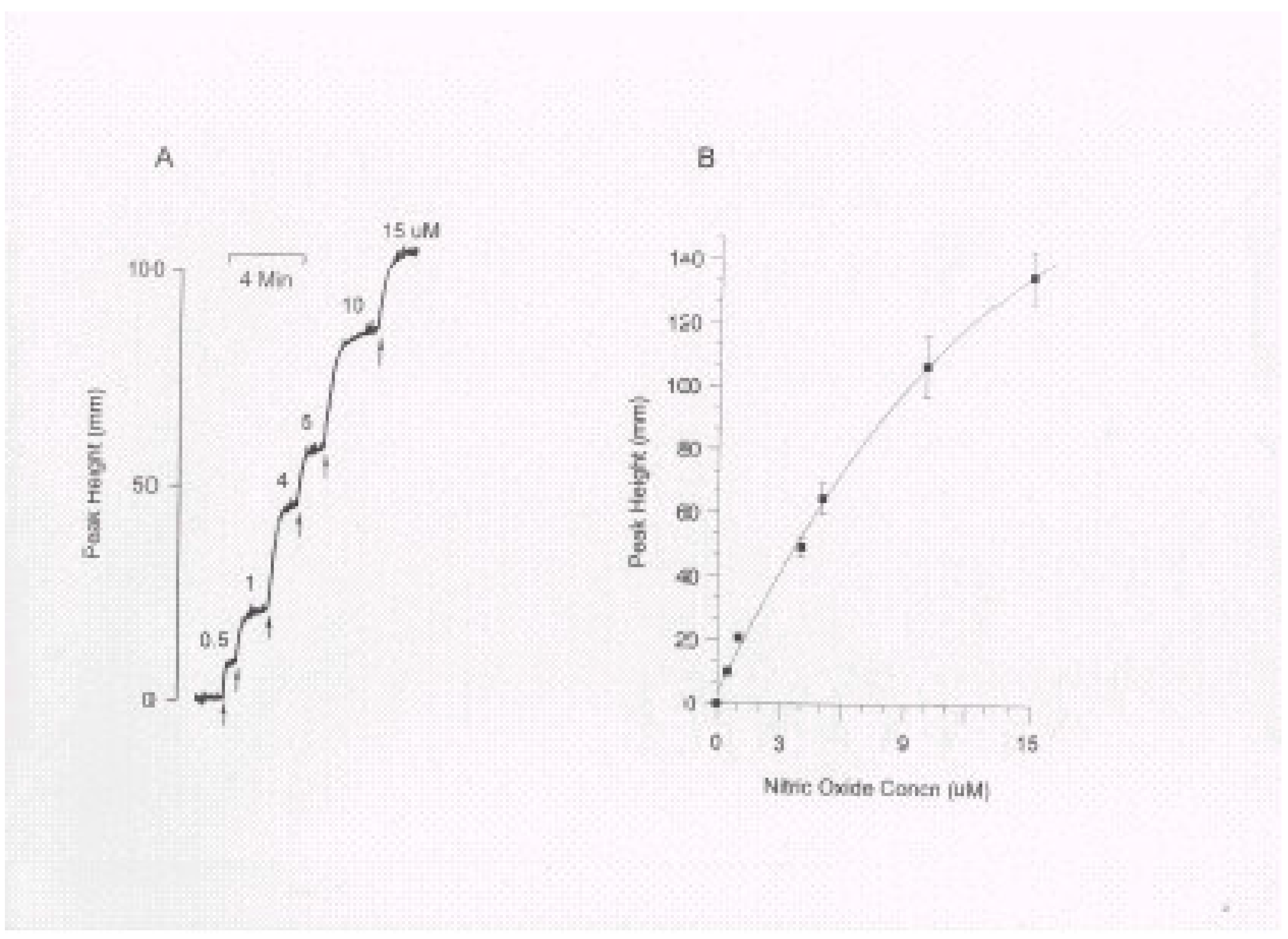
This procedure generates known concentrations of NO within a container supplied by WPI for calibration (calibration chamber). A calibration curve demonstrating changes in current or peak height (if a chart recorder is used) as a function of NO concentration can be produced (A typical chart recorder output is depicted in Figure 1).
The generation of NO is based on the following equation where a known amount of KNO2 (NaNO2 can be substituted for KNO2) is added to produce a known amount of NO.
2KNO2 + 2KI + 2H2SO4 → 2NO + I2 + 2H2O + 2K2SO2
Because stoichiometry exists between added KNO2 and the NO generated, and because KI and H2SO4 are added in excess, the final concentration of NO generated is equal to the concentration of KNO2 in the solution. The NO generated in this reaction persists long enough to calibrate the instrument with ease. Injection of each successive KNO2 as the current output reaches a plateau ensures an accurate determination. This is an easy and convenient method of calibration ideal for the standard sensor, however unfortunately because of the acidic nature of the solution used it is inappropriate for utilisation with the other NO sensors in the WPI range.
Calibration by decomposition of SNAP
This method has been developed by WPI and is based on the instantaneous decomposition of the NO donor SNAP to NO with the catalyst cuprous chloride. Cuprous (Cu I) chloride is a more efficient catalyst than Cu II and because Cu I is readily oxidised to Cu II which will appear during calibration as an apparent low sensitivity of the sensor some precautions are necessary to keep Cu I in its reduced state prior to calibration. Cuprous (Cu I) chloride can be stored under inert conditions and if used in solution it is important that these solutions be de-gased and oxygen free. The calibration method itself involves the preparation of a saturated solution of cuprous chloride (approx 2.4 mM at room temperature) in deoxygenated distilled water and a standard solution of SNAP (e.g. 5.6 mg) in 250 ml of EDTA in water (5 mg/250 ml, pH 9.0). This solution decomposes slowly and can be used for calibration in a single day. Using the stepped successive addition of samples of this solution (e.g. 100 nM, 400 nM, and 800 nM of equivalent NO) into a stirred 10 ml of the cuprous chloride solution (within an oxygen free environment) in the calibration chamber a calibration curve can be generated as described previously.
Figure 1.
NO Sensor Calibration. A: A representative calibration trace showing the responses of the electrode after successive injections (arrows) of NaNO2 substrate. B: NO calibration curve generated from 5 separate calibrations. Data are mean ± S.E.M. values.
Figure 1.
NO Sensor Calibration. A: A representative calibration trace showing the responses of the electrode after successive injections (arrows) of NaNO2 substrate. B: NO calibration curve generated from 5 separate calibrations. Data are mean ± S.E.M. values.

The purity of the NO donor SNAP in these calibrations is very critical for an accurate calibration. A high grade SNAP which is freshly made and has a minimal purity of 98 % is necessary. This calibration is suitable for all sensors but it is particularly useful for the microsensors offered by WPI. A second method of calibration with SNAP decomposition using copper (Cu II) sulphate or cupric (Cu II) chloride is available if the necessary storage conditions for cuprous (Cu I) chloride is not possible.
Example of the Utility of the NO Sensor – Use of a Dual Oxygen/NO Sensor Chamber
Cytokine activation during inflammation can lead to the production of NO from a variety of cells which may be cytotoxic/regulatory to other cells in the locality. This may be of particular importance in the central nervous system as activated astrocytic cells are a major source of NO and neurons are known to be acutely sensitive to oxidative stress mediated damage [14,15]. Although the mechanisms of toxicity and possible regulation are presently being unravelled it is known that NO can inhibit certain components of the mitochondrial respiratory chain and prevent cellular respiration/limit cellular ATP production. In some of these investigations the NO sensor has been used in combination with the oxygen electrode to monitor cytokine activated NO production and respiration in a simultaneous manner [5]. Before the commercial generation of the sensor chamber by WPI we established a similar vessel to investigate the properties of a novel NO donor compound on cellular respiration and the possibility of NO mediated inhibition of endothelial and glial cell respiration [16,17]. The following descriptions demonstrate the utility of the NO sensor in these respects.
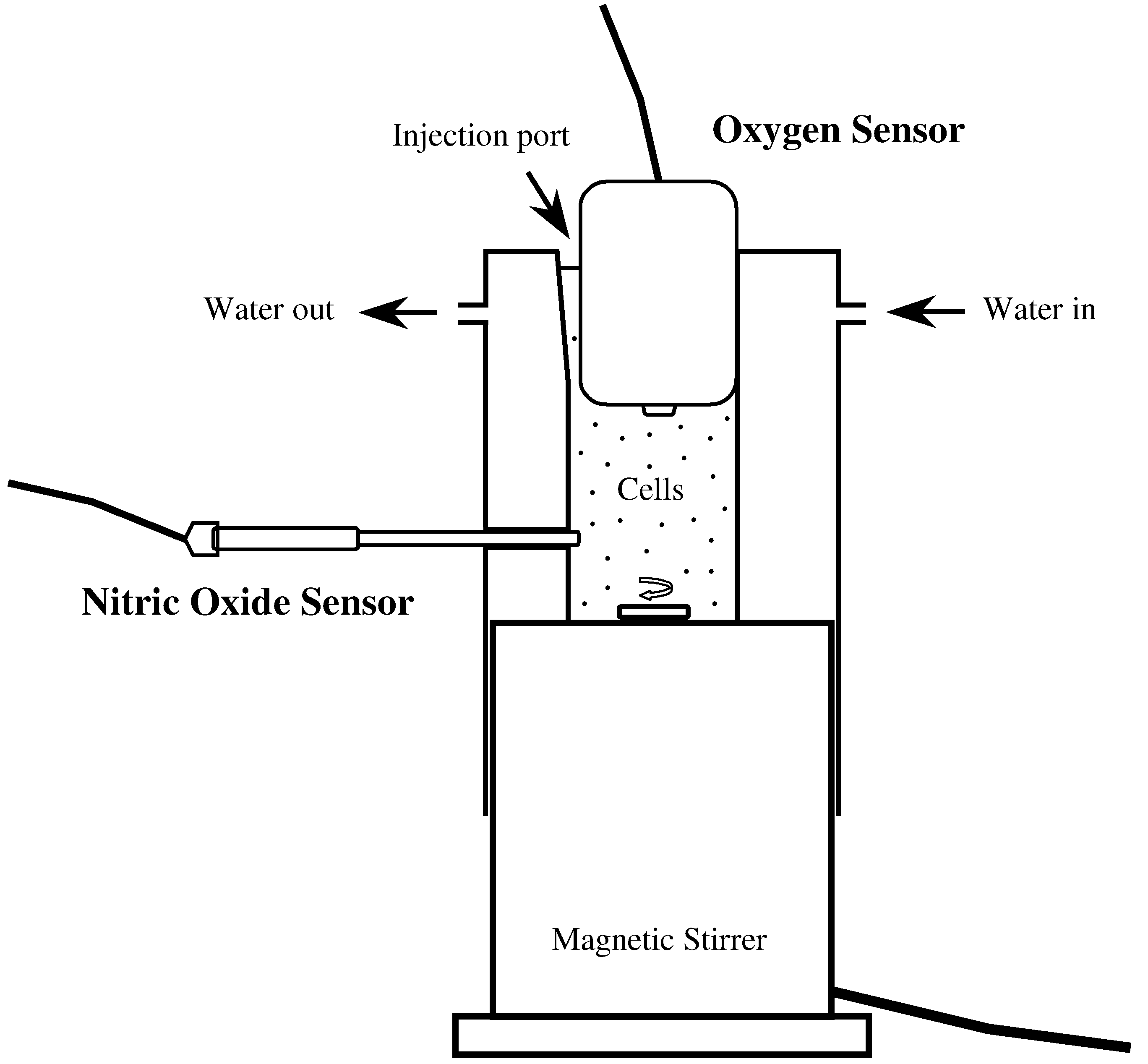
Figure 2.
Schematic representation of the dual NO/O2 sensor chamber used to monitor cellular oxygen consumption and NO levels. The perspex vessel has a volume of approx 250 μl, is water jacketed to allow temperature control by attachment to a waterbath. The chamber sits on a magneticstirrer and houses ports for both the NO (standard 2mm probe) and oxygen sensor. A small groove adjacent to the oxygen sensor housing allows the application of agents of interest to the cells. Small screws (not shown) secure and allow adjustment of the sensors in the vessel.

Figure 2 shows a schematic representation of the NO/O2 sensor chamber. For details of the equivalent chamber offered by WPI visit their web site. The vessel is made of Perspex, can be temperature regulated, attaches to a magnetic stirrer and has ports for the accommodation of both the oxygen and NO sensors.
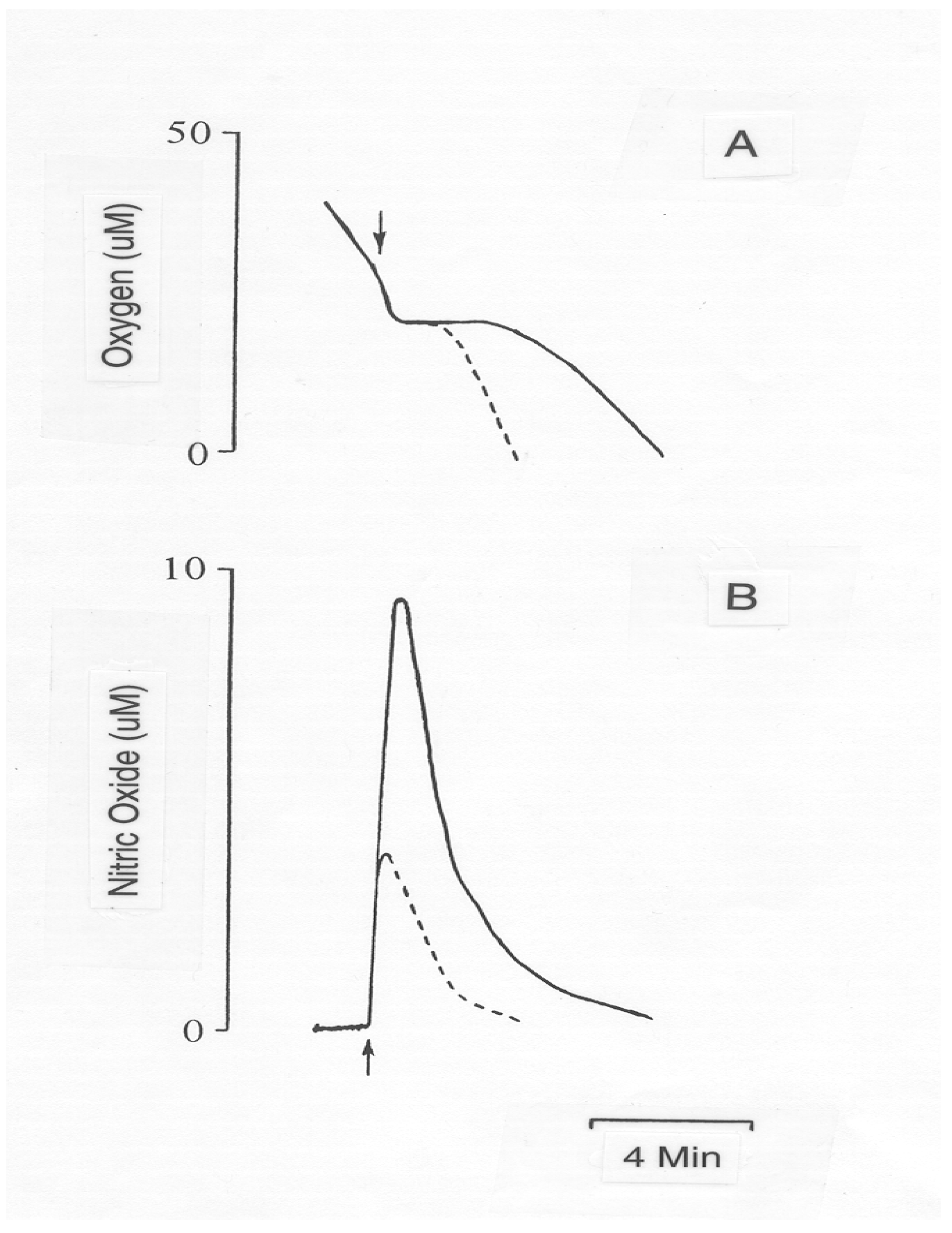
Figure 3 shows the simultaneous real time detection of oxygen consumption and NO levels in an endothelial cell line following exposure to a bolus of NO gas. Human vascular endothelial-like cells (ECV304, European Collection of Animal Cell Cultures, Wiltshire, UK) were grown to confluency in Hepes buffered M199 medium supplemented with fetal bovine serum (10 %). Endothelial cells were harvested by trypsinisation and kept on ice in M199 culture medium until required (<4 hours). 1 x 10⁶ cells in an oxygen saturated Hank’s balanced salt solution (supplemented with 2 mM CaCl2 and 20 mM Hepes, pH 7.4) were placed in the chamber. Following the addition of NO (from a NO gas saturated solution) at variable concentrations the peaks in NO result in a variable degree of respiration block with high concentrations leading toward permanent irreversible blockade.
Figure 3.
Chart recorder traces showing the effects of NO gas on endothelial cell oxygen consumption. Endothelial cell oxygen consumption (A) and NO concentration (B) were simultaneously measured at 37°C. Arrows indicate addition of NO at 10 (dotted line) and 20 μM (solid line).
Figure 3.
Chart recorder traces showing the effects of NO gas on endothelial cell oxygen consumption. Endothelial cell oxygen consumption (A) and NO concentration (B) were simultaneously measured at 37°C. Arrows indicate addition of NO at 10 (dotted line) and 20 μM (solid line).

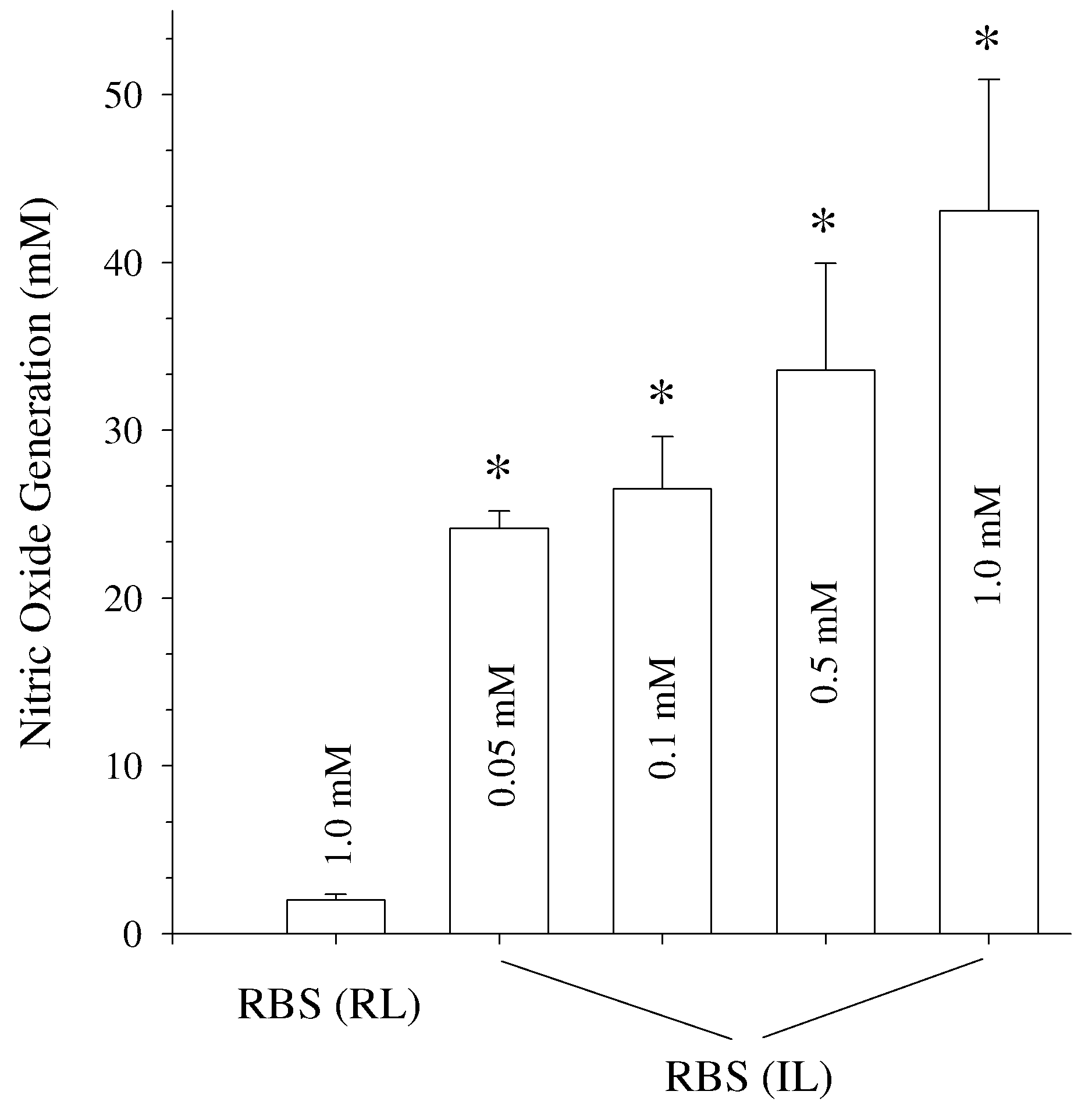
Figure 4 demonstrates the utility of the sensor and the chamber to evaluate the potential of new NO donor compounds. For example Roussin’s black salt an iron sulphur nitrosyl which contains seven potentially releasable moieties was recently evaluated by us as a compound which may be of use therapeutically in targeted NO delivery. Figure 4 shows the large and dose-dependent liberation of NO from this compound following exposure to intense white light. Further studies using Roussin’s black salt have suggested that upon cellular exposure the compound may access intracellular sites and liberate NO intracellularly [16,18].
In summary, electrochemical NO sensors provide an appealing and improved way to determine the level of NO in biomedical samples. They have significantly contributed to the comprehension of the action and effects of this important molecule in the few years since its discovery and the explosion of interest. These sensors provide accurate, sensitive, and real time measurements and with the advent of very small microchip sensors will further provide the tools necessary for more detailed and necessary in vivo monitoring.
Figure 4.
Donor NO production following intense white light exposure. The novel NO donor compound Roussin’s black salt (RBS) was dissolved in Hank’s balanced salt solution and placed in the NO sensor chamber. Maximal NO levels were determined from a range of donor concentrations under laboratory light conditions (RL) and following intense white light exposure (IL) from a cooled fiber optic placed at a distance of 2 cm from the centre of the vessel and equipped with a 150Watt bulb. Data are mean values ± SEM from 4 experiments.

* = p<0.001 versus NO generation from room light exposed donor.
References
- Malinski, T.; Taha, Z. Nitric oxide release from a single cell measured in situ by a porphyrinic-based microsensor. Nature 1992, 358, 676–678. [Google Scholar] [CrossRef] [PubMed]
- Abu-Soud, H.M.; Hazen, S.L. Nitric oxide is a physiological substrate for mammalian peroxidases. J. Biol. Chem. 2000, 275, 37524–37532. [Google Scholar] [CrossRef] [PubMed]
- Bellamy, T.C.; Wood, J.; Goodwin, D.A.; Garthwaite, J. Rapid desensitisation of the nitric oxide receptor soluble guanylate cycles; underlies diversity of cellular cGMP responses. PNAS 2000, 97, 2928–2933. [Google Scholar] [CrossRef] [PubMed]
- Thom, S.R.; Fisher, D.; Xu, Y.A.; Notarfrancesco, K.; Ischiropoulos, H. Adaptive responses and apoptosis in endothelial cells exposed to carbon monoxide. PNAS 2000, 97, 1305–1310. [Google Scholar] [CrossRef] [PubMed]
- Brown, G.C.; Bolaños, J.P.; Heales, S.J.R.; Clark, J.B. Nitric oxide produced by activated astrocytes rapidly and reversibly inhibits cellular respiration. Neurosci. Lett. 1995, 193, 201–204. [Google Scholar] [CrossRef]
- Tsukahara, H.; Ende, H.; Magazine, H.I.; Bahou, W.F.; Goligorsky, M.S. Molecular and functional characterization of the non-isopeptide-selective ETB receptor in endothelial cells. Receptor coupling to nitric oxide synthase. J. Biol. Chem. 1994, 269, 21778–21785. [Google Scholar] [PubMed]
- Liu, Y.; Shenouda, D.; Bilfinger, T.V.; Stefano, M.L.; Magazine, H.I.; Stefano, G.B. Morphine stimulates nitric oxide release from invertebrate microglia. Brain Res. 1996, 722, 125–131. [Google Scholar] [CrossRef]
- Engelman, D.T.; Watanabe, M.; Engelman, R.M.; Rousou, J.A.; Flack, J.E.; Deaton, D.W.; Das, D.K. Constitutive nitric oxide release is impaired after ischemia and reperfusion. J. Thoracic & Cardiovasc. Surg 1995, 110, 1047–1053. [Google Scholar]
- Engelman, D.T.; Watanabe, M.; Maulik, N.; Cordis, G.A.; Engelman, M.; Rousou, J.A.; Flack, J.E.; Deaton, D.W.; Das, D.K. L-arginine reduces endothelial inflammation and myocardial stunning during ischemia/reperfusion. Annals of Thoracic Surg 1995, 60, 1275–1281. [Google Scholar] [CrossRef]
- Rysz, J.; Luciak, M.; Kedziora, J.; Blaszczyk, J.; Sibinska, E. Nitric oxide release in the peripheral blood during hemodialysis. Kidney Int. 1997, 51, 294–300. [Google Scholar] [CrossRef] [PubMed]
- Clough, G.F. Role of nitric oxide in the regulation of microvascular perfusion in human skin in vivo. J. Physiol. 1999, 516, 549–557. [Google Scholar] [CrossRef] [PubMed]
- Levine, D.Z.; Iacovitti, M.; Burns, K.D.; Zhang, X.J. Real-time profiling of kidney tubular fluid nitric oxide concentrations in vivo. Am. J. Physiol. 2001, 281, F189–F194. [Google Scholar]
- Zhang, X.; Lin, J.; Cardoso, L.; Broderick, M.; Darley-Usmar, V. A novel microchip nitric oxide sensor with sub-nM detection limit. Electroanalysis. 2002, 14, 697–702. [Google Scholar] [CrossRef]
- Bolaños, J.P.; Almeida, A.; Stewart, V.; Peuchen, S.; Land, J.M.; Clark, J.B.; Heales, S.J.R. Nitric oxide-mediated mitochondrial damage in the brain: mechanisms and implications for neurodegenerative diseases. J. Neurochem. 1997, 68, 2227–2240. [Google Scholar] [CrossRef] [PubMed]
- Bolaños, J.P.; Heales, S.J.R.; Land, J.M.; Clark, J.B. Effect of peroxynitrite on the mitochondrial respiratory chain: differential susceptibility of neurones and astrocytes in primary culture. J. Neurochem. 1995, 64, 1965–1972. [Google Scholar]
- Hurst, R.D.; Chowdhury, R.; Clark, J.B. Investigations into the mechanism of action of a novel nitric oxide generator on cellular respiration. J. Neurochem. 1996, 67, 1200–1207. [Google Scholar] [CrossRef] [PubMed]
- Hurst, R.D.; Clark, J.B. Nitric oxide induced blood-brain barrier dysfunction is not mediated by inhibition of mitochondrial respiratory chain activity and/or energy depletion. Nitric Oxide Biol. & Chem. 1997, 1, 121–129. [Google Scholar]
- Flitney, F.W.; Megson, I.L.; Flitney, D.E.; Butler, A.R. Iron-sulphur cluster nitrosyls, a novel class of nitric oxide generator: mechanism of vasodilator action on rat isolated tail artery. Br. J. Pharmacol. 1992, 107, 842–848. [Google Scholar] [CrossRef] [PubMed]
- Sample Availability: Available from the authors.
© 2003 by MDPI (http://www.mdpi.org). Reproduction is permitted for noncommercial purposes.
Share and Cite
MDPI and ACS Style
Hurst, R.D.; Clark, J.B. The Utility of the Nitric Oxide Electrochemical Sensor in Biomedical Research. Sensors 2003, 3, 321-329. https://doi.org/10.3390/s30800321
AMA Style
Hurst RD, Clark JB. The Utility of the Nitric Oxide Electrochemical Sensor in Biomedical Research. Sensors. 2003; 3(8):321-329. https://doi.org/10.3390/s30800321
Chicago/Turabian StyleHurst, Roger D., and John B. Clark. 2003. "The Utility of the Nitric Oxide Electrochemical Sensor in Biomedical Research" Sensors 3, no. 8: 321-329. https://doi.org/10.3390/s30800321