Article

Studies on the Interaction Mechanism of 1,10-Phenanthroline Cobalt(II) Complex with DNA and Preparation of Electrochemical DNA Biosensor

College of Chemistry and Molecular Engineering, Qingdao University of Science and Technology, Qingdao 266042, P.R. China
*
Author to whom correspondence should be addressed.
Sensors 2006, 6(10), 1234-1244; https://doi.org/10.3390/s6101234
Submission received: 1 September 2006 / Accepted: 5 October 2006 / Published: 10 October 2006
(This article belongs to the Special Issue Amperometric Sensors and Techniques for Neurochemical Monitoring)

Abstract

:
Fluorescence spectroscopy and ultraviolet (UV) spectroscopy techniques coupled with cyclic voltammetry (CV) were used to study the interaction between salmon sperm DNA and 1,10-Phenanthroline cobalt(II) complex, [Co(phen)2(Cl)(H2O)]Cl·H2O, where phen = 1,10-phenanthroline. The interaction between [Co(phen)2(Cl)(H2O)]+ and double-strand DNA (dsDNA) was identified to be intercalative mode. An electrochemical DNA biosensor was developed by covalent immobilization of probe single-strand DNA (ssDNA) related to human immunodeficiency virus (HIV) on the activated glassy carbon electrode (GCE). With [Co(phen)2(Cl)(H2O)]+ being the novel electrochemical hybridization indicator, the selectivity of ssDNA-modified electrode was investigated and selective detection of complementary ssDNA was achieved using differential pulse voltammetry (DPV).

1. Introduction

Nowadays, considerable attention has been paid on electrochemical DNA biosensor in the diagnosis of genetic diseases and the detection of pathogenic biological species due to the highly sensitive, rapid yet accurate, simple and inexpensive detection technique it offers [1-5]. Electrochemical DNA biosensor is generally based on an electrode with oligonucleotide immobilization. The sequence-specific hybridization of nucleic acids could be detected directly [6-9] or by DNA intercalator [10-13], and thus different forms of electrochemical DNA biosensors have been developed now. Among them, the later fashion, using an intercalator as redox label or indicator for the hybridization events, attracts great interest.
Owing to the potential of interacting with the nitrogenous bases of DNA, metal complexes, especial transition metal complexes, have received considerable attention in recent years as remarkable and unique DNA-binding compounds. Researches conducted also showed that transition metal complexes have important biological activities. It was reported that the metal coordination compounds of 1,10-phenanthroline (phen) and its derivatives could inhibit tumor growth by interacting with DNA. In order to obtain more insight into the design of highly sensitive reactive probes, diagnostic reagents and new drugs, detailed understanding of the interaction between such complexes and DNA is potentially useful. Yang [14] reported that [Ni(phen)2dppz]2+ and [Co(phen)2dppz]3+ could bind strongly to the DNA and make DNA split in the light, which made instructive suggestions in the designing of new anti-cancer drugs. Although interaction between cobalt(III) complexes and DNA have been reported in many researches [15-17], interaction between 1,10-Phenanthroline cobalt(II) complex and DNA is seldom studied. To our best knowledge, research on the interaction between [Co(phen)2(Cl)(H2O)]Cl·H2O and DNA has not been reported.
In the present paper, fluorescence spectroscopy and ultraviolet (UV) spectroscopy techniques combined with cyclic voltammetry (CV) were used to study the interaction between [Co(phen)2(Cl)(H2O)]Cl·H2O and model DNA, salmon sperm DNA. Results showed that [Co(phen)2(Cl)(H2O)]+ could bind to double-strand DNA (dsDNA) through intercalative binding mode. After probe single-strand DNA (ssDNA) related to human immunodeficiency virus (HIV) was covalently immobilized on the activated glassy carbon electrode (GCD), an electrochemical DNA biosensor using [Co(phen)2(Cl)(H2O)]+ as the electrochemical hybridization indicator was developed and it demonstrated potential in selective detection of the complementary ssDNA. The work might bring further insight on the interaction mechanism between transition metal complexes and DNA and be helpful for further research for designing novel anti-tumor drugs and diagnosis disease.

2. Experimental Section

2.1. Instrumentation

The electrochemical measurement was carried out with Model CHI 832B Voltammetric Analyzer (ChenHua Instruments, China). A three-electrode system was employed with Pt wire as the auxiliary electrode, Ag/AgCl/KCl (sat) as reference electrode, and GCE or modified GCE as working electrode. Spectroscopic experiments were conducted on Hitachi F-4500 fluorospectrometer (Hitachi, Japan) and cary 50 UV/Vis spectrometer (Varian, Australian).

2.2 Reagents

Salmon sperm DNA (10 mg mL-1, A260/A280>1.8) were purchased from Huashun Biological Engineering Company (Shanghai, China) with the concentration being determined by the ultraviolet absorption at 260 nm (ε = 6600 L mol-1 cm-1). Ethidium bromide (EB), tris (hydroxymethy)ammomethane (Tris), ethylenediamine tetraacetic acid solution (EDTA), 1-ethyl-3-(3-dimethylaminopropyl)carbodiimide solution (EDC) and N-hydroxysuccinimide solution (NHS) were also obtained from Huashun Biological Engineering Company (Shanghai, China). The [Co(phen)2(Cl)(H2O)]Cl·H2O was synthesized according to Zhao et al [18]. Three 21-base oligonucleotides of HIV gene were purchased from SBS Genetech Company (Beijing, China). The base sequences were as follow: S1: 5′-ACT GCT AGA GAT TTT CCA CAT-3′; S2: 3′-TGA CGA TCT CTA AAA GGT GTA-5′; S3: 3′-AAA AGG TGT AAG CGT TTG CCG-5′. Amongst, S1 is complementary to S2. All oligonucleotide stock solutions of the three 21-base oligomers (100 μg mL-1) were prepared with Tris-HCl-EDTA buffer solution (TE solution, 1.00 × 10⁻² mol L-1 Tris-HCl plus1.00 × 10-3 mol L-1 EDTA, pH 8.0). Other chemicals employed were of analytical grade and doubly deionized water (DDW) was used throughout.

2.3 Spectroscopic study on the interaction between [Co(phen)2(Cl)(H2O)]+ and DNA

Fluorescence spectroscopy measurements were performed by maintaining a constant concentration of the [Co(phen)2(Cl)(H2O)]+ while varying concentration of salmon sperm DNA. 0.10 mL of 1.00 × 10⁻² mol L-1 [Co(phen)2(Cl)(H2O)]+ solution and different amounts of salmon sperm DNA solution were added to 10 mL colorimetric tubes. The mixture was diluted to the mark using 0.1 mol L-1 phosphate buffer solution (PBS) at pH 6.0 and reacted for 12 min at room temperature. Afterward, the measurement of fluorescence was performed using 298 nm as the excited wavelength. To investigate the binding mode between [Co(phen)2(Cl)(H2O)]+ and DNA, the fluorescence spectrums of EB-DNA system in the presence of [Co(phen)2(Cl)(H2O)]+ were recorded by maintaining a constant concentration of EB and [Co(phen)2(Cl)(H2O)]+ while varying concentration of salmon sperm DNA. The measurement of fluorescence was performed using 490 nm as the excited wavelength.
UV spectroscopy was also used to investigate the interaction between [Co(phen)2(Cl)(H2O)]+ and salmon sperm DNA. DNA solution and [Co(phen)2(Cl)(H2O)]+ was mixed and reacted for 12 min at room temperature. Afterwards, UV spectrum of the mixture was recorded with a scanning range from 200 nm to 400 nm.

2.4. Electrochemical study on the interaction between [Co(phen)2(Cl)(H2O)]+ and DNA

The changes on characteristics of cyclic voltammograms (CVs) and the differential pulse voltammograms (DPVs) of the [Co(phen)2(Cl)(H2O)]+ solutions in the absence and presence of salmon sperm DNA were investigated. For cyclic voltammetric scanning, the potential scanning ranged from -0.4 V to 0.6 V and the scanning rate was 0.10 V s-1. The sample interval was set as 0.001 V and the quiet time was 2 s. For DPV measurment, the initial potential was 0.8 V. The high potential was 0.8 V and the low potential was -0.4 V. The pulse scope was set as 0.004 V and the pulse extent was 0.05 s. The pulse cycle was 0.2 s. The sample interval and the quiet time were 0.001 V and 2 s respectively.

2.5. Preparation of the electrochemical DNA sensor

The developed electrochemical DNA biosensor was based on the covalent immobilization of human immunodeficiency virus (HIV) probe single-strand DNA (S1) on the activated electrode. Prior to experiment, the GCE was firstly polished using 1.0 μm, 0.3 μm and 0.05 μm α-Al2O3 suspension respectively and then extensively rinsed in DDW with ultrasonic. Afterwards, the electrode was oxidized at +0.50 V for 1 min in 5.00 × 10⁻² mol L-1 PBS at pH 7.4 followed by thoroughly rinsed with DDW. As previously reported [19], four-step procedure was used for the preparation of the DNA biosensor. Firstly, the electrode was activated by dropping 20 μL 5.00 × 10⁻² mol L-1 PBS (pH 7.4) containing 5.0 × 10⁻³ mol L-1 EDC and 8.0 × 10⁻³ mol L-1 NHS on the electrode surface. After the solution was air-dried, the activated electrode was rinsed with DDW for several times. Secondly, ssDNA (S1) was immobilized on the electrode surface by dropping ssDNA solution on the activated GCE. After dried under an infrared lamp, the electrode was rinsed with DDW to eliminate the DNA adsorbed. Thirdly, the S1-immobilized GCE was immersed in 20 mmol L-1 Tris-HCl buffer (pH 7.0) containing complementary S2 or uncomplementary S3 respectively to obtain S1-S2 hybridized GCE and S1-S3 hybridized GCE respectively. Such DNA interaction was allowed at 40 oC for 1 h with stirring. Afterwards, the obtained electrodes were thoroughly rinsed with the same Tris-HCl buffer and DDW successively to eliminate the adsorbed S2 or S3 and then dried at room temperature. Fourthly, [Co(phen)2(Cl)(H2O)]+ was accumulated onto the surface by immersing the resulted electrode in the Tris-HCl buffer solution containing [Co(phen)2(Cl)(H2O)]+ at room temperature for 5 min with stirring. After rinsed with the same Tris-HCl buffer and DDW successively, the DPV measurements were performed.

3. Results and Discussion

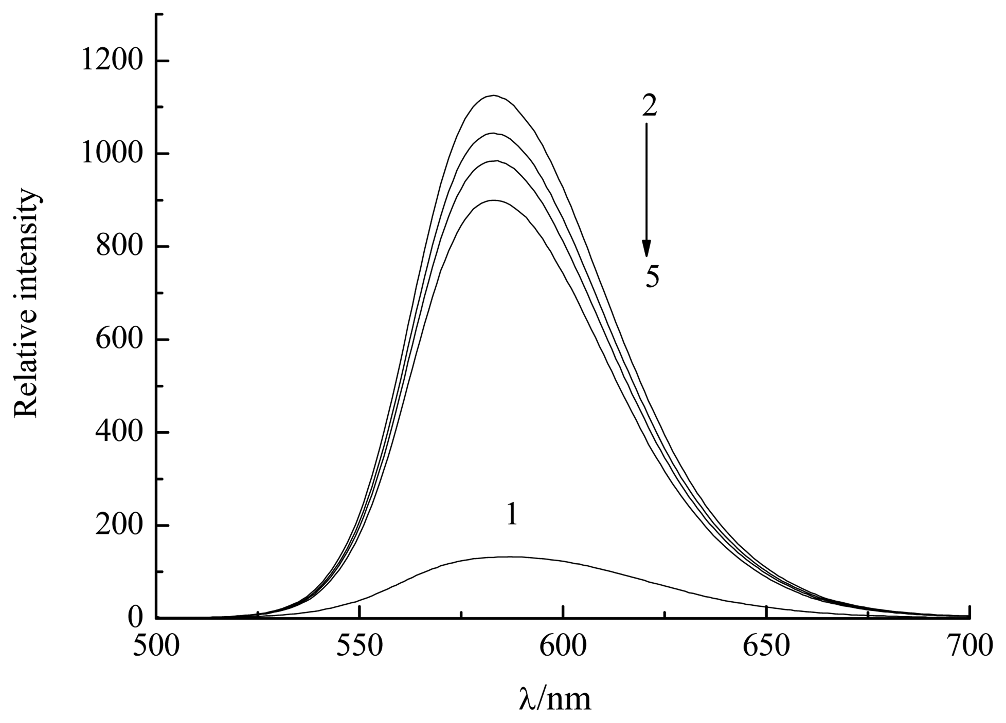
3.1. Fluorescence spectroscopic studies of the interaction between [Co(phen)2(Cl)(H2O)]+ and DNA

Fluorescence spectroscopy was used to investigate the interaction between [Co(phen)2(Cl)(H2O)]+ and salmon sperm DNA. Figure 1 displayed the fluorescence features of [Co(phen)2(Cl)(H2O)]+ in the absence and presence of DNA. It was observed from curve 1 that [Co(phen)2(Cl)(H2O)]+ had the maximal emission at 366 nm. We could find that the fluorescence intensity of [Co(phen)2(Cl)(H2O)]+ enhanced gradually with the amount of added DNA increased (curve 2-4), indicating the interaction between [Co(phen)2(Cl)(H2O)]+ and salmon sperm DNA. The phenomenon might be ascribed to the hydrophobic circumstance of DNA, which could enhance the fluorescence quantum yield of [Co(phen)2(Cl)(H2O)]+ and the corresponding fluorescence intensity.
To investigate the binding mode between [Co(phen)2(Cl)(H2O)]+ and dsDNA, the fluorescence spectrum of EB-DNA system in the presence of [Co(phen)2(Cl)(H2O)]+ was studied. As shown in Figure 2, EB itself emitted weak fluorescence emission (curve 1). After it intercalated into the double helix of DNA molecule, a significant increase in fluorescence intensity was observed (curve 2). However, distinct quenching of fluorescence occurred when [Co(phen)2(Cl)(H2O)]+ was added (curve 3-5). Moreover, the more the [Co(phen)2(Cl)(H2O)]+ added, the lower the fluorescence emitted. Such fluorescence quenching might be due to the competition between [Co(phen)2(Cl)(H2O)]+ and EB for the binding sites of dsDNA, which made the fluorescence intensity of EB-DNA system weaken [20]. Accordingly, the binding mode of [Co(phen)2(Cl)(H2O)]+ to dsDNA might be similar with EB. So, the intercalative binding between [Co(phen)2(Cl)(H2O)]+ and DNA was supposed.

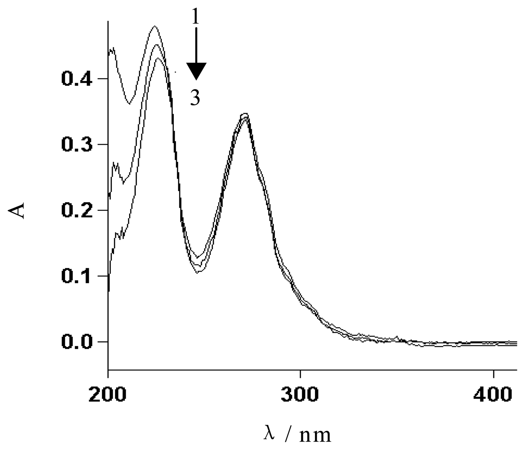
3.2. UV spectroscopic studies of the interaction between [Co(phen)2(Cl)(H2O)]+ and DNA

The hypochromic effect and the bathochromic effect were the identifying marks of the intercalation, as reported by Long et al [21]. To confirm our hypothesis, UV spectroscopy was used to further study the interaction between [Co(phen)2(Cl)(H2O)]+ and DNA. The variation of UV spectrum for [Co(phen)2(Cl)(H2O)]+ in the absence and presence of salmon sperm DNA was shown in Figure 3. When the amount of added DNA increased, the absorption peaks of [Co(phen)2(Cl)(H2O)]+ at 202.0, 224.0 and 268.0 nm decreased and a slight red shift was observed, indicating both the hypochromic effect and bathochromic effect. Therefore, the intercalative interaction between [Co(phen)2(Cl)(H2O)]+ and DNA was identified.

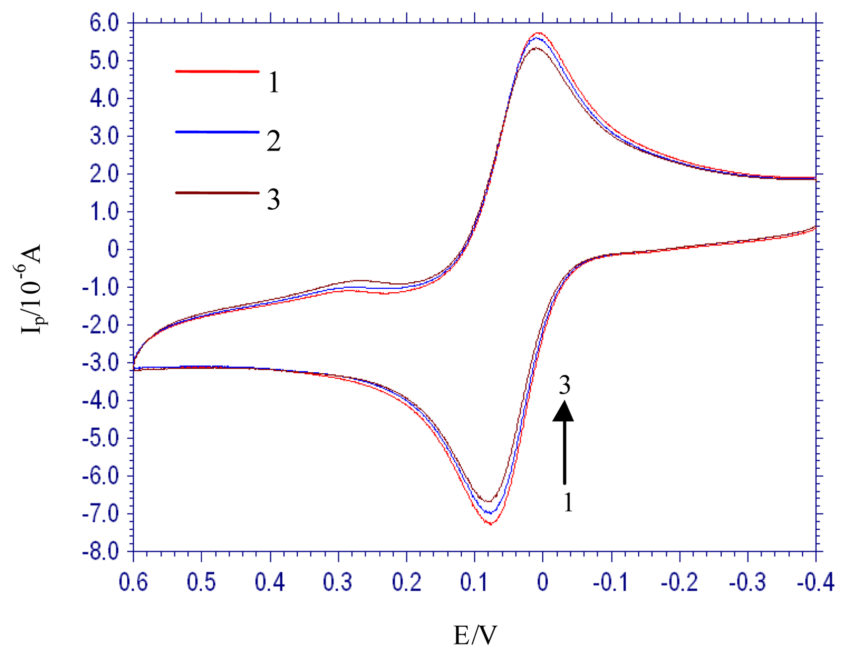
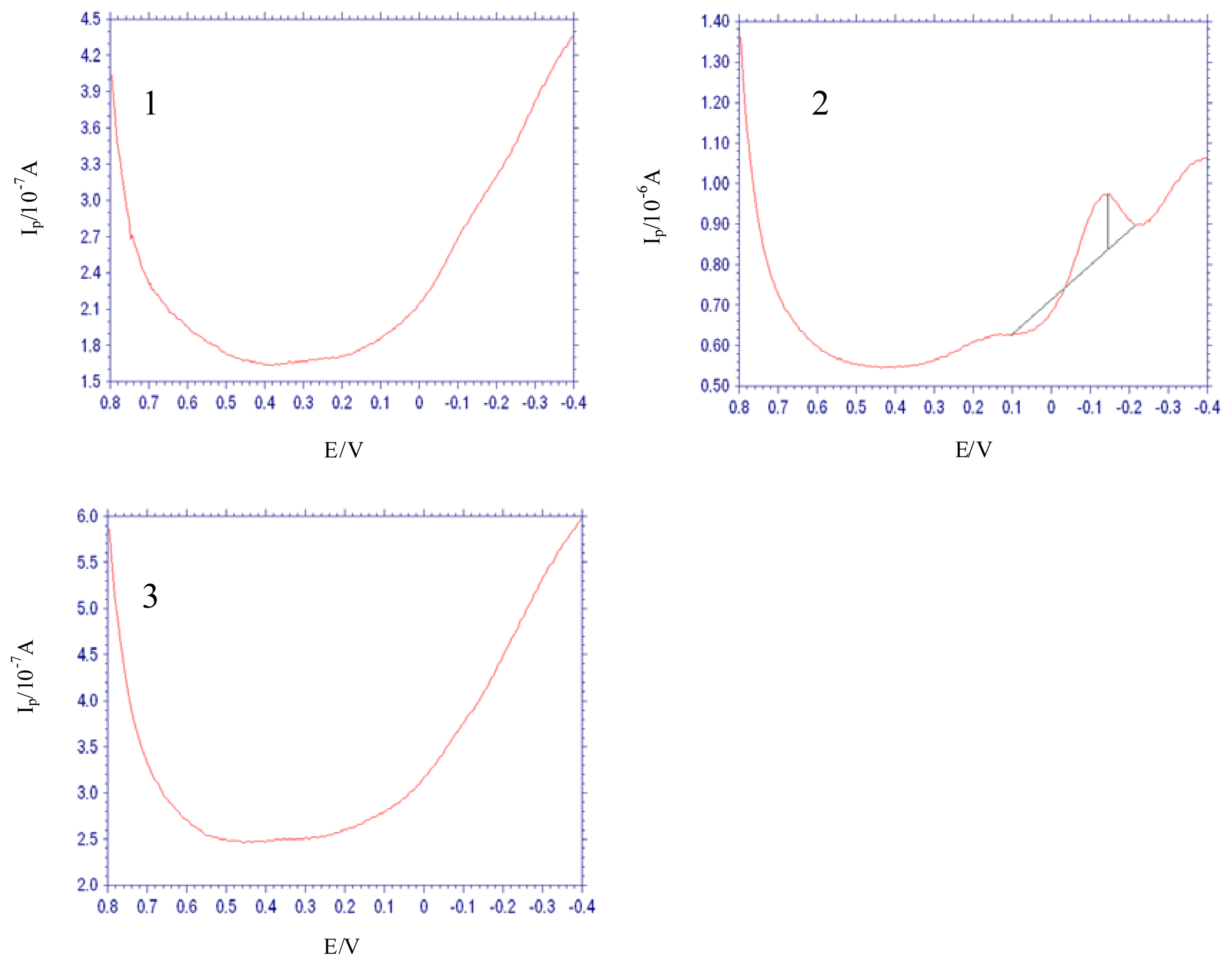
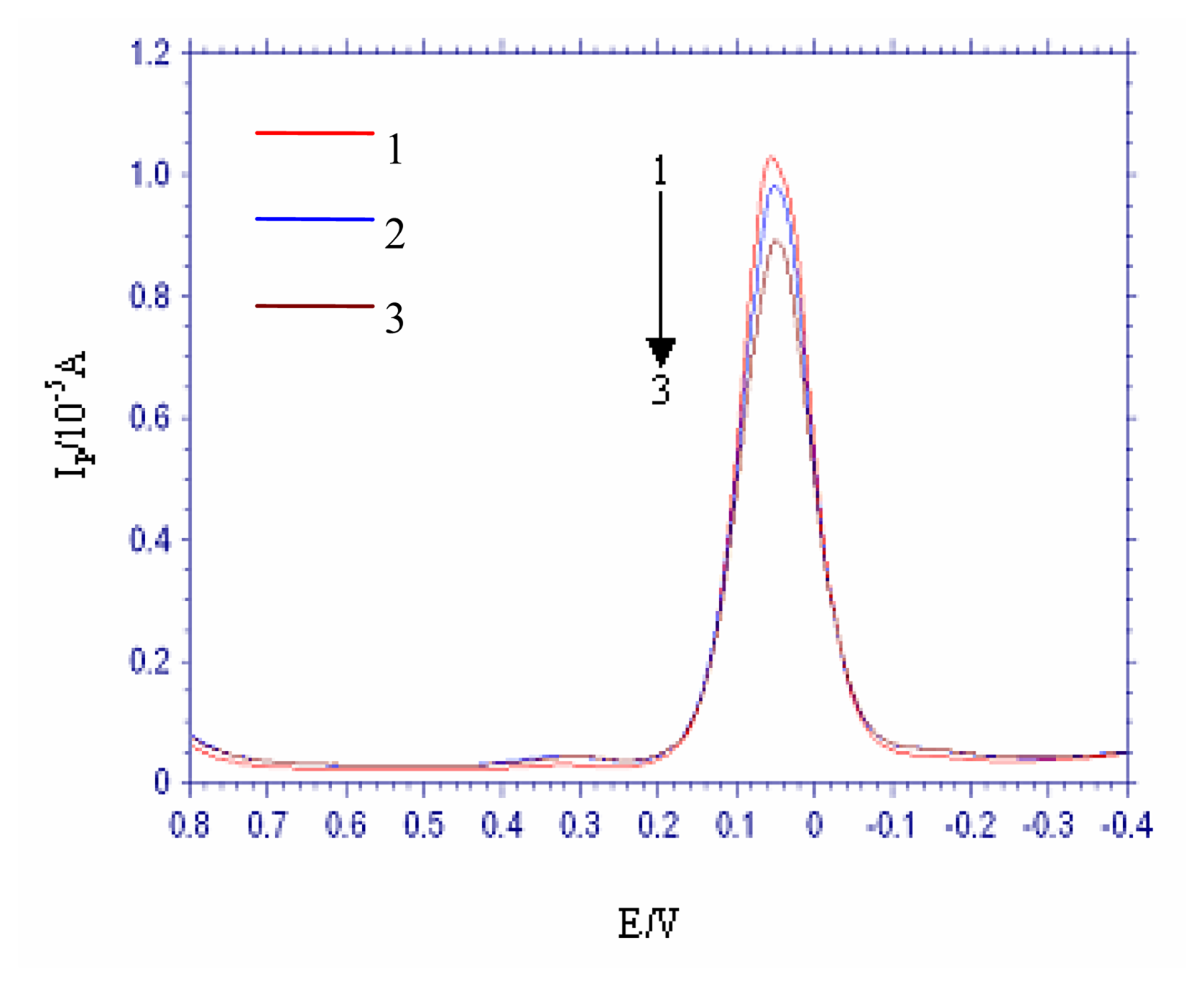
3.3. Electrochemical study on the interaction between [Co(phen)2(Cl)(H2O)]+ and DNA

To explore the application of [Co(phen)2(Cl)(H2O)]+ in electrochemical DNA biosensors, electrochemical study on [Co(phen)2(Cl)(H2O)]+ and its interaction with DNA were performed. CVs of [Co(phen)2(Cl)(H2O)]+ in the absence and presence of salmon sperm DNA were shown in Figure 4. We could see that there were a couple of quasi-reversible redox voltammetric peaks for [Co(phen)2(Cl)(H2O)]+ (curve 1), indicating the electrochemical activity of [Co(phen)2(Cl)(H2O)]+. The cathodic peak potential (Epc) and the anodic peak potential (Epa) were 0.010 V and 0.077 V respectively. Curves 2-3 were the CVs of [Co(phen)2(Cl)(H2O)]+ in the presence of different concentration of DNA. When the DNA concentration in the solution increased, the peak currents, both Ipc and Ipa, decreased slightly and small shift to positive potentials for both Epc and Epa occurred. The phenomena mentioned above were further studied by DPV and results were shown in Figure 5. Curve 1 was the differential pulse voltammogram of the [Co(phen)2(Cl)(H2O)]+ solution, while curve 2-3 were the voltagramms when different amounts of DNA were added to the solution. As can be seen, the peak currents also decreased with increasing of DNA. Therefore, intercalative binding mode of [Co(phen)2(Cl)(H2O)]+ to DNA drawn from electrochemical measurement was consistent with that obtained through spectroscopic studies [22].
The experimental conditions of the interaction between [Co(phen)2(Cl)(H2O)]+ and DNA, e.g. pH, reaction time, scan rate and the concentration of DNA, were optimized. Phosphate buffer solution (PBS) at pH 6.0 was chosen as the reaction medium and 12 min as the reaction time. Ipc is directly in proportion to the square root of the scan rate in the range from 0.02 V s-1 to 0.23 V s-1. The regression equation is Ipc = 17.339ν1/2 + 0.6043 with a correlation coefficient γ = 0.9991. This indicated that the electrochemical process of [Co(phen)2(Cl)(H2O)]+ was controlled by the diffusion of [Co(phen)2(Cl)(H2O)]+. It was also found that ΔIpc is in linear proportion to the concentration of DNA in the range from 2.10 × 10⁻⁴ mol L-1 to 2.77 × 10⁻³ mol L-1, with a regression equation ΔIpc = 0.072 CDNA-0.0165, and a correlation coefficient γ = 0.9997.

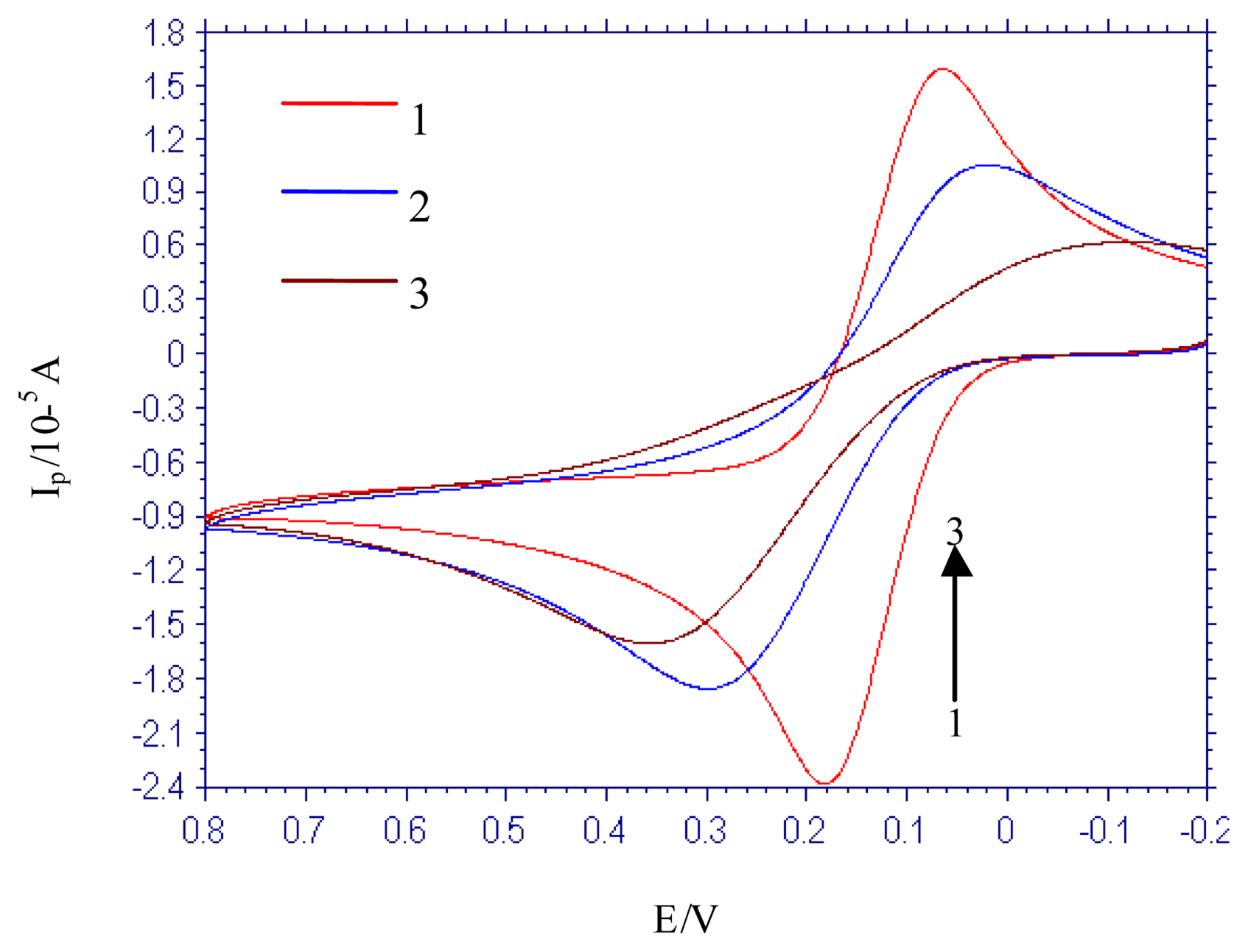
3.4. The electrochemical characterization of modified GCE

The DNA biosensor using [Co(phen)2(Cl)(H2O)]+ as electrochemical hybridization label was studied. The electrochemical probe molecules, Fe(CN)63-/Fe(CN)64-, were chosen to characterize the modified electrode. The bare GCE, S1-immobilied GCE, and S1-S2 hybridized GCE had been used as the working electrode respectively. The CVs of 5 × 10⁻³ mol L-1 K4Fe(CN)6 in 0.01 mol L-1 KCl solution were shown in Figure 6. Compared to bare GCE, redox peak currents of Fe(CN)63-/Fe(CN)64- on S1-immobilied GCE, and S1-S2 hybridized GCE decreased significantly. The negative charge of Fe(CN)64- and DNA immobilized on the electrode surface resulted in the static repulsive force and weakened the electrochemical reaction of Fe(CN)63-/Fe(CN)64- on the electrode, which lead to the decrease of the redox peak currents.

3.5. The selectivity of the DNA electrochemical biosensor

Differential pulse voltammetry was used to study the selectivity of the prepared electrochemical DNA biosensor. Figure 7 showed the representative differential pulse voltammograms obtained in PBS (pH 6.0) with the S1-immobilied GCE (curve 1), S1-S2 hybridized GCE (curve 2) and S1-S3 hybridized GCE (curve 3) being the working electrode respectively. S1-S2 hybridized GCE represented that S1-immobilied GCE hybridized with the complimentary DNA segment S2 and S1-S3 hybridized GCE referred to the electrode obtained after S1-immobilied GCE hybridized with uncomplimentary DNA segment S3. Before DPV measurement, [Co(phen)2(Cl)(H2O)]+ accumulation step for the three electrodes were all performed. No voltammetric response for the S1-immobilied GCE was found in curve 1. However, the reduction peak of the S1-S2 hybridized GCE appeared at about -0.144 V, as shown in curve 2, which was the same potential as the bare GCE in the [Co(phen)2(Cl)(H2O)]+ solution. The peak current was 1.378 × 10⁻⁷ A. The phenomenon indicated the intercalation of the indicator, [Co(phen)2(Cl)(H2O)]+, into the base pairs of the dsDNA resulted from S1-S2 hybridization. In the case of S1-S3 hybridized GCE, no dsDNA was formed because S3 was uncomplementary to S1 and no indicator could intercalate. As expected, S1-S3 hybridized GCE displayed no voltammetric response, as shown in curve 3, which indicated the favorable selectivity of the ssDNA modified GCE.
In summary, the interaction between [Co(phen)2(Cl)(H2O)]+ and the probing DNA, salmon sperm DNA, was studied by fluorescence spectroscopy, ultraviolet spectroscopy and voltammetry. [Co(phen)2(Cl)(H2O)]+ can bind to dsDNA by intercalative binding mode. By using [Co(phen)2(Cl)(H2O)]+ as the electrochemical hybridization indicator, the electrochemical DNA biosensor was prepared base on the covalent immobilization of ssDNA and it showed high selectivity in detection of complementary ssDNA.

Acknowledgments

The project was supported by the Program for New Century Excellent Talents in University (No. NCET-04-0649), the National Natural Science Foundation of China (No. 20475030) and the special project of Qingdao for generalship of Science and Techonology (No. 05-2-JC-80).

References and Notes

  1. Millan, K. M.; Saraullo, A.; Mikkelsen, S. R. Voltammetric DNA Biosensor for Cystic Fibrosis Based on a Modified Carbon Paste Electrode. Anal. Chem. 1994, 66, 943–2948. [Google Scholar]
  2. Wang, J.; Cai, X. H.; Rivas, G.; Shiraishi, H.; Farias, P. A. M.; Dontha, N. DNA Electrochemical Biosensor for the Detection of Short DNA Sequences Related to the Human Immunodeficiency Virus. Anal. Chem. 1996, 68, 2629–2934. [Google Scholar]
  3. Koji, H.; Keiko, I.; Yoshio, I. Novel DNA Sensor for Electrochemical Gene Detection. Anal. Chim. Acta 1994, 286, 219–224. [Google Scholar]
  4. Erdem, A.; Kosmider, B.; Osiecka, R.; Zyner, E.; Ochocki, J.; Ozsoz, M. Electrochemical genosensing of the Interaction Between the Potential Chemotherapeutic Agent, cis-bis(3-aminoflavone)dichloroplatinum(II) and DNA in Comparison with cis-DDP. J. Pharmaceut. Biomed. 2005, 38, 645–652. [Google Scholar]
  5. Rauf, S.; Gooding, J. J.; Akhtar, K.; Ghauri, M. A.; Rahman, M.; Anwar, M. A.; Khalid, A. M. Electrochemical Approach of Anticancer Drugs–DNA Interaction. J. Pharmaceut. Biomed. 2005, 37, 205–217. [Google Scholar]
  6. Wang, J.; Cai, X. H.; Rivas, G.; Shiraishi, H. Stripping Potentiometric Transduction of DNA Hybridization Processes. Anal. Chim. Acta 1996, 326, 141–147. [Google Scholar]
  7. Wang, J.; Rivas, G.; Cai, X. H.; Dontha, N.; Shiraishi, H.; Luo, D. B.; Valera, F. S. Sequence-specific Electrochemical Biosensing of M-tuberculosis DNA. Anal. Chim. Acta 1997, 337, 41–48. [Google Scholar]
  8. Ozkan, D.; Karadeniz, H.; Erdem, A.; Mascini, M.; Ozsoz, M. Electrochemical Genosensor for Mitomycin C–DNA Interaction Based on Guanine Signal. J. Pharmaceut. Biomed. 2004, 35, 905–912. [Google Scholar]
  9. Karadeniz, H.; Gulmez, B.; Sahinci, F.; Erdem, A.; Kaya, G. I.; Unver, N.; Kivcak, B.; Ozsoz, M. Disposable Electrochemical Biosensor for the Detection of the Interaction Between DNA and Lycorine based on Guanine and Adenine Signals. J. Pharmaceut. Biomed. 2003, 33, 295–302. [Google Scholar]
  10. Millan, K. M.; Mikkelsen, S. R. Sequence-selective Biosensor for DNA Based on Electroactive Hybridization Indicators. Anal. Chem. 1993, 65, 2317–2323. [Google Scholar]
  11. Pang, D. W.; Abruna, H. D. Interactions of Benzyl Viologen with Surface-Bound Single- and Double-Stranded DNA. Anal. Chem. 2000, 72, 4700–4706. [Google Scholar]
  12. Ju, H. X.; Ye, Y. K.; Zhao, J. H.; Zhu, Y. L. Hybridization Biosensor Using di(2,2′-bipyridine)osmium (III) as Electrochemical Indicator for Detection of Polymerase Chain Reaction Product of Hepatitis B Virus DNA. Anal. Biochem. 2003, 313, 255–261. [Google Scholar]
  13. Girousi, S. T.; Gherghi, I. C.; Karava, M. K. DNA-modified Carbon Paste Electrode Applied to the Study of Interaction Between Rifampicin (RIF) and DNA in Solution and at the Electrode Surface. J. Pharmaceut. Biomed. 2004, 36, 851–858. [Google Scholar]
  14. Fin, L.; Yang, P. Synthesis and DNA Binding Studies of Cobalt (III) Mixed-polypyridyl Complex. J. Inorg. Biochem. 1997, 68, 79–83. [Google Scholar]
  15. Jin, B. K.; Ji, X. P.; Nakamura, T. Voltammetric Study of Interaction of Co(phen)33+ with DNA at Gold Nanoparticle Self-assembly Electrode. Electrochimica Acta 2004, 50, 1049–1055. [Google Scholar]
  16. Srinivasan, S.; Annaraj, J.; Athappan, P. R. Spectral and Redox Studies on Mixed Ligand Complexes of Cobalt(III) Phenanthroline/bipyridyl and Benzoylhydrazones, Their DNA Binding and Antimicrobial Activity. J. Inorg Biochem. 2005, 99, 876–882. [Google Scholar]
  17. Sastri, C. V.; Eswaramoorthy, D.; Giribabu, L.; Maiya, B. G. DNA Interactions of New Mixed-ligand Complexes of Cobalt(III) and Nickel(II) that Incorporate Modified Phenanthroline Ligands. J. Inorg Biochem. 2003, 94, 128–145. [Google Scholar]
  18. Zhao, P. S.; Lu, L. D.; Jian, F. F. Structure of Chloro bis(1,10-phenanthroline)cobalt(II) Complex, [Co(phen)2(Cl)(H2O)]Cl·2H2O. J. Korean Chem. Soc. 2003, 47, 334–336. [Google Scholar]
  19. Zhang, S. S.; Niu, S. Y.; Qu, B.; Jie, G. F.; Xu, H.; Ding, C. F. Studies on the Interaction Mechanism Between Hexakis(imidazole) Manganese(II) Terephthalate and DNA and Preparation of DNA Electrochemical Sensor. J. Inorg. Biochem. 2005, 99, 2340–2347. [Google Scholar]
  20. Song, Y. M.; Yang, P. J.; Wang, L. F.; Yang, M. L.; Kang, J. W. Study on the Interactions Between Sm(RA)2·Ac·4H2O and DNA. Acta Chim. Sinica 2003, 61, 1266–1270. [Google Scholar]
  21. Long, E. C.; Barton, J. K. On Demonstrating DNA Intercalation. Accounts Chem. Res. 1990, 23, 271–273. [Google Scholar]
  22. Carter, M. T.; Rodriguez, M.; Bard, A. J. Voltammetric Studies of the Interaction of Metal Chelates with DNA. 2. Tris-chelated Complexes of Cobalt(III) and Iron(II) with 1,10-phenanthroline and 2,2′-bipyridine. J. Am. Chem. Soc. 1989, 111, 8901–8911. [Google Scholar]
Figure 1. Fluorescence spectrum of [Co(phen)2(Cl)(H2O)]+ in the absence and presence of different concentration of salmon sperm DNA. [Co(phen)2(Cl)(H2O)]+ concentration: 1.00 × 10⁻⁴ mol L-1; DNA concentration (mol L-1): (1): 0; (2): 3.12 × 10⁻⁴; (3): 4.68 × 10⁻⁴; (4): 5.46 × 10⁻⁴.
Sensors 06 01234f1
Figure 2. Fluorescence spectrum of EB-DNA system in the absence and presence of [Co(phen)2(Cl)(H2O)]+. (1): 6.35 × 10⁻⁵ mol L-1 EB; (2): (1) + 1.56 × 10⁻³ mol L-1 salmon sperm DNA; (3): (2) + 6.67 × 10⁻⁵ mol L-1 [Co(phen)2(Cl)(H2O)]+ (4): (2) + 1.33 × 10⁻⁴ mol L-1 [Co(phen)2(Cl)(H2O)]+ (5): (2) + 2.67 × 10⁻⁴ mol L-1 [Co(phen)2(Cl)(H2O)]+.
Sensors 06 01234f2
Figure 3. UV spectrum of [Co(phen)2(Cl)(H2O)]+ in the absence and presence of salmon sperm DNA. (1): 2.00 × 10⁻⁵ mol L-1 [Co(phen)2(Cl)(H2O)]+; (2): (1) + 1.87 × 10⁻⁵ mol L-1 DNA; (3): (1) + 3.74 × 10⁻⁵ mol L-1 DNA.
Sensors 06 01234f3
Figure 4. Cyclic voltammograms of [Co(phen)2(Cl)(H2O)]+ in the absence and presence of different concentration of DNA. [Co(phen)2(Cl)(H2O)]+ concentration: 1.00 × 10⁻³ mol L-1; DNA concentration (mol L-1): (1): 0; (2): 2.10 × 10⁻⁴; (3): 6.23 × 10⁻⁴.
Sensors 06 01234f4
Figure 5. Differential pulse voltammograms of [Co(phen)2(Cl)(H2O)]+ in the absence and presence of different concentration of DNA. [Co(phen)2(Cl)(H2O)]+ concentration: 1.00 × 10⁻³ mol L-1; DNA concentration (mol L-1): (1): 0; (2): 5.21 × 10⁻⁴; (3):1.53 × 10⁻³.
Sensors 06 01234f5
Figure 6. Cyclic voltammograms of Fe(CN)63-/Fe(CN)64- on different electrodes. (1): Bare GCE; (2): S1-immobilized GCE; (3): S1-S2 hybridized GCE.
Sensors 06 01234f6
Figure 7. Differential pulse voltammograms of different working electrode modified with [Co(phen)2(Cl)(H2O)]+ accumulation step. (1): S1-immobilized GCE; (2): S1-S2 hybridized GCE; (3): S1-S3 hybridized GCE.
Sensors 06 01234f7

