1. Introduction
The light dependent resistor (LDR) is a sensor whose resistance decreases when light impinges on it. This kind of sensor is commonly used in light sensor circuits in open areas, to control street lamps for example. Another possible use is in spectroscopic apparatus [
1]. In this kind of apparatus, continuous light or pulsed light can be used. Continuous light is used in common spectroscopic apparatus. The use of lock-in amplifiers made the use of pulsed light in spectroscopy easier, as is commonly used in photoacoustic spectroscopy [
2]. LDR's are made of semiconductors as light sensitive materials, on an isolating base. The most common semiconductors used in this system are cadmium sulphide, lead sulphide, germanium, silicon and gallium arsenide [
3]. A less known light sensor is the electret microphone. As the electret membrane functions as an absorbing black body, and as the electret microphone case has an air chamber that can be used as photoacoustic chamber, the electret microphone can be used as a detector of pulsed light [
4]. This type of microphone can be used to obtain the transmission spectrum of any transparent material.
The aim of this communication is to study the response of LDR to pulsed light and the analysis of the spectral curves obtained with a LDR and an electret microphone as light sensors in an optical spectroscopy device.
2. Experimental Section
To study the response of the LDR to luminous stimulus, it was used a voltage divider circuit, composed by a 4.7 kΩ resistance, a LDR and a 9 V battery. The voltage was measured on the LDR using a multimeter or a lock-in amplifier.
First the response of the LDR to continuous light was studied. This was done using a He-Ne laser as light source (UNIPHASE, mod. 1201-1) emitting at 633 nm with mean power output of 1.9 mW. To control the light power, two linear polarizers were used, crossing their polarizing axis at a fixed angle that permits the light power to be changed following the Malus' law. In this way, the light power was decreased and measured with a power meter (MELLES GRIOT, mod. 13 PEM 001). The curve of the voltage as function of light power was constructed, and analyzed using the software Microcal Origin.
After the continuous light analysis, a pulsed light analysis was done. In this case, the same light source was used. The laser power was constant (1.9 mW) and a mechanical chopper (Stanford Research Systems Mod. SRS540) was used to pulse the light beam. A two phase lock-in amplifier (Stanford Research Systems Mod. SR530) was used to measure the amplitude and phase of the LDR voltage.
Absorption spectra were obtained using a home-built photoacoustic spectrometer setup. A light beam supplied by a 1000 W Xenon lamp (model 66071, Oriel) was modulated at 17 Hz by a mechanical chopper (model 197, EG&G) and passed through a monochromator (model 77250, Oriel). Then the monochromatic beam was focused into the LDR or a commercial electret microphone using mirrors and lenses.
To record simultaneously the amplitude and phase of the signal, the voltage signal was fed to a two-phase lock-in amplifier (SR850, Stanford Research Systems). The lock-in amplifier was interfaced with a microcomputer to record the signal.
The electret microphone permits to obtain transmission spectra because it functions as a photoacoustic chamber. In this case the chamber is the frontal air gap of a cylindrical electret microphone, and the sample is always mounted directly on top of it. The front sound-inlet of the electret microphone is a 3 mm diameter hole, the front air chamber adjacent to the metallized face of the electret diaphragm has a diameter of 7 mm and is roughly 1 mm high. To obtain the transmission spectra, the air chamber of the electret microphone is tightened with a glass slide of thickness 150 μm, and the sample is put on it without good thermal contact between them. In these conditions the photoacoustic signal is due only to the light absorbed by the microphone membrane after passing through the sample, and is proportional to the amount of light reaching it.
To eliminate from our spectra the contribution of the spectral response of the optical apparatus, the measured signal was divided by the one measured at the same photon energy on a completely absorbing material. In both cases (LDR and microphone) the reference spectrum was obtained without the sample on the optical pathway.
As the spectrum obtained corresponds with the sample transmittance, absorption must be proportional to the negative of the natural logarithm, as stated by the Lambert-Beer law. So, the absorption spectra analyzed are in fact the graph of the negative natural logarithm of the normalized signal.
The samples used were two different cachaças that are distilled Brazilian beverages. The first one, identified as sample 1 was from the “Morro Velho” brand and has a yellowish color. The second one, identified as sample 2, is know as Aguardente de Cambuci. This beverage is made with Cambuci that is a Brazilian fruit, and the liquid has a brown-reddish color.
3. Results and Discussion
Figure 1 shows the LDR voltage as function of the continuous light power at log-log scale. It can be observed that after 1 mW the voltage has a linear behaviour with linear coefficient of −0.547 ± 0.008, that can be understood as a functional dependence as:
For voltages lower than 1 mW the functional dependence is more complicated, because in the log-log graph it follows a second order polynomial curve.
Figure 2 shows the LDR voltage as function of the light pulse frequency. As the signal must arise through non-radiative processes, the signal must follow approximately the function [
5]:
where
f is the light pulse frequency and τ is the non-radiative time decay, that can be related with the time response of the LDR.
The fit to the experimental values of
Fig. 2 using
Eq. (2) produce the following values: V
0 = 0.268±0.002 and (2πτ)
2 = 0.0024±0.0001. This produces a value for τ of 0.008 s or 8 ms. This means that a LDR must be insensible to frequencies higher than 125 Hz.
Figure 3 shows the amplitude of the electret microphone signal as function of the light pulse frequencies. The experimental points were also adjusted to the expression in
Equation (2). The fit produced the values V
0 = 0.0078 ± 0.00004 and (2πτ)
2 = 0.0023 ± 0.00004, accounting for a τ value of about 0.008 s or 8 ms. Both detectors have similar values for their time responses. It can be observed that the electret microphone has voltage values lower than that of the LDR. To obtain the curve in
Fig. 3 a different light source was used, because the power of the He-Ne laser was insufficient to produce a signal in the microphone. In this case, a black painted aluminum foil was closing the microphone aperture and a halogen tungsten lamp was used as light source. So, if similar light sources are used with both light detector, the LDR must produce higher voltages than the electret microphone.
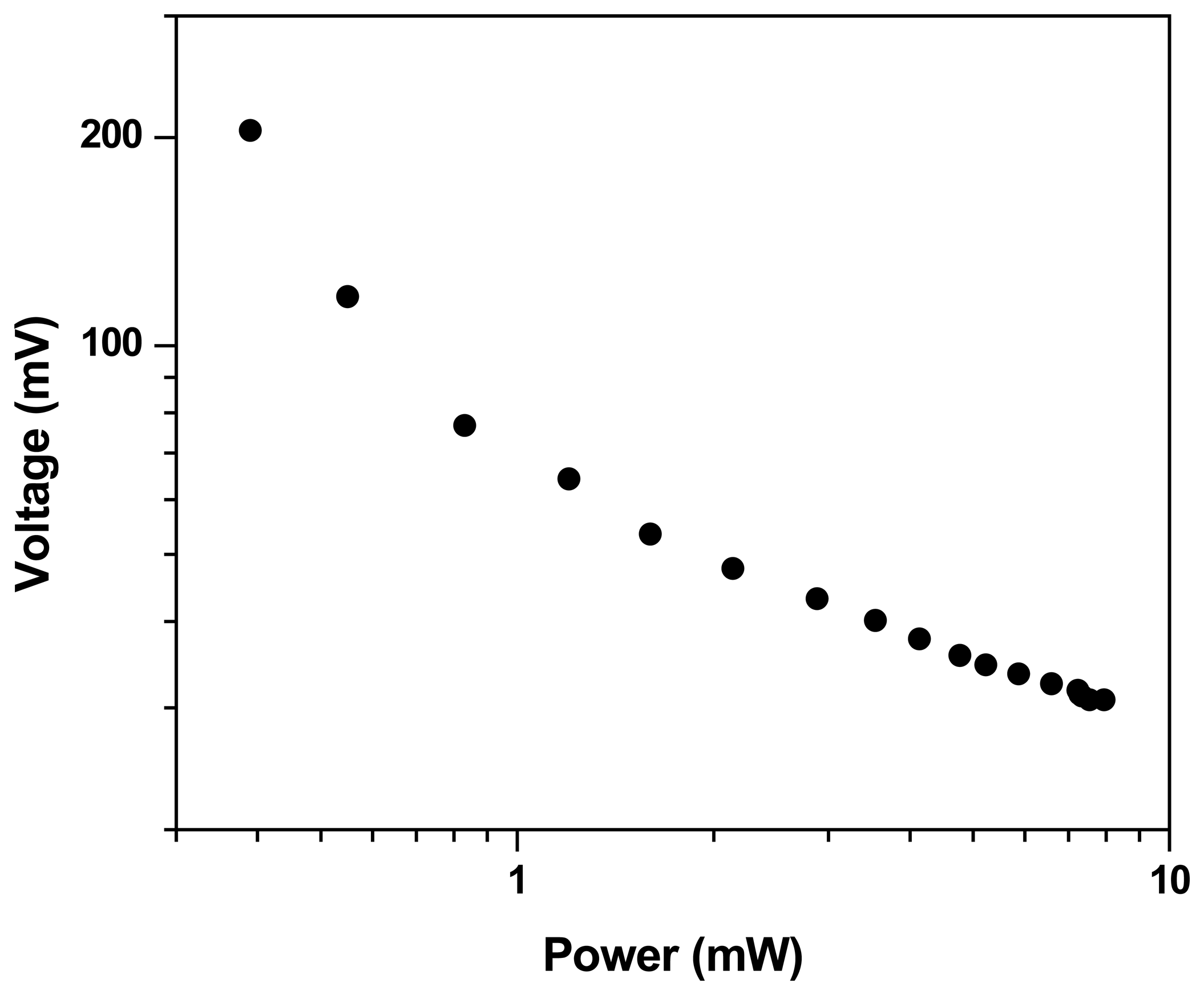
Figure 4 shows the LDR voltage as function of the light power with frequency of 71 Hz. The graph is in the log-log scale. As can be seen, after 1 mW it has a linear behavior, with linear coefficient of -0.345± 0.005 that it can be interpreted as a functional dependence as follows:
For voltages lower than 1 mW the functional dependence is more complicated, because in the log-log graph it follows a second order polynomial curve, similarly as in the case of continuous light.
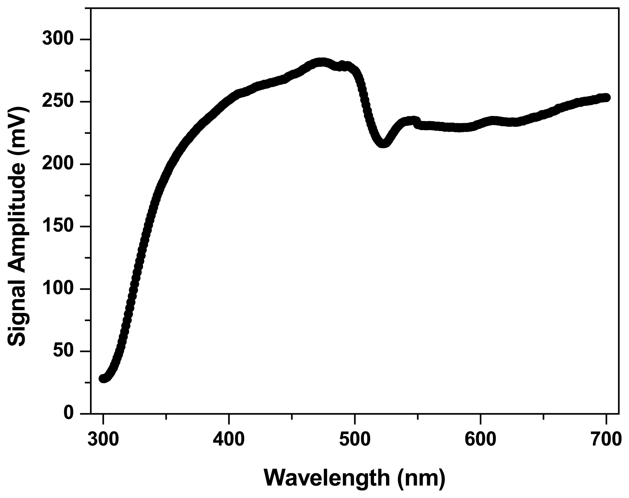
Figure 5 and
6 show the amplitude and phase, respectively, of the microphone signal as function of the wavelength. As the electret microphone functions as light detector through the photoacoustic effect, it only detects chopped light and not continuous light. As can be seen at
Fig. 5, the signal amplitude reproduces fairly well the spectral emission of the light source used [
6]. The phase must be a constant when the microphone air chamber is appropriately sealed with a glass window, giving a stable signal.
Fig. 6 shows that the phase is approximately stable with small oscillations on an average value of –176°.
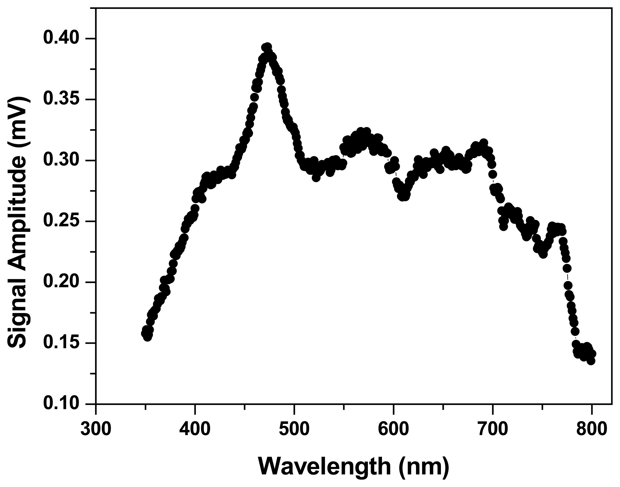
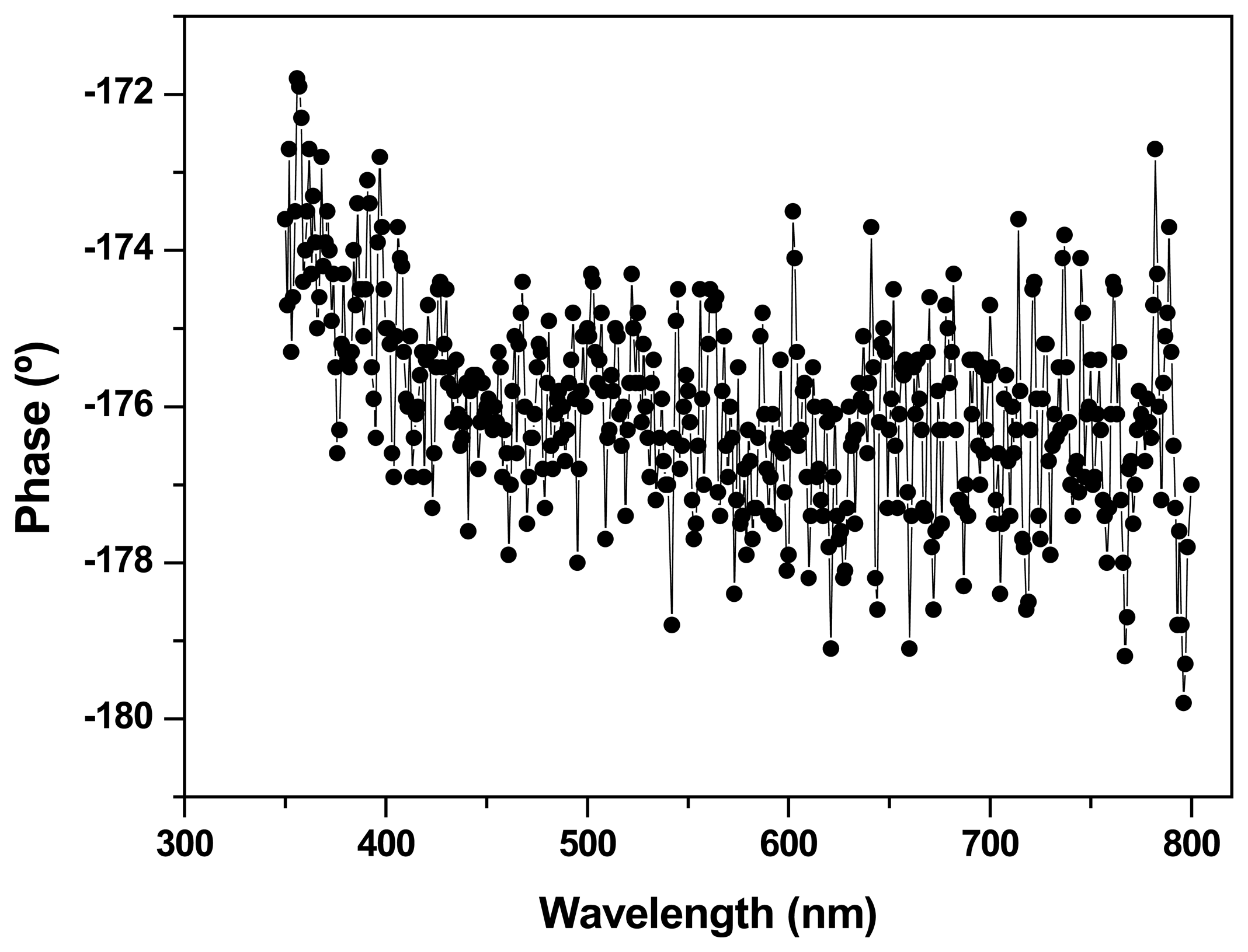
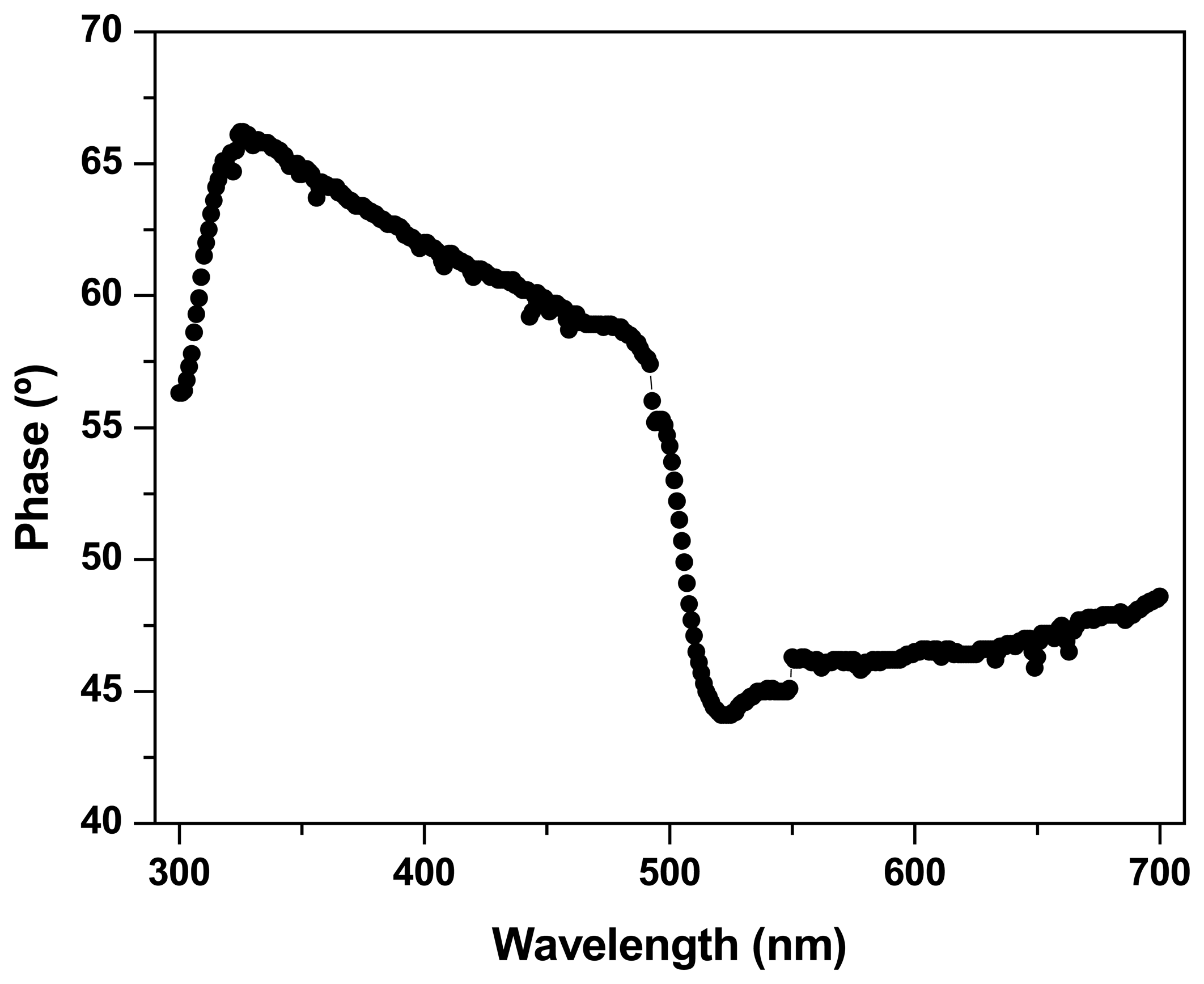
Figure 7 and
8 show the amplitude and phase, respectively, of the LDR signal as function of the wavelength. As can be seen, the amplitude of the LDR signal is not similar to the spectral emission of the lamp.
Fig. 7 corresponds with the spectral emission of the lamp multiplied by spectral response of the LDR coating surface, which is a semiconductor material. The phase of the signal shows one interesting characteristic of the absorbing semiconductor: the energy band gap. Cadmium sulfide (CdS) is the most common semiconductor used in LDR manufacturing. It has the energy band gap at 2.42 eV, that corresponds with a light wavelength of 513 nm. The phase curve at
Fig. 8 resembles that of the CdS absorption, as can be seen at reference [
7], with a signal drop at about 500 nm. The sudden change of the phase must be related to different mechanisms in the generation of the photocurrent below and above the energy related to band gap.
Figures 9 and
10 show the spectra obtained for sample 1 with the microphone and the LDR respectively. The use of the glass window on the microphone limits the spectral range used to wavelengths higher than 350 nm. LDR has not limitation and its spectrum beginning at 300 nm. Both spectra present absorption peaks at same positions: 420 nm and 490 nm. LDR spectrum at
Fig. 10 also shows a peak at 314 nm.
Figure 9 shows a noisy spectrum and
Fig. 10 shows a spectrum with less noise, that permits a better identification of the absorption peaks position.
Figures 11 and
12 show the spectra obtained for sample 2 with the microphone and LDR respectively. In the spectrum obtained using the electret microphone can be observed the presence of an absorption band at about 490 nm, and in that obtained using the LDR can be observed the presence of absorption bands at about 316 nm, 340 nm, 420 nm and 495 nm. Again, the spectrum obtained with the LDR shows a better signal-to-noise ratio when compared with the spectrum obtained with the electret microphone.
The results obtained show that LDR could be used as a sensor of pulsed light in optical spectroscopy devices. It is interesting that the phase of the LDR signal gives information about the energy band gap of the LDR semiconductor. Perhaps it could be used as a way to monitor changes of color in objects, because the phase of the signal does not depend on light intensity but depends on the photon energy related to the light wavelength. In case of
Figs. 10 and
12, above 500 nm it can be seen that a drop in the spectrum appears. This is not usual and must be understood as an error in normalization. Perhaps normalization above 500 nm must be in a different way. The functional behavior observed for the LDR voltage with pulsed light (
eq. 3) has a direct effect in the spectrum structure: as the light source has a distribution of light powers in function of wavelength, the peak height in the spectrum could be shorter than they must be. So, an optical spectrum obtained with this sensor must be used to determine the absorption peak position. To determine the intensity, care must be taken with the multiplicative factor that depends on the light power. About the beverages used in this study, the spectra obtained for them are different, accounting for the different color. The presence of absorption peaks at 420 and 490 nm for sample 1 are related to its yellowish color [
8]. In the case of sample 2, the electret microphone spectrum shows an absorption band at about 490 nm. In the case of LDR spectrum, other peaks can be observed after 400 nm, but the most notorious is that at 490 nm. This absorption peak is more related with its reddish color [
8]. The peaks at about 310 nm are more related with the presence of different organic molecules, in this case, different types of alcohol molecules.
In conclusion LDR light sensors could be used in optical spectroscopy setups, whenever appropriate normalization procedures be used. The use of pulsed light and lock-in amplifiers increases the signal-to-noise ratio, allowing a better definition of absorption peaks and bands. Electret microphones have worse signal to noise ratio when compared with LDR sensors. So, LDR must be the first choice in the construction of cheap homemade spectroscopy systems.


