Monitoring Transport Across Modified Nanoporous Alumina Membranes

Department of Chemistry, Missouri State University, 901 South National Avenue, Springfield, MO 65897, USA
*
Author to whom correspondence should be addressed.
Sensors 2007, 7(11), 2942-2952; https://doi.org/10.3390/s7112942
Submission received: 12 September 2007
/
Accepted: 22 November 2007
/
Published: 23 November 2007
Abstract
:This paper describes the use of several characterization methods to examine alumina nanotubule membranes that have been modified with specific silanes. The function of these silanes is to alter the transport properties through the membrane by changing the local environment inside the alumina nanotube. The presence of alkyl groups, either long (C18) or short and branched (isopropyl) hydrocarbon chains, on these silanes significantly decreases the rate of transport of permeant molecules through membranes containing alumina nanotubes as monitored via absorbance spectroscopy. The presence of an ionic surfactant can alter the polarity of these modified nanotubes, which correlates to an increased transport of ions. Fluorescent spectroscopy is also utilized to enhance the sensitivity of detecting these permeant molecules. Confirmation of the alkylsilane attachment to the alumina membrane is achieved with traditional infrared spectroscopy, which can also examine the lifetime of the modified membrane. The physical parameters of these silane-modified porous alumina membranes are studied via scanning electron microscopy. The alumina nanotubes are not physically closed off or capped by the silanes that are attached to the alumina surfaces.
1. Introduction
Thin alumina membranes containing cylindrical pores with diameters in the nanometer region have been utilized as sensor components in a wide variety of applications [1-2]. These films are synthesized via the electrochemical oxidation of thin aluminum foil in an anodization method that can be tailored to generate pore diameters of a chosen size. These porous alumina membranes can be used without modification as a size-selective separation filter and they are sold commercially for this purpose specifically. However, these porous membranes can be utilized in many ways to build on their native size-selective functionality. The Martin group developed the template method [3] which involves synthesizing nano-sized materials directly within the pores of another material. The porous alumina membrane serves as a solid support and as a foundation to build nanotubes and nanowires of many different compositions with uniform characteristics such as length and diameter. For example, carbon nanotubes have been grown within porous alumina membranes via a carbon vapor deposition process [4-6], while researchers have recently synthesized aligned liquid crystalline nanowires using these membranes [7].
Several reports have examined silica nanotubes that were produced via the template method [8-11]. These silica nanotubes are grown within the pores of alumina membranes using a sol-gel method [12-13] utilizing pure ethanol, tetraethyl orthosilicate and hydrochloric acid. An attractive feature of these nanotubes is silanol (Si-OH) groups present on all of the inner and outer surfaces of the nanotube. Indeed, many researchers have successfully attached hydrophobic, hydrophilic, fluorescent, and antibody molecules to these surfaces via this silanol group [8-11]. For example, Lee and co-workers added hydrophobic octadecyltrimethoxysilane to the interior of 200nm diameter silica nanotubes and then filled them with fluorescent tags (DiIC18) using sonication [9], which yielded modified nanotubes that were imaged using a fluorescent microscope. Another study functionalized the silica nanotubes with an aldehyde-terminated silane, which then served as a coupling agent with amino sites on an antibody [10]. Modified silica nanotubes have also been utilized in novel microarrays with excellent specificity [11].
While many research groups are utilizing porous alumina membranes as templates for generating nanostructures, these membranes can be used directly in sensor applications. The hydroxyl groups on the alumina surfaces are amenable to further functionalization in much the same way as the silanol groups in the aforementioned examples. One of the first published reports involved attaching octadecyltrimethoxysilane groups to the inner surfaces of a porous alumina membrane [14]. These C18-modified membranes were rendered hydrophobic and the amount of transport of ions through the membrane decreased dramatically. Furthermore, the pores of these membranes could be “re-opened” via the addition of an appropriate length surfactant, which permitted the transport of ions again. This behavior was likened to ligand gated ion-channels that function in either an “on” or “off” stage in response to the presence of very particular molecules. Recently, Smirnov and co-workers detected and separated DNA fragments with nanoporous alumina membranes that had single-strands of DNA immobilized within the pores using an amino-terminated silane as a linker [15-16].
Researchers have also examined some of the fundamental characteristics of C18-modified porous alumina membranes with regard to electrical, wetting, and molecular transport properties [17-21]. Applied current, electrolyte ionic strength, and solution pH were varied to determine the impact on electroosomotic flow through the nanopores and the native stability of the Al-O-Si bond [17-19]. Odom et al. elucidated a transport mechanism of neutral molecules through the hydrophobic nanopores based on Langmuir-type adsorption onto the C18 layers and subsequent solid-state diffusion along these layers [20]. Using a homologous series of permeant molecules, the transport rate across the membrane correlated to the hydrophobicity of the molecule. Ku and co-workers examined the nature of electrical conductivity across the C18-modified membranes [21]. They concluded that even though these nanopores were filled with air (due to the presence of the hydrophobic alkylsilane), electrical conduction was still possible through a small amount of “hydrophilically defective” pores, which they confirmed using AC impedance spectroscopy and 29Si NMR [21].
In this preliminary work, we examine methods of detecting molecular transport through modified porous alumina membranes, along with characterization studies. Membranes modified with either isobutyltrimethoxysilane or octadecyltrimethoxylsilane demonstrate significantly decreased ion transport which can be altered via the addition of an appropriate surfactant. Initial studies demonstrate that the utilization of fluorescent spectroscopy provides a more sensitive method for measuring molecular transport through the modified membranes. The presence of alkylsilanes to the surfaces was confirmed via infrared spectroscopy experiments, while scanning electron microscopy images visualize the surface and cross-section of the porous membranes.
2. Results and Discussion
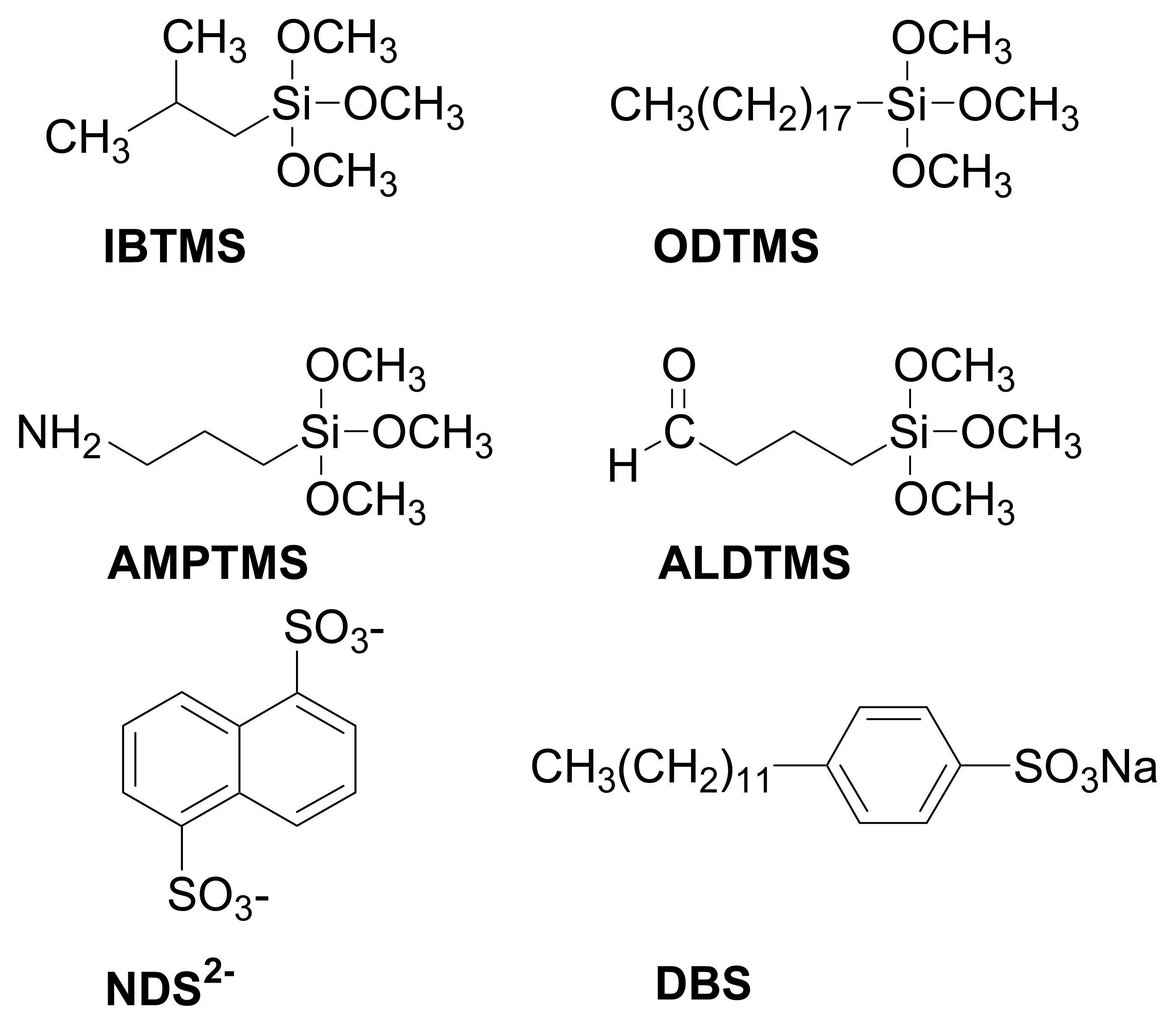
Alumina membranes can be modified by the addition of trimethoxysilane derivatives to form a strong Al–O–Si bond (see Experimental section 4.2 for details on the attachment method). Wide assortments of substituted trimethoxysilanes are available commercially for use in these experiments. Our initial experiments examined four separate groups (see Figure 1 for structures): a short alkyl chain (isobutyltrimethoxysilane, IBTMS), a long alkyl chain (octadecyltrimethoxysilane, ODTMS), an amino-terminated group (3-aminopropyl(trimethoxysilane), AMPTMS), and an aldehyde-terminated group (3-(trimethoxysilyl)propyl aldehyde, ALDTMS). The alkyl trimethoxysilanes were studied for their ability to change the hydrophobicity of the nanopores, and the amino and aldehyde-terminated silanes as groups that could serve as points of further attachment in future experiments.
2.1. Transport across modified porous alumina membranes
To quantify the molecular transport rate through these porous membranes, a U-tube configuration was constructed (see Experimental section 4.3 for specific details). As molecules traveled through the porous membrane and arrived in the permeant half-cell, the concentration of these molecules was measured via simple ultraviolet-visible absorbance spectroscopy at the proper wavelength of light. Indeed, the rate of transfer (absorbance vs. time) of the permeant ion naphthalene disulfonate (NDS2-, see Figure 1 for structure) was linear for the unmodified and alkyl trimethoxysilane-treated membranes. By correlating with Beer's Law plots, these absorbance values were converted to units of molarity and, by nature of a fixed volume, to moles of permeant ion transported. As shown in Table 1, the rate of transport decreased significantly when IBTMS or OBTMS was attached to the porous alumina membranes. In previous work, it was hypothesized that the hydrophobic nature of the attached alkyltrimethoxysilanes made the nanopores significantly more hydrophobic [14], as determined by AC impedance and ion current studies.
Furthermore, these hydrophobic membranes are also affected by species that may present within the permeant and feed solutions. The ionic surfactant dodecylbenzene sulfonate (DBS, see Figure 1 for structure) has been shown previously to affect the transport properties porous alumina membranes made hydrophobic by alkyltrimethoxysilanes [14]. The dodecyl group on the DBS molecule can interact with the alkyl chain on the silane, which in turn decreases the local effect of the hydrophobic alkyl chain on the nanopore. Indeed, this effect was likened to switching the nanotube from an “off” state to an “on” state [14]. In this work, two different molar concentrations of DBS were added to IBTMS and OBTMS-treated membranes and the transport rate of NDS2- was recorded. While the presence of 0.1 mM and 0.2 mM DBS did not restore the transport rates to the level demonstrated by the unmodified membrane, modest improvements in transport were observed (see Table 1).
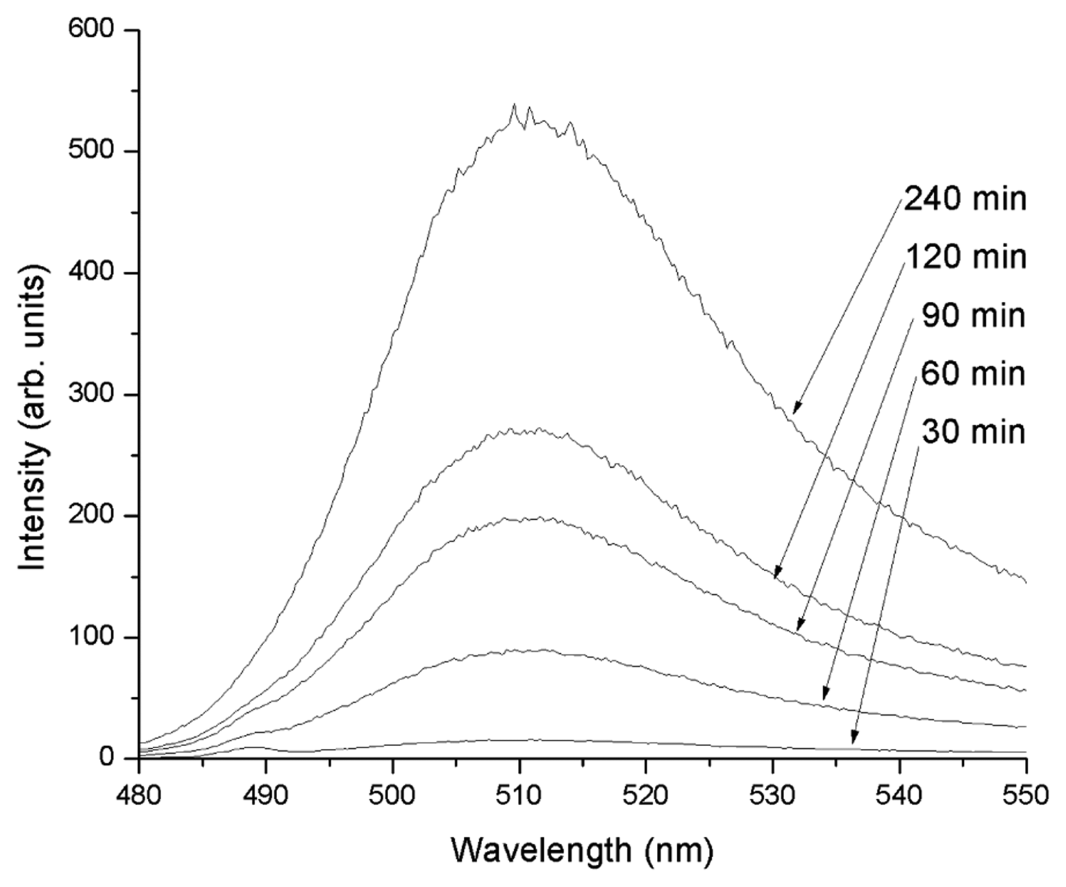
In order to improve the sensitivity of the measuring technique, molecular fluorescence spectrometry was utilized. For these experiments, the permeant ion was changed to fluorescein, which has a large quantum yield when present as a doubly charged anion in moderately basic pH solutions. The transport of 50 μM fluorescein through an unmodified membrane is depicted in Figure 2. Indeed, the transport rate is linear with time and can be converted to a unit of moles per second using an appropriate calibration curve. While this method is only feasible for permeant molecules with appreciable quantum efficiency, the native sensitivity advantages of fluorescence over absorption spectrometry permits improved detection of molecular transport. For example, when transport of 50 μM fluorescein through a porous alumina membrane modified with aminopropyl(trimethoxysilane) was measured, detection in the nanomolar concentration range was observed within one hour (data not shown).
One of the disadvantages to monitoring with absorbance and fluorescence spectrometry is the need to periodically remove aliquots from the permeant half-cell to place within the corresponding instrument for analysis. Possibilities for monitoring molecular transport include direct or “real-time” measurements such as potentiometric ion-selective electrodes and fiber-optic spectrometric probes. The instruments in these techniques could be inserted directly into the permeant half-cell to constantly measure and record increases in permeant concentration with a computer-controlled data acquisition system. Experiments utilizing these techniques are currently in progress in our laboratory.
2.2. Characterization of modified porous alumina membranes
One of the first basic methods of confirming the attachment of trimethoxysilanes to the porous alumina membrane is visual inspection of water drops on the membrane surface. The unmodified hydrophilic membranes adsorb water readily, while water droplets form on the surface on membranes treated with substituted trimethoxysilanes. Infrared (IR) absorption spectrometry is traditionally utilized as a qualitative technique for structure determination. The porous alumina membranes utilized in this study are optically transparent and can be examined in the infrared region of the wavelength spectrum without significant mechanical manipulations. By using the unmodified membranes as a means of comparison, the spectra of modified porous alumina membranes can be analyzed. Infrared spectra of unmodified, IBTMS, and ODTMS-treated porous alumina membranes are depicted in Figure 3. The useful IR range of alumina is from 50,000 to 1,250 cm-1, which precludes the measurement of Al-O-Si and Si-O-Si bonds at 963 and 1000 cm-1, respectively [22]. The broad peak around 3,500 cm-1 is due to the presence of O-H bonds and adsorbed moisture at the alumina surface, while the intense peaks from 1,250 to 1,650 cm-1 can be attributed to the adsorption of CO2 on the membrane [23].
The IR spectra of the IBTMS and ODTMS-treated membranes (Figure 3, inset) have several small bands from 2,800 to 3,000 cm-1 that are not present on the spectrum of the untreated membrane. The peaks at 2855, 2924, and 2961 cm-1 have been previously assigned to symmetric [νs(CH2)], antisymmetric [νa(CH2)], and antisymmetric [νa(CH3)] bands, respectively, when characterizing ODTMS and octadecyltrichlorosilane monolayers on silicon wafers [24]. The membrane treated with octadecyltrimethoxysilane has stronger peaks in this region as compared to isobutyltrimethoxysilane-treated membranes having many less carbons in the alkyl chain.
Furthermore, infrared spectroscopy can be utilized to examine the reusability of these modified porous alumina membranes. Typically, after each transport experiment, the membrane unit is rinsed several times with absolute ethanol and dried in an 80 °C oven for 3-4 minutes to remove the ethanol. The rinsing process removes any permeant molecule or surfactant that might be present on the surface or inside the nanopores of the membrane. The effectiveness of this rinsing can be monitored by comparing the IR spectrum of the membrane before and after rinsing, while paying special attention to regions of the IR spectrum where the permeant ion or surfactant would be expected to appear. The rinsing procedure does not remove the alkyltrimethoxysilanes, as the three distinctive bands from 2,800 to 3,000 cm-1 are always observed, even after several consecutive trials and rinses (data not shown).
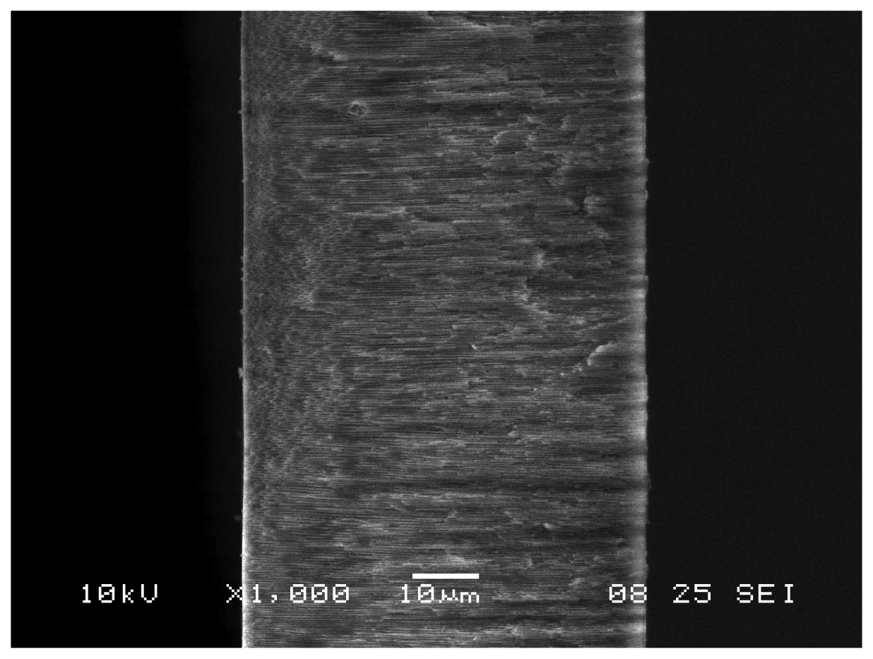
Another available method of membrane characterization is scanning electron microscopy (SEM). This imaging method can be utilized to determine whether the attachment of substituted silanes to porous alumina membranes alters any of the physical dimensions of the membrane. For this study, a series of unmodified and modified alumina membranes were examined with surface and cross-section SEM imaging. As shown in Figure 4, an alumina membrane treated with an aldehyde-terminated trimethoxysilane (ALDTMS) has uniformly sized pores with diameters of ∼200nm. A cross-section view of an amino-terminated trimethoxysilane (AMPTMS)-treated (Figure 5) confirms the thickness (60 μm) of the membrane and the straight nanopores that pass through the entirety of the membrane. In order to obtain the cross-section image, the extremely brittle alumina membrane had to be cut and some evidence of the cut (broken nanotubes and small particulate) is evident. Images of unmodified alumina membranes, both surface and cross-section, are nearly identical to the silane-treated membranes. The presence of the silane modifier does not appear to impact the physical parameters of the nanopore (i.e. closing off or capping the nanopore).
3. Conclusions
This work examines a variety of spectroscopic methods to detect transport through modified porous alumina membranes. Detection utilizing molecular absorbance can be applied to determine transport rates using common and inexpensive instrumentation. In comparison, fluorescence detection has more limited applicability but offers significantly improved sensitivity. The modified alumina membranes were examined via infrared spectroscopy in order to confirm the attachment of the alkyl trimethoxysilanes. Scanning electron microscopy imaging demonstrates that the diameters of nanopores are not significantly changed by the presence of the silane modifiers. Further experimentation will focus on employing these traditional techniques to examine the transport properties of modified porous alumina membranes in order to achieve unique separations and improved sensor applicability.
Practical applications of these modified nanomembranes as sensors could include a combination of selectivity based on molecular size, selectivity based on hydrophobic and hydrophilic properties, and sensitive detection via absorbance or fluorescence spectroscopy. Future experiments will focus on determining whether sensors developed with these modified nanomembranes can accurately calculate the concentration of a single analyte species within a complex mixture. Infrared spectroscopy and surface microscopy techniques will be utilized to confirm the modification of the alumina membranes with variable-substituted trimethoxysilanes.
4. Experimental Section
4.1. Materials
Octadecyltrimethoxysilane, isobutyltrimethoxysilane, 3-aminopropyl(trimethoxysilane), sodium dodecylbenzenesulfonate, naphthalene disulfonate, and fluorescein were obtained from Aldrich and used as received. 3-(trimethoxysilyl)propyl aldehyde was obtained from United Chemical Technologies (Bristol, PA). Anopore® Anodisc 47 filters made of 60 μm thick alumina membranes with 200 nm diameter pores were obtained from Whatman (Maidstone, England).
4.2. Preparation of modified porous alumina membranes
Following a prior method [14], a 5% (v/v) solution of a trimethoxysilane was prepared in pure ethanol. The solution was mixed with a 0.1M acetate buffer (pH = 5.1) to make a 5% (v/v) solution in the buffer. This solution was stirred mechanically for 5 min, and then an alumina membrane was added. After 2 h of soaking, the membrane was removed from the mixture, rinsed with pure ethanol several times, and then cured in a 150 °C oven for 20 min. Membranes described as “unmodified” were treated by the same method (soaking, rinsing, and curing), with the only difference being the lack of trimethoxysilane/ethanol in the soaking solution.
The membrane assembly used in the transport experiments consists of 2 pieces of fiberglass (37mm by 37mm squares) with 12mm diameter holes in the center. There were also 2 cut pieces of Parafilm (25mm by 25mm squares) with 6 mm diameter holes punched through the center. Sections of the treated alumina membranes were carefully cut from the brittle parent membrane and placed between the 2 Parafilm squares, which was then placed between the 2 fiberglass squares. The entire membrane assembly was placed in an 80 °C oven for less than 10 min to allow the Parafilm to melt and seal the entire assembly together.
4.3. Transport cell and measurements
A custom-designed U-tube permeation assembly was constructed by Ace Glass (Vineland, NJ). Each half-cell of the U-tube could accommodate a maximum volume of 20 mL. The membrane assembly described in the previous section was placed between the 2 U-tube half-cells and clamped in place. The half-cells were designated as either feed or permeant, with the only transport possible through a 0.28 cm2 area of porous alumina membrane (defined by the size of the hole in the parafilm). For transport experiments, aliquots of sample from the permeant half-cell were removed periodically, examined with the appropriate instrument, and returned to the permeant half-cell.
For the ultraviolet absorbance experiments, 50 μM of naphthalene disulfonate (NDS2-) was added to the feed half-cell of the U-tube assembly. The transport of this anion through the membrane was monitored using a Hitachi U-2001 spectrophotometer set at 286 nm. When the surfactant dodecyl benzenesulfonate (DBS) was used as a modify transport, it was added to both half-cells at a fixed concentration (either 0.1 or 0.2 mM).
For examining transport via fluorescence spectroscopy, 50 μM of fluorescein in 0.05M phosphate buffer (pH = 9.0) was added to the feed half-cell. The phosphate buffer was also placed in the permeant half-cell. Using a Shimadzu RF-5301 PC spectrofluorimeter set at an excitation wavelength of 489nm, fluorescent intensity was measured from 480 to 550 nm.
4.4. Membrane characterization
Infrared spectra were obtained in the range 500-4000 cm-1 using a FT-IR Nicolet Magna 550 spectrometer, equipped with OMNIC software. Scanning electron microscopy (SEM) images were acquired using a JEOL JSM-6360LV (JEOL USA Inc., Peabody, MA) instrument with an accelerating voltage of 10 or 15 kV.
Acknowledgments
The authors would like to thank the Center for Applied Science and Engineering (CASE) at Missouri State University for funding and support. We acknowledge Rishi J. Patel and Erin E. Sutton at CASE for performing the SEM imaging.
References and Notes
- Hillebrenner, H.; Buyukserin, F.; Stewart, J.D.; Martin, C.R. Biofunctionalization and Capping of Template Synthesized Nanotubes. J. Nanoscience and Nanotechnology 2007, 7, 2211–2221. [Google Scholar]
- Kohli, P.; Wirtz, M.; Martin, C.R. Nanotube Membrane Based Sensors. Electroanalysis 2004, 16, 9–18. [Google Scholar]
- Martin, C.R. Nanomaterials: A Membrane-Based Synthetic Approach. Science 1994, 266, 1961–1966. [Google Scholar]
- Miao, J.Y.; Cai, Y.; Chan, Y.F.; Sheng, P.; Wang, N. A Novel Carbon Nanotube Structure Formed in Ultra-long Nanochannels of Anodic Aluminum Oxide Templates. J. Phys. Chem. B 2006, 110, 2080–2083. [Google Scholar]
- Che, G.; Lakshmi, B.B.; Fisher, E.R.; Martin, C.R. Carbon Nanotubule Membranes for Electrochemical Energy Storage and Production. Nature 1998, 393, 346–349. [Google Scholar]
- Kyotani, T.; Tsai, L.; Tomita, A. Formation of Ultrafine Carbon Tubes by Using an Anodic Aluminum Oxide Film as a Template. Chem. Mater. 1995, 7, 1427–1428. [Google Scholar]
- Steinhart, M.; Zimmerman, S.; Göring, P.; Schaper, A.K.; Gösele, U.; Weder, C.; Wendorff, J.H. Liquid Crystalline Nanowires in Porous Alumina: Geometric Confinement versus Influence of Pore Walls. Nano Lett. 2005, 5, 429–434. [Google Scholar]
- He, B.; Son, S.J.; Lee, S.B. Shape-Coded Silica Nanotubes for Biosensing. Langmuir 2006, 22, 8263–8265. [Google Scholar]
- Jayaraman, K.; Okamoto, K.; Son, S.J.; Luckett, C.; Gopalani, A.H.; Lee, S.B.; English, D.S. Observing Capillarity in Hydrophobic Silica Nanotubes. J. Am. Chem. Soc. 2005, 127, 17385–17392. [Google Scholar]
- Mitchell, D.; Lee, S.B.; Trofin, L.; Li, N.; Nevanen, T.K.; Soderlund, H.; Martin, C.R. Smart Nanotubes for Bioseparations and Biocatalysis. J. Am. Chem. Soc. 2002, 124, 11864–11865. [Google Scholar]
- Kang, M.; Trofin, L.; Mota, M.O.; Martin, C.R. Protein Capture in Silica Nanotube Membrane 3-D Microwell Arrays. Anal. Chem. 2005, 77, 6243–6249. [Google Scholar]
- Lakshmi, B.B.; Patrissi, C.J.; Martin, C.R. Sol-Gel Template Synthesis of Semiconductor Oxide Micro- and Nanostructures. Chem. Mater. 1997, 9, 2544–2550. [Google Scholar]
- Kovtyukhova, N.I.; Mallouk, T.E.; Mayer, T.S. Templated Surface Sol-gel Synthesis of SiO2 Nanotubes and SiO2-Insulated Metal Nanowires. Adv. Mater. 2003, 15, 780–785. [Google Scholar]
- Steinle, E.D.; Mitchell, D.T.; Wirtz, M.; Lee, S.B.; Young, V.Y.; Martin, C.R. Ion Channel Mimetic Micropore and Nanotube Membrane Sensors. Anal. Chem. 2002, 74, 2416–2422. [Google Scholar]
- Vlassiouk, I.; Takmakov, P.; Smirnov, S. Sensing DNA Hybridization via Ionic Conductance Through a Nanoporous Electrode. Langmuir 2005, 21, 4776–4778. [Google Scholar]
- Vlassiouk, I.; Krasnoslobodtsev, A.; Smirnov, S.; Germann, M. “Direct” Detection and Separation of DNA using Nanoporous Alumina Filters. Langmuir 2004, 20, 9913–9915. [Google Scholar]
- Winkler, B.H.; Baltus, R.E. Modification of the Surface Characteristics of Anodic alumina Membranes using Sol-gel Precursor Chemistry. J. Membr. Sci. 2003, 226, 75–84. [Google Scholar]
- Chen, W.; Yuan, J.H.; Xia, X.H. Characterization and Manipulation of the Electroosmotic Flow in Porous Anodic Alumina Membranes. Anal. Chem. 2005, 77, 8102–8108. [Google Scholar]
- Szczepanski, V.; Vlassiouk, I.; Smirnov, S. Stability of Silane Modifiers on Alumina Nanoporous Membranes. J. Membr. Sci. 2006, 281, 587–591. [Google Scholar]
- Odom, D.J.; Baker, L.A.; Martin, C.R. Solvent-Extraction and Langmuir-Adsorption-Based Transport in Chemically Functionalized Nanopore Membranes. J. Phys. Chem. B 2005, 109, 20887–20894. [Google Scholar]
- Ku, A.Y.; Ruud, J.A.; Early, T.A.; Corderman, R.R. Evidence of Ion Transport through Surface Conduction in Alkylsilane-Functionalized Nanoporous Ceramic Membranes. Langmuir 2006, 22, 8277–8280. [Google Scholar]
- Naviroj, S.; Koenig, J.L.; Ishida, H. Diffuse Reflectance Fourier Transform Infrared Spectroscopic Study of Chemical Bonding and Hydrothermal Stability of an Aminosilane on Metal Oxide Surfaces. J. Adhesion 1985, 18, 93–110. [Google Scholar]
- Haneda, M.; Joubert, E.; Ménézo, J.C.; Duprez, D.; Barbier, J.; Bion, N.; Daturi, M.; Saussey, J.; Lavalley, J.C.; Hamada, H. Surface Characterization of Alumina-Supported Catalysts Prepared by Sol-gel Method. Part I. Acid-base Properties. Phys. Chem. Chem. Phys. 2001, 3, 1366–1370. [Google Scholar]
- Koga, T.; Morita, M.; Ishida, H.; Yakabe, H.; Sasaki, S.; Sakata, O.; Otsuka, H.; Takahara, A. Dependence of the Molecular Aggregation State of Octadecylsiloxane Monolayers on Preparation Methods. Langmuir 2005, 21, 905–910. [Google Scholar]
Figure 1.
Structures of silane modifiers, transport molecules, and surfactants.

Figure 2.
Transport profile of 50 μM fluorescein through an unmodified porous (200nm diameter) alumina membrane.
Figure 2.
Transport profile of 50 μM fluorescein through an unmodified porous (200nm diameter) alumina membrane.

Figure 3.
FT-IR spectra of untreated or silane-treated porous alumina membranes.

Figure 4.
SEM image of the surface of a 3-(trimethoxysilyl)propyl aldehyde (ALDTMS)-modified porous alumina membrane.
Figure 4.
SEM image of the surface of a 3-(trimethoxysilyl)propyl aldehyde (ALDTMS)-modified porous alumina membrane.

Figure 5.
SEM image of the cross section of an aminopropyl(trimethoxysilane) (AMPTMS)- modified porous alumina membrane
Figure 5.
SEM image of the cross section of an aminopropyl(trimethoxysilane) (AMPTMS)- modified porous alumina membrane


Table 1.
Transport rates of NDS2- through modified porous alumina membranes as determined by ultraviolet-visible absorption spectrometry.
| Membrane modifier | Surfactant concentration | Transport rate (moles / s) |
|---|---|---|
| No modifier (blank membrane) | -- | 1.22 7× 10⁻¹⁰ |
| IBTMS | -- | 9.74 × 10⁻¹² |
| IBTMS | 0.1 mM DBS | 3.61 × 10⁻¹¹ |
| IBTMS | 0.2 mM DBS | 4.74 × 10⁻¹¹ |
| ODTMS | -- | 1.28 × 10⁻¹² |
| ODTMS | 0.1 mM DBS | 2.34 × 10⁻¹¹ |
| ODTMS | 0.2 mM DBS | 2.56 × 10⁻¹¹ |
© 2007 by MDPI ( http://www.mdpi.org). Reproduction is permitted for noncommercial purposes.
Share and Cite
MDPI and ACS Style
Penumetcha, S.S.; Kona, R.; Hardin, J.L.; Molder, A.L.; Steinle, E.D. Monitoring Transport Across Modified Nanoporous Alumina Membranes. Sensors 2007, 7, 2942-2952. https://doi.org/10.3390/s7112942
AMA Style
Penumetcha SS, Kona R, Hardin JL, Molder AL, Steinle ED. Monitoring Transport Across Modified Nanoporous Alumina Membranes. Sensors. 2007; 7(11):2942-2952. https://doi.org/10.3390/s7112942
Chicago/Turabian StylePenumetcha, Sai S., Ravikanth Kona, Jonathan L. Hardin, Andrew L. Molder, and Erich D. Steinle. 2007. "Monitoring Transport Across Modified Nanoporous Alumina Membranes" Sensors 7, no. 11: 2942-2952. https://doi.org/10.3390/s7112942