Article

An Investigation of Glutathione-Platinum(II) Interactions by Means of the Flow Injection Analysis Using Glassy Carbon Electrode

by
Ondrej Zitka
1,
Dalibor Huska
1,
Sona Krizkova
1,
Vojtech Adam
1,
Grace J. Chavis
4,
Libuse Trnkova
5,
Ales Horna
6,
Jaromir Hubalek
7 and
Rene Kizek
1,*
1
Department of Chemistry and Biochemistry, Faculty of Agronomy, Mendel University of Agriculture and Forestry, Zemedelska 1, CZ-613 00 Brno, Czech Republic
2
Department of Biochemistry, Faculty of Science, Masaryk University, Kotlarska 2, CZ-611 37 Brno, Czech Republic
3
Department of Animal Nutrition and Forage Production, Faculty of Agronomy, Mendel University of Agriculture and Forestry, Zemedelska 1, CZ-613 00 Brno, Czech Republic
4
Department of Chemistry, University of California, One Shields Avenue, CA-956 16 Davis, USA
5
Department of Chemistry, Faculty of Science, Masaryk University, Kotlarska 2, CZ-611 37 Brno, Czech Republic
6
Department of Food Engineering, Faculty of Technology, Tomas Bata University, T.G. Masaryka 275, CZ-762 72 Zlin, Czech Republic Department of Food Engineering, Faculty of Technology, Tomas Bata University, T.G. Masaryka 275, CZ-762 72 Zlin, Czech Republi
7
Department of Microelectronics, Faculty of Electrical Engineering and Communication, Brno University of Technology, Udolni 53, CZ-602 00 Brno, Czech Republic
*
Author to whom correspondence should be addressed.
Sensors 2007, 7(7), 1256-1270; https://doi.org/10.3390/s7071256
Submission received: 15 June 2007 / Accepted: 13 July 2007 / Published: 20 July 2007

Abstract

:
Despite very intensive research in the synthesising of new cytostatics, cisplatin is still one of the most commonly used anticancer drugs. Therefore, an investigation of interactions of cisplatin with different biologically important amino acids, peptides and proteins is very topical. In the present paper, we utilized flow injection analysis coupled with electrochemical detection to study and characterize the behaviour of various forms of glutathione (reduced glutathione – GSH, oxidized glutathione – GSSG and S-nitroso glutathione – GSNO). The optimized conditions were as follows: mobile phase consisted of acetate buffer (pH 3) with a flow rate of 1 mL min-1. Based on results obtained we chose 850 mV as the optimal potential for detection of GSH and 1,100 mV as the optimal potential for detection of GSSG and GSNO. The detection limits of GSH, GSSG and GSNO were 100 pg mL-1, 50 ng mL-1 and 300 pg mL-1, respectively. Further, the optimized technique was used for investigation of interactions between cisplatin and GSH. We were able to observe the interaction between GSH and cisplatin via decrease in the signal corresponding to glutathione. Moreover, we evaluated the formation of the complex by spectrometry. The spectrometric results obtained were in good agreement with electrochemical ones.

1. Introduction

Glutathione was discovered by M. J. de Rey Pailhade at the end of the 19th century as the substance “hydrogénant le soufre”, which was renamed by F.G. Hopkins in 1921. Hopkins first characterised the compound as a dipeptide of glutamic acid and cysteine. Few years later he suggested the correct structure to be a tripeptide which also contains glycine [1]. GSH as a ubiquitous tripeptide thiol is a vital intra- and extra-cellular protective antioxidant. It plays a number of key roles in the controlling of signalling processes, detoxifying of some xenobiotics and heavy metals etc. Glutathione is found almost exclusively in its reduced form; since the enzyme, which reverts it from its oxidized form (GSSG) called glutathione reductase, is constitutively active and inducible upon oxidative stress. In fact, the ratio of reduced to oxidized glutathione within cells is often used as a marker of cytotoxicity [2-5]. In connection with this, many fundamental events of cell regulation such as protein phosphorylation and binding of transcription factors to consensus sites on DNA are driven by physiological oxidant-antioxidant homeostasis, especially by thiol-disulfide balance. Therefore endogenous glutathione and thioredoxin systems and may be considered to be effective regulators of redox-sensitive gene expression [6-9].
The sulfhydryl group of glutathione is highly reactive and is often found conjugated to other molecules such as nitric oxide (NO) [10-14]. Nitrosation of the glutathione may serve as a signal event and/or as a deposition of NO to S-nitrosoglutathione. Thus, GSH can serve also as a substrate in conjugation reactions [15].
Based on the facts mentioned above it is not surprising that GSH can influence the treatment of diseases, such as cancer [16-18]. In malignant tumours, as compared with normal tissues, the multidrug resistance associates mostly with higher GSH levels within these cancer cells [19]. Thus, approaches to cancer treatment based on modulation of GSH should control possible tumour growth-associated changes in GSH content and synthesis in these cells [20-23]. An investigation of the interaction of GSH with heavy metal based cytostatics can be one of the possible ways of studying of reasons how GSH can interrupt the cancer treatment [24]. The heavy metal based cytostatics, most of all platinum complexes, play an important role in the chemotherapy of various malignancies [25-31]. The biological activity of the first platinum based cytostatic drug – cisplatin (cis-diamminedichloroplatinum(II)), which is still one of the most frequently used cytotoxic agents, was discovered in 1965 by Rosenborg during his studies on the effects of an electric current on bacterial growth [32]. Since then hundreds of platinum(II) and platinum(IV) complexes have been synthesized and evaluated as anticancer agents over the past 40 years. Despite very intensive research in the synthesising of new cytostatics, cisplatin is still one of the most commonly used anticancer drugs. Therefore, an investigation of interactions of cisplatin with different biologically important amino acids, peptides and proteins is very topical. Many experimental or soft modelling methods have been proposed for these purposes,[33-39]. In the present paper, we attempted to utilize flow injection analysis coupled with electrochemical detection to study and characterize behaviour of various glutathiones (reduced – GSH, oxidized – GSSG and S-nitroso – GSNO, Fig. 1). Frther, the optimized technique was used for investigation of interactions between cisplatin and reduced GSH.

2. Experimental

2.1 Chemicals

Reduced glutathione (GSH), oxidised glutathione (GSSG), S-nitrosoglutathione (GSNO) sodium tetraborate and other chemicals used were purchased from Sigma Aldrich (St. Louis, USA) unless noted otherwise. Tris(2-carboxyethyl)phosphine (TCEP) is produced by Molecular Probes (Evgen, Oregon, USA). Stock standard solutions of GSH, GSSG and GSNO (100 mg mL-1) were prepared with ACS water (Sigma-Aldrich, USA) and stored in the dark at –20 °C. In addition GSH solutions were prepared in the presence of reduction agent TCEP to avoid oxidation [40]. Working standard solutions were prepared daily by dilution of the stock solutions. The pH value was measured using WTW inoLab (Weilheim, Germany). The pH-electrode (SenTix-H, pH 0–14/3M KCl) was regularly calibrated with WTW buffers (Weilheim, Germany).

2.2 Preparation of cisplatin solutions

The chemotherapeutic drug of cisplatin was synthesized and provided by Pliva-Lachema (Brno, Czech Republic) [41]. Stock standard solutions of cisplatin (500 μg mL-1) were prepared by sodium chloride solution (0.5 M, pH 6.4) and stored in the dark at -20 °C [38,42]. Working standard solutions were prepared daily by dilution of the stock solutions in phosphate buffer (100 mM).

2.3 Electrochemical measurements

Electrochemical measurements were performed with AUTOLAB Analyser (EcoChemie, Netherlands) connected to a VA-Stand 663 (Metrohm, Switzerland), using a standard cell with three electrodes. The working electrode was a carbon paste electrode (CPE). The reference electrode was the Ag/AgCl/3M KCl electrode and the auxiliary electrode was the graphite electrode. All experiments were carried out at room temperature. The carbon paste was made of 70 % graphite powder (Sigma Aldrich) and 30 % mineral oil (Sigma Aldrich). The carbon paste was housed in a Teflon body having a 2.5 mm diameter of active disk surface. The electrode surface was polished before each determination with a soft filter paper prior to measurement [43-45]. For smoothing and baseline correction [46], the software GPES 4.4 was employed.

2.4 Square wave voltammetry measurements of GSH, GSSG and GSNO

The GSH, GSSG and GSNO were measured using SWV at the surface CPE. The supporting electrolyte (Britton-Robinson buffer, pH 5) was purchased from Sigma Aldrich in ACS purity. SWV parameters were as follows: the initial potential of –0.2 V, the end potential 1.5 V, frequency 200 Hz and step potential 5 mV. For other experimental details see references [40,47].

2.5 Preparation of GS-Pt complexes

The GS-Pt complex was prepared according to Ishikawa and Ali-Osman [48] by incubation of GSH (60 μg mL-1) with cisplatin (30 μg mL-1) in 500 μl of 100 mM phosphate buffer pH 7.8 in the dark at 37 °C (thermostat BTV Brno) [49].

2.6 Flow electrochemical measurement

An FIA-ED system consisted of a solvent delivery pump operating in the range of 0.001-9.999 ml/min (Model 582 ESA Inc., Chelmsford, MA, USA), a guard cell (Model 5020 ESA, USA), a reaction coil (1 m) and an electrochemical detector. The amperometric electrochemical detector (ED) includes one low volume flow-through analytical cell (Model 5040, ESA, USA), which consists of a glassy carbon working electrode, a palladium electrode as a reference electrode and an auxiliary carbon electrode, and a Coulochem III as a control module. The sample (5 μl) was injected manually. The obtained data were treated by CSW 32 software. The experiments were carried out at room temperature (22 °C). A glassy carbon electrode was polished mechanically by 0.1 μm of alumina (ESA Inc., USA) and sonicated at room temperature for 5 min using a Sonorex Digital 10 P Sonicator (Bandelin, Berlin, Germany) at 40 W.

2.7 Spectroscopic analysis

Spectra of GSH, cisplatin and their complexes were recorded in the presence pf 100 mM phosphate buffer (pH 7.8) using a Helios spectrophotometer (Analytic Jena, Germany).

2.8 Statistical analysis

STATGRAPHICS® (Statistical Graphics Corp®, USA) was used for statistical analyses. Results are expressed as mean ± S.D. unless otherwise noted. Values of p < 0.05 were considered significant.

3. Results and Discussion

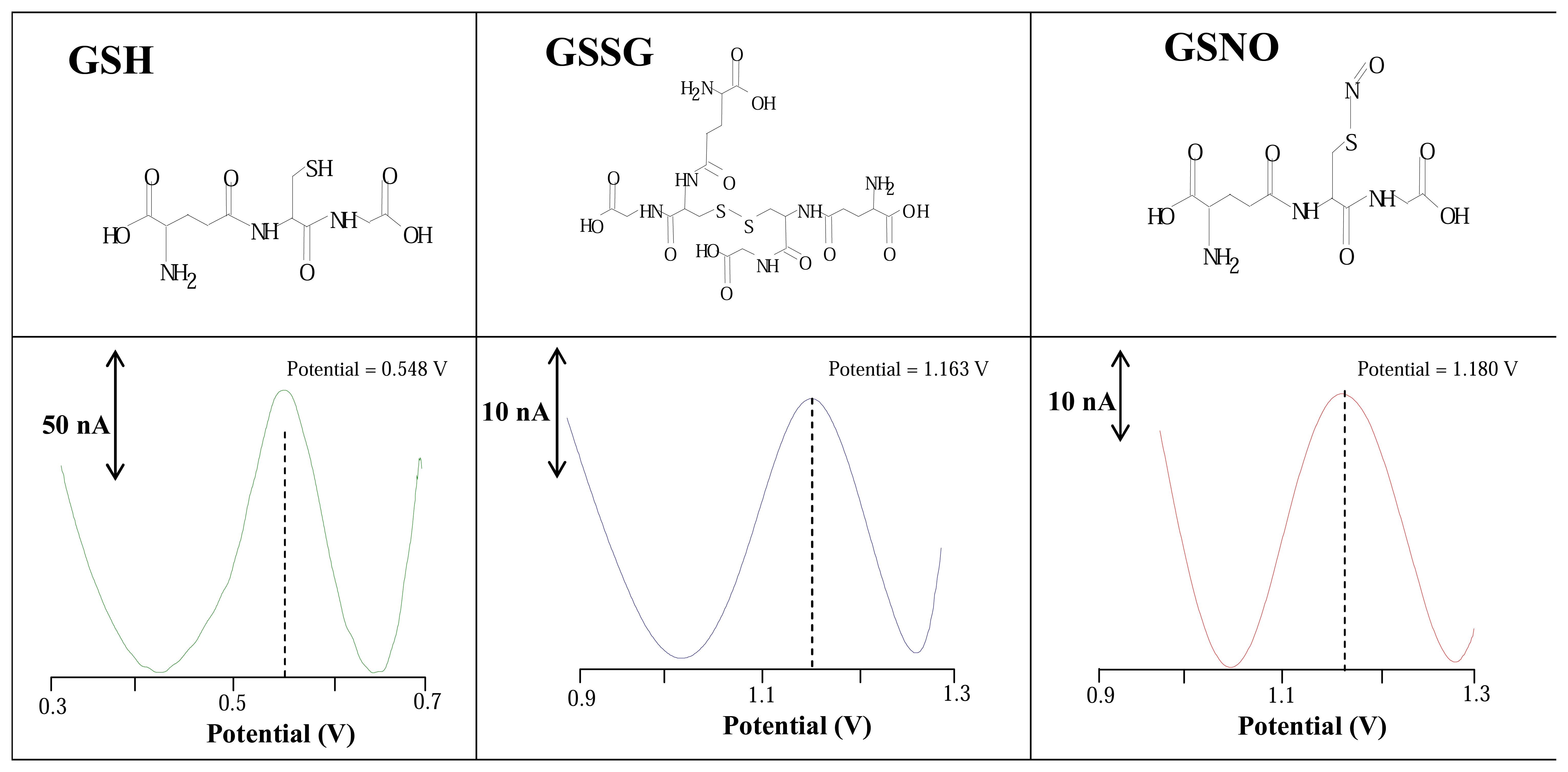
3.1 Stationary electrochemical system

Firstly we focused on studying the basic electrochemical properties of target molecules. Reduced (GSH), oxidised (GSSG) and S-nitroso (GSNO) glutathione were measured by square wave voltammetry using carbon paste electrode. We investigated how the compounds of interest behave in the presence of Britton-Robinson buffer (pH 5) as a supporting electrolyte. We found that certain glutathiones can be determined on CPE in concentrations below 1 μM. Reduced glutathione gave oxidation signals near 0.56 V, oxidized glutathione approximately 1.16 V and nitrosoglutathione at about 1.19 V. The shift of potentials of the target molecules to more positive values can be probably related with the higher energy necessary to oxidize stronger bonds as S-S and S-NO compared to free– SH group (Fig. 1).

3.2 Electrochemical analysis of glutathiones in flow system

When we assessed that the glutathiones can be measured at solid electrodes (Fig. 1), flow injection analysis using glassy carbon electrode as a working one was utilized for measurement of the compounds of the interest. The flow rate of the mobile phase (Britton-Robinson, pH 5.0) was the first experimental condition we optimized. The influence of flow rate on the electrochemical responses of the target molecules was investigated within the range from 0.4 to 1.5 mL min-1 at a working potential of 700 mV. The responses enhanced with increasing flow rate to 1.0 mL min-1 and then slightly decrease. Based on the obtained results we chose 1.0 mL min-1 as the optimal flow rate of the mobile phase. The observed signals were well developed and symmetrical (not shown).

3.2.1 Mobile phase composition and its pH

Other experimental conditions such as composition of mobile phase and its pH were also optimized. We choose three various buffers (acetate, phosphate and Britton-Robinson) and investiagted how their different pH values can affect the electrochemical responses of glutathiones (Fig. 2). It clearly follows from the results obtained that acetate buffer (pH 3) was the most suitable mobile phase to measure glutathiones with the highest sensitivity. If phosphate buffer was used, the lowest signals were measured. Particularly, the peak heights of glutathiones measured in the presence of phosphate buffer were about 80 % lower than in acetate buffer. Similarly, if we used Britton Robinson buffer as the mobile phase signals were about 20-60 % lower than in acetate buffer. It clearly follows from the obtained results that the electrochemical responses of glutathiones measured on a glassy carbon electrode are affected not only by the composition of the mobile phase but also by its pH (Fig. 2). Moreover it can be concluded that the higher pH values used, the lower the electrochemical responses of compounds of interest were observed.

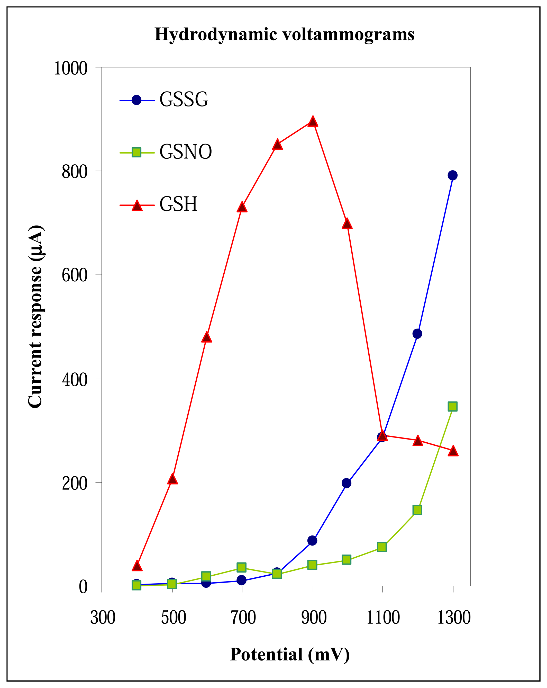
3.2.2 Applied potential on glassy carbon working electrode

As we have optimized flow rate of a mobile phase and its composition, a working electrode potential was the last experimental conditions we optimized. We measured glutathiones of the same concentration (30 μg mL-1) in the presence of acetate buffer (pH 3) with a flow rate of 1 mL min-1 at various working electrode potentials. The hydrodynamic voltammograms obtained are shown in Fig. 3. If GSH was measured, the rapid increase of the signal with maximum at 900 mV was observed. The increase of the electrochemical responses of GSSG and GSNO were observed at potentials higher than 800 mV. The peaks increased to 1,300 mV, where the highest peaks of GSSG and GSNO were observed. If we compared the potentials of the highest peaks of glutathiones measured by flow technique to those measured by SWV, we found that the potentials measured by flow technique shifted slightly to more positive values (Fig. 1 and Fig. 3). The shift can be associated with different reference and working electrodes used (hydrogen-palladium in flow system and Ag/AgCl, 3M KCl in stationary). Based on the results obtained we choose 850 mV as the optimal potential for detection of GSH and 1,100 mV as the optimal potential for detection of GSSG and GSNO. The signals of GSSG and GSNO measured under potentials higher than 1,100 mV were higher but the stabilizing of background currents lasted more than twenty minutes and standard deviations exceeded 10 %. These disadvantages encouraged us to utilize 1,100 mV for detection of GSSG and GSNO in the following experiments.

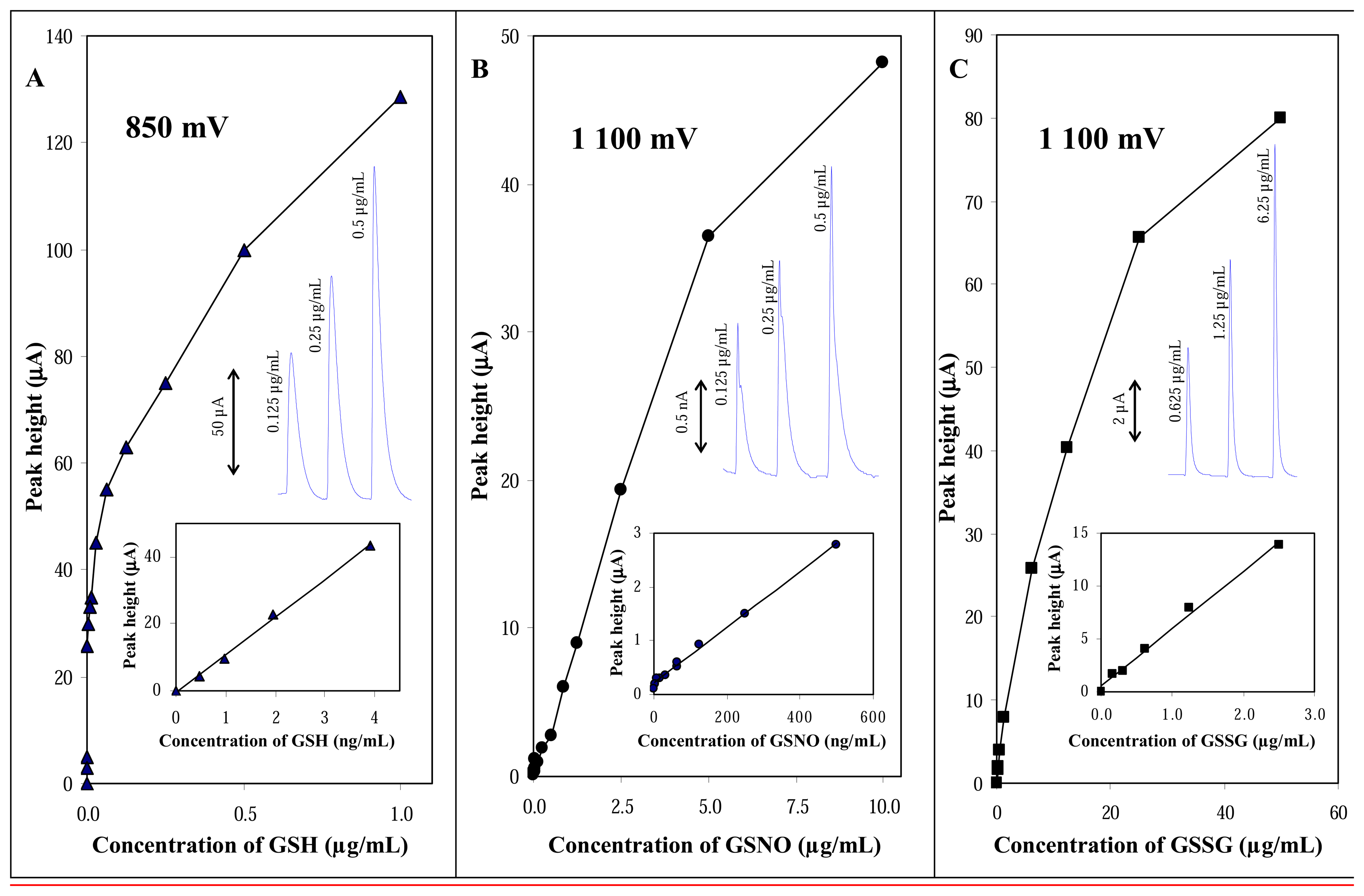
3.2.3 Calibration curves

Under the experimental conditions optimized above we investigated the influence of glutathiones concentration on their current responses. The dependences measured are shown in Figs. 4A,B,C. The dependences were nonlinear within the concentration ranges we studied, whereas the linear ranges of the dependences are shown in insets in Figs. 4A,B,C. The nonlinearity observed can be associated with an accumulation of analyte on the electrode surface, where poly-layers of analyte adsorbed could form [50]. In addition, the signals were very well reproducible with a relative standard deviation of 2-5%.
GSSG gave the lowest current responses in comparison with other compounds of interest. The concentration dependence of GSSG was linear (y = 5.4816x + 0.4914; R2 = 0.9939) within the concentration range from 0.1 to 2.5 μg mL-1, with a detection limit of 50 ng mL-1. If we measured GSNO, we obtained strictly linear concentration dependence (y = 0.0052x + 0.2099; R2 = 0.9957) within the range from 1 to 500 ng mL-1, whereas the detection limit was 300 pg mL-1. In addition, GSH concentration dependence was strictly linear (y = 11.315x - 0.5146; R2 = 0.9969) within the range from 0.5 to 4 ng mL-1. The detection limit of GSH was 100 pg mL-1 (Fig. 4). The detection limits were evaluated as 3 S/N ratio according to Long and Winefordner [51].

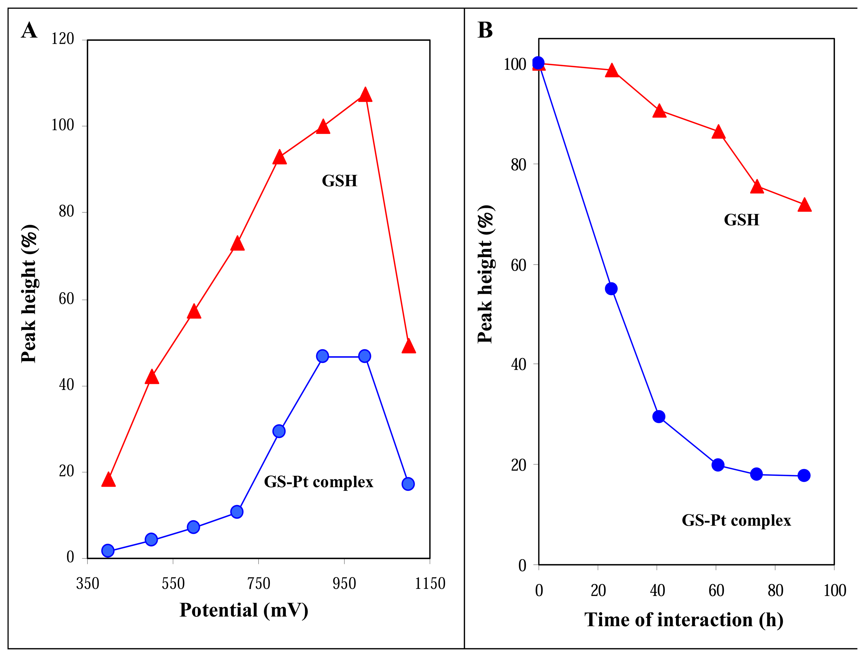
3.3 Study of GSH-cisplatin interaction

GSH belongs to group of thiols, like metalothioneins, which could most likely influence therapeutic concentration of platinum based cytostatics (e.g. cisplatin) within a body and, therefore, could play one of the key roles in formation of a resistance on the treatment [52-57]. A mechanism of action of GSH with cisplatin is still unclear, but the most accepted opinion is that the thiol decreases of therapeutic concentration in the place of the drug action [58-60]. The contribution of other forms of glutathiones including GSNO and/or GSSG to decrease of therapeutic concentration of platinum based cytostatics is thought to be negligible due to their few folds lower concentration and reactivity. Thus, studying of interactions between these low abundant glutathiones with cytostatics is of low clinical importance. Nevertheless, investigating of interactions between GSH and cytostatics if of high importance but it is very difficult and needs robust and high cost instruments as nuclear magnetic resonance.
Here, we propose a simple method to characterize the interactions of GSH with cisplatin. Therapeutic doses of cisplatin (3, 15 and 30 μg mL-1) were used for this purpose. GS-Pt complexes were prepared according to Ishikawa and Ali-Osman [48]. We observed the interaction between ciplatin and GSH by determining of changes in GSH signal. As expected, the formed GS-Pt complex gave lower responses compared to reduced glutathione (Fig. 5A). Therefore, the hydrodynamic voltammograms of GSH and GS-Pt complex measured in the range from 400 to 1,100 mV differed. The current response of GS-Pt complex increased slowly up to an applied potential of 750 mV, then the rapid increase of the signal with maximum at 900-950 mV occurred (Fig. 5A). Therefore we further measured the GS-Pt complex at 950 mV. Moreover, the signal of cisplatin (30 μg mL-1) under these experimental conditions was negligible.
During time scale investigating of formation complexes between GSH and cisplatin we also studied if the height of GSH peaks without cisplatin can change. We found the peak of GSH decreased gradually within the range of time we were interested in (Fig. 5B). In addition, the dependence of peak height of GS-Pt complex on times of interactions is shown in Fig. 5B. It clearly follows from the results obtained that dependences differed markedly, which confirmed that we really observed the interactions between cisplatin and GSH. The signal of complex decreased more than 40 % during the 20 h long interaction at 37 °C. Moreover, after a 60 h long interaction the maximum yield of complex had been reached. We also decided to evaluate the formation of complex by another reference technique. The complex formation was monitored spectrometrically as absorbance change at 280 nm [49]. The spectrometric results obtained were in good agreement with electrochemical ones.

Conclusion

Electrochemical detection represents an alternative method for determination of biologically active compounds [42,61-71]. Here, we have shown the flow injection analysis coupled with electrochemical detector could be a suitable technique not only for direct detection of various forms of glutathione but also for investigating of interactions between thiols and cytostatics. FIA-ED is very promising tool for such purposes because it is low cost and very sensitive to various electroactive species.

Acknowledgments

This work was supported from the grant of the Grant Agency of Academy of Sciences of the Czech Republic No. GAAV IAA401990701.

References

  1. Hopkins, F.G. On an autoxidisable constituent of the cell. Biochem. J. 1921, 15, 286–305. [Google Scholar]
  2. Carelli, S.; Ceriotti, A.; Cabibbo, A.; Fassina, G.; Ruvo, M.; Sitia, R. Cysteine and glutathione secretion in response to protein disulfide bond formation in the ER. Science 1997, 277, 1681–1684. [Google Scholar]
  3. Locigno, R.; Castronovo, V. Reduced glutathione system: Role in cancer development, prevention and treatment (Review). Int. J. Oncol. 2001, 19, 221–236. [Google Scholar]
  4. Noctor, G.; Foyer, C.H. Ascorbate and glutathione: Keeping active oxygen under control. Annu. Rev. Plant Biol. 1998, 49, 249–279. [Google Scholar]
  5. Townsend, D.M.; Tew, K.D.; Tapiero, H. The importance of glutathione in human disease. Biomed. Pharmacother. 2003, 57, 145–155. [Google Scholar]
  6. Droge, W. Free radicals in the physiological control of cell function. Physiol. Rev. 2002, 82, 47–95. [Google Scholar]
  7. Sen, C.K.; Packer, L. Antioxidant and redox regulation of gene transcription. Faseb J. 1996, 10, 709–720. [Google Scholar]
  8. Hayes, J.D.; Pulford, D.J. The glutathione S-Transferase supergene family: Regulation of GST and the contribution of the isoenzymes to cancer chemoprotection and drug resistance. Crit. Rev. Biochem. Mol. Biol. 1995, 30, 445–600. [Google Scholar]
  9. Yegorova, S.; Yegorov, O.; Lou, M.F. Thioredoxin induced antioxidant gene expressions in human lens epithelial cells. Exp. Eye Res. 2006, 83, 783–792. [Google Scholar]
  10. Sehlotho, N.; Griveau, S.; Nyokong, T.; Bedioui, F. Cobalt phthalocyanine molecular electrode for the electrochemical investigation of the release of glutathione upon copper-catalyzed decomposition of S-nitrosoglutathione. Electroanalysis 2007, 19, 103–106. [Google Scholar]
  11. Musameh, M.; Moezzi, N.; Schauman, L.M.; Meyerhoff, M.E. Glutathione peroxidase-based amperometric biosensor for the detection of S-nitrosothiols. Electroanalysis 2006, 18, 2043–2048. [Google Scholar]
  12. Balendiran, G.K.; Dabur, R.; Fraser, D. The role of glutathione in cancer. Cell Biochem. Function 2004, 22, 343–352. [Google Scholar]
  13. Clancy, R.M.; Levartovsky, D.; Leszczynskapiziak, J.; Yegudin, J.; Abramson, S.B. Nitric-oxide reacts with intracellular glutathione and activates the hexose-monophosphate shunt in human neutrophils - Evidence for S-nitrosoglutathione as a bioactive intermediary. P. Natl. Acad. Sci. USA 1994, 91, 3680–3684. [Google Scholar]
  14. Vitecek, J.; Petrlova, J.; Petrek, J.; Adam, V.; Potesil, D.; Havel, L.; Mikelova, R.; Trnkova, L.; Kizek, R. Electrochemical study of S-nitrosoglutathione and nitric oxide by carbon fibre NO sensor and cyclic voltammetry - possible way of monitoring of nitric oxide. Electrochim. Acta 2006, 51, 5087–5094. [Google Scholar]
  15. Meister, A. Glutathione Metabolism and Its Selective Modification. J. Biol. Chem. 1988, 263, 17205–17208. [Google Scholar]
  16. Brabec, V.; Kasparkova, J. Molecular aspects of resistance to antitumor platinum drugs. Drug Resist. Update 2002, 5, 147–161. [Google Scholar]
  17. Kasparkova, J.; Novakova, O.; Vrana, O.; Intini, F.; Natile, G.; Brabec, V. Molecular aspects of antitumor effects of a new platinum(IV) drug. Mol. Pharmacol. 2006, 70, 1708–1719. [Google Scholar]
  18. Ramos-Lima, F.J.; Vrana, O.; Quiroga, A.G.; Navarro-Ranninger, C.N.; Halamikova, A.; Rybnickova, H.; Hejmalova, L.; Brabec, V. Structural characterization, DNA interactions, and cytotoxicity of new transplatin analogues containing one aliphatic and one planar heterocyclic amine ligand. J. Med. Chem. 2006, 49, 2640–2651. [Google Scholar]
  19. Loe, D.W.; Almquist, K.C.; Deeley, R.G.; Cole, S.P.C. Multidrug resistance protein (MRP)-mediated transport of leukotriene C-4 and chemotherapeutic agents in membrane vesicles -Demonstration of glutathione-dependent vincristine transport. J. Biol. Chem. 1996, 271, 9675–9682. [Google Scholar]
  20. Estrela, J.M.; Ortega, A.; Obrador, E. Glutathione in cancer biology and therapy. Crit. Rev. Clin. Lab. Sci. 2006, 43, 143–181. [Google Scholar]
  21. Kauvar, L.M.; Morgan, A.S.; Sanderson, P.E.; Henner, W.D. Glutathione based approaches to improving cancer treatment. Chem.-Biol. Interact. 1998, 112, 225–238. [Google Scholar]
  22. Clynes, M.; Heenan, M.; Hall, K. Human Cell-Lines as Models for Multidrug-Resistance in Solid Tumors. Cytotechnology 1993, 12, 231–256. [Google Scholar]
  23. Mitchell, J.B.; Glatstein, E. Radiation Oncology - Past Achievements and Ongoing Controversies. Cancer Res. 1991, 51, S5065–S5073. [Google Scholar]
  24. Riddick, D.S.; Lee, C.; Ramji, S.; Chinje, E.C.; Cowen, R.L.; Williams, K.J.; Patterson, A.V.; Stratford, I.J.; Morrow, C.S.; Townsend, A.J.; Jounaidi, Y.; Chen, C.S.; Su, T.; Lu, H.; Schwartz, P.S.; Waxman, D.J. Cancer chemotherapy and drug metabolism. Drug Metab. Dispos. 2005, 33, 1083–1096. [Google Scholar]
  25. Raymond, E.; Faivre, S.; Chaney, S.; Woynarowski, J.; Cvitkovic, E. Cellular and molecular pharmacology of oxaliplatin. Mol. Cancer Ther. 2002, 1, 227–235. [Google Scholar]
  26. Raymond, E.; Chaney, S.G.; Taamma, A.; Cvitkovic, E. Oxaliplatin: A review of preclinical and clinical studies. Ann. Oncol. 1998, 9, 1053–1071. [Google Scholar]
  27. Fuertes, M.A.; Castilla, J.; Alonso, C.; Perez, J.M. Cisplatin biochemical mechanism of action: From cytotoxicity to induction of cell death through interconnections between apoptotic and necrotic pathways. Curr. Med. Chem. 2003, 10, 257–266. [Google Scholar]
  28. Ong, S.T.; Vogelzang, N.J. Chemotherapy in malignant pleural mesothelioma: A review. J. Clin. Oncol. 1996, 14, 1007–1017. [Google Scholar]
  29. Weiss, R.B.; Christian, M.C. New Cisplatin Analogs in Development - a Review. Drugs 1993, 46, 360–377. [Google Scholar]
  30. Pivonkova, H.; Brazdova, M.; Kasparkova, J.; Brabec, V.; Fojta, M. Recognition of cisplatin-damaged DNA by p53 protein: Critical role of the p53 C-terminal domain. Biochem. Biophys. Res. Commun. 2006, 339, 477–484. [Google Scholar]
  31. Kasparkova, J.; Pospisilova, S.; Brabec, V. Different recognition of DNA modified by antitumor cisplatin and its clinically ineffective trans isomer by tumor suppressor protein p53. J. Biol. Chem. 2001, 276, 16064–16069. [Google Scholar]
  32. Rosenberg, B.; Van Camp, L.; Krigas, T. Inhibition of cell division in Escherichia coli by electrolysis products from a platinum electrode. Nature 1965, 205, 698–699. [Google Scholar]
  33. Rietz, B.; Krarup-Hansen, A.; Rorth, M. Determination of platinum by radiochemical neutron activation analysis in neural tissues from rats, monkeys and patients treated with cisplatin. Anal. Chim. Acta 2001, 426, 119–126. [Google Scholar]
  34. Stergiou, V.A.; Thomas, L.V.; Adams, M.R. Interactions of nisin with glutathione in a model protein system and meat. J. Food Prot. 2006, 69, 951–956. [Google Scholar]
  35. Kikugawa, K.; Kawa, N.; Miyazawa, A.; Shindo, K.; Kato, T. Interaction of nitric oxide with glutathione or cysteine generates reactive oxygen species causing DNA single strand breaks. Biol. Pharm. Bull. 2005, 28, 998–1003. [Google Scholar]
  36. Spuches, A.M.; Kruszyna, H.G.; Rich, A.M.; Wilcox, D.E. Thermodynamics of the As(III)-thiol interaction: Arsenite and monomethylarsenite complexes with glutathione, dihydrolipoic acid, and other thiol ligands. Inorg. Chem. 2005, 44, 2964–2972. [Google Scholar]
  37. Pietrini, F.; Iannelli, M.A.; Pasqualini, S.; Massacci, A. Interaction of cadmium with glutathione and photosynthesis in developing leaves and chloroplasts of Phragmites australis (Cav.) Trin. ex steudel. Plant Physiol. 2003, 133, 829–837. [Google Scholar]
  38. Petrlova, J.; Potesil, D.; Zehnalek, J.; Sures, B.; Adam, V.; Trnkova, L.; Kizek, R. Cisplatin electrochemical biosensor. Electrochim. Acta 2006, 51, 5169–5173. [Google Scholar]
  39. Krizkova, S.; Adam, V.; Petrlova, J.; Zitka, O.; Stejskal, K.; Zehnalek, J.; Sures, B.; Trnkova, L.; Beklova, M.; Kizek, R. A suggestion of electrochemical biosensor for study of platinum(II)-DNA interactions. Electroanalysis 2007, 19, 331–338. [Google Scholar]
  40. Kizek, R.; Vacek, J.; Trnkova, L.; Jelen, F. Cyclic voltammetric study of the redox system of glutathione using the disulfide bond reductant tris(2-carboxyethyl)phosphine. Bioelectrochemistry 2004, 63, 19–24. [Google Scholar]
  41. Dolezel, P.; Kuban, V. Mass spectrometric study of platinum complexes based on cisplatin. Chem. Pap.-Chem. Zvesti 2002, 56, 236–240. [Google Scholar]
  42. Adam, V.; Zehnalek, J.; Petrlova, J.; Potesil, D.; Sures, B.; Trnkova, L.; Jelen, F.; Vitecek, J.; Kizek, R. Phytochelatin modified electrode surface as a sensitive heavy-metal ion biosensor. Sensors 2005, 5, 70–84. [Google Scholar]
  43. Kizek, R.; Masarik, M.; Kramer, K.J.; Potesil, D.; Bailey, M.; Howard, J.A.; Klejdus, B.; Mikelova, R.; Adam, V.; Trnkova, L.; Jelen, F. An analysis of avidin, biotin and their interaction at attomole levels by voltammetric and chromatographic techniques. Anal. Bioanal. Chem. 2005, 381, 1167–1178. [Google Scholar]
  44. Masarik, M.; Kizek, R.; Kramer, K.J.; Billova, S.; Brazdova, M.; Vacek, J.; Bailey, M.; Jelen, F.; Howard, J.A. Application of avidin-biotin technology and adsorptive transfer stripping square-wave voltammetry for detection of DNA hybridization and avidin in transgenic avidin maize. Anal. Chem. 2003, 75, 2663–2669. [Google Scholar]
  45. Petrlova, J.; Masarik, M.; Potesil, D.; Adam, V.; Trnkova, L.; Kizek, R. Zeptomole detection of streptavidin using carbon paste electrode and square wave voltammetry. Electroanalysis 2007, 19, 1177–1182. [Google Scholar]
  46. Bromba, M.U.A.; Ziegler, H. Application hints for Savitzky-Golay digital smoothing filtres. Anal. Chem. 1981, 53, 1583–1586. [Google Scholar]
  47. Kizek, R.; Vacek, J.; Trnkova, L.; Klejdus, B.; Havel, L. Application of catalytic reactions on a mercury electrode for metallothionein electrochemical detection. Chem. Listy 2004, 98, 166–173. [Google Scholar]
  48. Ishikawa, T.; Aliosman, F. Glutathione-Associated Cis-Diamminedichloroplatinum(Ii) Metabolism and Atp-Dependent Efflux from Leukemia-Cells - Molecular Characterization of Glutathione-Platinum Complex and Its Biological Significance. J. Biol. Chem. 1993, 268, 20116–20125. [Google Scholar]
  49. Arner, E.S.J.; Nakamura, H.; Sasada, T.; Yodoi, J.; Holmgren, A.; Spyrou, G. Analysis of the inhibition of mammalian thioredoxin, thioredoxin reductase, and glutaredoxin by cis-diamminedichloroplatinum (II) and its major metabolite, the glutathione-platinum complex. Free Radic. Biol. Med. 2001, 31, 1170–1178. [Google Scholar]
  50. Adam, V.; Petrlova, J.; Potesil, D.; Zehnalek, J.; Sures, B.; Trnkova, L.; Jelen, F.; Kizek, R. Study of metallothionein modified electrode surface behavior in the presence of heavy metal ions-biosensor. Electroanalysis 2005, 17, 1649–1657. [Google Scholar]
  51. Long, G.L.; Winefordner, J.D. Limit of Detection. Anal. Chem. 1983, 55, A712, -&. [Google Scholar]
  52. Cole, S.P.C.; Sparks, K.E.; Fraser, K.; Loe, D.W.; Grant, C.E.; Wilson, G.M.; Deeley, R.G. Pharmacological Characterization of Multidrug-Resistant Mrp-Transfected Human Tumor-Cells. Cancer Res. 1994, 54, 5902–5910. [Google Scholar]
  53. Prusa, R.; Svoboda, M.; Blastik, O.; Adam, V.; Zitka, O.; Beklova, M.; Eckschlager, T.; Kizek, R. Increase in content of metallothionein as marker of resistence to cisplatin treatment. Clin. Chem. 2006, 52, A174–A175. [Google Scholar]
  54. Prusa, R.; Petrlova, J.; Kukacka, J.; Adam, V.; Sures, B.; Beklova, M.; Kizek, R. Study of interaction of glutathiones and metallothionein with cytostatics. Clin. Chem. 2006, 52, A175–A175. [Google Scholar]
  55. Petrlova, J.; Potesil, D.; Mikelova, R.; Blastik, O.; Adam, V.; Trnkova, L.; Jelen, F.; Prusa, R.; Kukacka, J.; Kizek, R. Attomole voltammetric determination of metallothionein. Electrochim. Acta 2006, 51, 5112–5119. [Google Scholar]
  56. Prusa, R.; Blastik, O.; Potesil, D.; Trnkova, L.; Zehnalek, J.; Adam, V.; Petrlova, J.; Jelen, F.; Kizek, R. Analytic method for determination of metallothioneins as tumor markers. Clin. Chem. 2005, 51, A56–A56. [Google Scholar]
  57. Prusa, R.; Kizek, R.; Trnkova, L.; Vacek, J.; Zehnalek, J. Study of relationship between metallothionein and heavy metals by CPSA method. Clin. Chem. 2004, 50, A28–A29. [Google Scholar]
  58. Ciardiello, F.; Caputo, R.; Bianco, R.; Damiano, V.; Pomatico, G.; De Placido, S.; Bianco, A.R.; Tortora, G. Antitumor effect and potentiation of cytotoxic drugs activity in human cancer cells by ZD-1839 (Iressa), an epidermal growth factor receptor-selective tyrosine kinase inhibitor. Clin. Cancer Res. 2000, 6, 2053–2063. [Google Scholar]
  59. Rose, P.G.; Bundy, B.N.; Watkins, E.B.; Thigpen, J.T.; Deppe, G.; Maiman, M.A.; Clarke-Pearson, D.L.; Insalaco, S. Concurrent cisplatin-based radiotherapy and chemotherapy for locally advanced cervical cancer. N. Engl. J. Med. 1999, 340, 1144–1153. [Google Scholar]
  60. Eisenhauer, E.A.; Huinink, W.W.T.; Swenerton, K.D.; Gianni, L.; Myles, J.; Vanderburg, M.E.L.; Kerr, I.; Vermorken, J.B.; Buser, K.; Colombo, N.; Bacon, M.; Santabarbara, P.; Onetto, N.; Winograd, B.; Canetta, R. European-Canadian Randomized Trial of Paclitaxel in Relapsed Ovarian-Cancer - High-Dose Versus Low-Dose and Long Versus Short Infusion. J. Clin. Oncol. 1994, 12, 2654–2666. [Google Scholar]
  61. Babula, P.; Huska, D.; Hanustiak, P.; Baloun, J.; Krizkova, S.; Adam, V.; Hubalek, J.; Havel, L.; Zemlicka, M.; Horna, A.; Beklova, M.; Kizek, R. Flow injection analysis coupled with carbon electrodes as the tool for analysis of naphthoquinones with respect to their content and functions in biological samples. Sensors 2006, 11, 1466–1482. [Google Scholar]
  62. Prasek, J.; Adamek, M.; Hubalek, J.; Adam, V.; Trnkova, L.; Kizek, R. New hydrodynamic electrochemical arrangement for cadmium ions detection using thick-film chemical sensor electrodes. Sensors 2006, 11, 1498–1512. [Google Scholar]
  63. Supalkova, V.; Petrek, J.; Havel, L.; Krizkova, S.; Petrlova, J.; Adam, V.; Potesil, D.; Babula, P.; Beklova, M.; Horna, A.; Kizek, R. Electrochemical sensors for detection of acetylsalicylic acid. Sensors 2006, 11, 1483–1497. [Google Scholar]
  64. Supalkova, V.; Huska, D.; Diopan, V.; Hanustiak, P.; Zitka, O.; Stejskal, K.; Baloun, J.; Pikula, J.; Havel, L.; Zehnalek, J.; Adam, V.; Trnkova, L.; Beklova, M.; Kizek, R. Electroanalysis of plant thiols. Sensors 2007, 7, 932–959. [Google Scholar]
  65. Supalkova, V.; Petrek, J.; Baloun, J.; Adam, V.; Bartusek, K.; Trnkova, L.; Beklova, M.; Diopan, V.; Havel, L.; Kizek, R. Multi-instrumental investigation of affecting of early somatic embryos of Spruce by cadmium(II) and lead(II) ions. Sensors 2007, 7, 743–759. [Google Scholar]
  66. Trnkova, L.; Jelen, F.; Petrlova, J.; Adam, V.; Potesil, D.; Kizek, R. Elimination voltammetry with linear scan as a new detection method for DNA sensors. Sensors 2005, 5, 448–464. [Google Scholar]
  67. Adam, V.; Krizkova, S.; Zitka, O.; Trnkova, L.; Petrlova, J.; Beklova, M.; Kizek, R. A determination of apo-metallothionein using adsorptive transfer stripping technique in connection with differential pulse voltammetry. Electroanalysis 2007, 19, 339–347. [Google Scholar]
  68. Beklova, M.; Krizkova, S.; Supalkova, V.; Mikelova, R.; Adam, V.; Pikula, J.; Kizek, R. Determination of bromadiolone in pheasants and foxes by differential pulse voltammetry. Int. J. Environ. Anal. Chem. 2007, 87, 459–469. [Google Scholar]
  69. Huska, D.; Zitka, O.; Adam, V.; Beklova, M.; Krizkova, S.; Zeman, L.; Horna, A.; Havel, L.; Zehnalek, J.; Kizek, R. A sensor for investigating the interaction between biologically important heavy metals and glutathione. Czech J. Anim. Sci. 2007, 52, 37–43. [Google Scholar]
  70. Krizkova, S.; Zitka, O.; Adam, V.; Beklova, M.; Horna, A.; Svobodova, Z.; Sures, B.; Trnkova, L.; Zeman, L.; Kizek, R. Possibilities of electrochemical techniques in metallothionein and lead detection in fish tissues. Czech J. Anim. Sci. 2007, 52, 143–148. [Google Scholar]
  71. Zitka, O.; Stejskal, K.; Kleckerova, A.; Adam, V.; Beklova, M.; Horna, A.; Havel, L.; Kizek, R. Utilizing of electrochemical techniques for detection of biological samples. Chem. Listy 2007, 101, 225–231. [Google Scholar]
Figure 1. Chemical structures of reduced (GSH), oxidized (GSSG) and S-nitroso (GSNO) glutathiones. Square wave voltammograms of GSH, GSSG and GSNO measured on carbon paste electrode using AUTOLAB Analyser. The supporting electrolyte: Britton-Robinson buffer, pH 5. SWV parameters were as follows: the initial potential of –0.2 V, the end potential 1.5 V, frequency 200 Hz and step potential 5 mV. The concentration of thiols was 100 μM.
Sensors 07 01256f1
Figure 2. Influence of pH of acetate, phosphate and Britton-Robinson buffers on electrochemical responses of GSH, GSSG and GSNO. Concentration of the buffers: 0.2 M. Flow rate of electrolyte was 1.0 ml.min-1, potential 700 mV. Signal of GSH measured in the presence of acetate buffer (pH 3.0) of height 468 μA corresponds to 100 %. The concentration of thiols was 30 μg mL-1.
Sensors 07 01256f2
Figure 3. Hydrodynamic voltammograms of GSH, GSSG and GSNO measured on glassy carbon electrode. Mobile phase: 0.2 M acetate buffer. For other details see Fig. 2.
Sensors 07 01256f3
Figure 4. Dependences of peak heights of GSH (A), GSSG (B) and GSNO (C) on their concentrations; insets: calibration dependences of the thiols and real chromatograms. For other details see Fig. 3.
Sensors 07 01256f4
Figure 5. Hydrodynamic voltammograms of GSH and GS-Pt complex (1 μg mL-1) after 60 h long interactions (A). Dependences of height of GSH and GS-Pt complex on time and time of interaction, respectively (B). Signal of GSH of height 46.7 μA (A) and of height 1,620 μA (B) corresponds to 100 %. Cisplatin (30 μg mL-1) and GSH (60 μg mL-1) interacted in the presence of phosphate buffer (pH 7.8) in the dark at 37 °C. For other details see Fig. 4.
Sensors 07 01256f5

