Molecular Recognition and Specific Interactions for Biosensing Applications


Abstract
:1. Introduction
2. Surface functionalization for immobilization of functional biomolecules
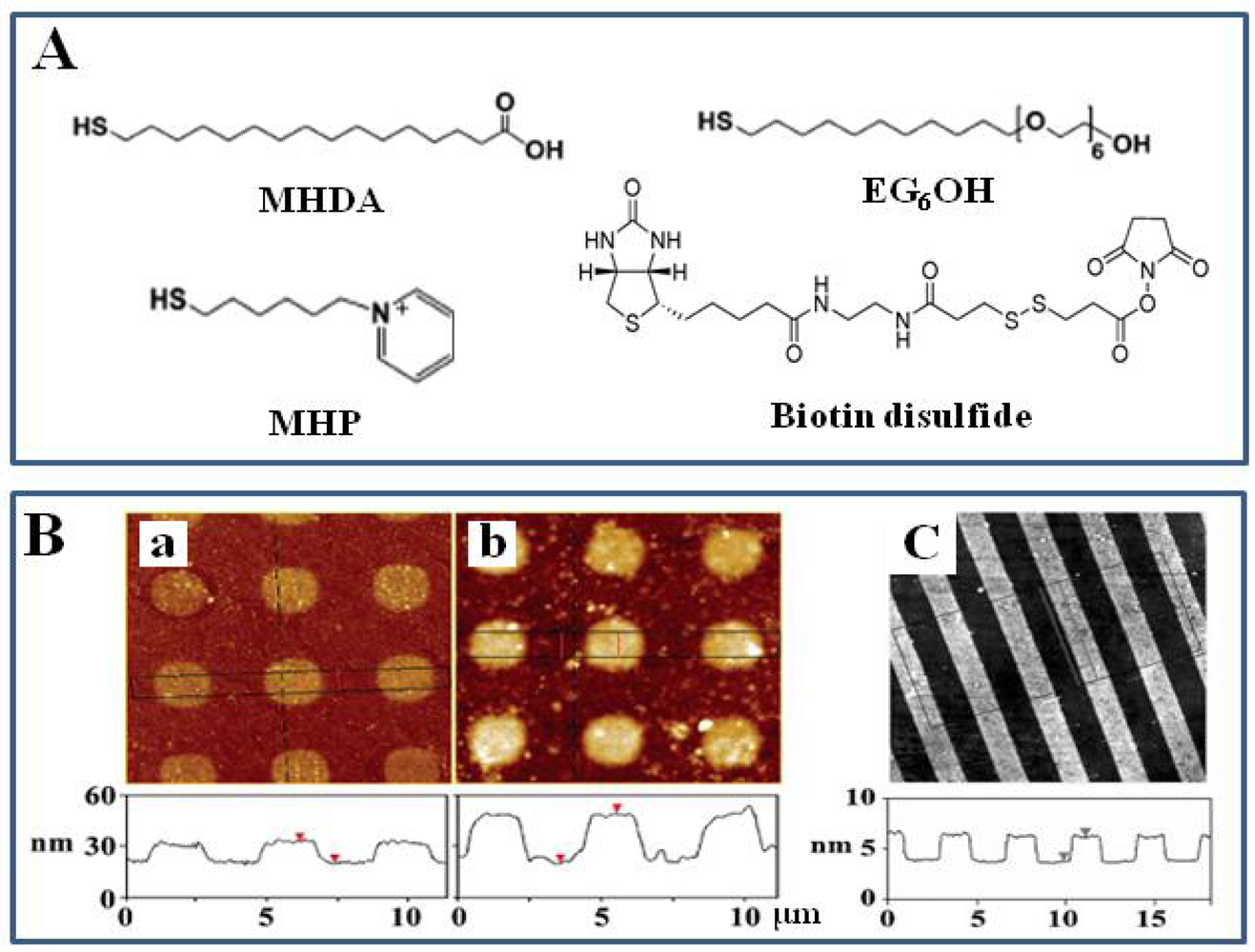
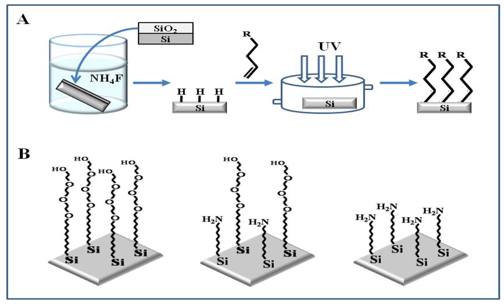
2.1. Self-assembled monolayer (SAM) and surface functionalization
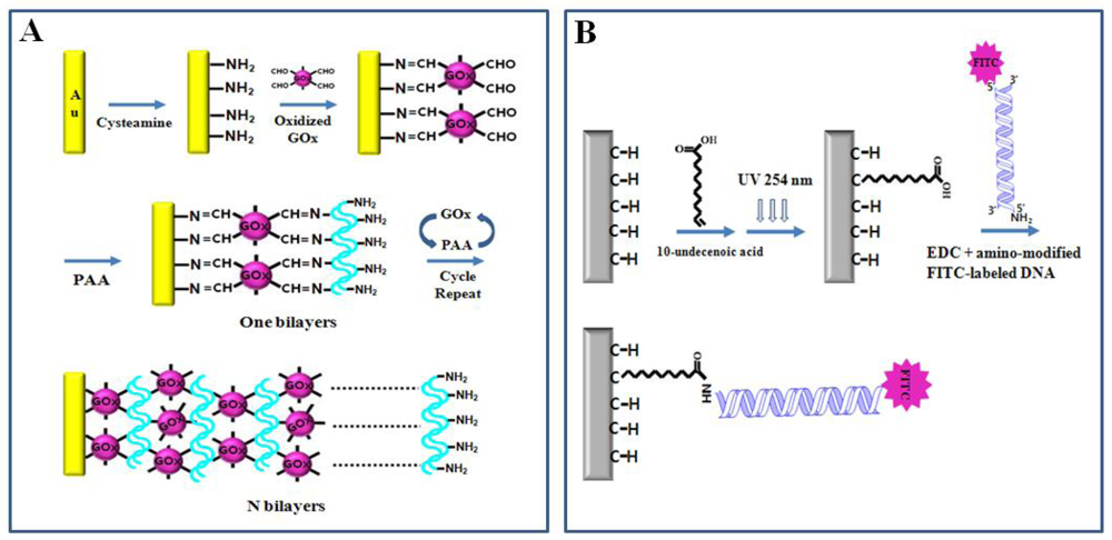
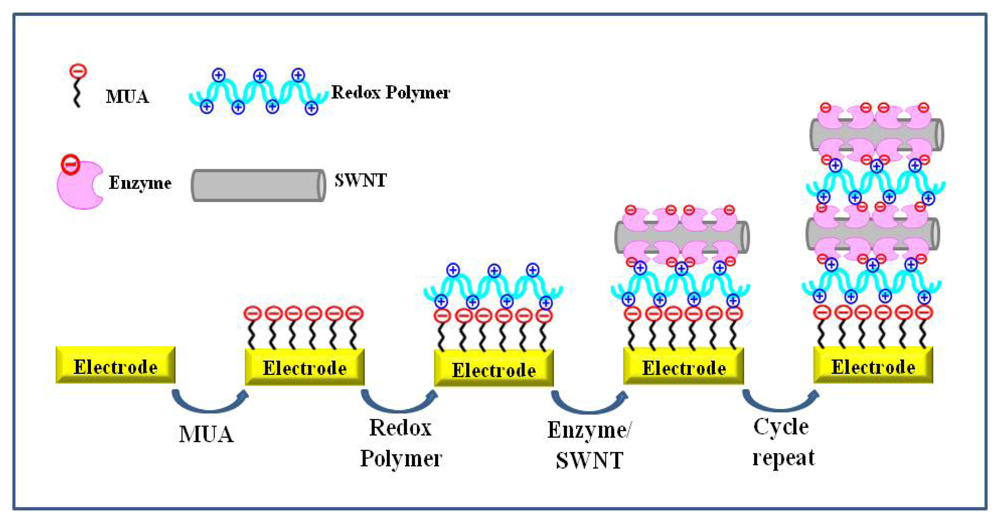
2.2. Layer-by-layer assembly
3. Immobilization of functional biomolecules on functionalized surfaces
3.1. Physical adsorption
3.1.1. Electrostatic interaction
3.1.2. Hydrophobic interaction
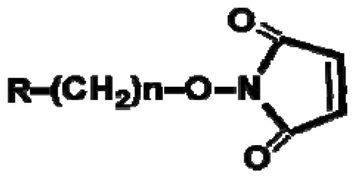
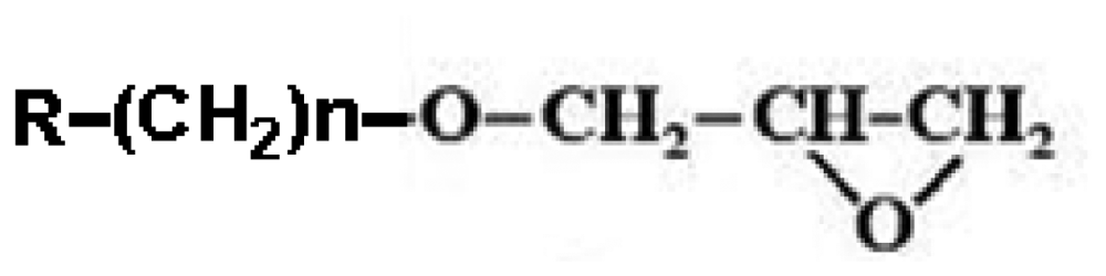
3.2. Covalent bonding
3.2.1. Schiff-base reaction
3.2.2. EDC-mediated and NHS-mediated chemical reaction
3.3. Specific interaction
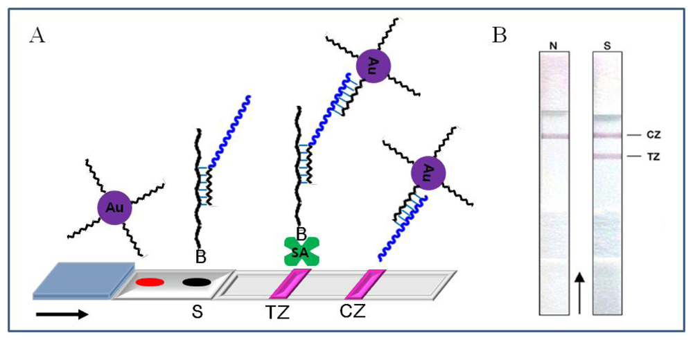
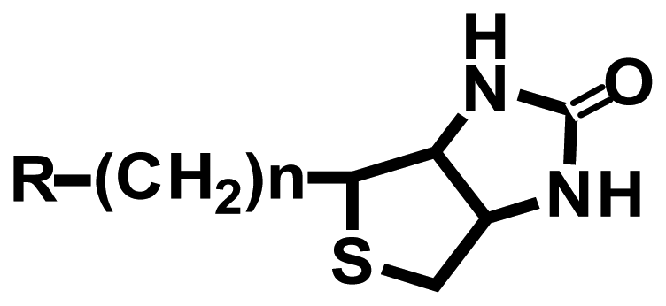
3.3.1. Molecular recognition between biotin and avidin
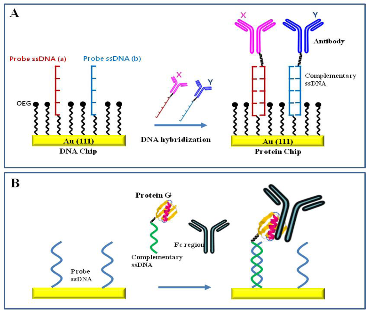
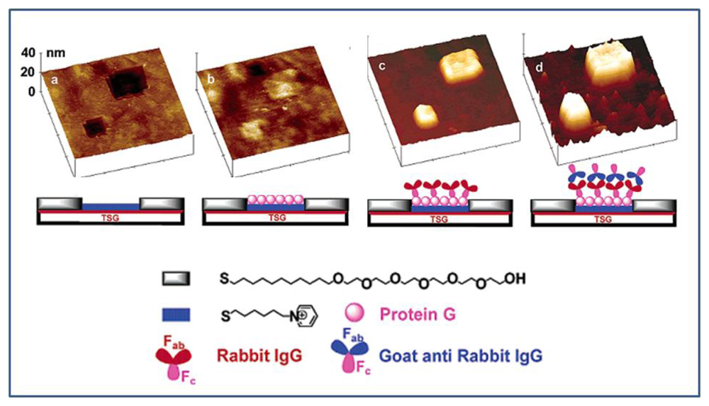
3.3.2. Molecular recognition between protein A/G and antibody
3.3.3. Sequence-specific DNA hybridization
4. Nanoscale patterning of biomolecules using molecular recognition and specific interactions
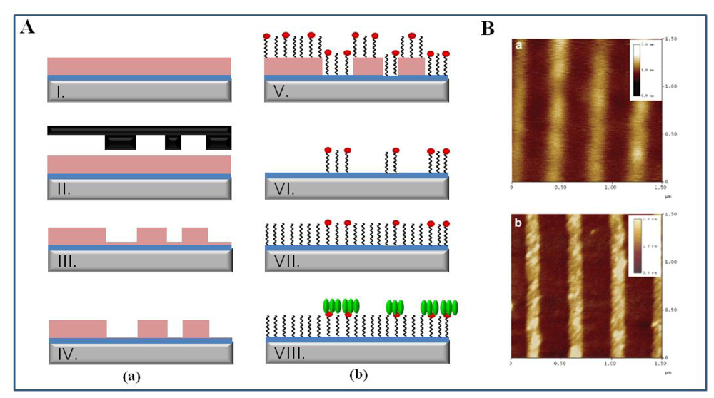
4.1. Electron-beam lithography (EBL)
4.2. Soft lithography
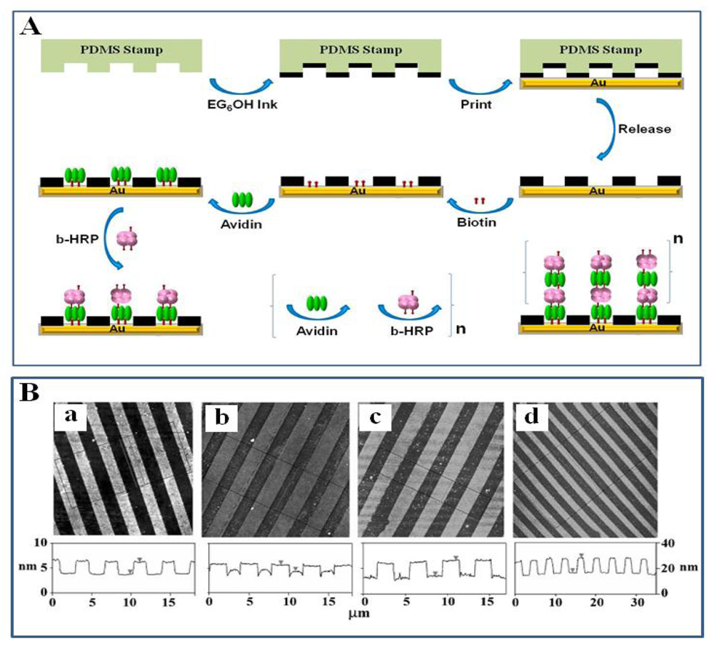
4.2.1. Nanocontact printing (NCP)
4.2.2. Nanoimprint lithography (NIL)
4.3. Nanografting and Dip-pen nanolithography
4.3.1. Nanografting and Nanoshaving
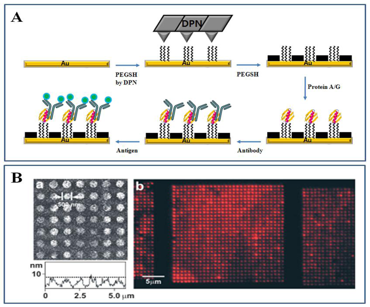
4.3.2. Dip-pen nanolithography (DPN)
4.4. Molecular recognition and specific interaction of biomolecules on nanomaterials
4.4.1. Nanotubes
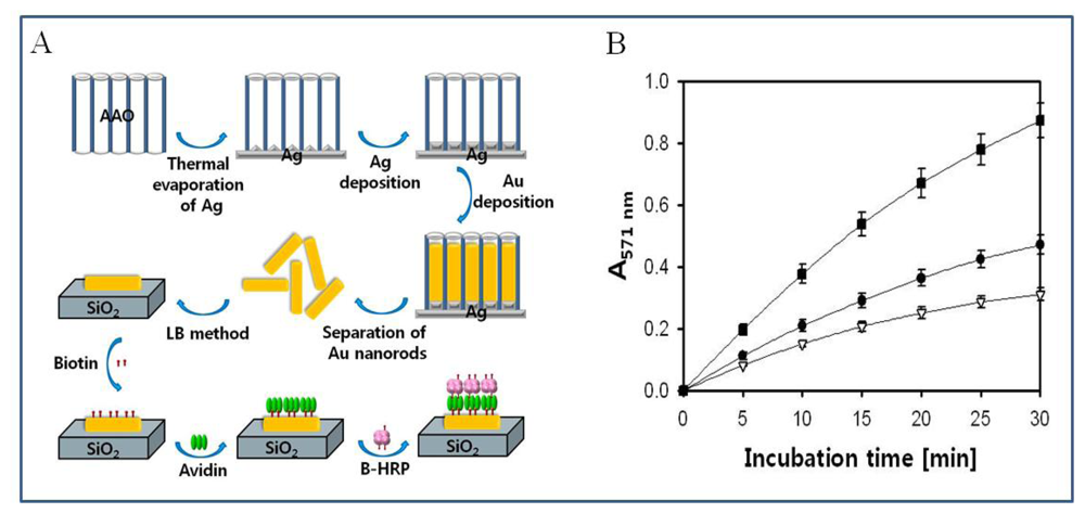
4.4.2. Nanorods and nanoparticles
5. Nanoscale detection of molecular recognition and specific interactions
5.1. Advantages of nanoscale detection
5.2. Detection of molecular binding at the nanoscale
5.2.1. DNA hybridization
5.2.2. Antigen-antibody
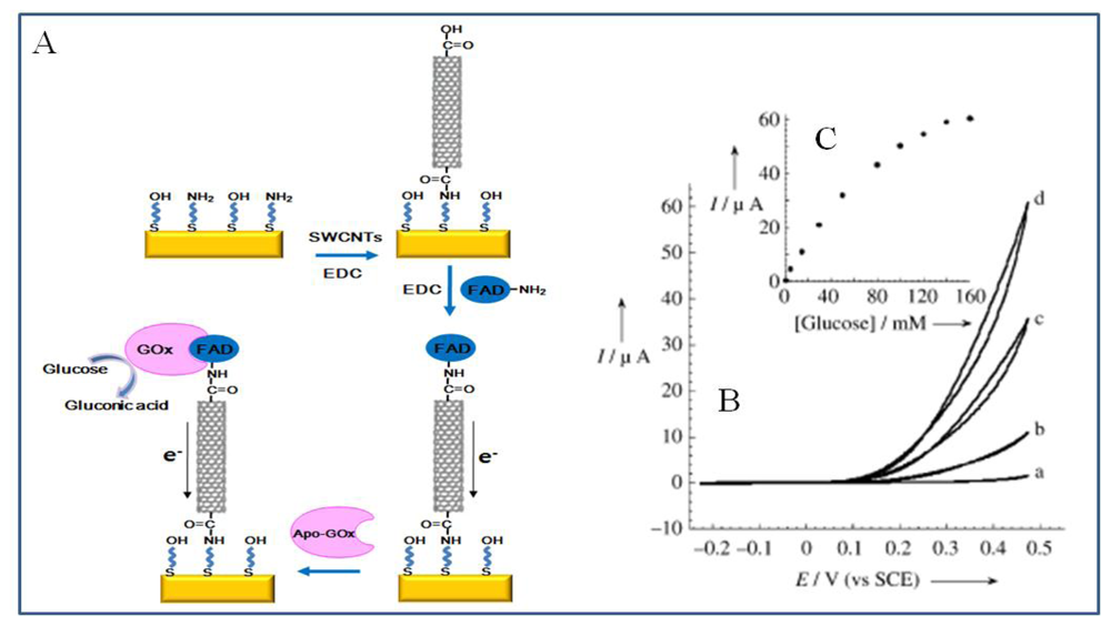
5.2.3. Enzyme reaction
5.2.4. Specific molecular recognitions
6. Future directions
Acknowledgments
References and Notes
- McFadden, P. Broadband biodetecion: holmes on a chip. Science 2002, 297, 2075–2076. [Google Scholar]
- Jung, Y.; Jeong, J.Y.; Chung, B.H. Recent advances in immobilization methods of antibodies on solid supports. Analyst 2008, 133, 697–701. [Google Scholar]
- Rauf, S.; Zhou, D.; Abell, C.; Klenerman, D.; Kang, D.J. Building three-dimensional nanostructures with active enzymes by surface template layer-by-layer assembly. Chem. Commun. 2006, 1721–1723. [Google Scholar]
- Sun, Y.; Yan, F.; Yang, W.; Sun, C. Multilayered construction of glucose oxidase and silica nanoparticles on Au electrodes based on layer-by-layer covalent attachment. Biomaterials 2006, 27, 4042–4049. [Google Scholar]
- Miyake, T.; Tanii, T.; Kato, K.; Zako, T.; Funatsu, T.; Ohdomari, I. Selective improvement in protein nanopatterning with a hydroxyl-terminated self-assembled monolayer template. Nanotechnology 2007, 18, 305304. [Google Scholar]
- Mrksich, M.; Whitesides, G.M. Using self-assembled monolayers to understand the interactionsof man-made surfaces with proteins and cells. Annu. Rev. Biophys. Biomol. Struct. 1996, 25, 55–78. [Google Scholar]
- Hyun, J.; Ahn, S.J.; Lee, W.K.; Chilkoti, A.; Zauscher, S. Molecular recognition-mediated fabrication of protein nanostructures by dip-pen lithography. Nano Lett. 2002, 2, 1203–1207. [Google Scholar]
- Lee, J.; So, H.; Jeon, E.; Chang, H.; Won, K.; Kim, Y.H. Aptamers as molecular recognition elements for electrical nanobiosensors. Anal. Bioanal. Chem. 2008, 390, 1023–1032. [Google Scholar]
- Mrksich, M. A surface chemistry approach to studying cell adhesion. Chem. Soc. Rev. 2000, 29, 267–273. [Google Scholar]
- Kong, D.Y.; Wang, Z.L.; Lin, C.K.; Quan, Z.W.; Li, Y.Y.; Li, C.X.; Lin, J. Biofunctionalization of CeF3:Tb3+ nanoparticles. Nanotechnology 2007, 18, 075601. [Google Scholar]
- Love, J.C.; Estroff, L.A.; Kriebel, J.K.; Nuzzo, R.G.; Whitesides, G.M. Self-assembled monolayers of thiolates on metals as a form of nanotechnology. Chem. Rev. 2005, 105, 1003–1169. [Google Scholar]
- Huie, J.C. Guided molecular self-assembly: a review of recent efforts. Smart Mater. Struct. 2003, 12, 264–271. [Google Scholar]
- Howarter, J.A.; Youngbood, J.P. Optimization of silica silanization by 3-aminopropyltriethoxy-silane. Langmuir 2006, 22, 11142–11147. [Google Scholar]
- Whitesides, G.M.; Ostuni, E.; Takayama, S.; Jiang, X.; Ingber, D.E. Soft lithography in biology and biochemistry. Annu. Rev. Biomed. Eng. 2001, 3, 335–373. [Google Scholar]
- Laibinis, P.E.; Whitesides, G.M.; Allara, D.L.; Tao, Y.; Parikh, A.N.; Nuzzo, R.G. Comparison of the structures and wetting properties of self-assembled monolayers of n-alkanethiols on the coinage metal surfaces, Cu, Ag, Au. J. Am. Chem. Soc. 1991, 113, 7152–7167. [Google Scholar]
- Malinsky, M.D.; Kelly, K.L.; Schatz, G.C.; Van Duyne, R.P. Chain length dependence and sensing capabilities of the localized surface plasmon resonance of silver nanoparticles chemically modified with alkanethiol self-assembled monolayers. J. Am. Chem. Soc. 2001, 123, 1471–1482. [Google Scholar]
- Porter, L.A.; Ji, D.; Westcott, S.L.; Graupe, M.; Czernuszewicz, R.S.; Halas, N.J.; Lee, T.R. Gold and silver nanoparticles functionalized by the adsorption of dialkyl disulfides. Langmuir 1998, 14, 7378–7386. [Google Scholar]
- Silberzan, P.; Léger, L.; Ausserré, D.; Benattar, J.J. Silanation of silica surfaces. A new method of constructing pure or mixed monolayers. Langmuir 1991, 7, 1647–1651. [Google Scholar]
- Suzdal'tsev, E.I.; Lesnikov, A.K. Amorphous silicon dioxide: preparation techniques and applications. Refrac. Ind. Ceram. 2005, 46, 189–192. [Google Scholar]
- Bhatt, V.; Chandra, S. Silicon dioxide films by RF sputtering for microelectronic and MEMS applications. J. Micromech. Microeng. 2007, 17, 1066–1077. [Google Scholar]
- Choi, Y.; Jeong, Y.; Chung, H.; Ito, E.; Hara, M.; Noh, J. Formation of ordered self-assembled monolayers by adsorption of octhylthiocyanates on Au(111). Langmuir 2008, 24, 91–96. [Google Scholar]
- Li, L.; Chen, S.; Jiang, S. Protein adsorption on alkanethiolate self-assembled monolayers: nanoscale surface structural and chemical effects. Langmuir 2003, 19, 2974–2982. [Google Scholar]
- Ostuni, E.; Grzybowski, B.A.; Mrksich, M.; Roberts, C.S.; Whitesides, G.M. Adsorption of proteins to hydrophobic sites on mixed self-assembled monolayers. Langmuir 2003, 19, 1861–1872. [Google Scholar]
- Ogawa, T.; Ding, B.; Sone, Y.; Shiratori, S. Super-hydrophobic surfaces of layer-by-layer structured film coated electrospun nanofibrous membranes. Nanotechnology 2007, 18, 165607. [Google Scholar]
- Zhou, D.; Brucbauer, A.; Batchelor, M.; Kang, D.J.; Abell, C.; Klenerman, D. Influence of the foundation layer on the layer-by-layer assembly of poly-L-lysine and poly(styrenesulfonate) and its usage in the fabrication of 3D microscale features. Langmuir 2004, 20, 9089–9094. [Google Scholar]
- Zhou, D.; Bruckbauer, A.; Ying, L.; Abell, C.; Klenerman, D. Building three-dimensional surface biological assemblies at the nanometer scale. Nano Lett. 2003, 3, 1517–1520. [Google Scholar]
- Biebricher, A.; Paul, A.; Tinnefeld, P.; Gölzhäuser, A.; Sauer, M. Controlled three-dimensional immobilization of biomolecules on chemically patterned surfaces. J. Biotechnol. 2004, 112, 97–107. [Google Scholar]
- Lasseter, T.L.; Clare, B.H.; Abbott, N.L.; Hamers, R.J. Covalently modified silicon and diamond surfaces: resistance to nonspecific protein adsorption and optimization for biosensing. J. Am. Chem. Soc. 2004, 126, 10220–10221. [Google Scholar]
- Sharma, S.; Johnson, R.W.; Desai, T.A. Evaluation of the stability of nonfouling ultrathin poly(ethylene glycol) films for silicon-based microdevices. Langmuir 2004, 20, 348–356. [Google Scholar]
- Faucheux, N.; Schweiss, R.; Lützow, K.; Werner, C.; Groth, T. Self-assembled monolayers with different terminating groups as model substrates for cell adhesion studies. Biomaterials 2004, 25, 2721–2730. [Google Scholar]
- Zhu, X.Y.; Jun, Y.; Staarup, D.R.; Major, R.C.; Danielson, S.; Boiadjiev, V.; Gladfelter, W.L.; Bunker, B.C.; Guo, A. Grafting of high-density poly(ethylene glycol) monolayers on Si(111). Langmuir 2001, 17, 7798–7803. [Google Scholar]
- Cicero, R.L.; Linford, M.R.; Chidsey, C.E.D. Photoreactivity of unsaturated compounds with hydrogen-terminated silicon(111). Langmuir 2000, 16, 5688–5695. [Google Scholar]
- Zhang, X.; Chen, H.; Zhang, H. Layer-by-layer assembly: from conventional to unconventional methods. Chem. Commun. 2007, 1395–1405. [Google Scholar]
- Ariga, K.; Hill, J.P.; Ji, Q. Layer-by-layer assembly as a versatile bottom-up nanofabrication technique for exploratory research and realistic application. Phys. Chem. Chem. Phys. 2007, 9, 2319–2340. [Google Scholar]
- Schneider, G.; Decher, G. Functional core/shell nanoparticles via layer-by-layer assembly.Investigation of the experimental parameters for controlling particle aggregation and for enhancing dispersion stability. Langmuir 2008, 24, 1778–1789. [Google Scholar]
- Wu, B.Y.; Hou, S.H.; Yin, F.; Zhao, Z.X.; Wang, Y.Y.; Wang, X.S.; Chen, Q. Amperometric glucose biosensor based on multilayer films via layer-by-layer self-assembly of multi-wall carbon nanotubes, gold nanoparticles and glucose oxidase on the Pt electrode. Biosens. Bioelectron. 2007, 22, 2854–2860. [Google Scholar]
- Wang, J.; Liu, G.; Lin, Y. Amperometric choline biosensor fabricated through electrostatic assembly of bienzyme/polyelectrolyte hybrid layers on carbon nanotubes. Analyst 2006, 131, 477–483. [Google Scholar]
- Wang, Y.; Joshi, P.P.; Hobbs, K.L.; Johnson, M.B.; Schmidtke, D.W. Nanostructured biosensors built by layer-by-layer electrostatic assembly of enzyme-coated single-walled carbon nanotubes and redox polymers. Langmuir 2006, 22, 9776–9783. [Google Scholar]
- Yan, X.B.; Chen, X.J.; Tay, B.K.; Khor, K.A. Transparent and flexible glucose biosensor via layer-by-layer assembly of multi-wall carbon nanotubes and glucose oxidase. Electrochem. Commun. 2007, 9, 1269–1275. [Google Scholar]
- Jung, Y.; Lee, J.M.; Jung, H.; Chung, B.H. Self-directed and self-oriented immobilization of antibody by protein G-DNA conjugate. Anal. Chem. 2007, 79, 6534–6541. [Google Scholar]
- Wohlstadter, J.; Wilbur, J.L.; Sigal, G.B.; Biebuyck, H.A.; Billadeau, M.A.; Dong, L.; Fisher, A.B.; Gudibande, S.R.; Jameison, S.H.; Kenten, J.H.; Leginus, J.; Leland, J.K.; Massdy, R.J.; Wohlstadter, S.J. Carbon nanotube-based biosensor. Adv. Mater. 2003, 15, 1184–1187. [Google Scholar]
- Zhdanov, V.P.; Kasemo, B. Monte Carlo simulation of denaturation of adsorbed proteins. Proteins: Struct. Func. Gen. 1998, 30, 168–176. [Google Scholar]
- Krishnan, A.; Liu, Y.; Cha, P.; Allara, D.; Vogler, E.A. Interfacial energetic of globular-blood protein adsorption to a hydrophobic interface from aqueous-buffer solution. J. R. Soc. Interface 2006, 3, 283–301. [Google Scholar]
- Karlsson, M.; Ekeroth, J.; Elwing, H.; Carlsson, U. Reduction of irreversible protein adsorption on solid surfaces by protein engineering for increased stability. J. Biol. Chem. 2005, 280, 25558–25564. [Google Scholar]
- Babacan, S.; Pivarnik, P.; Letcher, S.; Rand, A.G. Evaluation of antibody immobilization methods for piezoelectric biosensor application. Biosens. Bioelectron 2000, 15, 615–621. [Google Scholar]
- Decher, G. Fuzzy nanoassemblies: toward layered polymeric multicomposites. Science 1997, 277, 1232–1237. [Google Scholar]
- Hammond, P.T. Recent explorations in electrostatic multilayer thin film assembly. Curr. Opin. Coll. Inter. Sci. 2000, 4, 430–442. [Google Scholar]
- Xu, J.; Zhao, W.; Luo, X.; Chen, H. A sensitive biosensor for lactate based on layer-by-layer assembling MnO2nanoparticles and lactate oxidase on ion-sensitive field-effect transistors. Chem. Commun 2005, 792–794. [Google Scholar]
- Franchina, J.G.; Lackowski, W.M.; Dermody, D.L.; Crooks, R.M.; Bergbreiter, D.E. Electrostatic immobilization of glucose oxidase in a weak acid, polyelectrolyte hyperbranched ultrathin film on gold: fabrication, characterization, and enzymatic activity. Anal. Chem. 1999, 71, 3133–3139. [Google Scholar]
- Vakurov, A.; Simpson, C.E.; Daly, C.L.; Gibson, T.D.; Millner, P.A. Acetylecholinesterase-based biosensor electrodes for organophosphate pesticide detection. II. Immobilization and stabilization of acetylecholinesterase. Biosens. Bioelectron 2005, 20, 2324–2329. [Google Scholar]
- Chen, G.; Wu, G.; Wang, L.; Zhang, S.; Su, Z. Layer-by-layer assembly of single-charged ions with a rigid polyampholyte. Chem. Commum. 2008, 1741–1743. [Google Scholar]
- Guo, X.; Zhang, H.; Hu, N. Myoglobin-loaded layer-by-layer films containing SiO2 nanoparticles studied using electrochemistry. Nanotechnology 2008, 19, 055709. [Google Scholar]
- Weidinger, I.M.; Murgida, D.H.; Dong, W.; Möhwald, H.; Hildebrandt, P. Redox processes of cytochrome c immobilized on solid supported polyelectrolyte multilayers. J. Phys. Chem. 2006, 110, 522–529. [Google Scholar]
- Song, Y.; Wang, L.; Ren, C.; Zhu, G.; Li, Z. A novel hydrogen peroxide sensor based on horseradish peroxidase immobilibized in DNA films on a gold electrode. Sens. Actuat B. 2006, 114, 1001–1006. [Google Scholar]
- Goh, Y.Y.; Ho, B.; Ding, J.L. A novel fluorescent protein-based biosensor for gram-negative bacteria. Appl. Environ. Microb. 2002, 68, 6343–6352. [Google Scholar]
- Darder, M.; Casero, E.; Pariente, F.; Lorenzo, E. Biosensors based on membrane-bound enzymes immobilized in a 5-(octyldithio)-2-nitrobenzoic acid layer on gold electrodes. Anal. Chem. 2000, 72, 3784–3792. [Google Scholar]
- Catimel, B.; Scott, A.M.; Lee, F.T.; Hanai, N.; Ritter, G.; Welt, S.; Old, L.J.; Burgess, A.W.; Nice, E. Direct immobilization of gangliosides onto gold-carboxymethyldextran sensor surfaces by hydrophobic interaction: applications to antibody characterization. Glycobiology 1998, 8, 927–938. [Google Scholar]
- Frederix, F.; Bonroy, K.; Reekmans, G.; Laureyn, W.; Campitelli, A.; Abramov, M.A.; Dehaen, W.; Maes, G. Reduced nonspecific adsorption on covalently immobilized protein surfaces using poly(ethylene oxide) containing blocking agents. J. Biochem. Biophys. Methods 2004, 58, 67–74. [Google Scholar]
- Lu, Z.; Li, C.M.; Z, Q.; Bao, Q.; Cui, X. Covalently linked DNA/protein multilayered film for controlled DNA release. J. Coll. Inter. Sci. 2007, 314, 80–88. [Google Scholar]
- Duan, L.; He, Q.; Yan, X.; Cui, Y.; Wang, K.; Li, J. Hemoglobin protein hollow shells fabricated through covalent layer-by-layer technique. Biochem. Biophys. Res. Commun. 2007, 354, 357–362. [Google Scholar]
- Zhang, S.; Yang, W.; Niu, Y.; Sun, C. Multilayered construction of glucose oxidase on gold electrodes based on layer-by-layer covalent attachment. Anal. Chim. Acta. 2004, 523, 209–217. [Google Scholar]
- Christiaens, P.; Vermeeren, V.; Wenmackers, S.; Daenen, M.; Haenen, K.; Nesladek, M.; vandeVen, M.; Ameloot, M.; Michiels, L.; Wagner, P. EDC-mediated DNA attachment to nanocrystalline CVD diamond films. Biosens. Bioelectron 2006, 22, 170–177. [Google Scholar]
- Nicolau, D.V.; Taguchi, T.; Taniguchi, H.; Yoshikawa, S. Protein patterning via radiation-assisted surface functionalization of conventional microlithographic materials. Coll. Surf. A: Physicochem. Eng. Aspects 1999, 155, 51–62. [Google Scholar]
- Cooper, M. Optical biosensors in drug discovery. Nature Rev. 2002, 1, 515–528. [Google Scholar]
- Tlili, A.; Ali Jarboui, M.; Abdelghani, A.; Fathallah, D.M.; Maaref, M.A. A novel silicon nitride biosensor for specific antibody–antigen interaction. Mater. Sci. Eng C. 2005, 25, 490–495. [Google Scholar]
- Gao, Q.; Yang, F.; Ma, Y.; Yang, X. The modification of screen-printed carbon electrodes with amino group and its application to construct a H2O2 biosensor. Electronanalysis 2004, 16, 730–735. [Google Scholar]
- Bhatnagar, P.; Mark, S.S.; Kim, I.; Chen, H.; Schmidt, B.; Lipson, M.; Batt, C.A. Dendrimer-scaffold-based electron-beam patterning of biomolecules. Adv. Mater. 2006, 18, 315–319. [Google Scholar]
- Maraldo, D.; Mutharasan, R. Optimization of antibody immobilization for sensing using piezoelectrically excited-millimeter-sized cantilever (PEME) sensors. Sens. Actuat B. 2007, 123, 474–479. [Google Scholar]
- Masson, J.; Kranz, C.; Booksh, K.S.; Mizaikoff, B. Improved sensitivity and stability of amperometric enzyme microbiosensors by covalent attachment to gold electrodes. Biosens. Bioelectron 2007, 23, 355–361. [Google Scholar]
- Davis, J.J.; Halliwell, C.M.; Hill, H.A.O.; Canters, G.W.; van Amsterdam, M.C.; Ph. Verbeet, M. Protein adsorption at a gold electrode studied by in situ scanning tunnelling microscopy. New J. Chem. 1998, 22, 1119–1123. [Google Scholar]
- Bae, Y.M.; Oh, B.; Lee, W.; Lee, W.H.; Chol, J. Detection of insulin-antibody binding on a solid surface using imaging ellipsometry. Biosens. Bioelectron 2004, 20, 895–902. [Google Scholar]
- Boozer, C.; Ladd, J.; Chen, S.; Jiang, S. DNA-directed protein immobilization for simultaneous detection of multiple analytes by surface plasmon resonance biosensor. Anal. Chem. 2006, 78, 1515–1519. [Google Scholar]
- Padeste, C.; Steiger, B.; Grubelnik, A.; Tiefenauer, L. Molecular assembly of redox-conductive ferrocene-streptavidin conjugates-towards bio-electrochemical devices. Biosens. Bioelectron. 2004, 20, 545–552. [Google Scholar]
- Danczyk, R.; Krieder, B.; North, A.; Webster, T.; HogenEsch, H.; Rundell, A. Comparison of antibody functionality using different immobilization methods. Biotechnol. Bioeng. 2003, 84, 215–223. [Google Scholar]
- Vianello, F.; Zennaro, L.; Di Paolo, M.L.; Rigo, A.; Malacarne, C.; Scarpa, M. Preparation, morphological characterization, and activity of thin films of horseradish peroxidase. Biotechnol. Bioeng. 2000, 68, 488–495. [Google Scholar]
- Fowler, J.M.; Stuart, M.C.; Wong, D.K.Y. Self-assembled layer of thiolated protein G as an immunosensor scaffold. Anal. Chem. 2007, 79, 350–354. [Google Scholar]
- Ha, T.H.; Jung, S.O.; Lee, J.M.; Lee, K.Y.; Lee, Y.; Park, J.S.; Chung, B.H. Oriented immobilization of antibodies with GST-fused multiple Fc-specific B-domains on an gold surface. Anal. Chem. 2007, 79, 546–556. [Google Scholar]
- Gao, D.; McBean, N.; Schultz, J.S.; Yan, Y.; Mulchandani, A.; Chen, W. Fabrication of antibody arrays using thermally responsive elastin fusion proteins. J. Am. Chem. Soc. 2006, 128, 676–677. [Google Scholar]
- Boozer, C.; Ladd, J.; Chen, S.; Yu, Q.; Homola, J.; Jiang, S. DNA directed protein immobilization on mixed ssDNA/Oligo(ethylene glycol) self-assembled monolayers for sensitive biosensors. Anal. Chem. 2004, 76, 6967–6972. [Google Scholar]
- Niemeyer, C.M.; Koehler, J.; Wuerdemann, C. DNA-directed assembly of bienzymic complexes from in vivobiotinylated NAD(P)H:FMN oxidoreductase and luciferase. ChemBioChem. 2002, 2/3, 242–245. [Google Scholar]
- Fruk, L.; Müller, J.; Weber, G.; Nárvaez, A.; Domínguez, E.; Niemeyer, C.M. DNA-directed immobilization of horseradish peroxidase-DNA conjugates on microelectrode arrays: towards electrochemical screening of enzyme libraries. Chem. Eur. J. 2007, 13, 5223–5231. [Google Scholar]
- Niemeyer, C.M.; Wacker, R.; Adler, M. Combination of DNA-directed immobilization and immune-PCR: very sensitive antigen detection by means of self-assembled DNA-protein conjugates. Nucleic Acids Res. 2003, 31, e90. [Google Scholar]
- Zhang, G.; Tanii, T.; Funatsu, T.; Ohdomari, I. Patterning of DNA nanostructures on silicon surface by electron beam lithography of self-assembled monolayer. Chem. Commun. 2004, 786–787. [Google Scholar]
- Blättler, T.; Huwiler, C.; Ochsner, M.; Städler, B.; Solak, H.; Vörös, J.; Grandin, H.M. Nanopatterns with biological functions. J. Nanosci. Nanotechnol 2006, 6, 2237–2264. [Google Scholar]
- Mendes, P.M.; Yeung, C.L.; Preece, J.A. Bio-nanopatterning of surfaces. Nanoscale Res. Lett. 2007, 2, 373–384. [Google Scholar]
- Whitesides, G.M. The once and future nanomachine. Scient. Amer. 2001, 285, 78–83. [Google Scholar]
- Yu, B.; Meyyappan, M. Nanotechnology: role in emerging nanoelectronics. Solid-State Electron. 2006, 50, 536–544. [Google Scholar]
- Christman, K.L.; Enriquez-Rios, V.D.; Maynard, H.D. Nanopatterning proteins and peptides. Soft Matter 2006, 2, 928–939. [Google Scholar]
- Denis, F.A.; Pallandre, A.; Nysten, B.; Jonas, A.M.; Dupont-Gillain, C.C. Alignment and assembly of adsorbed collagen molecules induced by anisotropic chemical nanopatterns. Small 2005, 1, 984–991. [Google Scholar]
- Li, H.; Muir, B.V.O.; Fichet, G.; Huck, W.T.S. Nanocontact printing: a route to sub-50-nm-sale chemical and biological patterning. Langmuir 2003, 19, 1963–1965. [Google Scholar]
- Hoff, J.D.; Cheng, L.; Meyhöfer, E.; Guo, L.J.; Hunt, A.J. Nanoscale protein patterning by imprint lithography. Nano Lett. 2004, 4, 853–857. [Google Scholar]
- Liu, M.; Amro, N.A.; Chow, C.S.; Liu, G. Production of nanostructures of DNA on surfaces. Nano Lett. 2002, 2, 863–867. [Google Scholar]
- Wilson, D.L.; Martin, R.; Hong, S.; Cronin-Golomb, M.; Mirkin, C.A.; Kaplan, D.L. Surface organization and nanopatterning of collagen by dip-pen nanolithography. Proc. Natl. Acad. Sci USA 2001, 98, 13660–13664. [Google Scholar]
- Zhang, G.; Tanii, T.; Zako, T.; Hosaka, T.; Miyake, T.; Kanari, Y.; Funatsu, T.; Ohdomari, I. Nanoscale patterning of protein using electron beam lithography of organosilane self-assembled monolayers. Small 2005, 1, 833–837. [Google Scholar]
- Niwa, D.; Omichi, K.; Motohashi, N.; Homma, T.; Osaka, T. Formation of micro and nanoscale patterns of monolayer templates for position selective immobilization of oligonucleotide using ultraviolet and electron beam lithography. Chem. Lett. 2004, 33, 176–177. [Google Scholar]
- Harnett, C.K.; Satyalakshmi, K.M.; Craighead, H.G. Bioactive templates fabricated by low-energy electron beam lithography of self-assembled monolayers. Langmuir 2001, 17, 178–182. [Google Scholar]
- Xia, Y.; Whitesides, G.M. Soft lithography. Annu. Rev. Mater. Sci. 1998, 28, 153–184. [Google Scholar]
- Chou, S.Y.; Krauss, P.R.; Renstrom, P.J. Imprint lithography with 25-nanometer resolution. Science 1996, 272, 85–87. [Google Scholar]
- Visconti, P.; Pisignano, D.; Torre, A.D.; Persano, L.; Maruccio, G.; Biasco, A.; Cingolani, R.; Rinaldi, R. Electron beam and mechanical lithographies as enabling factors for organic-based device fabrication. Mater. Sci. Eng C. 2005, 25, 848–852. [Google Scholar]
- Renault, J.P.; Bernard, A.; Bietsch, A.; Michel, B.; Bosshard, H.R.; Delamarche, E. Fabricating arrays of single protein molecules on glass using microcontact printing. J. Phys. Chem B. 2003, 107, 703–711. [Google Scholar]
- Truskett, V.N.; Watts, M.P.C. Trends in imprint lithography for biological applications. Trends Biotechnol. 2006, 24, 312–317. [Google Scholar]
- Guo, L.J. Nanoimprint lithography: methods and material requirements. Adv. Mater. 2007, 19, 495–513. [Google Scholar]
- Ohtake, T.; Nakamatsu, K.; Matsui, S.; Tabata, H.; Kawai, T. DNA nanopatterning with self-organization by using nanoimprint. J. Vac. Sci. Technol B. 2004, 22, 3275–3278. [Google Scholar]
- Falconnet, D.; Pasqui, D.; Park, S.; Eckert, R.; Schift, H.; Gobrecht, J.; Barbucci, R.; Textor, M. A novel approach to produce protein nanopatterns by combining nanoimprint lithography and molecular self-assembly. Nano Lett. 2004, 4, 1909–1914. [Google Scholar]
- Salaita, K.; Wang, Y.; Mirkin, C.A. Applications of dip-pen nanolithography. Nature nanotech. 2007, 2, 145–155. [Google Scholar]
- Wadu-Mesthrige, K.; Xu, S.; Amro, N.A.; Liu, G. Fabrication and imaging of nanometer-sized protein patterns. Langmuir 1999, 15, 8580–8583. [Google Scholar]
- Zhou, D.; Sinniah, K.; Abell, C.; Rayment, T. Label-free detection of DNA hybridization at the nanoscale: a highly sensitive and selective approach using atomic-force microscopy. Angew. Chem. Int. Ed. 2003, 42, 4934–4937. [Google Scholar]
- Zhou, D.; Wang, X.; Birch, L.; Rayment, T.; Abell, C. AFM study on protein immobilization on charged surfaces at the nanoscale: toward the fabrication of three-dimensional protein nanostructures. Langmuir 2003, 19, 10557–10562. [Google Scholar]
- Nuraje, N.; Banerjee, I.A.; MacCuspie, R.I.; Yu, Lingtao; Matsui, H. Biological bottom-up assembly of antibody nanotubes on patterned antigen arrays. J. Am. Chem. Soc. 2004, 126, 8088–8089. [Google Scholar]
- Piner, R.D.; Zhu, J.; Xu, F.; Hong, S.; Mirkin, C.A. “Dip-pen” nanolithography. Science 1999, 283, 661–663. [Google Scholar]
- Lee, K.; Park, S.J.; Mirkin, C.A.; Smith, J.C.; Mrksich, M. Protein nanoarrays generated by dip-pen nanolithography. Science 2002, 295, 1702–1705. [Google Scholar]
- Lee, S.W.; Oh, B.; Sanedrin, R.G.; Salaita, K.; Fujigaya, T.; Mirkin, C.A. Biologically active protein nanoarrays generated using paralled dip-pen nanolithography. Adv. Mater 2006, 18, 1133–1136. [Google Scholar]
- Demers, L.M.; Ginger, D.S.; Park, S.J.; Chun, S.W.; Mirkin, C.A. Direct patterning of modified oligonucleotides on metals and insulators by dip-pen nanolithography. Science 2002, 296, 1836–1838. [Google Scholar]
- Chung, S.; Ginger, D.S.; Morales, M.W.; Zhang, Z.; Chandrasekhar, V.; Ratner, M.A.; Mirkin, C.A. Top-down meets bottom-up: dip-pen nanolithography and DNA-directed assembly of nanoscale electrical circuits. Small 2005, 1, 64–69. [Google Scholar]
- Anandan, V.; Rao, Y.L.; Zhang, G. Nanopillar array structures for enhancing biosensing performance. Int. J. Nanomed. 2006, 1, 73–79. [Google Scholar]
- Yang, H.; Zhu, Y. A high performance glucose biosensor enhanced via nanosized SiO2. Anal. Chim. Acta. 2005, 554, 92–97. [Google Scholar]
- Zhu, X.; Yuri, I.; Gan, X.; Suzuki, I.; Li, G. Electrochemical study of the effect of nano-zinc oxide on microperoxidase and its application to more sensitive hydrogen peroxide biosensor preparation. Biosens. Bioelectron. 2007, 22, 1600–1604. [Google Scholar]
- Maehashi, K.; Katsura, T.; Kerman, K.; Takamura, Y.; Matsumoto, K.; Tamiya, E. Label-free protein biosensor based on aptamer-modified carbon nanotube field-effect transistors. Anal. Chem. 2007, 79, 782–787. [Google Scholar]
- Dekvaux, M.; Walcarius, A.; Demoustier-Champagne, S. Electrocatalytic H2O2 amperometric detection using gold nanotube electrode ensembles. Anal. Chim. Acta. 2004, 525, 221–230. [Google Scholar]
- Zheng, G.; Patolsky, F.; Cui, Y.; Wang, W.U.; Lieber, C.M. Multiplexed electrical detection of cancer markers with nanowire sensor arrays. Nature Biotechnol. 2005, 23, 1294–1301. [Google Scholar]
- O'Connor, M.; Kim, S.N.; Killard, A.J.; Forster, R.J.; Smyth, M.R.; Papadimitrakopoulos, F.; Rusling, J.F. Mediated amperometric immunosensing using single walled carbon nanotube forests. Analyst 2004, 129, 1176–1180. [Google Scholar]
- Siwy, Z.; Trofin, L.; Kohi, P.; Baker, L.A.; Trautmann, C.; Martin, C.R. Protein biosensors based on biofunctionalized conical gold nanotubes. J. Am. Chem. Soc. 2005, 127, 5000–5001. [Google Scholar]
- Balasubramanian, K.; Burghard, M. Biosensors based on carbon nanotubes. Anal. Bioanal. Chem. 2006, 385, 452–468. [Google Scholar]
- Patolsky, F.; Weizmann, Y.; Willner, I. Long-range electrical contacting of redox enzymes by SWCNT connectors. Angew. Chem. Int. Ed. 2004, 43, 2113–2117. [Google Scholar]
- Chen, R.J.; Bangsaruntip, S.; Drouvalakis, K.A.; Kam, N.W.S.; Shim, M.; Li, Y.; Kim, W.; Utz, P.J.; Dai, H. Noncovalent functionalization of carbon nanotubes for highly specific electronic biosensors. Proc. Natl. Acad. Sci USA 2003, 100, 4984–4989. [Google Scholar]
- Kim, D.C.; Jang, A.R.; Kang, D.J.; Yoo, S.H.S. Park. Fabrication of LBL-assembled bio-architecture on gold nanorods. J. Korean Phys. Soc. 2008, 53, 886–891. [Google Scholar]
- Endo, T.; Yamamura, S.; Nagatani, N.; Morita, Y.; Takamura, Y.; Tamiya, E. Localized surface plasmon resonance based optical biosensor using surface modified nanoparticle layer for label-free monitoring of antigen-antibody reaction. Sci. Technol. Adv. Mater. 2005, 6, 491–500. [Google Scholar]
- Xiao, Y.; Patolsky, F.; Katz, E.; Hainfeld, J.F.; Willner, I. “Plugging into Enzymes”: nanowiring of redox enzymes by a gold nanoparticle. Science 2003, 299, 1877–1881. [Google Scholar]
- Yamanoi, Y.; Nishihara, H. Assembly of nanosize metallic particles and molecular wires on electrode surfaces. Chem. Commun. 2007, 3983–3989. [Google Scholar]
- Lee, K.; Kim, E.; Mirkin, C.A.; Wolinsky, S.M. The use of nanoarrays for highly sensitive and selective detection of human immunodeficiency virus type 1 in plasma. Nano Lett. 2004, 4, 1869–1872. [Google Scholar]
- Lee, K.; Lim, J.; Mirkin, C.A. Protein nanostructures formed via direct-write dip-pen nanolithography. J. Am. Chem. Soc. 2003, 125, 5588–5589. [Google Scholar]
- Yun, S.; Park, Y.K.; Kim, S.K.; Park, S. Linker-molecule-free gold nanorod layer-by-layer films for surface-enhanced raman scattering. Anal. Chem. 2007, 79, 8584–8589. [Google Scholar]
- Sreeprasad, T.S.; Samal, A.K.; Pradeep, T. Body- or tip-controlled reactivity of gold nanorods and their conversion to particles through other anisotropic structures. Langmuir 2007, 23, 9463–9471. [Google Scholar]
- Kichambare, P.D.; Star, A. Biosensing using carbon nanotube field-effect transistors. In Nanomaterials for biosensors, 1st Edition; Kumar, C., Ed.; Wiley-VCH Verlag GmbH & Co. KGaA: Weinheim, Germany, 2007; pp. 1–26. [Google Scholar]
- Gooding, J.J. Nanoscale biosensors: significant advantages over larger devices? Small 2006, 2, 313–315. [Google Scholar]
- Stern, E.; Klemic, J.F.; Routenberg, D.A.; Wyrembak, P.N.; Turner-Evans, D.B.; Hamilton, A.D.; LaVan, D.A.; Fahmy, T.M.; Reed, M.A. Label-free immunodetection with CMOS-compatible semiconducting nanowires. Nature 2007, 445, 519–522. [Google Scholar]
- So, H.; Won, K.; Kim, Y.H.; Kim, B.; Ryu, B.H.; Na, P.S.; Kim, H.; Lee, J. Single-walled carbon nanotube biosensors using aptamers as molecular recognition elements. J. Am. Chem. Soc. 2005, 127, 11906–11907. [Google Scholar]
- Kasili, P.M.; Song, J.M.; Vo-Dinh, T. Optical sensor for the detection of Caspase-9 activity in a single cell. J. Am. Chem. Soc. 2004, 126, 2799–2806. [Google Scholar]
- Li, B.; Zhao, L.; Zhang, C.; Hei, X.; Li, F.; Li, X.; Shen, J.; Li, Y.; Huang, Q.; Xu, S. Ultrasensitive colorimetric method to quantitate hundreds of polynucleotide molecules by gold nanoparticles with silver enhancement. Anal. Sci. 2006, 22, 1367–1370. [Google Scholar]
- Chang, Z.; Fan, H.; Chen, M.; He, P.; Fang, Y. Electrochemical DNA biosensors based on palladium nanoparticles combined with carbon nanotubes. Electroanalysis 2008, 20, 131–136. [Google Scholar]
- Kalogianni, D.P.; Koraki, T.; Christopoulos, T.K.; Ioannou, P.C. Nanoparticle-based DNA biosensor for visual detection of genetically modified organisms. Biosens. Bioelectron. 2006, 21, 1069–1076. [Google Scholar]
- Lin, Y.; Wang, J.; Liu, G.; Wu, H.; Wai, C.M.; Lin, Y. A nanoparticle label/ immunochromatographic electrochemical biosensor for rapid and sensitive detection of prostate-specific antigen. Biosens. Bioelectron. 2008, 23, 1659–1665. [Google Scholar]
- Liu, Q.; Lu, X.; Li, J.; Yao, X.; Li, J. Direct electrochemistry of glucose oxidase and electrochemical biosensing of glucose on quantum dots/carbon nanotubes electrodes. Biosens. Bioelectron. 2007, 22, 3203–3209. [Google Scholar]
- Xiao, Y.; Piorek, B.D.; Plaxco, K.W.; Heeger, A.J. A reagentless signal-on architecture for electronic, aptamer-based sensors via target-induced strand displacement. J. Am. Chem. Soc. 2005, 127, 17990–17991. [Google Scholar]


| Functional groups | Surface property | Interaction or reaction with biomolecules |
|---|---|---|
| R-(CH2)n-NH3+ | (+) Charge |
|
| R-(CH2)n-COO- | (-) Charge |
|
 | Hydrophobic |
|
 | Aldehyde NHS |
|
 | Maleimide |
|
 | Epoxy |
|
 | Biotin |
|
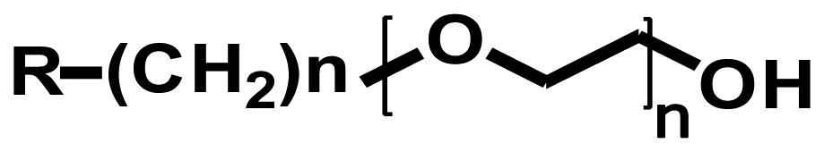 | Ethylene glycol |
|
| Routes | Advantages | Drawbacks | Bond energy (kcal/mol) |
|---|---|---|---|
| Electrostatic interaction | Simple, Fast, Reversible, Direct method (no linker molecules), Retention of the natural 3D structure | Desorption by change of ionic strength or pH, Random orientation | < 5 (individually weak but collectively strong) |
| Hydrophobic interaction | Simple, Fast, Direct method (no linker molecules), Suitable to lipophilic biomolecules | Desorption by detergent, Random orientation, Denaturation of soluble biomolecules | |
| Covalent bonding | Good stability, High binding strength Use during long term | Random orientation, Use of linker molecules, Slow, Irreversible | ∼100 |
| Specific interaction | Improved orientation, High specificity and functionality, Well-controlled, Reversible | Use of biocompatible linker molecules, Expensive, Slow | – |
| Technique | Advantages | Limitations | Highest Resolution |
|---|---|---|---|
| Electron beam lithography (EBL) | Maskless, Stampless, High-resolution, Arbitrary patterning with different shapes and sizes | Slow (serial process), Complicated, Expensive (Requiring equipment, clean room and vacuum condition), Small area patterning | ∼30 nm [89] |
| Nanocontact printing (NCP) | Simple (direct patterning), Parallel, Cheap, Fast process, Large area patterning | Preparing nanoscale stamp with high feature density, Mechanical stability of stamp, Diffusion of SAM inks | ∼70 nm [90] |
| Nanoimprint lithography (NIL) | Large area patterning with a high-throughput and low-cost, Parallel | Stress and wear of mold, Use of polymer, Slow (molding, demolding, and etching process) | ∼75 nm [91] |
| Nanografting/ Nanoshaving | High-resolution, Ambient, Quick change of fabricated patterns | Small area patterning | ∼10 nm [92] |
| Dip-pen nanolithography (DPN) | High-resolution, Ambient, Variety of inks usable, Parallelization possible | Slow (serial process), Small area patterning | ∼30 nm [93] |
| Nanostructures | Description | Detection | Ref. |
|---|---|---|---|
| DPN-generated nanoarray | Two-sequence DNA array High-throughput screening | Epifluorescence | [113] |
| Gold nanoparticle + silver enhancement | Ultra-sensitive colorimetric biosensor | Absorbance at 630 nm | [139] |
| CNT + phalladium nanoparticle + indicator | Ultra-sensitive electrochemical biosensor | Differential pulse voltammetry | [140] |
| Gold nanoparticle | Visual detection of genetically modified organisms (GMO) | Colored band | [141] |
| Nanostructures | Description | Detection | Ref. |
|---|---|---|---|
| DPN-generated nanoarray + gold nanoparticle | Detection of human immunodeficiency virus type 1 (HIV-1) | RT (Reverse transcriptase)-PCR-based assay | [130] |
| CNT + poly(ethylene vinylacetate) (EVA) | Use of ruthenium(II) tris(2,2′-bipyridine)-conjugated antibody | Electrochemiluminescen ce (ECL)based assay | [139] |
| Surface modified gold- capped nanoparticle layer | Optical biosensor with 10 ng/ml of detection limit | Localized surface plasmon resonance | [127] |
| CdSe@ZnS QD as a signal-amplifier vehicle | Rapid, sensitive detection of PSA in human serum | Electrochemical | [142] |
| Nanostructures | Enzyme | Description | Detection | Ref. |
|---|---|---|---|---|
| Gold nanoparticle | GOx | Use of apo-GOx-FAD cofactor specific interaction | Electrochemical | [128] |
| ZnO nanoparticle | Micro- peroxidase | Ultra-sensitive colorimetric biosensor | Electrochemical | [117] |
| SWNT forests | HRP | Ultra-sensitive electrochemical biosensor | Electrochemical | [121] |
| CdTe QD + CNT | GOx | High sensitivity | Electrochemical | [143] |
| CNT | GOx | Use of apo-GOx-FAD cofactor specific interaction | Electrochemical | [124] |
| Nanostructures | Molecular recognition | Detection | Ref. |
|---|---|---|---|
| NIL-generated nanopattern | Biotin-streptavidin | Epifluorescence | [91] |
| EBL-generated nanopattern | Biotin-streptavidin Streptavidin-biotin-GFP | Epifluorescence | [94] |
| Gold nanotube | Biotin-streptavidin Streptavidin-biotin-protein G Protein G-antibody Antibody-antigen | Electrochemical | [122] |
| CNT | Biotin-streptavidin Protein A-antibody | Electrochemical | [125] |
| CNT-FET | Aptamer-Immunoglobulin E | Electrochemical | [118] |
| SWCNT-FET | Aptamer-thrombin | Electrochemical | [137] |
© 2008 by the authors; licensee Molecular Diversity Preservation International, Basel, Switzerland. This article is an open access article distributed under the terms and conditions of the Creative Commons Attribution license (http://creativecommons.org/licenses/by/3.0/).
Share and Cite
Kim, D.C.; Kang, D.J. Molecular Recognition and Specific Interactions for Biosensing Applications. Sensors 2008, 8, 6605-6641. https://doi.org/10.3390/s8106605
Kim DC, Kang DJ. Molecular Recognition and Specific Interactions for Biosensing Applications. Sensors. 2008; 8(10):6605-6641. https://doi.org/10.3390/s8106605
Chicago/Turabian StyleKim, Dong Chung, and Dae Joon Kang. 2008. "Molecular Recognition and Specific Interactions for Biosensing Applications" Sensors 8, no. 10: 6605-6641. https://doi.org/10.3390/s8106605