Article

Biomarkers of Contaminant Exposure in Chub (Leuciscus cephalus L.) – Biomonitoring of Major Rivers in the Czech Republic

by
Marcela Havelkova
1,*,
Jana Blahova
1,
Hana Kroupova
2,
Tomas Randak
2,
Iveta Slatinska
1,
Drahomira Leontovycova
3,
Roman Grabic
4,
Richard Pospisil
5 and
Zdenka Svobodova
1,2
1
University of Veterinary and Pharmaceutical Sciences Brno, Department of Veterinary Public Health and Toxicology, Palackeho 1-3, 612 42 Brno, Czech Republic
2
University of South Bohemia Ceske Budejovice, Research Institute of Fish Culture and Hydrobiology Vodnany, Zatisi 728/II, 389 25 Vodnany, Czech Republic
3
Czech Hydrometeorological Institute, Na Sabatce 17, 143 06 Praha 4, Czech Republic
4
Institute of Public Health Ostrava, Partyzanske nam. 7, 702 00 Ostrava, Czech Republic
5
Palacky University Olomouc, Faculty of Law, Department of Financial Law and Economics, Tr. 17. listopadu 8, 771 00 Olomouc, Czech Republic
*
Author to whom correspondence should be addressed.
Sensors 2008, 8(4), 2589-2603; https://doi.org/10.3390/s8042589
Submission received: 31 March 2008 / Accepted: 10 April 2008 / Published: 11 April 2008

Abstract

:
Biochemical analysis of organisms to assess exposure to environmental contaminants is of great potential use. Biochemical markers, specifically liver enzymes of the first and the second phase of xenobiotic transformation - cytochrome P450 (CYP 450), ethoxyresorufin-O-deethylase (EROD), glutathione-S-transferase (GST) and tripeptide reduced glutathione (GSH) - were used to assess contamination of the aquatic environment at 12 locations near the mouths of major rivers in the Czech Republic. These rivers were the Lužnice, Otava, Sázava, Berounka, Vltava, Labe, Ohře, Svratka, Dyje, Morava and Odra. The indicator species selected was the Chub (Leuciscus cephalus L.). The highest levels of CYP 450 and EROD catalytic activity were found in livers of fish from the Labe (Obříství; (0.32±0.10 nmol mg−1 protein and 1061.38±545.51 pmol min−1 mg−1 protein, respectively). The highest levels of GST catalytic activity and GSH content were found in fish from the Otava (35.39±13.35 nmol min−1 mg−1 protein and 4.29±2.10 nmol GSH mg−1 protein, respectively). They were compared with levels of specific inductors of these biochemical markers in muscle. The results confirmed contamination of some river locations (Labe Obříství, Svratka;.

1. Introduction

The need for assessment of aquatic ecosystem contamination and of its impact on water dwelling organisms has developed in response to rising aquatic environmental pollution, by agricultural and industrial contaminants, in the past several decades. Many of these pollutants are widespread in the environment, making the results of data collection complicated to interpret. These contaminants alter the physicochemical properties and stability of the entire aquatic ecosystem. Fish are susceptible to environmental contamination and are widely used as bioindicators for water quality assessment in both marine and freshwater environments [15].
Because environmental contaminants can have a broad spectrum of sublethal effects on organisms, bioindicators are useful tools for assessing the presence and levels of chemical pollution. Such effects in organisms sensitive to contaminant exposures can be used as early warning signs for the degradation of the environment [69].
Among the widely used biochemical markers in fish is the cytochrome P450 system, especially the 1A subfamily [9, 10]. Cytochrome P450s represent a large family of enzymes, included in phase I of xenobiotic metabolism, that oxidate both endogenous and exogenous substrates. A subfamily, CYP 4501A is present in all vertebrates and is especially important for metabolism of pollutants in aquatic ecoystems, where it is highly inducible by exposure to polycyclic aromatic hydrocarbons (PAH), polychlorinated biphenyls (PCB), dioxins (2,3,7,8-tetrachloro-dibenzodioxin; TCCD), and furans [1114]. Levels of CYP 450 in liver can be assessed by measuring ethoxyresorufin-O-deethylase catalytic activity, a highly sensitive biomarker [9].
Enzymes of phase II of xenobiotic metabolism catalyze the conjugation of both endogenous and exogenous substrates with several highly hydrophilic compounds that occur at high levels in cells (i.e., tripeptide glutathione - GSH). The reaction between tripeptide glutathione and the electrophilic centre of pollutants represents the main reaction of phase II xenobiotic detoxification. The conjugate reaction is catalyzed by glutathione-S-transferases (GST) enzymes. These reactions increase the water solubility of the substrate and facilitate its excretion [15]. Increased GST activity in fish liver has been demonstrated in various fish species as the result of exposure to PCBs [16-18], PAHs [19, 20], and pesticides [9].
The aim of the study was to assess levels of aquatic environment contamination at eleven major rivers in the Czech Republic through biochemical markers in liver tissue of Chub, Leuciscus cephalus L. (Figure 1), and to compare the results of these biochemical analyses with the results of chemical analysis, of fish muscle, to identify and quantify specific inductors of these biomarkers. Selected biochemical markers were enzymes of phase I and II xenobiotic metabolism and tripeptide glutathione. Contamination levels of the locations were assessed on the basis of the results, and the most highly contaminated localities were predicted.

2. Materials and Methods

In May and June 2006, male chub were caught at, or near, the mouths of 11 major rivers in the Czech Republic: the Lužnice (Bechyně), Otava (Topělec), Sázava (Nespeky), Berounka (Srbsko), Vltava (Zelčín), Labe (Obříství), Ohře (Terezín), Labe (Děčín), Svratka (Židlochovice), Dyje (Pohansko), Morava (Lanžhot), Odra (Bohumín;. The municipalites near which the samples were taken are given in brackets. The sampling sites are shown in Figure 2. Chub were selected as the most suitable species, being sensitive bioindicators of freshwater pollution and occuring at all test locations.
At most sites, eight male chub were captured by electrofishing. The number and biometric characteristics of fish captured are given in Table 1.
Following capture, fish were killed by severing the spinal cord after stunning, weighed and aged from scales. Individual liver samples were taken for analysis of biochemical markers (CYP P450, EROD, GST, tripeptide GSH). Muscle samples were pooled on site to create a combined sample for chemical analyses of polychlorinated dibenzo-p-dioxins (PCDD), polychlorinated dibenzofurans (PCDF) and PCB. Immediately after collection, all samples were placed into liquid nitrogen in cryotubes and stored at −80 °C until use.

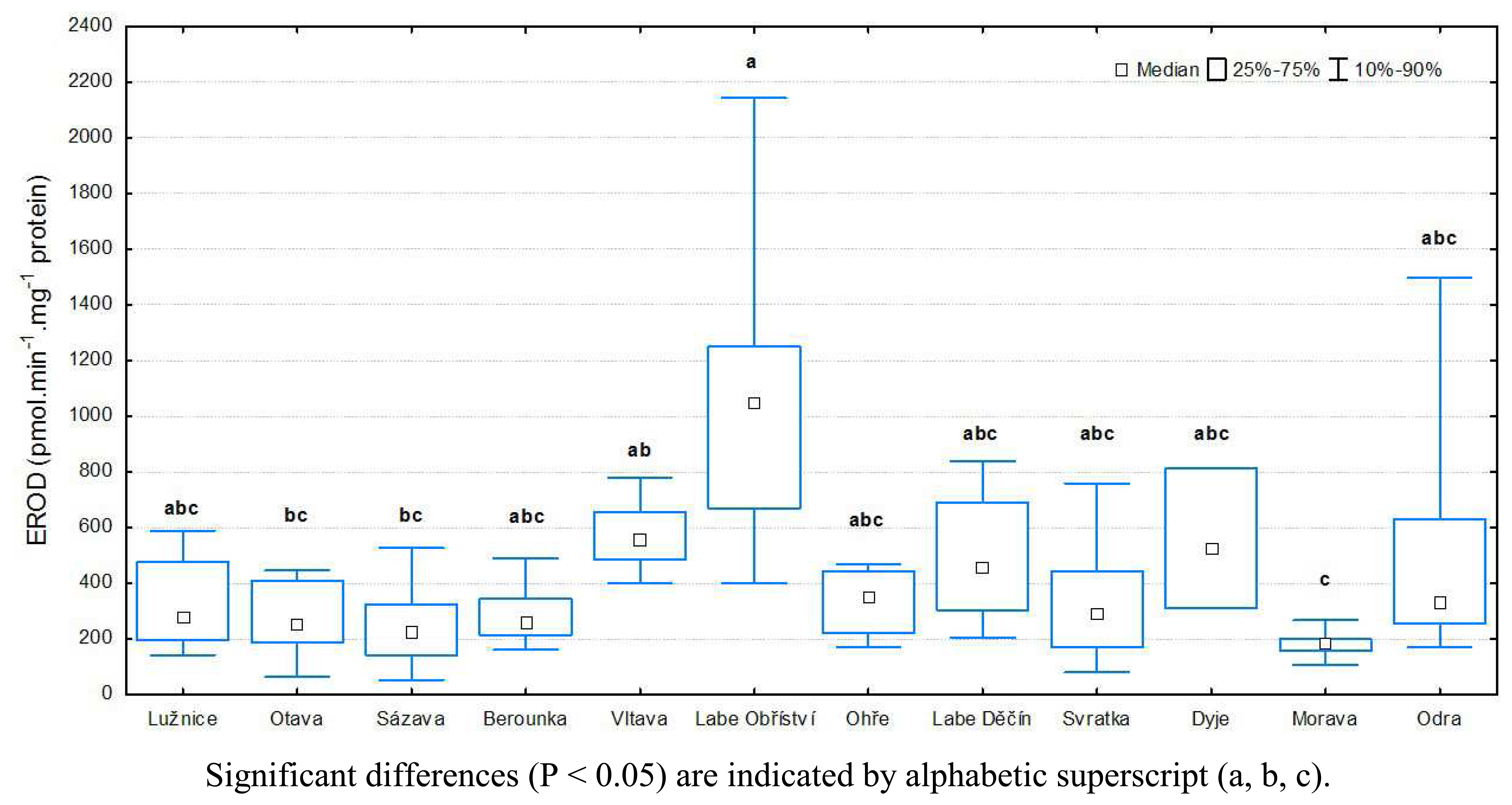
2.1 Determination of CYP 450 and EROD activity

Liver samples were homogenized in buffer (0.25 M saccharose, 0.01 M TRIS and 0.1 mM EDTA) and centrifuged at 10,000 g for 20 min at 4 °C. The supernatant was transferred to ultracentrifugation tubes and centrifuged again at 100,000 g for 1 h at 4 °C. The supernatant was drained, pellets washed with buffer and resuspended in buffer. This suspension was put into individual Eppendorf tubes and stored at –80 °C until use. Before the enzymes were assayed, microsomal protein concentrations were determined by the Lowry method [21].
Total cytochrome P450 was determined by visible light spectrophotometry at 400–490 nm on the basis of the difference between absorbance readings at 450 and 490 nm, and the values obtained were then transformed to final concentrations. Measurements were made after cytochrome reduction by sodium dithionite and after the complex with carbon oxide was formed. The method is described in detail in study Siroka et al. [22].
Catalytic activity of the enzyme ethoxyresorufin-O-deethylase was measured by spectrofluorometry. The method is described in detail in study Siroka et al. [22]. In the presence of NADPH, EROD transforms the substrate ethoxyresorufin to resorufin. Measurements were made using the Perkin-Elmer Fluorescence Spectrophotometer 203.

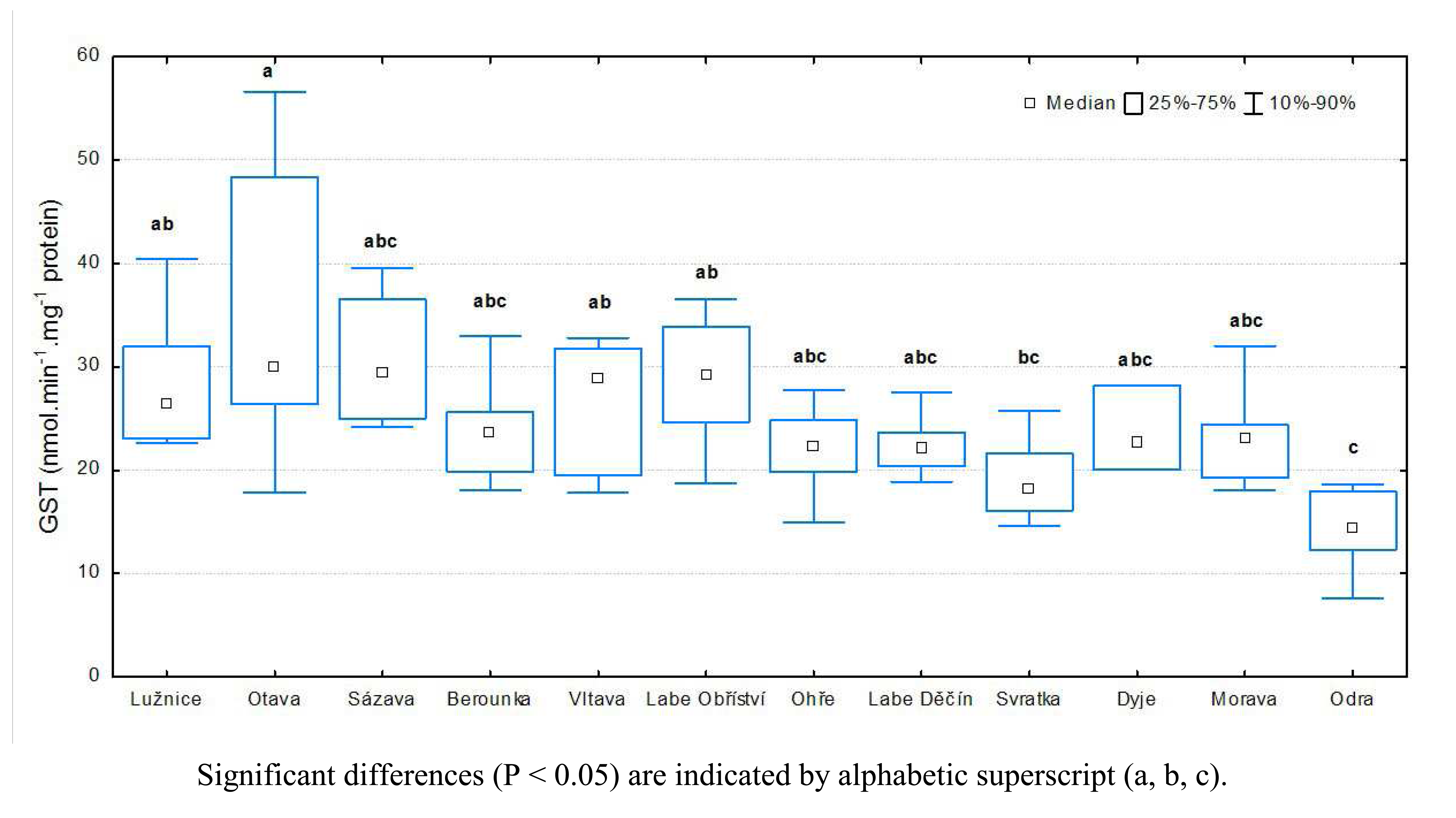
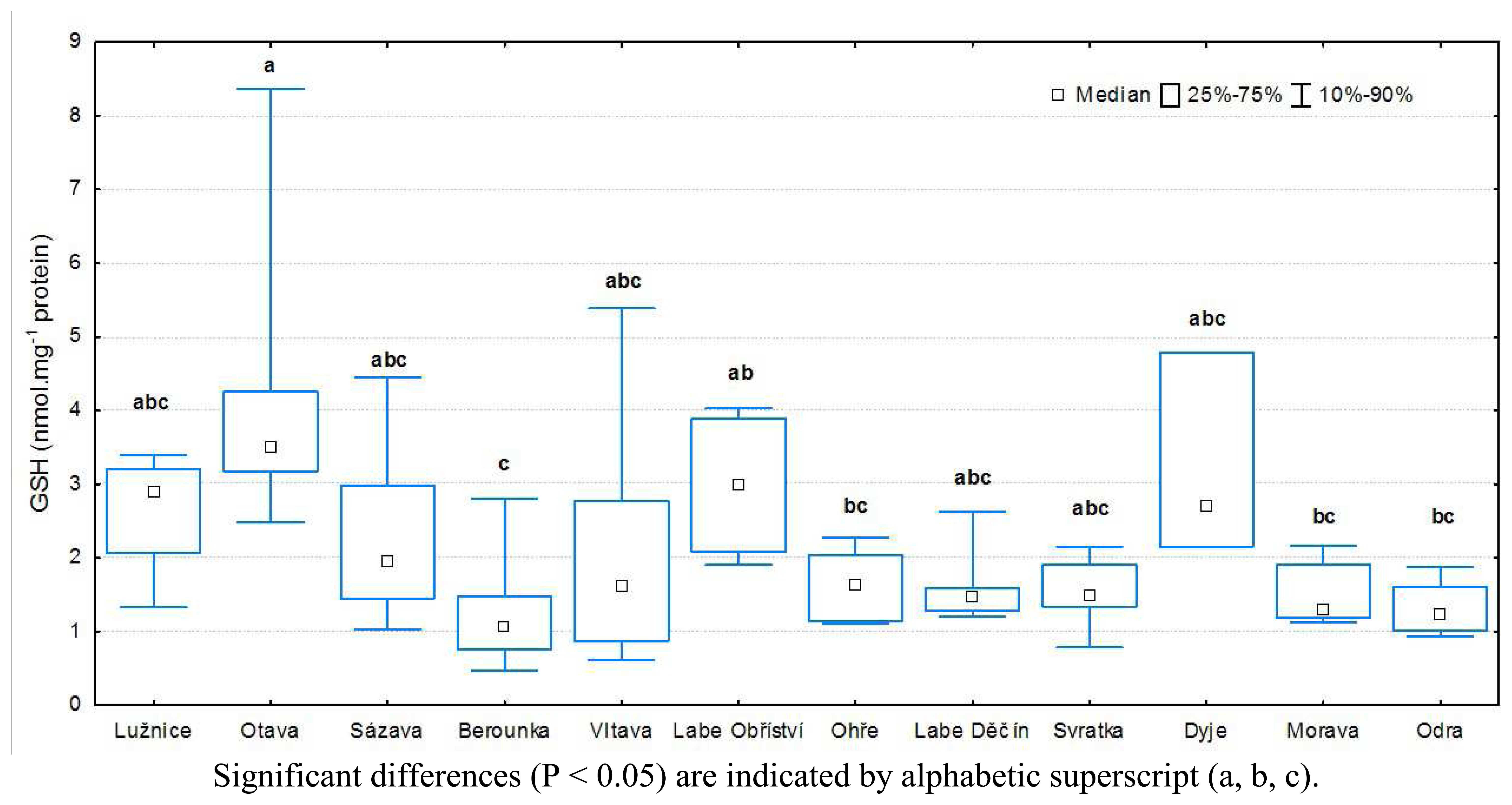
2.2 Determination of GST activity and tripeptide GSH

The liver samples were extracted with phospate buffer (pH 7.2). The homogenate of liver was centrifuged (2,400 g for 10 min, 4 °C) and the supernatant used for determination of glutathione-S-transferase (GST), reduced tripeptide glutathione (GSH), and protein concentration.
The catalytic activity of GST was measured spectrophotometrically (at 340 nm) by a modified method of Habig et al. [23] using a Cobas Emira biochemical analyzer. Supernatant with phosphate buffer (pH 7.2), 0.02 M CDNB (1-Cl-2,4-dinitrobenzene), and 0.1 M reduced glutathione was pipetted into the cuvette of the biochemical analyzer. The specific activity was expressed as nmol of formed product per minute per milligram of protein.
Tripeptide glutathione was determined by the method of Ellman [24] using the Cobas Emira biochemical analyzer. Absorbance of coloured product was determined at 405 nm and concentrations (nmol GSH mg−1 protein) calculated according to a standard calibration. The protein concentration was determined with the Bicinchoninic Acid Protein Essay Kit (Sigma-Aldrich) using bovine serum albumin as standard.

2.3 Determination of PCDD/PCDF and PCB

The pooled samples of fish muscle were homogenized and dried by lyophilization. Internal standards (15 13C12, labeled PCDD/PCDF; 11 13C12, labeled dioxin-like PCB; and 10 13C12, labeled mono- to deca- PCBs) were added to the lyophilized sample. The samples were Soxhlet-extracted with a 3:1 hexane-acetone mixture for 24 h. Fat removal, consequential clean up, and fractionation were performed by a combination of dialysis with a semipermeable membrane and a modified EPA 1613 method [25].
PCDD/PCDFs and dioxin-like PCBs were analyzed using gas chromatography/high-resolution mass spectrometry (GC/HRMS) (Thermo Electron MAT95XP) in MID scan mode. Two ions were monitored for both native and isotope labeled compounds. Quantification masses were chosen in accordance with EPA 1613 and EPA 1668. Full congener PCB analyses were performed by GC/MS/MS (Thermo Electron PolarisQ). The DB5ms column was used for separation of congeners. Gas chromatography conditions were set as follows: splitles injection at 260°C for 1 min; He as carrier gas at 1.1 mL/min constant flow mode; temperature of the transfer line, 275°C; temperature program of GC oven, 70°C for 1 min, 20°C min−1 to 180°C, then 2.5°C to 300°C, followed by 5 min isothermal.
The experiments were performed under constant conditions: ion source temperature, 200°C; flow of dumping He to ion trap – 1.05 mL/min; isolation time, 8 min; excitation time, 10 min; number of microscans, 3.
Toxic equivalents (TEQs) for fish were used to express levels of PCDD/PDCF and PCB concentrations in male chub muscle, as reported by Van den Berg et al. [26].
QA/QC: the laboratory is accredited by Czech Accreditation Institute in accordance to ISO 17 025 norm. The method was validated using certified reference materials NIST 1588a, NIST 1944, WMF, WMS (Wellington Laboratories). In accordance with our standard operating procedures, we obtain recoveries of IS ranging from 50-130%.

2.4 Statistical methods

Statistical analysis of the data was performed using the program STATISTICA 6.1 for Windows (StatSoft CR). Data were assessed by non-parametric methods because data normality was not proven. The Kruskal-Wallis test was used to compare biochemical markers of contamination among individual profiles. Whenever the Kruskal-Wallis test revealed significant differences between profiles (P < 0.05), multiple comparisons of all profiles were subsequently performed. Relationships among individual parameters were assessed using Spearman's correlation coefficient (R).

3. Results

3.1 Results of biochemical analyses

The highest CYP 450 levels in liver were found in fish from the Labe (Obříství; (0.32±0.10 nmol mg−1 protein), while the lowest concentration was in the Lužnice (0.11±0.08 nmol mg−1 protein) (Figure 3). Statistical analysis of CYP 450 levels showed significant differences between Labe (Obříství) and following locations: the Lužnice, the Sázava, the Berounka, the Svratka and the Morava (P < 0.05).
The highest EROD activity in liver was in fish from the Labe (Obříství; (1061.38±545.51 pmol min−1 mg−1 protein), and the lowest EROD activity was in the Morava (183.04±48.20 pmol min−1 mg−1 protein) (Figure 4). Significant differences were found (P < 0.05) between the Labe (Obříství) and the Otava, the Sázava, and the Morava. A significant difference (P < 0.05; was also found between the Morava and the Vltava.
The highest GST catalytic activity in fish liver was detected in fish from the Otava (35.39±13.35 nmol min−1 mg−1 protein). The lowest was in those from the Odra site (14.43±3.80 nmol min−1 mg−1 protein). Statistical analysis of GST activity showed significant differences between the location Odra and the locations Lužnice, Otava, Vltava, and Labe (Obříství) (P < 0.05), and also between the Svratka and the Otava (P < 0.05; (Figure 5).
The highest GSH tripeptide content in fish liver was detected in the Otava (4.29±2.10 nmol GSH mg−1 protein), the lowest GSH content was in the Berounka (1.23±0.73 nmol GSH mg−1 protein). Statistical analysis of GSH content in fish liver showed significant differences between the Otava and: the Berounka, the Ohře, the Morava, and the Odra (P < 0.05); statistical analysis also showed significant differences between the Labe (Obříství) and the Berounka (P < 0.05; (Figure 6).
Significant correlations (R = 0.571) of EROD activity with CYP 450 and of GST with GSH (R = 0.595) in liver tissue were found in fish from all locations (P < 0.01).

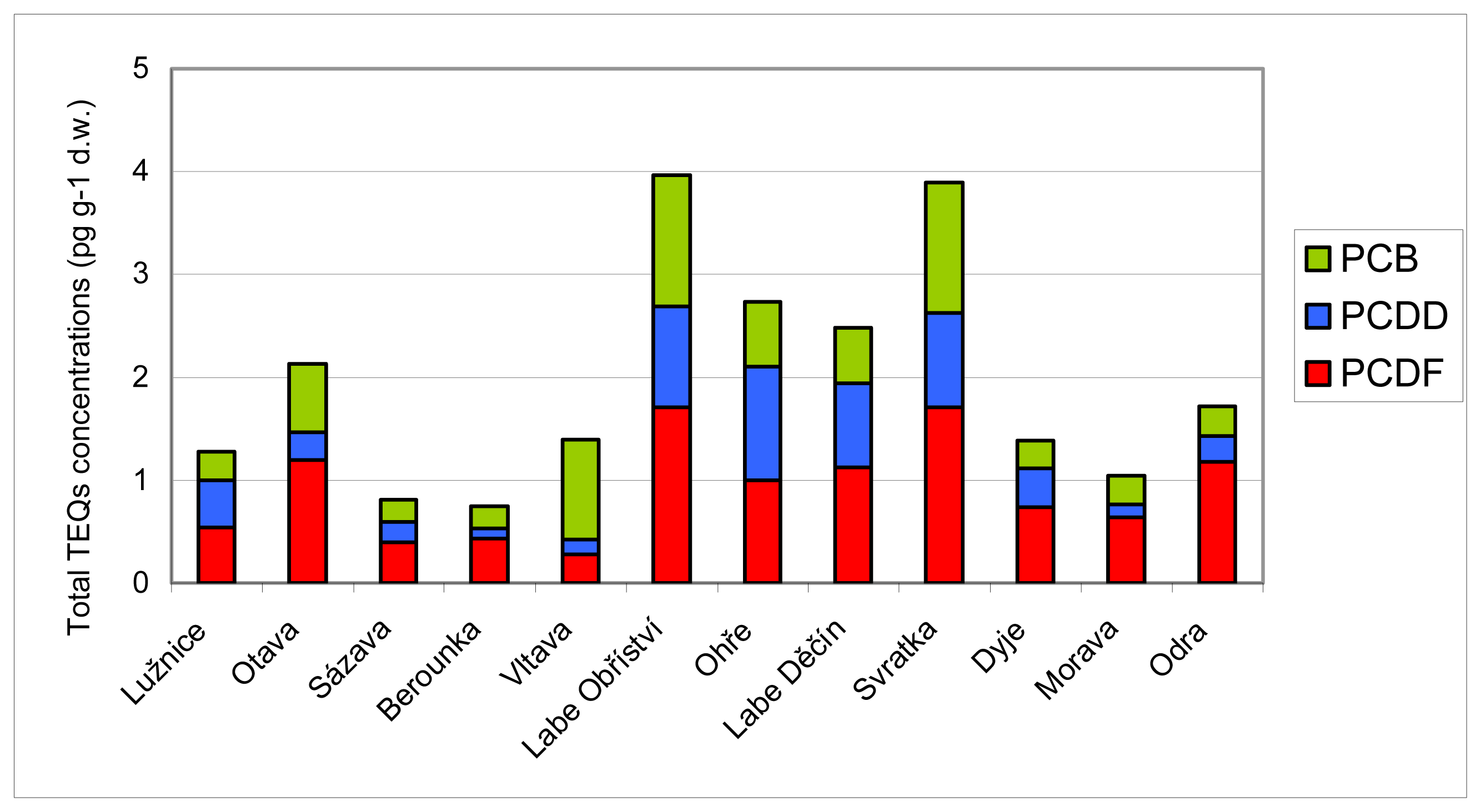
3.2 Results of chemical analyses

The concentrations of PCCD/PCDF TEQs in male chub muscle ranged from 0.10 (Berounka) to 1.10 pg g−1 dry weight (d.w.) (Ohře) and from 0.27 (Vltava; to 1.71 pg g−1 d.w. (Labe Obříství and Svratka), respectively. The concentrations of PCB TEQs in male chub muscle ranged from 0.21 (Berounka) to 1.28 pg g-1 d. w. (Labe Obříství). Total TEQs concentrations (Σ of PCDD/PCDF and PCB TEQs concentrations) in male chub muscle ranged from 0.74 (Berounka) to 3.97 pg g-1 d. w. (Labe Obříství;. The toxic equivalents of analysed chemicals are shown in Figure 7. No correlation with biochemical markers from individual sites was performed because of the combined chub muscle samples.

4. Discussion

Phase I enzyme levels (CYP 450 content and EROD activity) show the highest contamination to be at localities downstream from major chemical factories (Labe Obříství, Labe Děčín), urban concentrations (Vltava), and heavy industry (coal mining) (Odra;. The major inducers of commonly monitored biomarkers are contaminants that belong to a large group of organic pollutants, PAH, PCB, and PCDD/PCDF, persistent in the environment. Induction of the cytochrome P450 system and EROD activity is well documented; a large number of laboratory studies have been carried out measuring effects of organic trace pollutants on hepatic enzymes of various fish species [19, 27-30]. Results of laboratory studies have been confirmed in a great number of field studies [i.e., 31-34]. Our results indicate the presence, at observed locations, of inductors of the observed biochemical markers. The highest levels of these inductors were detected in fish muscle from the sites on the Labe (Obříství) and the Svratka. The source of organic chemical pollution at the Labe (Obříství; site is the chemical manufacturing industry. The River Labe is one of the most highly polluted European rivers [35-38]. Numerous chemical plants are located along its banks as well as along the tributaries of the upper Labe in the Czech Republic. Presumably as a result of dilution, the toxic substances are homogeneously distributed along the River Labe [39]. Despite efforts to reduce intentional and incidental releases, organic pollutants are frequently detected in environmental samples. The presence of organic pollutants in fish muscle and the relation to specific biomarkers were also determined by Siroka et al. [22] and Havelkova et al. [40] at the Labe (Obříství; and Vltava. Siroka et al. [22] found the highest PCB levels (E 7 PCB) in fish muscle and the greatest PAH levels in bottom sediment at sites on the River Labe and the Vltava. High levels of organic chemical pollutants at Svratka in the presented study may be related to the densely populated urban area of Brno.
Measurements of phase II enzymes may be useful in the context of the balance between phase I activation and phase II detoxification. The balance between phase I activation reactions and phase II conjugation pathways can underlie the toxicity of many organic xenobiotics, such as the PAHs.
The highest activity of GST and the highest levels of tripeptide reduced GSH were detected at the locations Otava, Labe (Obříství) and Lužnice. Major inductors of the enzymes of the phase II xenobiotic detoxification and tripeptide GSH are also considered to be persistent organic pollutants: PCBs, PAHs and PCCD/PCDFs [16, 18]. Increased concentrations of total liver glutathione relative to reference sites, have been observed in English sole (Pleuronectes vetulus) collected from a PCB- and PAH- polluted portion of the Puget Sound in Washington [41] and in brown bullhead (Ameiurus nebulosus) from a PAH-polluted section of the Buffalo River in New York [42].
Some studies have not confirmed those which show higher GST activity related to organic pollutants [17, 4345]. Significant differences in enzyme activity in fish from polluted and reference sites have been reported in some field studies [4649]. This variation in findings could be the result of fish species' differing sensitivity to the presence of xenobiotics in the environment [50]. The presence of GST catalytic activity inhibitors could play an important role in the biochemical responses of aquatic organisms [51].
The highest total TEQ concentrations of chemical pollutants (PCCD/PCDFs and PCB) were detected at the locations Labe (Obříství) and Svratka. The present study revealed a positive relationship between concentrations of chemical pollutants (PCDD/PCDF and PCB) in fish muscle and levels of biomarkers in indicator fish liver from e.g. Labe (Obříství). Decreased levels of phase I hepatic enzymes, despite increased levels of the specific inductors in muscle may indicate the presence of specific inhibitors of these enzymes in the aquatic environment (i.e., Svratka). There is extensive literature on the biomarker inhibition of metallic compounds (i.e., copper, lead, zinc, cadmium, arsenic, nickel, chromium, and tin; and their effects on fish populations [5256]. It is difficult to find an appropriate reference site with no contaminants. Moreover, several natural factors influence EROD activity, including sex, reproductive status, seasons, species, and water temperature, making data sometimes difficult to interpret [12, 5758]. Ecological and biological factors should be taken into account to explain variations in enzymatic biotransformation activities in fish.

5. Conclusions

Methods of biochemical monitoring for exposure to environmental contaminants are of great potential use. In the present study, CYP 450, catalytic activities of EROD and GST and tripeptide GSH were used for assessment of contamination by organic pollutants at twelve locations in the Czech Republic. The highest level of CYP 450 and EROD activity were found at the Labe (Obříství); the highest GST activity and levels of tripeptide GSH were found at the location Otava. The results of analysis of muscle for specific inductors of the measured biomarkers confirmed high levels of biochemical contamination at the Labe (Obříství;. However, high content of the inductors in muscle of fish from the Svratka coupled with low levels of liver biochemical markers may indicate the presence of specific inhibitors of biomarkers in the aquatic environment.

Acknowledgments

This research was supported by the Ministry of the Education, Youth and Sports of the Czech Republic (Projects MSM 6215712402, MSM 6007665809), by the Ministry of Environment of the Czech Republic (Project SP/2e7/229/07) and by the Biomonitoring programme of the Czech Hydrometeorological Institute.

References

  1. Bamgbose, O.; Jinadu, K.; Osibanjo, O. Galeoides-decadactylus - A bioindicator for chlorina hydrocarbons in the Nigerian marine-environment. J. Environ. Sci. Heal. A. 1993, 28, 321–337. [Google Scholar]
  2. Linde, A.R.; Arribas, P.; SanchezGalan, S.; GarciaVazquez, F. Eel (Anguilla anguilla) and brown trout (Salmo trutta) target species to assess the biological impact of trace metal pollution in freshwater ecosystems. Arch. Environ. Con. Tox. 1996, 31, 297–302. [Google Scholar]
  3. Ueno, D.; Iwata, H.; Tanabe, S.; Ikeda, K.; Koyama, J.; Yamada, H. Specific accumulation of persistent organochlorines in bluefin tuna collected from Japanese coastal waters. Mar. Pollut. Bull. 2002, 45, 254–261. [Google Scholar]
  4. Ueno, D.; Inoue, S.; Takahashi, S.; Ikeda, K.; Tanaka, H.; Subramanian, A.N.; Fillmann, G.; Lam, P.K.S.; Zheng, J.; Muchtar, M.; Prudente, M.; Chung, K.; Tanabe, S. Global pollution monitoring of butyltin compounds using skipjack tuna as a bioindicator. Environ. Pollut. 2004, 127, 1–12. [Google Scholar]
  5. Widianarko, B.; Van Gestel, C.A.M.; Verweij, R.A.; Van Straalen, N.M. Associations between trace metals in sediment, water, and guppy, Poecilia reticulata (Peters), from urban streams of Semarang, Indonesia. Ecotox. Environ. Safe. 2000, 46, 101–107. [Google Scholar]
  6. Adams, S.M.; Shepard, K.L.; Greeley, M.S.; Jimenez, B.D.; Ryon, M.G.; Shugart, L.R.; McCarthy, J.F.; Hinton, D.E. The use of bioindicators for assessing the effects of pollutant stress on fish. Mar. Environ. Res. 1989, 28, 459–464. [Google Scholar]
  7. Huska, D.; Krizkova, S.; Beklova, M.; Havel, L.; Zehnalek, J.; Diopan, V.; Adam, V.; Zeman, L.; Babula, P.; Kizek, R. Influence of cadmium(II) ions and brewery sludge on metallothionein level in earthworms (Eisenia fetida) - Biotransforming of toxic wastes. Sensors 2008, 8, 1039–1047. [Google Scholar]
  8. Krizkova, S.; Ryant, P.; Krystofova, O.; Adam, V.; Galiova, M.; Beklova, M.; Babula, P.; Kaiser, J.; Novotny, K.; Novotny, J.; Liska, M.; Malina, R.; Zehnalek, J.; Hubalek, J.; Havel, L.; Kizek, R. Multi-instrumental analysis of tissues of sunflower plants treated with silver(I) ions - Plants as bioindicators of environmental pollution. Sensors 2008, 8, 445–463. [Google Scholar]
  9. van der Oost, R.; Beyer, J.; Vermeulen, N.P.E. Fish bioaccumulation and biomarkers in environmental risk assessment: a review. Environ. Toxicol. Phar. 2003, 13, 57–149. [Google Scholar]
  10. Machala, M.; Nezveda, K.; Petrivalsky, M.; Jarosova, A.; Piacka, V.; Svobodova, Z. Monooxygenase activities in carp as biochemical markers of pollution by polycyclic and polyhalogenated aromatic hydrocarbons: choice of substrates and effects of temperature, gender and capture stress. Aquat. Toxicol. 1997, 37, 113–123. [Google Scholar]
  11. Bucheli, T.D.; Font, K. Induction of cytochrome-P450 as a biomarker for environmental contamination in aquatic ecosystems. Crit. Rev. Env. Sci. Tec. 1995, 25, 201–268. [Google Scholar]
  12. Goksoyr, A.; Forlin, L. The cytochrome-P-450 system in fish, aquatic toxicology and environmental monitoring. Aquat. Toxicol. 1992, 22, 287–311. [Google Scholar]
  13. Hahn, M.E.; Stegeman, J.J. Regulation of cytochrome P4501A1 in teleosts - sustained induction of CYP1A1 messenger-RNA, protein, and catalytic activity by 2,3,7,8 tetrachlorodibenzofuran in the marine fish. Stenotomus chrysops. Toxicol. Appl. Pharm. 1994, 127, 187–198. [Google Scholar]
  14. Payne, J.F.; Fancey, L.L.; Rahimtula, A.D.; Porter, E.L. Review and perspective on the use of mixed-function oxygenase enzymes in biological monitoring. Comp. Biochem. Phys. C. 1987, 86, 233–245. [Google Scholar]
  15. Eaton, D.L.; Bammler, T.K. Concise review of the glutathione S-transferases and their significance to toxicology. Toxicol. Sci. 1999, 49, 156–164. [Google Scholar]
  16. Gadagbui, B.K.M.; Goksoyr, A. CYP1A and other biomarker responses to effluents from a textile mill in the Volta River (Ghana) using caged tilapia (Oreochromis niloticus) and sediment-exposed mudfish (Clarias anguillaris). Biomarkers 1996, 1, 252–261. [Google Scholar]
  17. Otto, D.M.E.; Moon, T.W. Phase I and II enzymes and antioxidant responses in different tissues of brown bullheads from relatively polluted and non-polluted systems. Arch. Environ. Con. Tox. 1996, 31, 141–147. [Google Scholar]
  18. Pedrajas, J.R.; Peinado, J.; LopezBarea, J. Oxidative stress in fish exposed to model xenobiotics. Oxidatively modified forms of Cu,Zn-superoxide dismutase as potential biomarkers. Chem-Biol. Interact. 1995, 98, 267–282. [Google Scholar]
  19. Bello, S.M.; Franks, D.G.; Stegeman, J.J.; Hahn, M.E. Acquired resistance to Ah receptor agonists in a population of Atlantic killifish (Fundulus heteroclitus) inhabiting a marine superfund site: In vivo and in vitro studies on the inducibility of xenobiotic metabolizing enzymes. Toxicol. Sci. 2001, 60, 77–91. [Google Scholar]
  20. Celander, M.; Leaver, M.J.; George, S.G.; Forlin, L. Induction of cytochrome P450-1A1 and conjugating enzymes in rainbow-trout (Oncorhynchus-mykiss) liver - a time-course study. Comp. Biochem. Phys. C. 1993, 106, 343–349. [Google Scholar]
  21. Lowry, O.H.; Rosebrough, N.J.; Farr, A.L.; Randall, R.J. Protein measurement with the Folin phenol reagent. J. Biol. Chem. 1951, 193, 265–275. [Google Scholar]
  22. Siroka, Z.; Krijt, J.; Randak, T.; Svobodova, Z.; Peskova, G.; Fuksa, J.; Hajslova, J.; Jarkovsky, J.; Janska, M. Organic pollutant contamination of the River Elbe as assessed by biochemical markers. Acta Vet. Brno 2005, 74, 293–303. [Google Scholar]
  23. Habig, W.H.; Pabst, M.J.; Jakoby, W.B. Glutathione S-transferases. First enzymatic step in mercapturic acid formation. J. Biol. Chem. 1974, 249, 7130–7139. [Google Scholar]
  24. Ellman, G.L. Tissue sulfhydryl groups. Arch. Biochem. Biophys. 1959, 82, 70–77. [Google Scholar]
  25. Grabic, R.; Novak, J.; Pacakova, V. Optimization of a GC-MS/MS method for the analysis of PCDDs and PCDFs in human and fish tissue. HRC J. High. Res. Chromatogr. 2000, 23, 595–599. [Google Scholar]
  26. Van den Berg, M.; Birnbaum, L.; Bosveld, A.T.C.; Brunström, B.; Cook, P.; Feeley, M.; Giesy, J.P.; Hanberg, A.; Hasegawa, R.; Kennedy, S.W.; Kubiak, T.; Larsen, J.C.; van Leeuwen, F.X.R.; Liem, A.K.D.; Nolt, C.; Peterson, R.E.; Poellinger, L.; Safe, S.; Schrenke, D.; Tillitt, D.; Tysklind, M.; Younes, M.; Wærn, F.; Zacharewski, T. Toxic Equivalency Factors (TEFs) for PCBs, PCDDs, PCDFs for humans and wildlife. Environ. Health. Persp. 1989, 106, 775–792. [Google Scholar]
  27. Agradi, E.; Baga, R.; Cillo, F.; Ceradini, S.; Heltai, D. Environmental contaminants and biochemical response in eel exposed to Po river water. Chemosphere 2000, 41, 1555–1562. [Google Scholar]
  28. Schlezinger, J.J.; Stegeman, J.J. Induction of cytochrome P450 1A in the American Eel by model halogenated and non-halogenated aryl hydrocarbon receptor agonists. Aquat. Toxicol. 2000, 50, 375–386. [Google Scholar]
  29. Stagg, R.M.; Rusin, J.; McPhail, M.E.; McIntosh, A.D.; Moffat, C.F.; Craft, J.A. Effects of polycyclic aromatic hydrocarbons on expression of CYP1A in salmon (Salmo salar) following experimental exposure and after the Braer oil spill. Environ. Toxicol. Chem. 2000, 19, 2797–2805. [Google Scholar]
  30. Vanderweiden, M.E.J.; Vanderkolk, J.; Bleumink, R.; Seinen, W.; Vandenberg, M. Concurrence of P450-1A1 induction and toxic effects after administration of a low-dose of 2,3,7,8-tetrachlorodibenzo-p-dioxin (TCDD) in the rainbow-trout (Oncorhynchus-mykiss). Aquat. Toxicol. 1992, 24, 123–142. [Google Scholar]
  31. Al-Arabi, S.A.M.; Adolfsson-Erici, M.; Waagbo, R.; Ali, M.S.; Goksoyr, A. Contaminant accumulation and biomarker responses in caged fish exposed to effluents from anthropogenic sources in the Karnaphuly River, Bangladesh. Environ. Toxicol. Chem. 2005, 24, 1968–1978. [Google Scholar]
  32. Behrens, A.; Segner, H. Cytochrome P4501A induction in brown trout exposed to small streams of an urbanised area: results of a five-year-study. Environ. Pollut. 2005, 136, 231–242. [Google Scholar]
  33. Curtis, L.R.; Carpenter, H.M.; Donohoe, R.M.; Williams, D.E.; Hedstrom, O.R.; Deinzer, M.L.; Bellstein, M.A.; Foster, E.; Gates, R. Sensitivity of cytochrome-P450-1A1 induction in fish as a biomarker for distribution of TCDD and TCDF in the Willamette River, Oregon. Environ. Sci. Technol. 1993, 27, 2149–2157. [Google Scholar]
  34. Monod, D.; Devaux, A.; Riviere, J.L. Effects of chemical pollution on the activities of hepatic xenobiotic metabolizing enzymes in fish from the River Rhone. Sci. Total. Environ. 1988, 73, 189–201. [Google Scholar]
  35. Adams, M.S.; Kausch, H.; Gaumert, T.; Kruger, K.E. The effect of the reunification of Germany on the water chemistry and ecology of selected rivers. Environ. Conserv. 1996, 23, 35–43. [Google Scholar]
  36. Gandrass, J.; Zoll, M. Chlorinated hydrocarbons in sediments of the Elbe catchment area -Analytical methods and status of pollution. Acta Hydroch. Hydrob. 1996, 24, 212–217. [Google Scholar]
  37. Heininger, P.; Pelzer, J. Trends and patterns in the contamination of sediments from federal waterways in Eastern Germany. Acta Hydroch. Hydrob. 1998, 26, 218–225. [Google Scholar]
  38. Lehmann, A.; Rode, M. Long-term behaviour and cross-correlation water quality analysis of the river Elbe, Germany. Water. Res. 2001, 35, 2153–2160. [Google Scholar]
  39. Oetken, M.; Stachel, B.; Pfenninger, M.; Oehlmann, J. Impact of a flood disaster on sediment toxicity in a major river system - the Elbe flood 2002 as a case study. Environ. Pollut. 2005, 134, 87–95. [Google Scholar]
  40. Havelkova, M.; Randak, T.; Zlabek, V.; Krijt, J.; Kroupova, H.; Pulkrabova, J.; Svobodova, Z. Biochemical markers for assessing aquatic contamination. Sensors 2008, 7, 2599–2611. [Google Scholar]
  41. Nishimoto, M.; Leeberhart, B.T.; Sanborn, H.R.; Krone, C.; Varanasi, U.; Stein, J.E. Effects of a complex mixture of chemical contaminants on hepatic glutathione, L-cysteine and gamma-glutamylcysteine synthetase in English sole (Pleuronectes vetulus). Environ. Toxicol. Chem. 1995, 14, 461–469. [Google Scholar]
  42. Eufemia, N.A.; Collier, T.K.; Stein, J.E.; Watson, D.E.; DiGiulio, R.T. Biochemical responses to sediment-associated contaminants in brown bullhead (Ameriurus nebulosus) from the Niagara River ecosystem. Ecotoxicology 1997, 6, 13–34. [Google Scholar]
  43. Petrivalsky, M.; Machala, M.; Nezveda, K.; Piacka, V.; Svobodova, Z.; Drabek, P. Glutathione-dependent detoxifying enzymes in rainbow trout liver: Search for specific biochemical markers of chemical stress. Environ. Toxicol. Chem. 1997, 16, 1417–1421. [Google Scholar]
  44. Vanderoost, R.; Vangastel, L.; Worst, D.; Hanraads, M.; Satumalay, K.; Vanschooten, F.J.; Heida, H.; Vermeulen, N.P.E. Biochemical markers in feral roach (Rutilus-rutilus) in relation to the bioaccumulation of organic trace pollutants. Chemosphere 1994, 29, 801–817. [Google Scholar]
  45. Vigano, L.; Arillo, A.; Melodia, F.; Bagnasco, M.; Bennicelli, C.; Deflora, S. Hepatic and biliary biomarkers in rainbow-trout injected with sediment extracts from the River Po (Italy). Chemosphere 1995, 30, 2117–2128. [Google Scholar]
  46. Armknecht, S.L.; Kaattari, S.L.; Van Veld, P.A. An elevated glutathione S-transferase in creosote-resistant mummichog (Fundulus heteroclitus). Aquat. Toxicol. 1998, 41, 1–16. [Google Scholar]
  47. Lenartova, V.; Holovska, K.; Javorsky, P. The influence of environmental pollution on the SOD and GST-isoenzyme patterns. Water. Sci. Technol. 2000, 42, 209–214. [Google Scholar]
  48. Tuvikene, A.; Huuskonen, S.; Koponen, K.; Ritola, O.; Mauer, U.; Lindstrom-Seppa, P. Oil shale processing as a source of aquatic pollution: Monitoring of the biologic effects in caged and feral freshwater fish. Environ. Health. Persp. 1999, 107, 745–752. [Google Scholar]
  49. Vigano, L.; Arillo, A.; Melodia, F.; Arlati, P.; Monti, C. Biomarker responses in cyprinids of the middle stretch of the River Po, Italy. Environ. Toxicol. Chem. 1998, 17, 404–411. [Google Scholar]
  50. Hamed, R.R.; Farid, N.M.; Elowa, S.H.E.; Abdalla, A.M. Glutathione related enzyme levels of freshwater fish as bioindicators of pollution. Environmentalist 2003, 23, 313–322. [Google Scholar]
  51. Mannervik, B.; Danielson, U.H. Glutathione transferases - structure and catalytic activity. CRC Cr. Rev. Bioch. Mol. 1988, 23, 283–337. [Google Scholar]
  52. Bozcaarmutlu, A.; Arinc, E. Inhibitory effects of divalent metal ions on liver microsomal 7-ethoxyresorufin-O-deethylase (EROD) activity of leaping mullet. Mar. Environ. Res. 2004, 58, 521–524. [Google Scholar]
  53. Fent, K.; Bucheli, T.D. Inhibition of hepatic-microsomal monooxygenase system by organotins in-vitro in fresh-water fish. Aquat. Toxicol. 1994, 28, 107–126. [Google Scholar]
  54. Forlin, L.; Haux, C.; Karlsson-Norrgren, L.; Runn, P.; Larsson, A. Biotransformation enzyme-activities and histopathology in rainbow trout, Salmo-Gairdneri, treated with cadmium. Aquat. Toxicol. 1986, 8, 51–64. [Google Scholar]
  55. Risso-de Faverney, C.; Lafaurie, M.; Girard, J.P.; Rahmani, R. Effects of heavy metals and 3-methylcholanthrene on expression and induction of CYP1A1 and metallothionein levels in trout (Oncorhynchus mykiss) hepatocyte cultures. Environ. Toxicol. Chem. 2000, 19, 2239–2248. [Google Scholar]
  56. Stien, X.; Risso, C.; GnassiaBarelli, M.; Romeo, M.; Lafaurie, M. Effect of copper chloride in vitro and in vivo on the hepatic EROD activity in the fish Dicentrarchus labrax. Environ. Toxicol. Chem. 1997, 16, 214–219. [Google Scholar]
  57. Flammarion, P.; Garric, J. Cyprinids EROD activities in low contaminated rivers: A relevant statistical approach to estimate reference levels for EROD biomarker? Chemosphere 1997, 35, 2375–2388. [Google Scholar]
  58. Koivusaari, U.; Harri, M.; Hanninen, O. Seasonal-variation of hepatic biotransformation in female and male rainbow-trout (Salmo-gairdneri). Comp. Biochem. Phys. C. 1981, 70, 149–157. [Google Scholar]
Figure 1. Indicator species – Chub (Leuciscus cephalus L.).
Sensors 08 02589f1
Figure 2. Map of the Czech Republic and locations of sampling sites (1. Lužnice (Bechyně), 2. Otava (Topělec), 3. Sázava (Nespeky), 4. Berounka (Srbsko), 5. Vltava (Zelčín), 6. Labe (Obříství), 7. Ohře (Terezín), 8. Labe (Děčín), 9. Svratka (Židlochovice), 10. Dyje (Pohansko), 11. Morava (Lanžhot), 12. Odra (Bohumín);.
Sensors 08 02589f2
Figure 3. Content of cytochrome P450 in male chub (Leuciscus cephalus L.) livers.
Sensors 08 02589f3
Figure 4. EROD activity in male chub (Leuciscus cephalus L.) livers.
Sensors 08 02589f4
Figure 5. GST catalytic activity in male chub (Leuciscus cephalus L.) livers.
Sensors 08 02589f5
Figure 6. Content of tripeptide GSH in male chub (Leuciscus cephalus L.) livers.
Sensors 08 02589f6
Figure 7. Total TEQ concentrations for PCDD/PCDFs and PCBs (pg g−1 d. w.) in male chub (Leuciscus cephalus L.) muscle.
Sensors 08 02589f7
Table 1. Characteristics of male chub (Leuciscus cephalus L.) from sampling sites; n = number of fish.
Location River (Municipality) (distance from site to mouth of river)nWeight±SD (g)Age (years) mean (range)
Lužnice (Bechyně) (11 kms)8614.4±155.04.9 (4–6)
Otava (Topělec) (20 kms)9360.6±253.04.2 (3–6)
Sázava (Nespeky) (27.5 kms)8278.1±63.93.8 (3–4)
Berounka (Srbsko) (29 kms)8292.5±99.53.6 (3–5)
Vltava (Zelčín) (5 kms)10383.5±199.34.0 (3–7)
Labe (Obříství) (122 kms)*8306.3±144.94.1 (3–6)
Ohře (Terezín) (3 kms)10540.5±201.84.3 (3–7)
Labe (Děčín) (21 kms)*8546.3±211.04.8 (4–7)
Svratka (Židlochovice) (23 kms)8243.8±94.23.5 (3–4)
Dyje (Pohansko) (16 kms)3626.7±558.24.7 (3–7)
Morava (Lanžhot) (9.5 kms)8304.4±110.63.1 (2–4)
Odra (Bohumín) (9 kms)**8149.4±78.12.8 (2–4)
*Distances between Labe sites and the border with Germany
**Distance between Odra site and the border with Poland

