Review

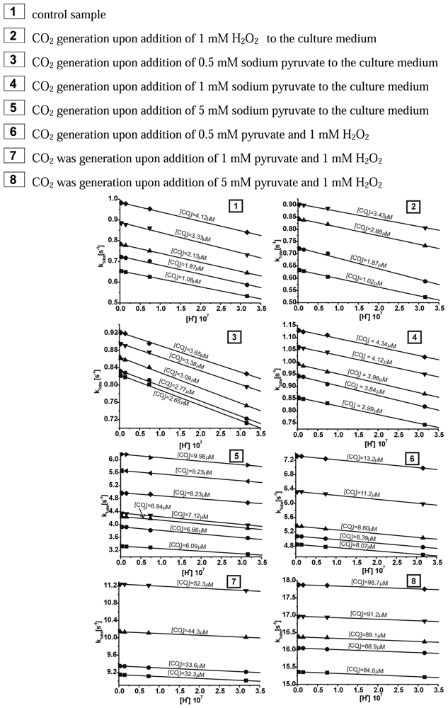
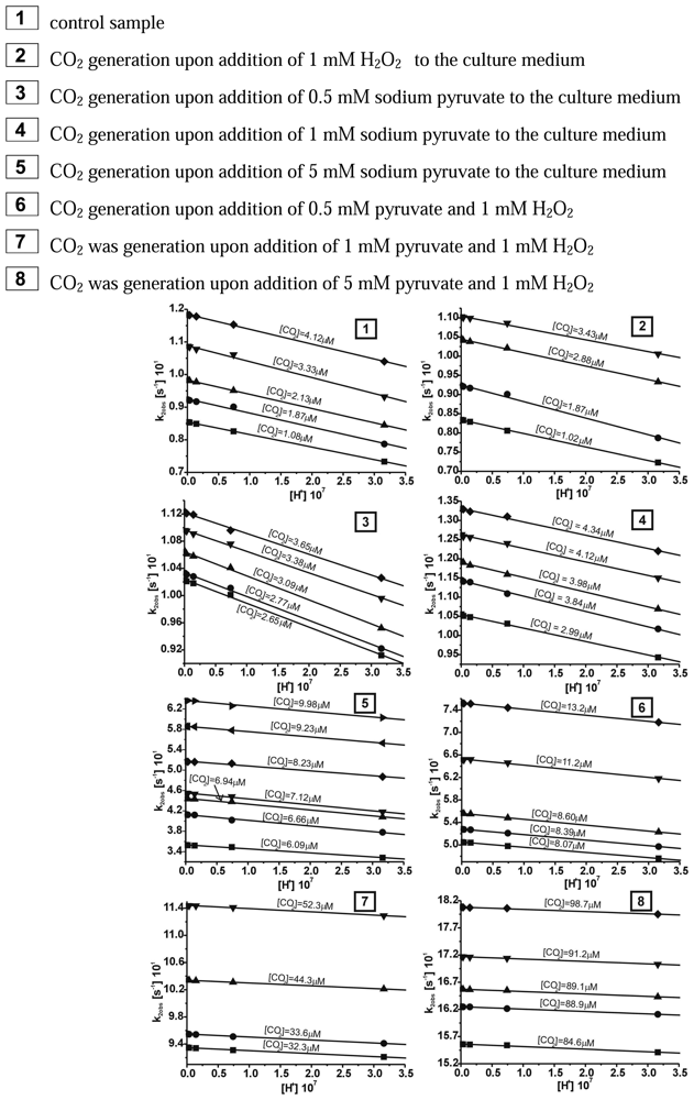
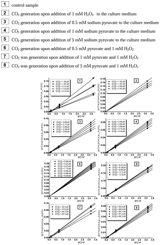
Coordinate cis-[Cr(C2O4)(pm)(OH2)2]+ Cation as Molecular Biosensor of Pyruvate’s Protective Activity Against Hydrogen Peroxide Mediated Cytotoxity

by
Dagmara Jacewicz
1,
Michał Szkatuła
2,
Agnieszka Chylewska
1,
Aleksandra Dąbrowska
1,
Michał Woźniak
2 and
Lech Chmurzyński
1,*
1
Department of General and Inorganic Chemistry, University of Gdańsk, Sobieskiego 18/19, 80-952 Gdańsk, Poland
2
Department of Medical Chemistry, Medical University of Gdańsk, Dębinki 1, 80-211, Gdańsk, Poland
*
Author to whom correspondence should be addressed.
Sensors 2008, 8(8), 4487-4504; https://doi.org/10.3390/s8084487
Submission received: 1 July 2008 / Revised: 28 July 2008 / Accepted: 28 July 2008 / Published: 1 August 2008

Abstract

:
In this paper instrumental methods of carbon dioxide (CO2) detection in biological material were compared. Using cis-[Cr(C2O4)(pm)(OH2)2]+ cation as a specific molecular biosensor and the stopped-flow technique the concentrations of CO2 released from the cell culture medium as one of final products of pyruvate decomposition caused by hydrogen peroxide were determined. To prove the usefulness of our method of CO2 assessment in the case of biological samples we investigated protective properties of exogenous pyruvate in cultured osteosarcoma 143B cells exposed to 1 mM hydrogen peroxide (H2O2) added directly to culture medium. Pyruvic acid is well known scavenger of H2O2 and, moreover, a molecule which is recognized as one of the major mediator of oxidative stress detected in many diseases and pathological situations like ischemia-reperfusion states. The pyruvate's antioxidant activity is described as its rapid reaction with H2O2, which causes nonenzymatic decarboxylation of pyruvate and releases of CO2, water and acetate as final products. In this work for the first time we have correlated the concentration of CO2 dissolved in culture medium with pyruvate's oxidant-scavenging abilities. Moreover, the kinetics of the reaction between aqueous solution of CO2 and coordinate ion, cis-[Cr(C2O4)(pm)(OH2)2]+ was analysed. The results obtained enabled determination of the number of steps of the reaction studied. Based on the kinetic equations, rate constants were determined for each step.

1. Introduction

Oxygen-derived free radical anion formed by mono-, di- or trivalent reduction of molecular oxygen have been involved in many disease such as diabetes, hypertension, ischemia-reperfusion injury, neurodegenerative disorders, atherosclerosis and others [1-4]. The presence of reactive oxygen species (ROS) including: superoxide anion (O2·-), hydrogen peroxide (H2O2) and more potent oxidant hydroxyl radical (HO·) can lead to irreversible damages of cell's components - proteins, lipids and DNA [5-7]. Since mammalian cells even in physiological state are constantly exposed to free radicals, they require a functional system of antioxidants to protect themselves from the toxic actions of ROS [8]. The most characteristic elements of this system are superoxide dismutase (SOD), catalase, peroxiredoxins, α-tocopherol, glutathione and ascorbic acid [9]. Among others also pyruvic acid belongs to this battery of anti-oxidants and it seems to be useful and powerful scavenger of hydrogen peroxide and other peroxides.
H2O2 is considered as a one of the most important mediators of oxidative stress. It can be produced intracellularly, especially by respiratory chain reaction and by extracellular oxidative burst mechanism used by activated inflammatory cells. In the presence of reduced transition metals, H2O2 can be transformed to more aggressive hydroxyl radical, which at least partially contributes to the cytotoxity of H2O2 [10]. Moreover, the ability of H2O2 to penetrate biological membranes allows to affect not only cells in which it was produced but also neighbouring cells [9]. The excessive production of H2O2 was noted in aging brain, in ischemia-reperfusion situations and the increase the level of H2O2 probably participates in the neuronal damage observed in Parkinson's disease [11-13]. Taking into consideration all those facts it seems that antioxidant properties of pyruvic acid (CH3COCOOH), which is also recognised as compound involved in energy production can play an important protective role against the toxity of hydrogen peroxide [14-16]. Furthermore, the pyruvate can be considered as an endogenous, as well as a specific exogenous antioxidant since many types of cells including neurons release into plasma and serum where it can protect them against the acting of H2O2 [17]. According to Mallet RT et. all for the antioxidant properties of pyruvic acid are responsible its chemical structure and the patterns of its cellular metabolism [18]. The α-ketocarboxylate structure enables pyruvate to detoxify H2O2 in a direct, nonenzymatic reaction in which H2O2 is reduced to H2O and pyruvic acid undergoes transformation to CO2 and acetate [19].
pyruvate + H 2 O 2 acetate + CO 2 + H 2 O
Despite the fact that the above presented reaction was described in 1904 by Holleman the mechanism of the antioxidant action of the pyruvate has not been fully elucidated to date. In the present study, using a novel specific molecular biosensor - coordinate ion, cis-[Cr(C2O4)(pm)(OH2)2]+, where pm denotes pyridoxamine and stopped-flow spetrophotometry method we were able to correlate the amount of liberated CO2 as one of final products of the chemical reaction between exogenous pyruvate and exogenous H2O2 with the cytoprotective activity of pyruvate. We also analyzed kinetics of this reaction using both biological and chemical models. Moreover, it was proved that reactive oxygen species could modulate activity of mitochondrial enzyme pyruvate dehydrogenase (PDH) [20]. It has been although found that no change in PDH activity in the brain occurred following ischemia but as much as 65% inhibition following 24h of reperfusion and H2O2 generation was detected [21]. This is why we decided to measure the protection of pyruvate, as well as CO2 level after 24 hours of H2O2 treatment. Thus it seems highly probable that nonenzymatic activity between pyruvate and H2O2 can substitute deficiency of PDH activity.
The structure of the synthesised complex of chromium(III) is shown in Figure 1 (A). It can be seen that the metal ion is coordinated by two oxygen atoms from oxalato anion, two oxygen atoms from two aqua ligands and one oxygen and one nitrogen atoms from pyridoxamine in cis-[Cr(C2O4)(pm)(OH2)2]+ ion.
The pyridoxamine acts as a bidentate ligand coordinating through the deprotonated phenolic-oxygen and the nitrogen donor atom from the amine group. Pyridinium nitrogen is protonated and it does not participate in binding of coordination center. For pyridoxamine, two types of tautomer forms are possible, as shown in Figure 1 (B). The structure of the ligand in pyridoxamine corresponds to type b. This structure was proposed also on the basis of the already published solution studies [22-25].
The selection of chromium(III) as the coordination center allows to obtain inert complexes undergoing slow transformations at ambient temperature, thus enabling investigation of the kinetics and mechanism of the processes under favorrable conditions. Furthermore, studies on the kinetics and mechanism of CO2 uptake by the coordination compounds consisting of the inert Cr(III) ion and a ligand molecule of pyridoxamine are aimed at elucidation of the mechanism of action of one of the metabolic steps. Carbon dioxide is caught by complexes of transition metal ions from biological material stoichiometrically as carbonate anion. This anion can be removed in the presence of acid as carbon dioxide (hydrolysis - reverse reaction to uptake) stoichiometrically too. The course of these two reactions can be presented as follows:
CO 2 + [ M trans ] + H + hydrolysis uptake [ M trans ( O 2 CO ) ] n

2. Results and Disscusion

The coordination compound of Cr(III) with as bidendate ligand - pyridoxamine turned out to be successfully applied in the case of the detection of CO2 generated in the reaction of decarboxylation of pyruvate caused by 1 mM H2O2. The reaction between the cis-[Cr(C2O4)(pm)(OH2)2]+ ion and carbon dioxide in aqueous solutions was observed between 340 – 700 nm by using stopped-flow method. In the first step of our studies the chemical model for this reaction was adopted, which had been already described in earlier paper [26]. While carrying out the measurements of CO2 uptake reaction by the cis-[Cr(C2O4)(pm)(OH2)2]+ cation it was noted that for all probes studied the approximated curve decayed bi-exponentially. It should be stressed that the reaction studied proceeded in two steps. Observable rate constants k1obs for the carbon dioxide uptake and k2obs for the ring closure for this compound were obtained by fitting the rate data to the pseudo-first order kinetic equation for consecutive reaction model (A→B→C). The results showing the dependence [H+] from observable constant rates (kobs) are demonstrated in Figures 2 and 3, respectively.
The results of calculations for reaction studied showed that at increasing [H+] and T=const the observable rate constant increased for carbon dioxide uptake (k1obs [s-1]) and the ring closure stages (k2obs [s-1]).
These results could be also treated as a confirmation of bistage reaction type. In the first step, an intermediate compound B is formed and subsequently converted to a final product C, characteristic for the second step. The results of global analysis, for reaction of CO2 uptake by the cis-[Cr(C2O4)(pm)(OH2)2]+ ion within the consecutive reaction model are presented in Figure 4.
The chemical and biological models of the reaction of CO2 uptake have been suggested for cis-[Cr(C2O4)(pm)(OH2)2]+ complex ion based on absorption spectra which are shown in Figure 4 (A). During the carbon dioxide uptake (where both CO2 was generated in chemical reaction; as well as) by cis-[Cr(C2O4)(pm)(OH2)2]+ complex ion the most significant changes of absorbance could be seen at λ = 560 nm. Furthermore, in the reaction between pyruvate and H2O2 in culture medium) It can be treated as a confirmation that the proposed chemical model fits to the biological system. This testifies about very. At the first step (carbon dioxide uptake), kinetics data were fitted by simple A→B reaction model (where B denotes moving-product). At the second step (the closure of the ring of carbonate ion), the reaction was monitored at maximum differences in molar absorptivities between the moving-products and products (B→C reaction model). It should be pointed that using two methods; namely the singular value decomposition (SVD) analysis and global analysis (GA), the same results were obtained. Next the mathematical model for CO2 uptake by cis-[Cr(C2O4)(pm)(OH2)2]+ was proposed. Based on the determined acidity constants, K1 and K2 and the observable rate constants, k1obs the final equation was obtained:
k 1 obs ( [ H + ] 2 + K 1 [ H + ] + K 1 K 2 K 1 [ CO 2 ] = k 1 [ H + ] + K 2 K 2
Then, based on the linear relationship between ([H+]2 + K1[H+] + K1K2)k1obs/K1[CO2] and [H+], rate constants k1[s-1M-1] and k2[s-1M-1] were determined for each concentration of CO2 (T=15°C) in the whole pH range between the measured and calculated pK1 and pK2 values. Plots of ([H+]2 + K1[H+] + K1K2)k1obs/K1[CO2] vs. [H+] yield straight lines with slopes k1 and intercepts of k2K2 as shown in Figure 5.
As it is evident from Figure 5, the intercepts at each temperature are negligibly small. This phenomenon is certainly not due to the inactivity of the cis-[Cr(C2O4)(pm)(OH)2]- ion species, but rather to the large uncertainty of k1obs at higher pH owing to the slow concurrent hydrolysis of CO2 at high pH [27-28]. The rate constants k1 [s-1M-1] and k2 [s-1M-1] at various concentrations of carbon dioxide for cis-[Cr(C2O4)(pm)(OH)2]- ion are listed in Table 1.
The results, which are collected in Table 1, show that rate constant k1 [s-1M-1] (involves the reaction of CO2 with the monohydroxo complex) is larger than k2 [s-1M-1] (involves the reaction of CO2 with the bishydroxo species) at all concentrations of carbon dioxide.
It has been found that during the second step a final product, cis-[Cr(C2O4)(pm)(O2CO2)]-, is formed from the intermediate, cis-[Cr(C2O4)(pm)(O2COH)(OH2)]0. Since at increasing [CO2] and pH the rate constant k2obs (Figure 3) decreases, it can be concluded, that among the three protolytic forms existing in solution, the ring closure occurs more readily in the cis-[Cr(C2O4)(pm)(OCO2H)(OH2)]0 compound. For the second step only the observable rate constant k2obs was determined whereas both the kinetic equations and the acidity constant were not determined. This was due to the fact that the second step was disturbed by the hydrolysis reaction of the product, cis-[Cr(C2O4)(pm)(O2CO)]- anion.
Proposed mechanism assumes that the reaction proceeds in two steps. In the first rapid step, CO2 is captured to cis-[Cr(C2O4)(pm)(OH2)2]+ ion and forms an intermediate product, in which the carbonate ion is linked to chromium(III) through one oxygen atom. In the second step new bond is formed between the hydrogen atom of the OH group (donor) and the oxygen atom of CO2 (acceptor).
Described method of CO2 detection allowed us to correlate the amount of released CO2 to culture medium with cytoprotective properties of pyruvate. Desagher et al. proved that extracellular pyruvate, as well as some other α - ketoacids were able to protect neurons against both exogenous and endogenous produced H2O2. They also confirmed that in scavenging activity of pyruvic acid direct reaction with oxidant – H2O2 played crucial role and it was completely independent of pyruvate's influence on energy state of cells [29]. In our experiments we also confirmed that the addition of sodium pyruvate to the culture medium protected osteosarcoma 143B cells in a dose-dependent manner. Moreover, we proved that the relation between the level of CO2 and cell survival could be useful method for the assessment of the antioxidant activities of purivic acid.
In our experiments we examined the capacity of sodium of pyruvate (we used three concentrations of 0.5 mM, 1 mM and 5 mM) to protect cultured osteosarcoma 143B cells exposed to 1 mM H2O2. The significant protection against H2O2 induced toxity was noted only for 1 mM and 5 mM concentrations of pyruvate what is in agreement with published data [30, 31]. When cells were preincubated with 0.5 mM sodium pyruvate (95% confidence intervals [CI], median 9.9%, range 9.51-12.54 of viable cells in compare to control) we observed slightly differences in cells survival in comparison to cells treated with H2O2 alone (95% CI, median 8.87%, range 8.13-9.77 of control) [Figure 6 (A)]. The level of CO2 increased only 4-times in comparison to control [Figure 6 (B)]. Among concentration used in our experiments the 0.5 mM sodium pyruvate was the closest to physiological concentration of endogenous pyruvate which is between 0.1 and 0.2 mM in arterial plasma [32, 33]. 1 mM sodium pyruvate was much more effective but it was still unable to protect completely osteosarcoma cells form injury caused by H2O2 (95% CI, median 74.99%, range 60.37-85.02 of control) [Figure 6 (A)]. It is well known that pyruvate in the milimolar concentrations reacts with H2O2 in a 1:1 stoichiometry. However, the observed ineffectiveness in protection can be explained by the reactivity of H2O2 which may react with exogenous pyruvate, as well as with crucial elements of the cells in the same time [34]. The level of CO2 generated when both reactants were added at 1 mM concentration increased 16-times in comparison to control [Figure 6 (B)]. The highest concentration of CO2 was detected for 5 mM sodium pyruvate, which was also the most effective protectant against cell injury caused by H2O2 (95% CI, median 106,61% of control, range 100.59 – 117.98) [Figure 6 (A)]. After incubation with 5 mM sodium pyruvate the CO2 concentration in the medium was about 36-times higher than in control [Figure 6 (B)]. This result is in a good agreement with observation that 5 mM sodium pyruvate not only completely protected cells but even induced cell proliferation [Figure 6 (A)]. The statistic analyses did not reveal significant differences between CO2 generation in the medium with or without cells. However, it is possible that for the slight increase in CO2 concentration observed in the presence of cells the metabolic transformation by pyruvate dehydrogenase is responsible. The pyruvic acid is known as an energy substrate and excess of it could have probably caused improvement in cellular metabolism and thus stimulation of cells growth [35]. The comparison of control media with and without cells indicated that for the initially 1,16 μM (95% CI, range 1,11-1,19) concentration of CO2 in the control medium is responsible not only cellular respiration and activity of pyruvate dehydrogenase an enzyme which transforms pyruvate to Acetyl-CoA and CO2[36] but also the presence of CO2 which comes from the 5% CO2 in the culture air. CO2 is in the equilibrium with the HCO3- (NaHCO3 was a component of media) in the medium to ensure a pH value close to 7.2 necessary to proper cells growth. Furthermore, our experiments showed that after addition of pyruvate into cell culture medium one might observe evolution of CO2 from reactivity of pyruvate with endogenous produced H2O2 [Figure 6 (C)] Thus it is very important to note that pyruvate supplementation not only protects cells exposed to oxidative stress but also prevents artefactual response of cell culture system from unexpected stress generation from cell culture medium components [37].

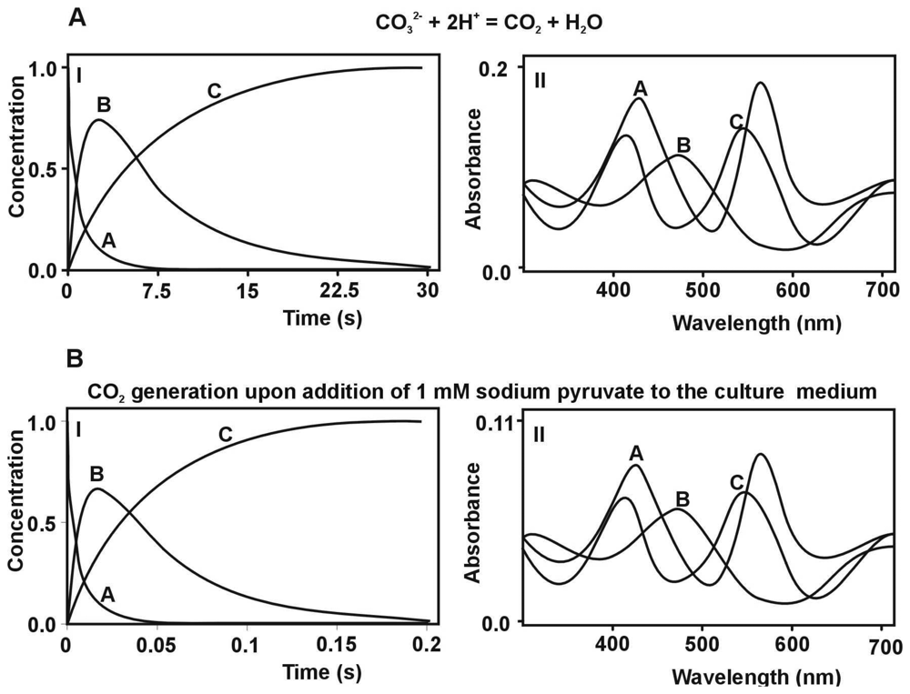
3. Materials and Methods

3.1. Reagents

Dihydrochloride pyridoxamine was purchased from Sigma. The cis form of diaquapyroxamino-oxalatochromate(III) was prepared according to standard literature procedures [25]. The final products, cis-[Cr(C2O4)(L-L)(O2CO)]- (where L-L denotes bidentate ligand – pyridoxamine (pm)) was synthesised by a modification of the method described in [38]. An aqueous solution of K2Cr2O7 (4.0 g, 8 mL) was gradually added to H2C2O4.2H2O solution (12 g, 17 mL). The precipitated crystals of trans-K[Cr(C2O4)2(OH2)2].3H2O were filtered off and flushed with ice-cold water and ethanol. Next, the solution of trans-K[Cr(C2O4)2(OH2)2].3H2O (1.96 g, 40 mL) was heated for 15 minutes (70°C – 75°C), after which its pH was adjusted to around 9. A stoichiometric quantity of pyridoxamine (5 mmol, 10 mL, pH ≈ 9) was added to this solution and then the mixture was stirred for 15 minutes, cooled and acidified with 0.5 M HClO4 to pH ≈ 2. The complex anion was separated by ion-exchange column chromatography (DOWEX 1 × 8 anionite). Fe(NO3)3 (0.2 M, 25 mL) and HNO3 (2 M, 15 mL) were added to the cis-[Cr(C2O4)2(pm)]- ion solution (180 mL). After being heated for 20 minutes (318 K) the solution was left to cool. The post-reaction mixture was gradient-eluted in a chromatographic column. Next, the pH of the solution of cis-[Cr(C2O4)(pm)(OH2)2]+ (1 g, 10 mL) was adjusted to 8.5 by the portion-wise addition of aq. K2CO3 (32 mg, 10 mL). The solution was stirred for 10 minutes, then was cooled to 0°C. The composition of the product, blue crystals of cis-K[Cr(C2O4)(pm)(O2CO)], was determined by elemental analysis. The results of analytical calculations for KC11H12CrN2O9: C, 32.43; H, 2.95; N, 6.88 were in good agreement with those obtained from the elemental analysis: C, 32.44.; H, 3.00; N, 6.87. Using analytical techniques the molar ratios for components of the complex cis-[Cr(C2O4)(pm)(O2CO)]- ion were obtained: Cr(III) : C2O42- : pm : CO32- = 1 : 1 : 1 : 1. The complex ion, cis-[Cr(C2O4)(pm)(O2CO)]-, was decomposed into its components in the presence of the Cr(II)(aq) ion in an argon atmosphere [39]. Chromium(III) and pyridoxamine were quantitatively characterised spectrophotometrically [40]. To identify CO32- anion the potentiometry titration method was used. A standard solution of HCl (0.102 M) in the presence of 1% aq. methyl orange [41] was used in each titration.

3.2. Cell culture

The osteosarcoma 143B cell line (ATCC-8303) was cultured at 37°C in a humidified atmosphere with 5% CO2 in Dulbecco's Modified Eagle's Medium supplemented with 10% heat-inactivated fetal bovine serum and penicillin (100 μg/ml) / streptomycin (100 μg/ml) without sodium pyruvate (Sigma Chemicals Co., St. Louis, MO, U.S.A).

3.3. Cell treatment

The cells passed by standard methods of trypsinization using 0.25% trypsin and 0.02% EDTA solution, cultured under conditions described above for 24 hours before replacing with experimental medium containing different concentrations of sodium pyruvate (0.5 mM, 1 mM and 5 mM). After 30 minutes of preincubation 1 mM H2O2 (Sigma Chemicals, U.S.A) was added. Cells were incubated for next 24 hours before the level of CO2 in the medium, as well as cell viability were measured.

3.4. Preincubation of medium with catalase

To exclude possible involvement of culture medium as a source of H2O2 which may cause decarboxylation of pyruvate 1 unit/ml of catalase (Sigma Chemicals, U.S.A) was added to the medium for 30 minutes before addition of sodium puryvate. Then, after 24 hours of incubation the CO2 was measured.

3.5. Cell viability: MTT assay

The cytoprotective properties of pyruvate were determined using MTT assay. Briefly, the osteosarcoma 143B cells were seeded onto 96-well plates at the density of 4 × 10³ per well and cultured for 24 hours, then cells were treated with sodium pyruvate and H2O2. After 24 h 0,5 mg/ml of 3-[4,5-dimethylthiazol-2-yl]-2,5-diphenyltetrazolium bromide (MTT) was added. The plates were incubated for 4 h and supernatant was removed after centrifugation (700 xg for 10 min). Finally, 100 μL of DMSO (Sigma Chemicals, U.S.A) was added. The absorbance was recorded using an Jupiter ELISA reader (ASYS Hitech) at 550 nm wavelength with references at wavelength of 630 nm and the cells survival was calculated. Percentage viability was defined as a 100% times the ratio of absorbance in the samples to the average absorbance in the control (untreated cells).

3.6. Kinetic measurements and simulation

Two buffer solutions were used: 0.2 M MES [2-(4-morpholino)ethanesulfonic acid] and 0.2 M TRIS [tris(hydroxymethyl)-aminomethane], both prepared by dissolving appropriate samples in MiliQ water. pH was measured by using a CX 731 pH-meter (reading accuracy of 0.01 pH unit) and a combined electrode manufactured by Hanna. Solutions of the studied complex were prepared by mixing 0.5 mL of the cis-[Cr(C2O4)(L-L)(OH2)2]+ (C = 10-3 M) with 2 mL of 0.2 M MES or TRIS and 2 mLof 2 M NaClO4 solutions. The reaction studied was investigated over the pH range 6.50 < pH < 9.12 and at T = 15° C. The cells were seeded onto 100-mm culture plates at the density of 1.106 per plate and incubated and treated as described before, than the culture medium was collected and used for CO2 concentration analysis by using stopped-flow spectrophotometry method.

3.7. Instrumentation

Spectral measurements were recorded in the UV-Vis region using a Perkin-Elmer Lambda 18 Instrument with the scan accuracy of 1 nm and 1 nm slit width at a scanning rate of 120.00 nm min-1. The pK1 and pK2 values in the ground state were computed by using Origin 6.0 program, based on absorbance variations at a selected wavelength and by using non-linear least squares method according to the equation described in [42]. Kinetic measurements were carried out using a stopped-flow technique and an Applied Photophysics SX-17MV spectrophotometer. The observable rate constants were computed using a “Glint” program based on global analysis [43-46].

3.8. Statistical Analysis

Data were computed using Statistica 7.1 program (Statsoft, Poland). Parametric and non-parametric distribution was assessed by Shapiro-Wilk test. The analysis was based on non-parametric statistic Wilcoxon test signed-rank as indicated by data distribution.

4. Conclusion

In this paper we described a new method of carbon dioxide detection in the physiological cell culture medium. Moreover, we have presented usefulness of our method, which is based on the interaction of CO2 with the coordination compound of cis-[Cr(C2O4)(pm)(OH2)2]+ ion for the CO2 detection in biological samples. Furthermore, the kinetic studies of carbon dioxide uptake by cis-[Cr(C2O4)(pm)(OH2)2]+ ion using the stopped-flow method enabled us to conclude that the reaction studied proceeded in two steps, namely carbon dioxide uptake (first step) and the closure of the ring of carbonate ion (the second step). The presented method above seems also to be handy tool to analyse scavenging reaction of H2O2 by pyruvate in biological samples. The measurement of CO2 concentration can be used not only to analyse chemical reaction but it can be also a marker of pyruvate's protection efficiency against oxidative stress.
Our results provided arguments for usefulness of pyruvate application for cell culture studies where culture media could produce artefectually significant levels of H2O2 before treatment of cells. On the other hand endogenous and exogenous source of H2O2 being implicated in cytotoxity in variety of human diseases can be safety prevented by pyruvate. The efficiency of this scavenger was clearly demonstrated by novel application of CO2 molecular biosensor.

Acknowledgments

This work was financially supported by Polish Ministry of Education and Science in years 2006-2009, under grants 2 P05A 062 29 and BW/8000-5-0448-8, BW/8000-5-0110-8 and Foundation for Polish Science.

References and Notes

  1. Friedman, J.; Peleg, E.; Kagan, T.; Shnizer, S.; Rosenthal, T. Oxidative stress in hypertensive, diabetic, and diabetic hypertensive rats. Am. J. Hyper. 2003, 16, 1049–1052. [Google Scholar]
  2. Ferrari, R.; Ceconi, C.; Curello, S.; Cargnoni, A.; Alfieri, O.; Pardini, A.; Marzollo, P.; Visioli, O. Oxygen free radicals and myocardial damage: protective role of thiol-containing agents. Am. J. Med. 1991, 91, 95–105. [Google Scholar]
  3. Reynolds, A.; Laurie, C.; Lee, C.; Mosley, R.; Gendelman, H.E. Oxidative stress and the pathogenesis of neurodegenerative disorders. Int. Rev. Neurobiol. 2007, 82, 297–325. [Google Scholar]
  4. Schleicher, E.; Friess, U. Oxidative stress, AGE, and atherosclerosis. Kidney Int. 2007, 106, 17–26. [Google Scholar]
  5. Standman, E.R.; Levine, R.L. Protein oxidation. Ann. NY Acad. Sci. 2000, 899, 191–208. [Google Scholar]
  6. Sultana, R.; Perluigi, M.; Butterfield, D.A. Protein oxidation and lipid peroxidation in brain of subjects with Alzheimer's disease: insights into mechanism of neurodegeneration from redox proteomics. Antioxid. Redox Signal. 2006, 8, 2021–2037. [Google Scholar]
  7. Dizdaroglu, M.; Jaruga, P.; Birincioglu, M.; Rodriguez, H. Free radical-induced damage to DNA: mechanisms and measurement. Free Radic. Biol. Med. 2002, 32, 1102–1115. [Google Scholar]
  8. Chance, B.; Sies, H.; Boveris, A. Hydroperoxide metabolism in mammalian organs . Physiol. Rev. 1979, 59, 527–605. [Google Scholar]
  9. Halliwell, B. Antioxidants in human health and disease. Ann. Rev. Nutr. 1996, 16, 33–50. [Google Scholar]
  10. Gutteridge, J.M.; Halliwell, B. Iron toxicity and oxygen radicals . Baillieres Clin. Haematol. 1989, 2, 195–256. [Google Scholar]
  11. Auerbach, J.M.; Segal, M. Peroxide modulation of slow onset potentiation in rat hippocampus. J. Neurosci. 1997, 17, 8695–8701. [Google Scholar]
  12. Makazan, Z.; Saini, H.K.; Dhalla, N.S. Role of oxidative stress in alterations of mitochondrial function in ischemic-reperfused hearts. Am. J. Physiol. Heart Circ. Physiol. 2007, 292, 1986–1994. [Google Scholar]
  13. Olanow, C.W. A radical hypothesis for neurodegeneration. Trends Neurosci. 1993, 16, 439–444. [Google Scholar]
  14. Cicalese, L.; Lee, K.; Schraut, W. Pyruvate prevents ischemia-reperfusion mucosal injury of rat small intestine. Am. J. Surg. 1999, 171, 97–100. [Google Scholar]
  15. Boer, L.W.; Bekx, P.A.; Han, L.; Steinke, L. Pyruvate enhances recovery of rat hearts after ischemia and reperfusion by preventing free radical generation. Am. J. Physiol. Heart Circ. Physiol. 1993, 265, 1571–1576. [Google Scholar]
  16. Crestanello, J.A.; Lingle, D.M.; Millili, J.; Whitman, G.J. Pyruvate improves myocardial tolerance to reperfusion injury by acting as an antioxidant: a chemiluminescence study. Surgery 1998, 124, 92–99. [Google Scholar]
  17. O'Donnell-Tormey, J.; Nathan, C.F.; Lanks, K.; DeBoer, C.J.; de la Harpe, J. Secretion of pyruvate. An antioxidant defense of mammalian cells. J. Exp. Med. 1987, 165, 500–514. [Google Scholar]
  18. Mallet, R.T.; Sun, J. Antioxidant properties of myocardial fuels. Mol. Cell Biochem. 2003, 253, 103–111. [Google Scholar]
  19. Constantopoulos, G.; Barranger, J.A. Nonenzymatic decarboxylation of pyruvate. Anal. Biochem. 1984, 139, 353–358. [Google Scholar]
  20. Tabatabaie, T.; Potts, J.D.; Floyd, R.A. Reactive Oxygen Species-Mediated Inactivation of Pyruvate Dehydrogenase. Arch. Biochem. Biophy. 1996, 32, 336–339. [Google Scholar]
  21. Bogaret, Y.E.; Rosenthal, R.E.; Fiskum, G. Postischemic Inhibition of Cerebral Cortex Pyruvate Dehydrogenase. Free Rad. Biol. Med. 1994, 16, 811–820. [Google Scholar]
  22. Kita, E.; Kachniarz, E. Model quasi-enzyme compounds of chromium(III) with vitamins B6 and histamine. Part VIII: Kinetics of acid catalyzed liberation of pyridoxylideneglycine (pl-gly) from [Cr(C2O4)2(pl-gly)]- ion. Pol. J. Chem. 1991, 65, 1925–1931. [Google Scholar]
  23. Hussain, I.; Kita, E.; Kita, P. Studies on base hydrolysis mechanism of chromium(III) complexes. Part X. Preparation and kinetics of aquation of [Cr(NCS)4(histamine)]- ion in aqueous acidic and alkaline solutions. Pol. J. Chem. 1991, 65, 2111– 2119. [Google Scholar]
  24. Kita, E. Model quasi-enzyme compounds of chromium(III) with vitamins B6 and histamine. Part IX: Kinetics and products of Fe(III)-promoted aquation of [Cr(C2O4)2(pyridoxamine)]- anion. Pol. J. Chem. 1991, 65, 1933–1940. [Google Scholar]
  25. Kita, E. Model quasi-enzyme compounds of chromium(III) with vitamins B6 and histamine. Part IV: Kinetics of Fe3+ (aq) promoted aquation of [Cr(C2O4)2(histamine)]- anion. Pol. J. Chem. 1987, 61, 361–367. [Google Scholar]
  26. Jacewicz, D.; Dąbrowska, A.; Banecki, B.; Kolisz, I.; Woźniak, M.; Chmurzyński, L. A stopped-flow study on the kinetics and mechanism of CO2 uptake by chromium(III) complexes with histamine and pyridoxamine. Trans. Met. Chem. 2005, 30, 209–216. [Google Scholar]
  27. Forster, J.; Edsall, A.; Otis, K.J. CO2 Biochemical Aspects, Roughton, ed.; Washington: D.C., 1969; pp. 487–496. [Google Scholar]
  28. Dąbrowska, A.; Jacewicz, D.; Łapińska, A.; Banecki, B.; Figarski, A.; Szkatuła, M.; Lehman, J.; Krajewski, J.; Kubasik-Juraniec, J.; Woźniak, M.; Chmurzyński, L. Pivotal participation of nitrogen dioxide in L-arginine induced acute necrotizing pancreatitis; protective role of superoxide scavenger 4-OH TEMPO. Biochem. Biophys. Res. Comm. 2005, 326, 313–32. [Google Scholar]
  29. Desagher, S.; Glowinski, J.; Prémont, J. Pyruvate protects neurons against hydrogen peroxide-induced toxicity. J Neurosci. 1997, 17, 9060–9067. [Google Scholar]
  30. Lee, Y.J.; Kang, I.J.; Bünger, R.; Kang, Y.H. Enhanced survival effect of pyruvate correlates MAPK and NF-kappaB activation in hydrogen peroxide-treated human endothelial cells. J. Appl. Physiol. 2004, 2, 793–801. [Google Scholar]
  31. Lee, Y.J.; Kang, I.J.; Bünger, R.; Kang, Y.H. Mechanisms of pyruvate inhibition of oxidant-induced apoptosis in human endothelial cells. Microvasc. Res. 2003, 2, 91–101. [Google Scholar]
  32. Owen, O.E.; Mozzoli, M.A.; Boden, G.; Patel, M.S.; Reichard, G.A., Jr; Trapp, V.; Shuman, C.R.; Felig, P. Substrate, hormone, and temperature responses in males and females to a common breakfast. Metabolism 1980, 29, 511–523. [Google Scholar]
  33. Meierhenrich, R.; Jedicke, H.; Voigt, A.; Lange, H. The effect of erythropoietin on lactate, pyruvate and excess lactate under physical exercise in dialysis patients. Clin. Nephrol. 1996, 45, 90–97. [Google Scholar]
  34. O′ Donnell-Tormey, J.; Nathan, C.F.; Lanks, K.; DeBoer, C.J.; de la Harpe, J. Secretion of pyruvate. An antioxidant defense of mammalian cells. J. Exp. Med. 1987, 165, 500–514. [Google Scholar]
  35. Wasilenko, W.J.; Marchok, A.C. Pyruvate regulation of growth and differentiation in primary cultures of rat tracheal epithelial cells. Exp. Cell. Res. 1984, 155, 507–17. [Google Scholar]
  36. Reed, L.J. A trail of research from lipoic acid to á-keto acid dehydrogenase complexes. J. Biol. Chem. 2001, 276, 38329–38336. [Google Scholar]
  37. Dijkstra, U.; Gabreëls, F.; Joosten, E.; Wevers, R.; Lamers, K.; Doesburg, W.; Renier, W. Friedreich's ataxia: intravenous pyruvate load to demonstrate a defect in pyruvate metabolism. Neurology 1984, 34, 1493–1497. [Google Scholar]
  38. van Eldik, R.; Palmer, D.A.; Harris, G.M. Kinetics and Mechanism of Aquation and Formation Reactions of Carbonato Complexes. 18. Carbon Dioxide Uptake by and Decarboxylation of the trans-Bis(ethylenediamine)rhodium(III) System in Aqueous Solution. Inorg. Chem. 1980, 19, 3673–3679. [Google Scholar]
  39. de Castro, B.; Freire, C. Spectroscopic and Magnetic Properties of Low Spin Chromium(II) Tris Complexes with the Bidentate -Diimine Ligands: 2,2′-Pyridylbenzimidazole, 2,2′-Pyridylimidazole and 2,2′-Pyridylimidazoline. Synth. React. Inorg. Met. Org. Nano-Met. Chem. 1990, 20, 1–12. [Google Scholar]
  40. Haupt, G.W. An alkaline solution of potassium chromate as a transmittancy standard in the ultraviolet. J. Research Nat. Bur. Standards 1952, 48, 414–416. [Google Scholar]
  41. Beetz, I. Microanalytisches Laboratorium; Kronach, 1989; pp. 33–39. [Google Scholar]
  42. Szabelski, M.; Guzow, K.; Rzeska, A.; Malicka, J.; Przyborowska, M.; Wiczk, W. Acidity of carboxyl group of tyrosine and its analogues and derivatives studies by steady-state fluorescence spectroscopy. J. Photochem. Photobiol. A: Chemistry 2002, 152, 73–77. [Google Scholar]
  43. Johanson, M.L.; Correira, J.J.; Yphantis, D.A.; Halvorson, H.R. Analysis of data from the analytical ultracentrifuge by nonlinear least- squares techniques. Biophys. J. 1981, 36, 575–588. [Google Scholar]
  44. Nagel, J.F.; Parodi, L.A.; Lozier, R.H. Procedure for testing kinetic models of the photocycle of bacteriorhodopsin. Biophys. J. 1982, 38, 161–174. [Google Scholar]
  45. Knutson, J.R.; Beechem, J.M.; Brand, L. Simultaneous analysis of multiple fluorescence decay curves: a global approach. Chem. Phys. Lett. 1983, 102, 501–507. [Google Scholar]
  46. Maeder, M.; Zuberbuchler, A. Nonlinear Least- Squares Fitting of Multivariate Absorption Data. Anal. Chem. 1990, 64, 2220–2224. [Google Scholar]
Figure 1. (A) Structure of the synthesized coordination compound of Cr(III); (B) Tautomers of pyridoxamine.
Sensors 08 04487f1
Figure 2. Dependence rate constants (k1obs) from [H+] for the carbon dioxide uptake by the cis-[Cr(C2O4)(pm)(OH2)2]+ ion in the T=15°C.
Sensors 08 04487f2
Figure 3. Dependence rate constants (k2obs) from [H+] for the ring closure by the cis-[Cr(C2O4)(pm)(OH2)2]+ ion in the T=15°C.
Sensors 08 04487f3
Figure 4. The comparison of kinetic and spectral characteristics of reactants in simple inorganic CO2 generating system. (A) (I) Curves of concentration decay and buildup of the substrate A (which is the cis-[Cr(C2O4)(pm)(OH2)2]+ ion), product C as cis-[Cr(C2O4)(pm)(O2CO)]- ion, and intermediate product B. (II) Absorption spectra of the reactants A, B and C in pH = 7.13, [CO2] = 0.01M, T=20°C. (B) (I) Curves of concentration changes for reaction of CO2 uptake observed for the substrate A (which is the cis-[Cr(C2O4)(pm)(OH2)2]+ ion), product C as cis-[Cr(C2O4)(pm)(O2CO)]- ion, and intermediate product B. (II) Absorption spectra of the reactants A, B and C in pH = 6.5, [CO2] = 8.23M T=20°C.
Sensors 08 04487f4
Figure 5. Plots of rate expression (eq. 3) for different concentration carbon dioxide uptake by cis-[Cr(C2O4)(pm)(H2O)2]+.
Sensors 08 04487f5
Figure 6. (A) Comparison of antioxidant protective effects of three concentration of pyruvate (0.5 mM, 1 mM and 5 mM) on viability of Osteosarcoma 143B cells exposed to 1 mM H2O2. The strongest protection was observed after 30 min of preincubation cells with 5 mM sodium pyruvate. The cytoprotective effect of 1 mM concentration was only partial. The lowest 0.5 mM sodium pyruvate was unable to protect against cell injury caused by H2O2. The results were analyzed by the Wilcoxon signed-rank test. The results are presented as median, 5th percentile/95th percentile. (*) P<0.05 were considered statistically significant when compared to the cell viability upon addition 1 mM H2O2 alone;(B) CO2 assessment in cell culture media upon addition of pyruvate and H2O2 The concentration was measured using novel stopped-flow method and specific CO2 detector - cis-[Cr(C2O4)(pm)(OH2)2]+. The results were analyzed by the Wilcoxon signed-rank test. The data are presented as median, 5th percentile/95th percentile. (*) P<0.05 were considered statistically significant when compared with respective control;(C) Pyruvate scavenging of endogenously produced H2O2 in cell media. The effect of catalase addition. The results of CO2 concentration upon addition pyruvate to the medium were compared with respective data obtained after preincubation with 1U/ml of catalase. The results were analyzed by the Wilcoxon signed-rank test. The data are presented as median, 5th percentile/95th percentile. (*) P<0.05 were considered statistically significant.
Sensors 08 04487f6
Table 1. Rate parameters for the carbon dioxide uptake reaction by cis-[Cr(C2O4)(pm)(OH2)2]+ Ion in T=20°C.
CO2 [M]k1 [ms-1 mM-1]k2 [s-1 M-1]
CO2 was generated control sample

1.085.61E-17.88E-1
1.875.68E-17.89E-1
2.135.7E-17.91E-1
3.335.94E-18.07E-1
4.126.03E-18.13E-1

CO2 was generated upon addition of 1 mM H2O2 to the culture medium

1.025.61E-17.82E-1
1.875.68E-17.89E-1
2.885.88E-18.03E-1
3.435.95E-18.08E-1

CO2 was generated upon addition of 0.5 mM sodium pyruvate to the culture medium

2.655.81E-17.95E-1
2.775.87E-17.98E-1
3.095.92E-18.06E-1
3.385.94E-18.07E-1
3.655.96E-18.09E-1

CO2 was generated upon addition of 1 mM sodium pyruvate to the culture medium

2.995.91E-18.05E-1
3.845.98E-18.10E-1
3.986.01E-18.11E-1
4.126.03E-18.13E-1
4.346.05E-18.15E-1
CO2 [M]k1 [ms-1 mM-1]k2 [ms-1 mM-1]

CO2 was generated upon addition of 5 mM sodium pyruvate to the culture medium

6.096.07E-18.18E-1
6.666.12E-18.21E-1
6.946.14E-18.23E-1
7.126.17E-18.25E-1
8.236.28E-18.29E-1
9.236.35E-18.55E-1
9.986.41E-18.63E-1

CO2 was generated upon addition of 0.5 mM pyruvate and 1 mM H2O2

8.076.23E-18.27E-1
8.396.29E-18.32E-1
8.606.31E-18.47E-1
11.26.53E-18.91E-1
13.27.22E-19.31E-1

CO2 was generated upon addition of 1 mM pyruvate and 1 mM H2O2

32.38.73E-11.09
33.69.02E-11.11
44.31.011.26
52.31.091.31

CO2 was generated upon addition of 5 mM pyruvate and 1 mM H2O2

84.61.411.67
88.91.431.68
89.11.441.71
91.21.481.74
98.21.541.81
The error in k1[ms-1mM-1] and k2[ms-1mM-1] oscillates from 0.8% to 2.8%.

