Article

Coronatine Gene Expression In Vitro and In Planta, and Protein Accumulation During Temperature Downshift in Pseudomonas syringae

by
Yvonne Braun
1,2,*,†,
Angela V. Smirnova
1,†,
Helge Weingart
1,
Alexander Schenk
1 and
Matthias S. Ullrich
1
1
Jacobs University Bremen, School of Engineering and Science, Campus Ring 1, 28759 Bremen, Germany
2
National Institute for Medical Research, The Ridgeway, London NW7 1AA, UK
*
Author to whom correspondence should be addressed.
Both authors contributed equally to the work
Sensors 2009, 9(6), 4272-4285; https://doi.org/10.3390/s90604272
Submission received: 1 April 2009 / Revised: 22 May 2009 / Accepted: 26 May 2009 / Published: 3 June 2009
(This article belongs to the Section Biosensors)

Abstract

:
The plant pathogenic bacterium Pseudomonas syringae PG4180 synthesizes high levels of the phytotoxin coronatine (COR) at the virulence-promoting temperature of 18 °C, but negligible amounts at 28 °C. Temperature-dependent COR gene expression is regulated by a modified two-component system, consisting of a response regulator, CorR, the histidine protein kinase CorS, and a third component, termed CorP. We analyzed at transcriptional and translational levels the expression of corS and the cma operon involved in COR biosynthesis after a temperature downshift from 28 to 18 °C. Expression of cma was induced within 20 min and increased steadily whereas corS expression was only slightly temperature-dependent. Accumulation of CmaB correlated with accumulation of cma mRNA. However, cma transcription was suppressed by inhibition of de novo protein biosynthesis. A transcriptional fusion of the cma promoter to a promoterless egfp gene was used to monitor the cma expression in vitro and in planta. A steady induction of cma::egfp by temperature downshift was observed in both environments. The results indicate that PG4180 responds to a temperature decrease with COR gene expression. However, COR gene expression and protein biosynthesis increased steadily, possibly reflecting adaptation to long-term rather than rapid temperature changes.

1. Introduction

In order to survive, bacteria constantly have to adapt to changing environmental conditions. Environmental stimuli can serve pathogenic bacteria as signals for expression of virulence factors, for example, temperature often serves as a trigger for the induction of virulence gene expression. While temperatures of warm-blooded hosts (37-41 °C) are important signals for the induction of virulence gene expression in humans and animal pathogens [1], plant pathogens generally express their virulence genes in response to lower temperatures [2-4]. Further, the later organisms adjust their virulence gene expression in response to diurnal and seasonal temperature fluctuations.
COR is a non-host specific phytotoxin, which is an important virulence factor of several pathovars of Pseudomonas syringae, precisely pv. alisalensis, atropurpurea, glycinea, maculicola, morsprunorum, porri, and tomato. Biosynthesis of COR is positively regulated by low temperature in P. syringae pv. glycinea PG4180 [4, 5]. PG4180 synthesizes COR in a temperature-dependent manner, with a maximum at 18 °C [5]. At 28 °C, the optimal growth temperature of P. syringae, COR biosynthesis is negligible. COR consists of the polyketide coronafacic acid (CFA) and the cyclised amino acid coronamic acid (CMA), an isoleucin derivative. CFA and CMA are linked by the formation of an amide bond. The genes required for CFA and CMA biosynthesis are organized as two biosynthetic operons, cfl/CFA and CMA, respectively [6,7]. Nucleotide sequence analysis of the CMA biosynthetic genes has suggested that this precursor is synthesized by a mechanism similar to the one used in the biosynthesis of non-ribosomal peptides [7-9]. In the CMA cluster seven genes have been identified. Six of them, cmaABCDET, encode the enzymes for biosynthesis of CMA from the progenitor L-allo-isoleucine [10]. Until today no function has been assigned to the seventh gene, cmaU. CFA is a polyketide and its synthesis resembles the biosynthesis of polyketides in Streptomyces and Bacillus spp. [11]. The CFA biosynthetic cluster contains nine genes. Two encode type I polyketide synthase proteins (cfa6 and cfa7), the genes cfa1, cfa2, and cfa3 encode proteins similar to the acyl carrier protein, dehydratase, and beta-ketoacyl:acyl carrier protein synthase of type II fatty acid synthases and polyketide synthases [12].
One means to sense and respond to environmental stimuli are two-component regulatory systems (TCSs). A modified TCS consisting of a histidine protein kinase (HPK), CorS, a response regulator (RR), CorR, and a third component, CorP, regulates COR production at the transcriptional level in P. syringae [5]. CorP showed high similarity to CorR but lacks a DNA binding domain characteristic for RRs. Mutational analysis demonstrated that CorP is necessary for induction of COR biosynthesis [5], but its exact function remained to be determined. The HPK, CorS, is believed to respond to a temperature change via autophosphorylation of a conserved histidine residue, and transduces the signal to the cognate RR CorR via phosphorylation of its conserved aspartate residue [13]. In vitro results indicated that CorR was able to bind to the CMA and CFA biosynthetic promoter regions in a temperature- and corS-dependent manner [14,15]. Additionally, the alternative sigma factor RpoN (σ54) which is required for the expression of a variety of virulence determinants and metabolic functions was shown to be essential for COR biosynthesis in P. syringae [16]. Consequently, COR gene expression seems to be regulated by its specific TCS and by at least one global regulator.
In natural settings PG4180 encounters temperature fluctuations and adapts COR gene expression accordingly. In this study, we investigated effects of a temperature shift from 28 to 18 °C on transcription of CMA biosynthetic genes in vitro as well as in planta and on CmaB protein biosynthesis. We also evaluated transcription of the regulatory gene corS after the temperature shift and studied whether de novo protein biosynthesis is required for transcriptional activation of COR biosynthetic genes. Taking into account that stability of mRNA contributes to mRNA levels and that mRNA stability is also influenced by temperature, we investigated the stability of the cma transcript at 18 and 28 °C after inhibition of transcriptional initiation. The HPK CorS is a membrane-associated protein, which possesses a hydrophobic N-terminus comprising six transmembrane domains (TMDs) [17]. HPKs of this structure are generally believed to sense environmental stimuli by means of their periplasmic loops. In some sensory proteins periplasmic domains for substrate binding were identified, e.g. the nitrate binding Nit domain of the sensor histidine kinases NarX and NarQ of E. coli [18]. Another example is the citrate binding CitAP domain in the CitA sensor kinase of Klebsiella pneumoniae [19]. In the case of CorS, a temperature decrease results in COR biosynthesis. Whether the temperature change on its own or an additional signal activates the regulatory cascade remains to be elucidated.

2. Materials and Methods

2.1. Bacterial strains, plasmids, and growth conditions

P. syringae pv. glycinea PG4180 [20] was maintained at 28 °C on MG agar plates [21]. For liquid cultures bacteria were grown at 18 and 28 °C with constant shaking at 280 rpm in HSC medium [4]. For comparison of the cma expression in vitro and in planta, PG4180 carrying plasmid pHW01 was used [22]. Plasmid pHW01 contains a transcriptional fusion of a promoterless enhanced green fluorescent protein (egfp) gene to the cma promoter region of PG4180 in the broad-host range vector pBBR1MCS [23]. The size of the cloned cma promoter region is 2.9 kb located at positions -717 to +2,135 with respect to the cmaA transcriptional start site [7].

2.2. Plant material and inoculation procedures

Soybean plants (Glycine max (L.) Merr. cv. Maple Arrow) were grown in a greenhouse at 22 to 25 °C, 60% humidity, with supplementary light for a 14-h photoperiod (350 μE m-2 s-1). To study expression of the cma promoter in planta, P. syringae PG4180 carrying plasmid pHW01 was infiltrated into soybean leaves using a needleless syringe at an OD600 of 0·05 (approx. 5 × 10⁷ CFU per mL). Following infiltration, plants were transferred to growth chambers at 18 or 28 °C. To recover bacteria after inoculation, 60 discs (7 mm diameter) containing infected leaf tissue were excised using a cork borer and macerated in 4 mL of isotonic NaCl. All greenhouse experiments were repeated at least three times to confirm reproducibility.

2.3. Confocal laser scanning microscopy

Fluorescence of bacterial cells was detected with a confocal laser scanning microscope, DMR XE, type TCS NT (Leica) using a PL-Fluotar objective (× 63; 1.32; numerical aperture, oil immersion). Images were analyzed with Leica software package TCS NT, version 1.5.451.

2.4. RNA isolation and spot blot analysis

Bacteria were cultured in HSC medium at 18 and 28 °C until OD600 of 1·3 or until selected time points after temperature shift or addition of antibiotics. 15 mL of bacterial culture were mixed with an equal volume of chilled killing buffer (20 mM Tris-HCl [pH 7.5], 20 mM NaN3) and subsequently centrifuged at 4 °C for 15 min at 4,000 rpm.
Total RNA was isolated from 5 mL of bacterial cells by acid phenol/chloroform extraction as described by Schenk et al. [24]. Extracted RNA was analysed using an Agilent 2,100 Bioanalyzer. The yield of RNA was determined spectrophotometrically at 260 nm using an Ultrospec 2,100 pro UV/Visible Spectrophotometer (Amersham). Aliquots of total RNA (200 ng per dot) were transferred to positively charged nylon membranes (Pall) using the Minifold® I Spot-Blot System (Schleicher & Schuell BioScience) according to the manufacturer's recommendations. Successful transfer of the RNA was verified by reversible staining of the membrane with methylene blue [25]. The digoxygenin-labelled specific RNA probes were synthesized by in vitro transcription using T7 RNA polymerase and specific PCR products as templates. Synthesis of the templates by PCR was performed using the following pairs of oligonucleotides: for the cmaA probe, primers cmaA-fwd (5′-TTTGAGTCGGTCTGCACGCA-3′) and cmaA-revT7 (5′-TAATACGACTCACTATAGGGAGGGCTGTACGTTGTCTACTAG-3′), for corS, corS-fwd (5′-AATACGGCGCGCTGTCAGTT-3′) and corS-revT7 (5′-TAATACGACTCACTATAGGGAGGAATGGAT GGCCTAATAGGCG-3′) for ssb, ssb-fwd (5′-ATGGCCCGTGGGGTTAACAAAGTC-3′) and ssb-revT7 (5′-TAATACGACTACTATAGGGAGGTGTCAAAGTCAGCAGCAGGC-3′). For the synthesis of DIG-labelled RNA probes, the Strip-EZ™ RNA Probe Synthesis & Removal kit (Ambion) and digoxygenin-11-UTP (Roche) were used. Before hybridization with a probe, RNA was cross-linked with the membrane under a UV-light using a CL-1000 Crosslinker (UVP). After 1 h of pre-hybridization at 68 °C the probe was hybridized to the membrane by incubation in hybridization solution (50% (v/v) formamide, 7% (w/v) SDS, 2% (w/v) blocking reagent, 0.1% (v/v) N-laurylsarcosine, 5 × SSC) at 68 °C for 12 – 16 h. After hybridization, the membrane was washed twice for 5 min at room temperature in 2 × SSC containing 0.1% (w/v) SDS, followed by two washes for 15 min at 68 °C in 0.2 × SSC containing 0.1% (w/v) SDS. Hybridization signals were detected by incubation with anti-digoxygenin-AP Fab fragments (Roche) and a fluorescence substrate for alkaline phosphatase (ECF) (Amersham-Pharmacia Biotech) using a FLA-3000 phosphoimager (Raytest, Strabenhardt, Germany). Signals were quantified using the AIDA Image Analyzer software package (Raytest). The specificity of the RNA probes was evaluated by Northern Blot analysis (data not shown).

2.5. Immunodetection of CmaB accumulation

Total protein extracts were isolated from cell pellets of bacterial cultures (1.5 mL) and were quantified by the Bradford assay [26]. Equal amounts of protein (30 μg per lane) were separated by 10%-SDS-PAGE on two gels running in the same chamber. Subsequently, proteins from one gel were electrotransferred to a Hybond-C nitrocellulose membrane (Amersham-Pharmacia Biotech). The second gel was stained with GelCode Blue Stain Reagent (Perbio Science). Polyclonal mouse antiserum raised against CmaB (dilution 1:1,000) was used to monitor the accumulation of CmaB after a temperature shift from 28 to 18 °C. Secondary anti-mouse IgG antibodies conjugated to alkaline phosphatase (Sigma) at 1:10,000 dilution, along with ECF (Amersham-Pharmacia Biotech), a fluorescence substrate for alkaline phosphatase, were used for detection of signals. Fluorescence was detected using a FLA-3000 phosphoimager (Raytest). Signals were quantified using the AIDA Image Analyzer software package (Raytest).

3. Results and Discussion

The plant pathogen P. syringae PG4180 often encounters temperature fluctuations during its epiphytic growth on plant surfaces. Besides diurnal changes, temperature decreases are often associated with rainy or humid weather conditions, which favour bacterial infection of the host plant (soybean) and affect the subsequent disease development [27,28]. In PG4180, a low-temperature stimulus is known to be sensed and to cause coordinated COR gene expression via the CorRSP regulatory system [5]. However, important questions that remained to be addressed were how the temperature stimulus is transmitted to COR gene expression and how rapidly the COR gene expression is induced in vitro as well as in planta when temperature changes. In this study, a significant induction of the cma biosynthetic operon and an increased synthesis of CmaB following a temperature downshift from 28 to 18 °C were demonstrated.

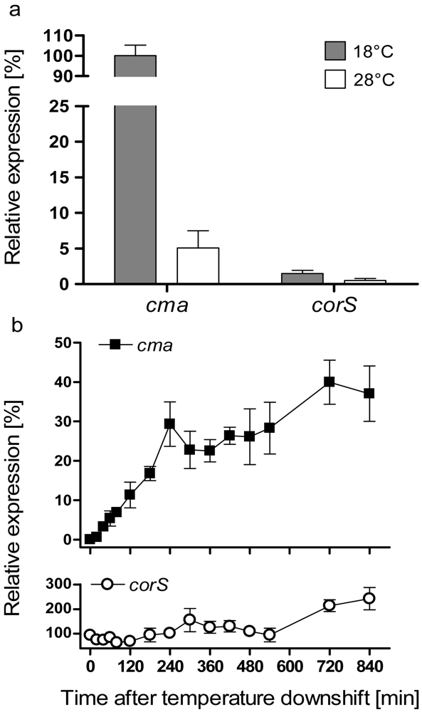
3.1. Analysis of cma and corS mRNA levels upon a temperature shift from 28 to 18 °C

In order to investigate how rapidly the induction of transcription of COR biosynthetic genes occurs after a temperature change, levels of cma and corS mRNA after a temperature shift from 28 to 18 °C were examined by RNA spot blot analysis. PG4180 cultures were grown in HSC medium at 28 °C to an OD600 of 0·5. Subsequently, one set of aliquots was shifted to 18 °C. Cultures continuously kept at 28 °C were used as a control to define the minimal level of expression. Additionally, cultures grown continuously at 18 °C were used as a control to define the maximal level of expression. Determination of the minimal and maximal levels, respectively, showed that the level of cma transcript was 20-fold higher at 18 than at 28 °C (Figure 1a).
However, temperature induction of the corS transcript was only 4-fold (Figure 1a) indicating that corS expression was only moderately affected by temperature in comparison to cma. Upon downshift in temperature, a slight increase of mRNA levels for the cma transcript occurred within 20 min (Figure 1b). Subsequently, the cma transcript accumulated gradually and reached a maximum concentration after 12 hours of growth at 18 °C. During the following hours cma mRNA remained at this level with minor fluctuations. 24 hours after the downshift the expression of cma had not reached levels measured in cultures constantly grown at 18 °C. The levels of mRNA in the 28 °C cultures remained at basal level (data not shown). The level of corS mRNA increased transiently upon temperature-downshift. 14 h after the temperature downshift a 2.5-fold increase of this transcript was determined and remained constant for the analyzed period of 24 h. Thus, transcription of the regulatory gene corS was not markedly affected by temperature. This is not surprising since regulatory genes such as corS need to be transcribed constitutively to allow their gene products to quickly react to environmental changes.
It had previously been reported that low temperature induces synthesis of cold-shock proteins [29] and desaturases in Bacillus and Synechocystis species, respectively [30,31]. Expression studies for the des gene encoding Δ5 desaturase of B. subtilis demonstrated a transient increase in des mRNA levels in response to a sudden change in temperature. The levels of the des transcript reached a maximum within 60 min and subsequently decreased after continuous growth at low temperature [30]. The synthesis of cold-shock proteins is also transiently induced and then re-adjusted to new steady-state levels within 4 h of a temperature downshift [32]. In contrast, the cma mRNA of PG4180 accumulated steadily and gradually after the shift from 28 to 18 °C over a period of 24 h (Figure 1b, data shown up to 14 h) indicating that it does not resemble a typical cold shock response.
To examine whether or not the induction of the cma transcription in PG4180 cultures shifted from 28 to 18 °C required de novo protein synthesis, levels of cma mRNA were measured after addition of the protein biosynthesis inhibitor, chloramphenicol (100 μg mL-1). After addition of chloramphenicol the level of cma mRNA decreased in the shifted culture (Figure 2). In parallel, samples were also probed for the ssb transcript encoding single-strand binding protein. The levels of ssb mRNA were not affected by addition of chloramphenicol. Thus, inhibition of de novo protein synthesis suppressed transcription of cma but not of ssb.
To determine whether or not a change in mRNA stability contributed to the temperature-induced increase in mRNA levels, the decay of the cma mRNA was monitored in cultures of PG4180 grown at 18 °C or shifted from 18 to 28 °C after addition of rifampicin. This antibiotic inhibits initiation of transcription. Rifampicin was added at 50 μg mL-1 to cultures of PG4180 at OD600 of 1·0. As seen from Figure 3, the cmaA transcript was degraded faster at 28 as compared to 18 °C.
The half-life of cma mRNA was 9.9 min at 18 °C and 7.3 min at 28 °C as calculated by linear regression. This result indicated that the stability of the cma transcript was rather moderately affected by a temperature change. Transcripts are generally more stable at lower temperatures. The slightly higher stability of the cma transcript at 18 compared to 28 °C seems to be due to normal slower degradation. Therefore, the increase in cma transcript at 18°C is rather a effect of accelerated transcription than of increased stability.

3.2. Analysis of CmaB protein levels following a temperature shift from 28 to 18 °C

The genes in the cma operon encode enzymes which resemble non-ribosomal peptide synthetases [7]. CmaB belongs to a new class of O2-dependent non-heme iron halogenases [36] that catalyzes a chlorination reaction of the CMA precursor [9]. To monitor CmaB production after the temperature downshift, 1.5 mL samples were drawn from PG4180 cultures as previously done for RNA spot blot experiments. Protein extracts were subjected to Western blot analysis using a CmaB polyclonal antiserum (Figure 4). In the first five hours after the temperature downshift, levels of CmaB increased gradually and reached 65% of the CmaB levels detected in cultures continuously grown at 18 °C (Figure 4). Generally, the accumulation of CmaB was consistent with the increase of cma mRNA levels. As controls, the level of CmaB was determined in cultures constantly grown at 28 °C at optical densities corresponding to the optical densities of the shifted culture. The amount of CmaB detected in this cultures remained always on a basal level (Figure 4). The highest level of CmaB, detected 5 h after the temperature downshift, was 9-fold higher in the shifted culture as compared to the 28 °C control. As shown in Figure 4, the CmaB antiserum also immuno-reacted non-specifically resulting in unspecific signals. However, intensities of the unspecific signals did not increase significantly, thus confirming a specific temperature-dependent accumulation of CmaB.
The steady accumulation of CmaB over the investigated period of 5 h together with the obtained mRNA accumulation (Figure 1) suggests that the effect of temperature downshift does not resemble a cold-shock response in P. syringae. These results were consistent with previously observed effects of temperature on other temperature-responsive genetic loci in PG4180 [33].

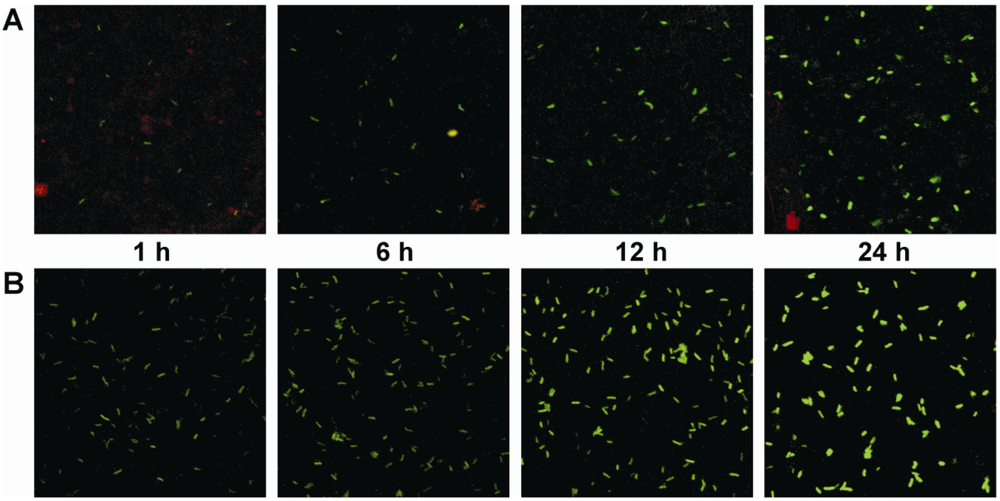
3.3. Analysis of cma promoter activity in planta following a temperature shift from 28 to 18 °C

In order to test whether temperature dependence of cma expression can also be seen in bacterial cells infecting host plants, the effects of a temperature downshift on in planta expression of cma were analyzed using cells of PG4180 (pHW01) isolated from soybean leaves. Plasmid pHW01 [22] allowed fluorescence-based monitoring of EGFP, a red-shifted variant of green fluorescent protein expressed as a transcriptional fusion of a promoterless egfp to the cma promoter region of PG4180. Soybean plants were inoculated with PG4180 (pHW01) grown at 28 °C, and subsequently kept in growth chambers at 18 °C. Bacteria were recovered from soybean leaves 1, 6, 12 and 24 hours after the inoculation. As in vitro control, a PG4180 (pHW01) culture grown in HSC medium at 28 °C was shifted to 18 °C at an OD600 of 0·5 and kept growing in HSC medium. Cells were harvested from the shifted culture after 1, 6, 12 and 24 hours of growth, resuspended in phosphate-buffered saline (pH 7.4) and adjusted to an OD600 of 0·5. EGFP fluorescence of bacteria grown in vitro and in planta was determined by confocal laser scanning microscopy. Fluorescence of cells constantly grown at 28 °C as control remained at a very low level throughout the entire growth of the bacteria in vitro and in planta [22].
After 1 h, fluorescence signals of the cells were barely detectable in planta (Figure 5a). A slight increase in the fluorescence intensity occurred after 6 h of growth in planta at 18 °C. Fluorescence signals were significantly enhanced after 12 h, and a maximal increase of cell fluorescence was detected after 24 hours of growth in planta at 18 °C. Similar enhancement of fluorescence was observed for cells grown in HSC medium after the temperature downshift (Figure 5b). These results clearly demonstrated that the temperature effects observed in vitro were also relevant for PG4180 cells grown under in planta conditions. However, the intensity of the transcriptional signal as visualized with egfp from 12 to 24 h in planta seemed to be less pronounced in vitro when mRNA was quantified. One potential reason for this could be the intrinsic stability of EGFP. It had previously been shown that GFP has a half-life of more than 1 day in vivo [34]. Thus, the protein may accumulate in bacterial cells and be present even after the respective promoter became less active. Alternatively, the stability of the mRNA might have been influenced by the transcriptional fusion. Moreover, the promoter probing construct includes the N-terminus of the amino acid coding region of CmaA, therefore it can be considered as a CmaA:EGFP protein fusion as well. This might also be responsible for the altered signal levels. Additionally, it cannot be ruled out that plant-derived signals may have contributed to the stronger induction of COR gene expression.

4. Conclusions

The plant pathogen P. syringae PG4180 produces COR, an important virulence factor, in a temperature-dependent manner with a maximum at 18 °C. Temperature fluctuations occur constantly on the plant surface. Thus, it is likely that the plant pathogen PG4180 is adapted to alter its gene expression in response to an average (low or high) daily temperature. A decrease in the average temperature favours a steady increase of COR gene expression. COR is a secondary metabolite and therefore not essential for bacterial survival at low temperatures [8]. By this adaptation to long-term temperature decreases the bacteria may presumably induce the energy-consuming COR biosynthesis only under conditions which favour the infection of the host plant. COR is synthesized from two precursor molecules, CFA and CMA. Genes for CFA and CMA biosynthesis are encoded in two distinct operons, separated by three regulatory genes, corR, corS, and corP. The present study demonstrated a significant increase in cma expression in response to a temperature downshift in vitro and in planta. Additionally, accumulation of CmaB protein was observed after temperature downshift. Taken together, the results demonstrate that COR gene expression in PG4180 was efficiently activated by temperature downshifts in vitro as well as in planta. On the contrary, expression of the regulatory gene corS was only moderately affected by temperature change. Budde et al. [8] previously reported that the cma transcript was not detected in a corR corS mutant of PG4180. We suggest that the induction of cma transcription was governed by a “switch” in the signal transduction pathway performed by CorRSP in response to the temperature downshift.
At low temperature CorS might undergo a conformational change to an active form in order to be efficiently autophosphorylated and to subsequently interact with its cognate RR, CorR, to allow initiation of transcription of COR biosynthetic genes [17,35]. Inhibition of de novo protein synthesis prior to the temperature downshift resulted in a subsequent decrease of cma transcription. This result supports the hypothesis that activation of cma transcription requires de novo synthesis and membrane incorporation of an altered form of CorS. Alternatively, suppressed synthesis of RpoN after inhibition of de novo biosynthesis might affect cma transcription indirectly via CorR or another σ54-dependent activator [16]. In both cases, COR gene expression seems to be dependent on de novo biosynthesis of regulatory proteins after a temperature downshift.

Acknowledgments

This work was supported by grants from the Deutsche Forschungsgemeinschaft and the Max-Planck-Gesellschaft. We are grateful to Carol Bender and Annette Wensing for stimulating discussions.

References

  1. Eriksson, S.; Hurme, R.; Rhen, M. Low-temperature sensors in bacteria. Phil. Trans. R. Soc. Lond. B 2002, 357, 887–893. [Google Scholar]
  2. Hugouvieux-Cotte-Pattat, N.; Dominguez, H.; Robert-Baudouy, J. Environmental conditions affect transcription of the pectinase genes of Erwinia chrysanthemi 3937. J. Bacteriol. 1992, 174, 7807–7818. [Google Scholar]
  3. Jin, S.; Song, Y.N.; Deng, W.Y.; Gordon, M.P.; Nester, E.W. The regulatory VirA protein of Agrobacterium tumefaciens does not function at elevated temperatures. J. Bacteriol. 1993, 175, 6830–6835. [Google Scholar]
  4. Palmer, D.A.; Bender, C.L. Effects of environmental and nutritional factors on production of the polyketide phytotoxin coronatine by Pseudomonas syringae pv. glycinea. Appl. Environ. Microbiol 1993, 59, 1619–1626. [Google Scholar]
  5. Ullrich, M.; Peñaloza-Vázquez, A.; Bailey, A.M.; Bender, C.L. A modified two-component regulatory system is involved in temperature-dependent biosynthesis of the Pseudomonas syringae phytotoxin coronatine. J. Bacteriol. 1995, 177, 6160–6169. [Google Scholar]
  6. Liyanage, H.; Palmer, D.A.; Ullrich, M.; Bender, C.L. Characterization and transcriptional analysis of the gene cluster for coronafacic acid, the polyketide component of the phytotoxin coronatine. Appl. Environ. Microbiol. 1995, 61, 3843–3848. [Google Scholar]
  7. Ullrich, M.; Bender, C.L. The biosynthetic gene cluster for coronamic acid, an ethylcyclopropyl amino acid, contains genes homologous to amino acid-activating enzymes and thioesterases. J. Bacteriol. 1994, 176, 7574–7586. [Google Scholar]
  8. Budde, I.P.; Rohde, B.H.; Bender, C.L.; Ullrich, M.S. Growth phase and temperature influence promoter activity, transcript abundance, and protein stability during biosynthesis of the Pseudomonas syringae phytotoxin coronatine. J. Bacteriol. 1998, 180, 1360–1367. [Google Scholar]
  9. Couch, R.; O'Connor, S.E.; Seidle, H.; Walsh, C.T.; Parry, R. Characterization of CmaA, an adenylation-thiolation didomain enzyme involved in the biosynthesis of coronatine. J. Bacteriol. 2004, 186, 35–42. [Google Scholar]
  10. Kelly, W.L.; Boyne, M.T., II; Yeh, E.; Vosburg, D.A.; Galonic, D.P.; Kelleher, N.L.; Walsh, C.T. Characterization of the aminocarboxycyclopropane-forming enzyme CmaC. Biochemistry 2007, 46, 359–368. [Google Scholar]
  11. Rangaswamy, V.; Jiralerspong, S.; Parry, R.; Bender, C.L. Biosynthesis of the Pseudomonas polyketide coronafacic acid requires monofunctional and multifunctional polyketide synthase proteins. Proc. Natl. Acad. Sci. USA 1998, 95, 15469–15474. [Google Scholar]
  12. Seidle, H.; Rangaswamy, V.; Couch, R.; Bender, C.L.; Parry, R.J. Characterization of Cfa1, a monofunctional acyl carrier protein involved in the biosynthesis of the phytotoxin coronatine. J. Bacteriol. 2004, 186, 2499–2503. [Google Scholar]
  13. Rangaswamy, V. ; Bender C.L. Phosphorylation of CorS and CorR, regulatory proteins that modulate production of the phytotoxin coronatine in Pseudomonas syringae. FEMS Microbiol. Lett. 2000, 193, 13–18. [Google Scholar]
  14. Peñaloza-Vázquez, A.; Bender, C.L. Characterization of CorR, a transcriptional activator which is required for biosynthesis of the phytotoxin coronatine. J. Bacteriol. 1998, 180, 6252–6259. [Google Scholar]
  15. Wang, L.; Bender, C.L.; Ullrich, M.S. The transcriptional activator CorR is involved in biosynthesis of the phytotoxin coronatine and binds to the cmaABT promoter region in a temperature-dependent manner. Mol. Gen. Genet. 1999, 262, 250–260. [Google Scholar]
  16. Alarcón-Chaidez, F.J.; Keith, L.; Zhao, Y.; Bender, C.L. RpoN (σ54) is required for plasmid-encoded coronatine biosynthesis in Pseudomonas syringae. Plasmid 2003, 49, 106–117. [Google Scholar]
  17. Smirnova, A.V.; Ullrich, M.S. Topological and deletion analysis of CorS, a Pseudomonas syringae sensor kinase. Microbiology 2004, 150, 2715–2726. [Google Scholar]
  18. Shu, C.J.; Ulrich, L.E.; Zhulin, I.B. The NIT domain: a predicted nitrate-responsive module in bacterial sensory receptors. Trends Biochem. Sci. 2003, 28, 121–124. [Google Scholar]
  19. Gerharz, T.; Reinelt, S.; Kaspar, S.; Scapozza, L.; Bott, M. Identification of basic amino acid residues important for citrate binding by the periplasmic receptor domain of the sensor kinase CitA. Biochemistry 2003, 42, 5917–5924. [Google Scholar]
  20. Bender, C.L.; Liyanage, H.; Palmer, D.; Ullrich, M.; Young, S.; Mitchell, R. Characterization of the genes controlling the biosynthesis of the polyketide phytotoxin coronatine including conjugation between coronafacic and coronamic acid. Gene 1993, 133, 31–38. [Google Scholar]
  21. Keane, P.J.; Kerr, A.; New, P.B. Crown gall of stone fruit. 2. Identification and nomenclature of Agrobacterium isolates. Aust. J. Biol. Sci. 1970, 23, 585–595. [Google Scholar]
  22. Weingart, H.; Stubner, S.; Schenk, A.; Ullrich, M.S. Impact of temperature on in planta expression of genes involved in synthesis of the Pseudomonas syringae phytotoxin coronatine. Mol. Plant-Microbe Interact. 2004, 17, 1095–1102. [Google Scholar]
  23. Kovach, M.E.; Phillips, R.W.; Elzer, P.H.; Roop, R.M.; Peterson, K.M. pBBR1MCS: a broad-host-range cloning vector. BioTechniques 1994, 16, 800–802. [Google Scholar]
  24. Schenk, A.; Weingart, H.; Ullrich, M.S. Extraction of high-quality bacterial RNA from infected leaf tissue for bacterial in planta gene expression analysis by multiplexed fluorescent Northern hybridization. Mol. Plant Pathol. 2008, 9, 227–235. [Google Scholar]
  25. Herrin, D.L.; Schmidt, G.W. Rapid, reversible staining of northern blots prior to hybridization. BioTechniques 1988, 6, 196–200. [Google Scholar]
  26. Bradford, M.M. Rapid and sensitive method for quantitation of microgram quantities of protein utilizing principle of protein-dye binding. Anal. Biochem. 1976, 72, 248–254. [Google Scholar]
  27. Dunleavy, J.M. Bacterial, fungal, and viral diseases affecting soybean leaves. In Soybean Diseases of the North Central Region; Wylie, T.D., Scott, D.H., Eds.; American Phytopathological Society: St. Paul, MN, USA, 1988; pp. 40–46. [Google Scholar]
  28. Smirnova, A.; Li, H.; Weingart, H.; Aufhammer, S.; Burse, A.; Finis, K.; Schenk, A.; Ullrich, M. S. Thermoregulated expression of virulence factors in plant-associated bacteria. Arch. Microbiol. 2001, 176, 393–399. [Google Scholar]
  29. Graumann, P.; Marahiel, M.A. Some like it cold: response of microorganisms to cold shock. Arch. Microbiol. 1996, 166, 293–300. [Google Scholar]
  30. Aguilar, P.S.; Lopez, P.; de Mendoza, D. Transcriptional control of the low-temperature-inducible des gene, encoding the Δ5 desaturase of Bacillus subtilis. J. Bacteriol. 1999, 181, 7028–7033. [Google Scholar]
  31. Los, D.A.; Ray, M.K.; Murata, N. Differences in the control of the temperature-dependent expression of four genes for desaturases in Synechocystis sp. PCC 6803. Mol. Microbiol. 1997, 25, 1167–1175. [Google Scholar]
  32. Thieringer, H.A.; Jones, P.G.; Inouye, M. Cold shock and adaptation. BioEssays 1998, 20, 49–57. [Google Scholar]
  33. Ullrich, M.S.; Schergaut, M.; Boch, J.; Ullrich, B. Temperature-responsive genetic loci in the plant pathogen Pseudomonas syringae pv. glycinea. Microbiology 2000, 146, 2457–2468. [Google Scholar]
  34. Andersen, J.B.; Sternberg, C.; Poulsen, L.K.; Bjørn, S.P.; Givskov, M.; Molin, S. New unstable variants of green fluorescent protein for studies of transient gene expression in bacteria. Appl. Environ. Microbiol. 1998, 64, 2240–2246. [Google Scholar]
  35. Smirnova, A.V.; Wang, L.; Rohde, B.; Budde, I.; Weingart, H.; Ullrich, M.S. Control of temperature-responsive synthesis of the phytotoxin coronatine in Pseudomonas syringae by the unconventional two-component system CorRPS. J. Mol. Microbiol. Biotechnol. 2002, 4, 191–196. [Google Scholar]
  36. Vaillancourt, F.H.; Yeh, E.; Vosburg, D.A.; O'Connor, S.E.; Walsh, C.T. Cryptic chlorination by a non-haem iron enzyme during cyclopropyl amino acid biosynthesis. Nature 2005, 436, 1191–1194. [Google Scholar]
Figure 1. Synthesis of cma and corS mRNAs at 18 °C, 28 °C and after a temperature downshift from 28 to 18 °C, as determined by RNA spot blot analysis. (A) RNA spot blot data for cma and corS mRNAs at 18 and 28 °C. Samples were withdrawn from cultures at OD600 of 1·0. mRNA levels at 18°C were defined as 100%. (B) RNA spot blot analysis for cma and corS mRNAs after a temperature downshift from 28 to 18°C. Bacteria were grown in HSC medium at 28 °C to an OD600 of 0·5 and subsequently shifted to 18 °C. Total RNA was isolated from samples withdrawn from PG4180-cultures at different time points after the downshift in temperature and subjected to RNA spot blot analysis. Quantities represent averages of two experiments with six replicates. The relative mRNA level is related to the level of the respective mRNA synthesized at 18 °C (A), which was defined as 100%.
Sensors 09 04272f1
Figure 2. Synthesis of cma and ssb mRNAs following a temperature downshift from 28 to 18 °C and addition of chloramphenicol (100 μg mL-1). Total RNA was isolated from samples withdrawn from PG4180 cultures at different time points and subjected to RNA spot blot analysis using cma- and ssb-specific probes. The amount of detected mRNA at zero minutes was considered as 100%. Quantitative data represent averages of two experiments with six replicates.
Sensors 09 04272f2
Figure 3. Stability of the cma transcript as a function of temperature. Transcription was blocked by addition of rifampicin (0 min) at 50 μg mL-1 to the 18 °C-culture and the culture shifted from 18 to 28 °C at an OD600 of 1·0. Total RNA was isolated from aliquots harvested at different time points after rifampicin addition and subjected to RNA spot blot analysis. The amount of detected mRNA at zero minutes was considered as 100%. The percentage of transcript remaining after rifampicin addition was plotted as a function of time.
Sensors 09 04272f3
Figure 4. Effect of temperature downshift on the levels of CmaB. Quantitative Western blot analysis of cells shifted from 28 to 18 °C using polyclonal mouse antiserum against CmaB. The time points of sampling are indicated in minutes. The levels of CmaB and of an unknown protein that immunoreacted nonspecifically with the CmaB antiserum were quantified. The relative protein level is related to the level of CmaB synthesized at 18 °C, which was defined as 100%. Quantitative data represent averages of two experiments with two replicates.
Sensors 09 04272f4
Figure 5. Fluorescence of P. syringae PG4180 (pHW01) after a temperature downshift in planta and in vitro imaged by confocal laser scanning microscopy. (A) Fluorescence of P. syringae PG4180 (pHW01) after a temperature downshift in planta. Plants were inoculated with bacteria grown at 28°C and subsequently kept in growth chambers at 18 °C. (B) Fluorescence of P. syringae PG4180 (pHW01) after a temperature downshift in vitro. Bacteria were grown in HSC medium at 28 °C to an OD600 of 0·5 and subsequently shifted to 18 °C. Cells were harvested at the indicated time points.
Sensors 09 04272f5

