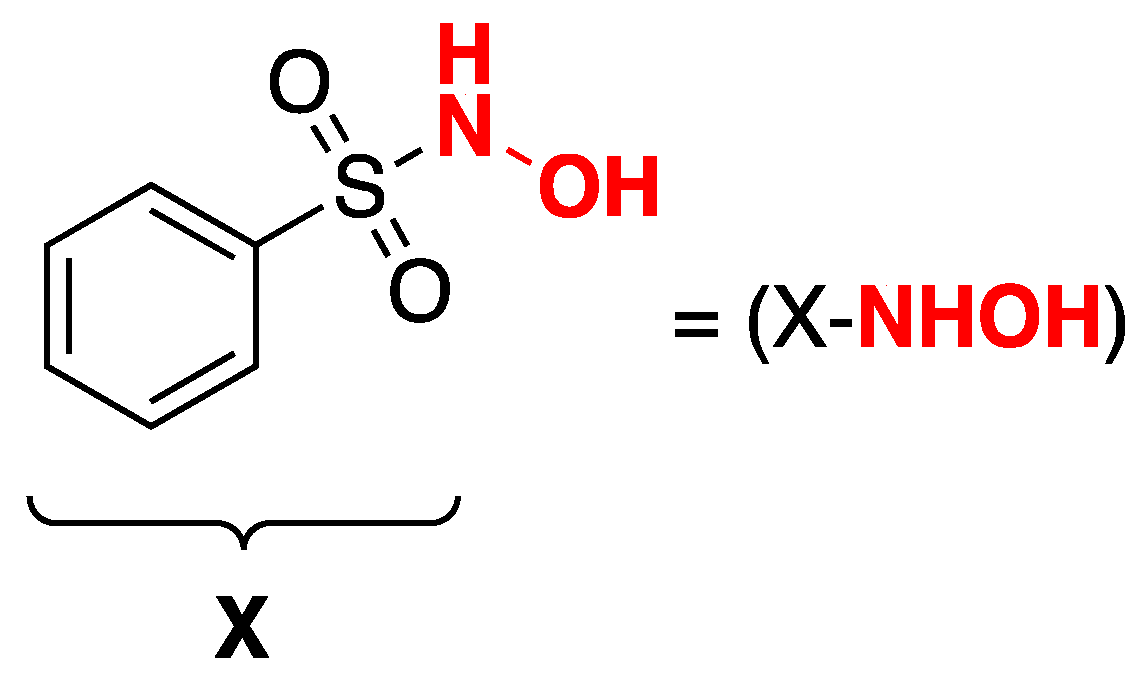
Synthesis of Nitric Oxide Donors Derived from Piloty’s Acid and Study of Their Effects on Dopamine Secretion from PC12 Cells


 , and
, and
Abstract
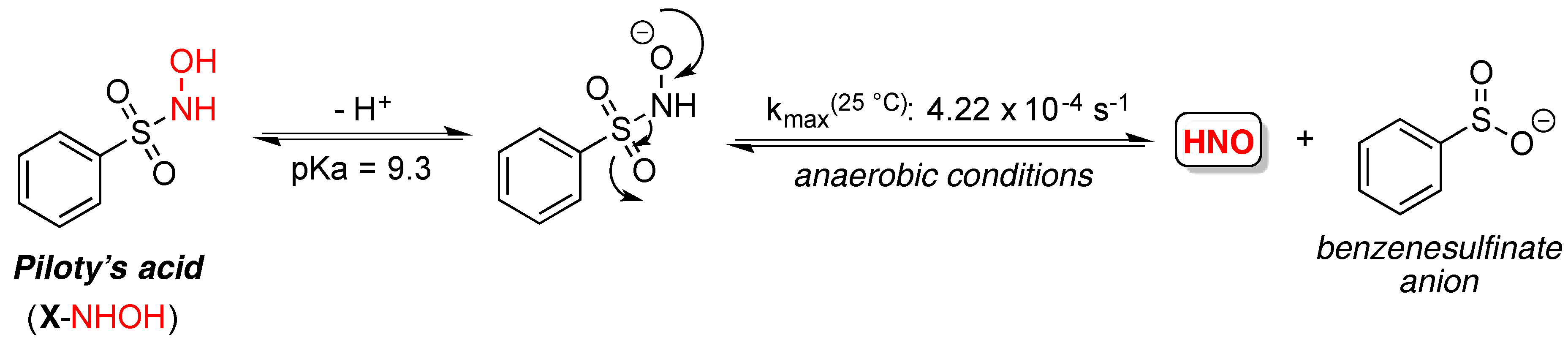
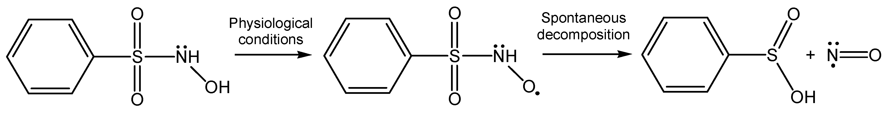
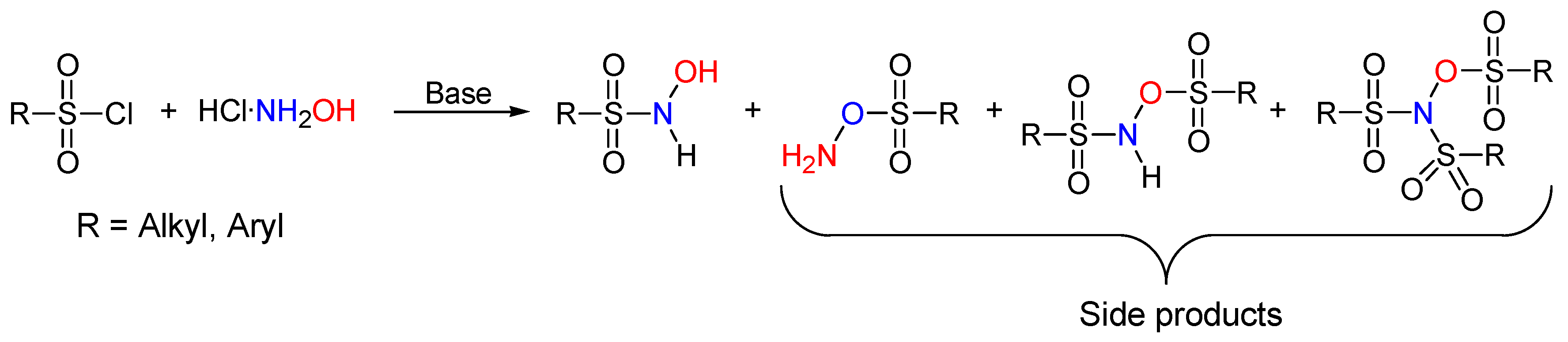
:1. Introduction
2. Results
2.1. Chemicals
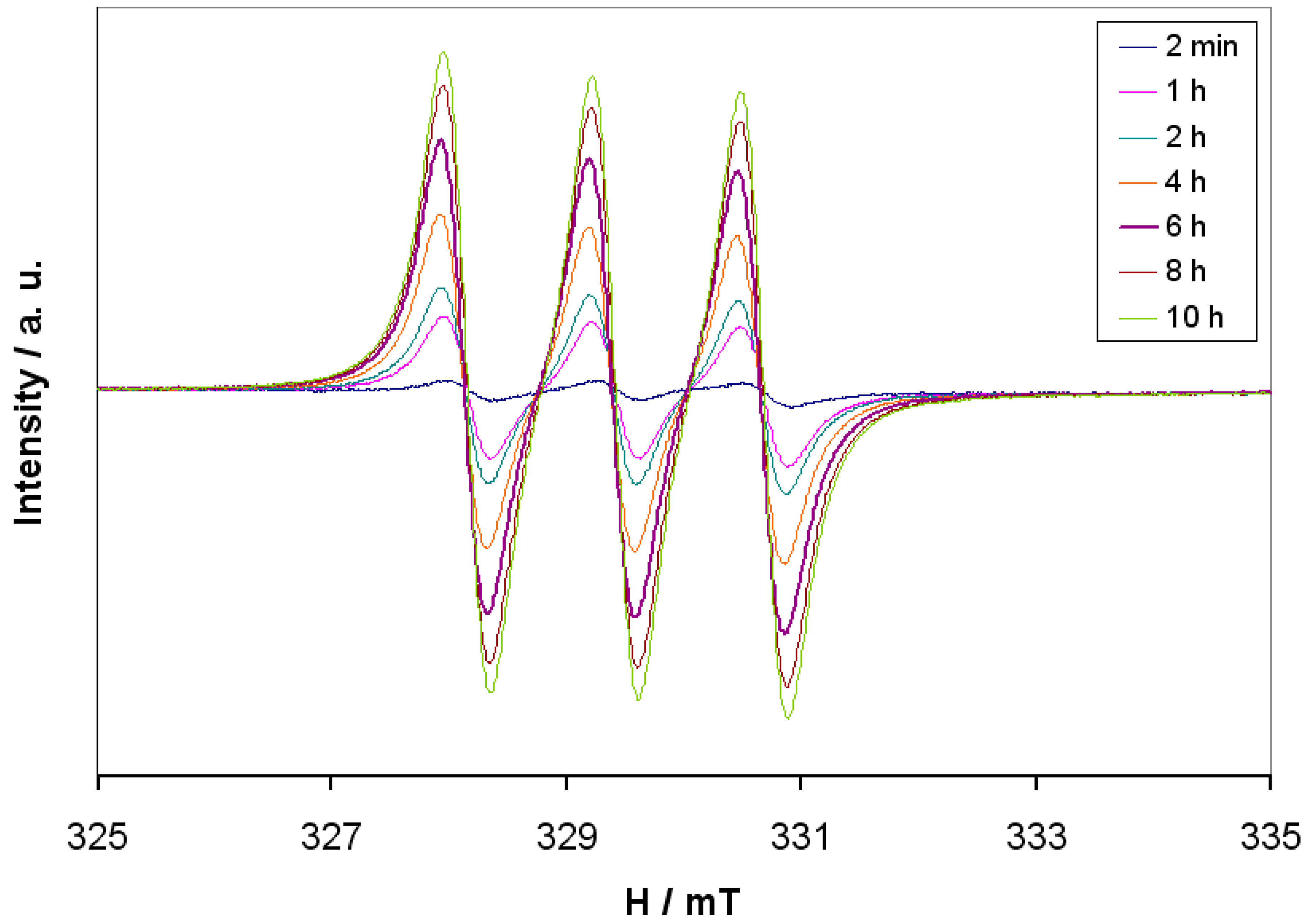
2.2. EPR Characterization of NO-Donors
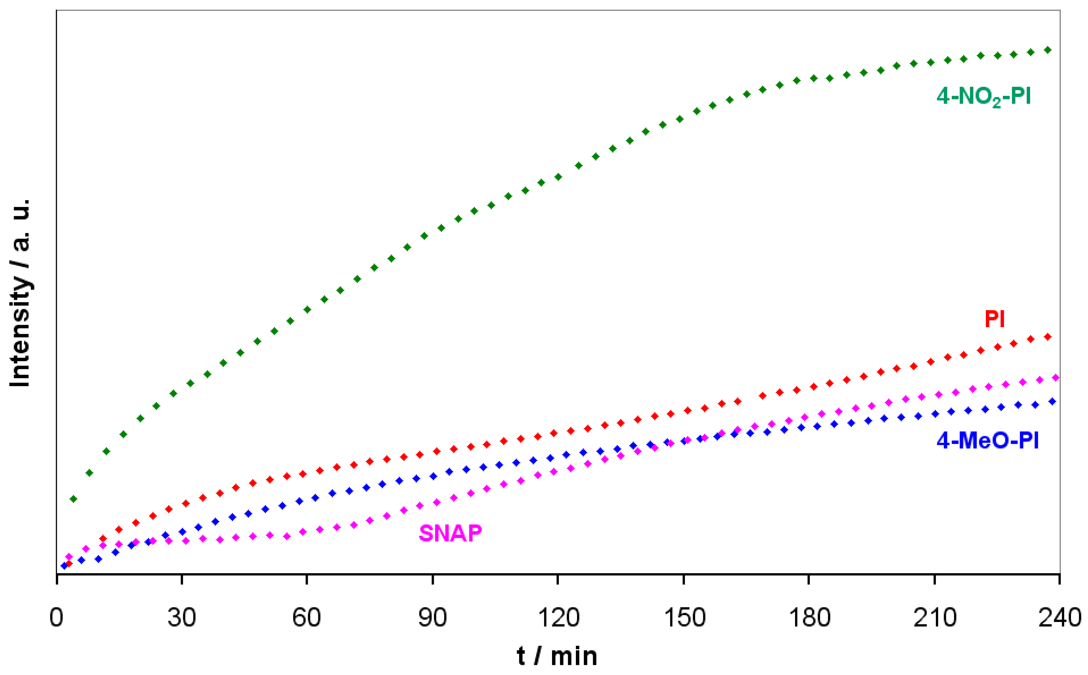
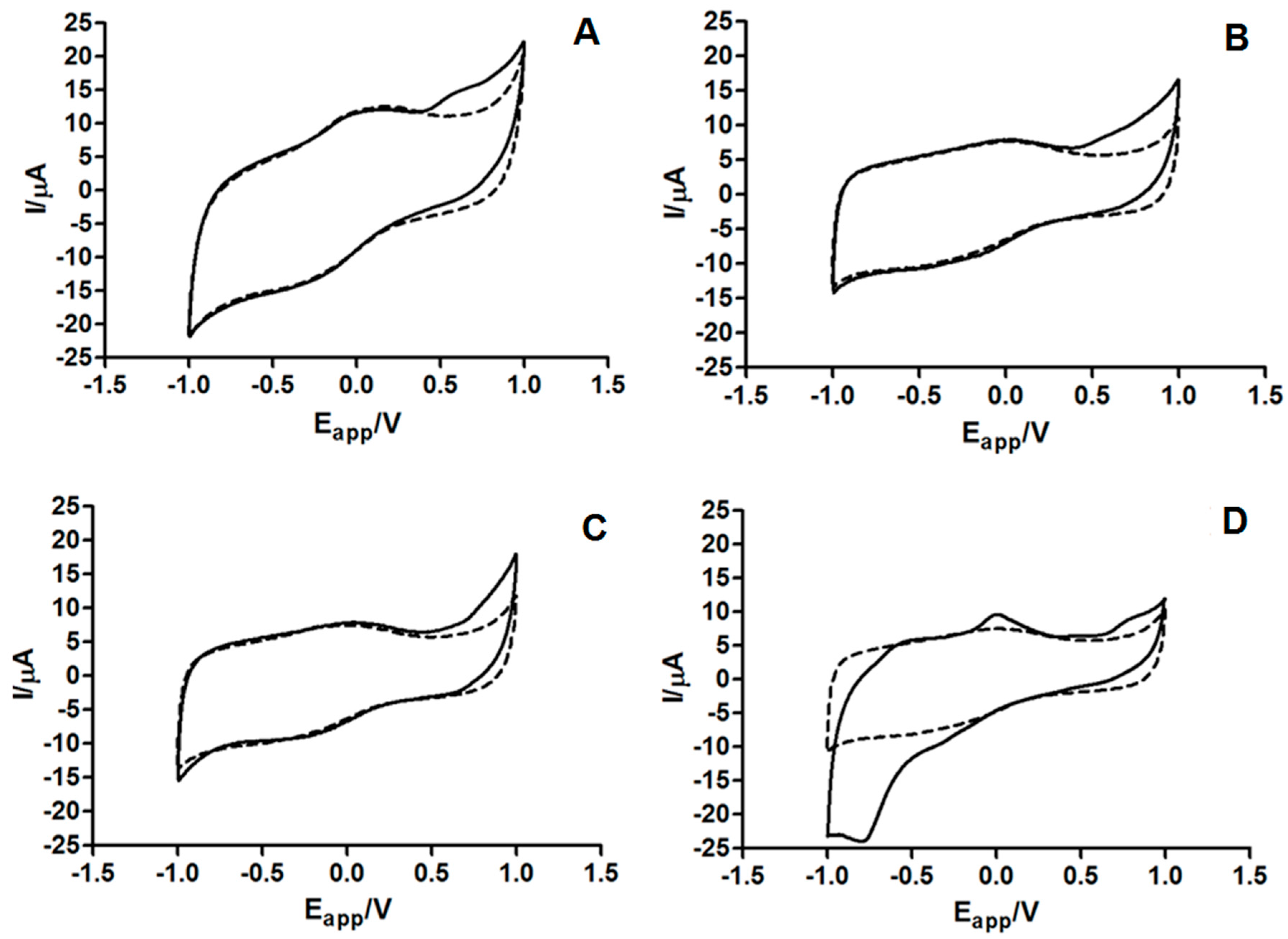
2.3. Electrochemical Characterization of NO-Donors
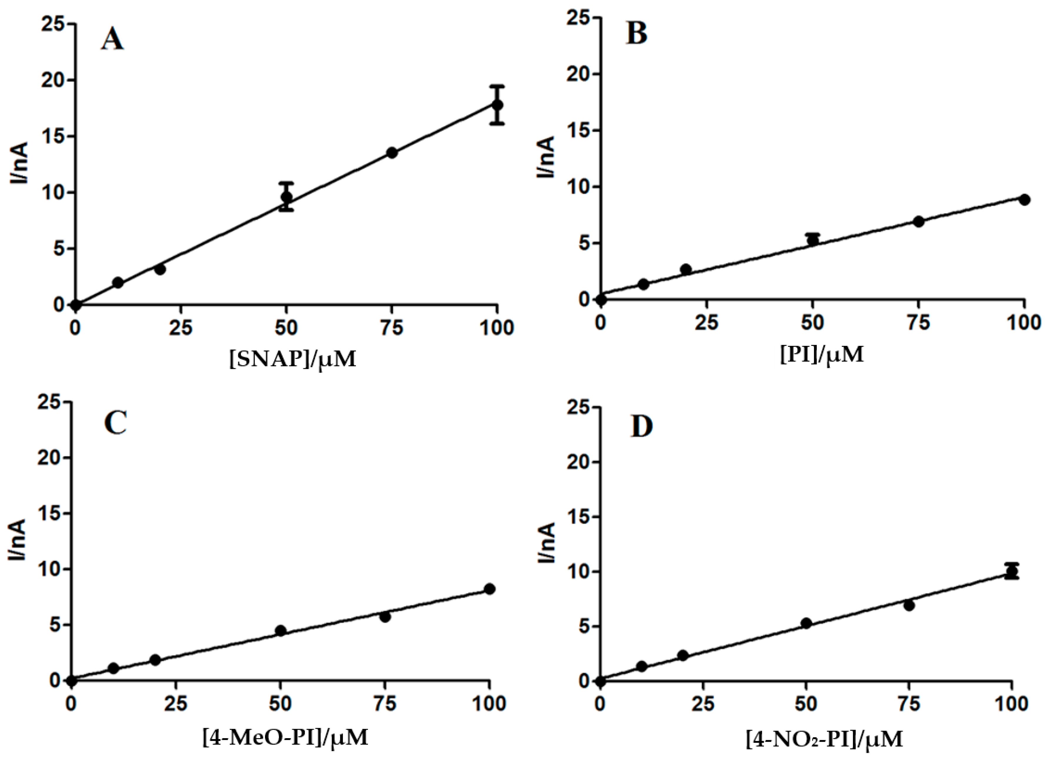
2.4. In Vitro Calibration of NO Microsensor
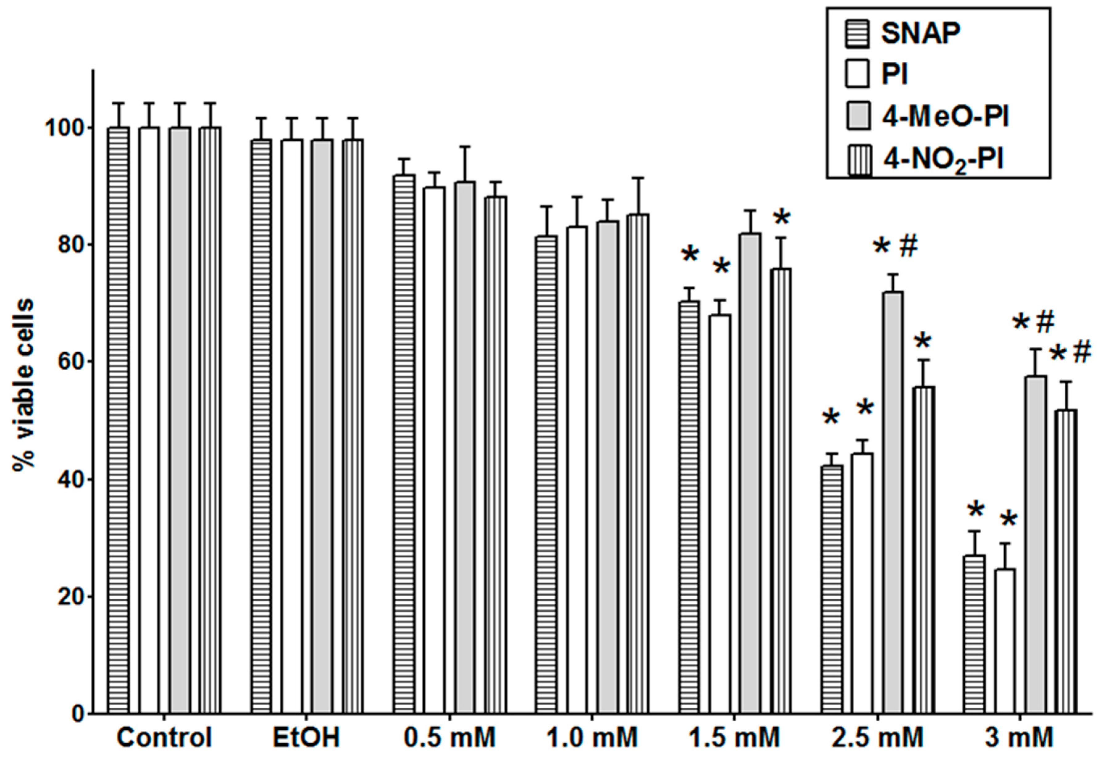
2.5. PC12 Cell Viability
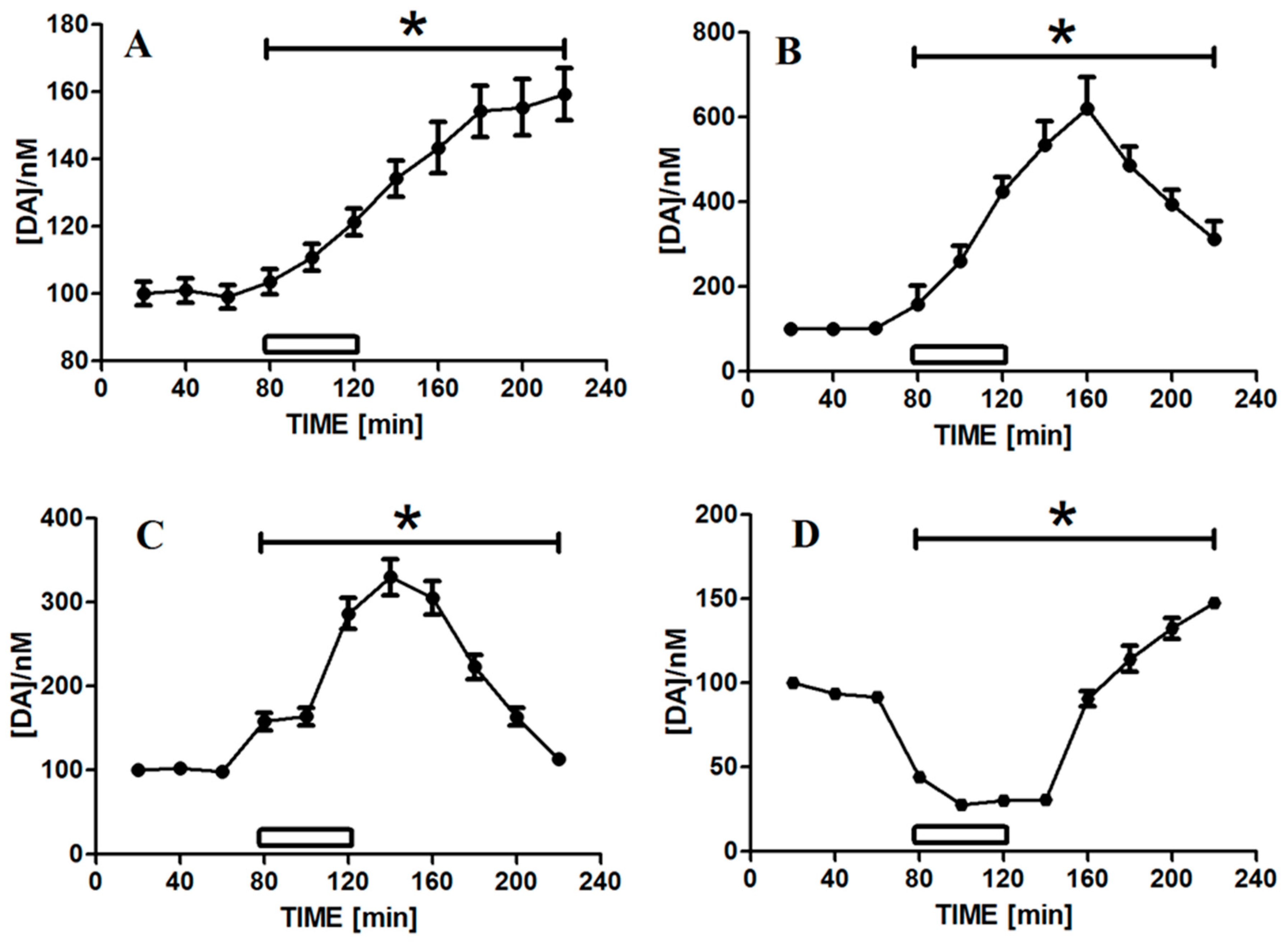
2.6. Effect of NO-Donor Molecules on Dialysate Levels of DA
3. Materials and Methods
3.1. Chemicals
3.2. Organic Synthesis
3.2.1. Synthesis of N-Methyl-d-glucamine Dithiocarbamate (MGD)
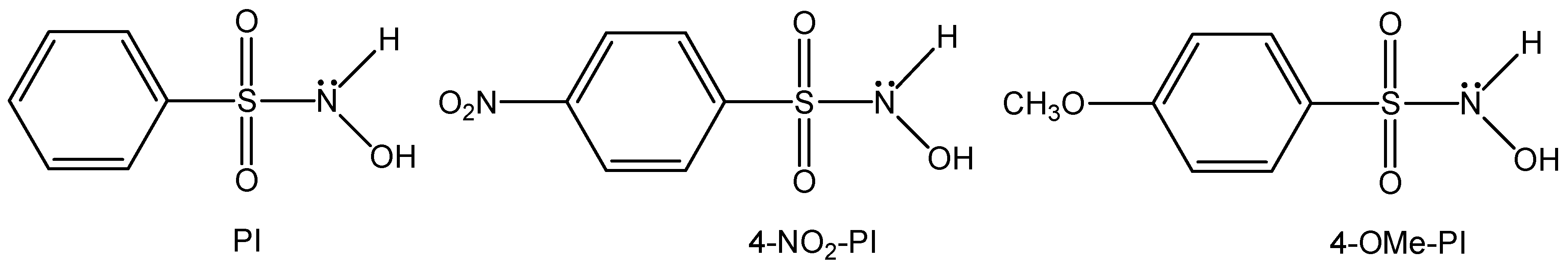
3.2.2. Synthesis of N-Hydroxy-4-methoxybenzenesulfonamide
3.2.3. Synthesis of N-Hydroxy-4-nitrobenzenesulfonamide
3.3. Electron Paramagnetic Spectroscopy
3.3.1. Theory Background and Spin Trapping
3.3.2. Experimental
3.4. NO Microsensors
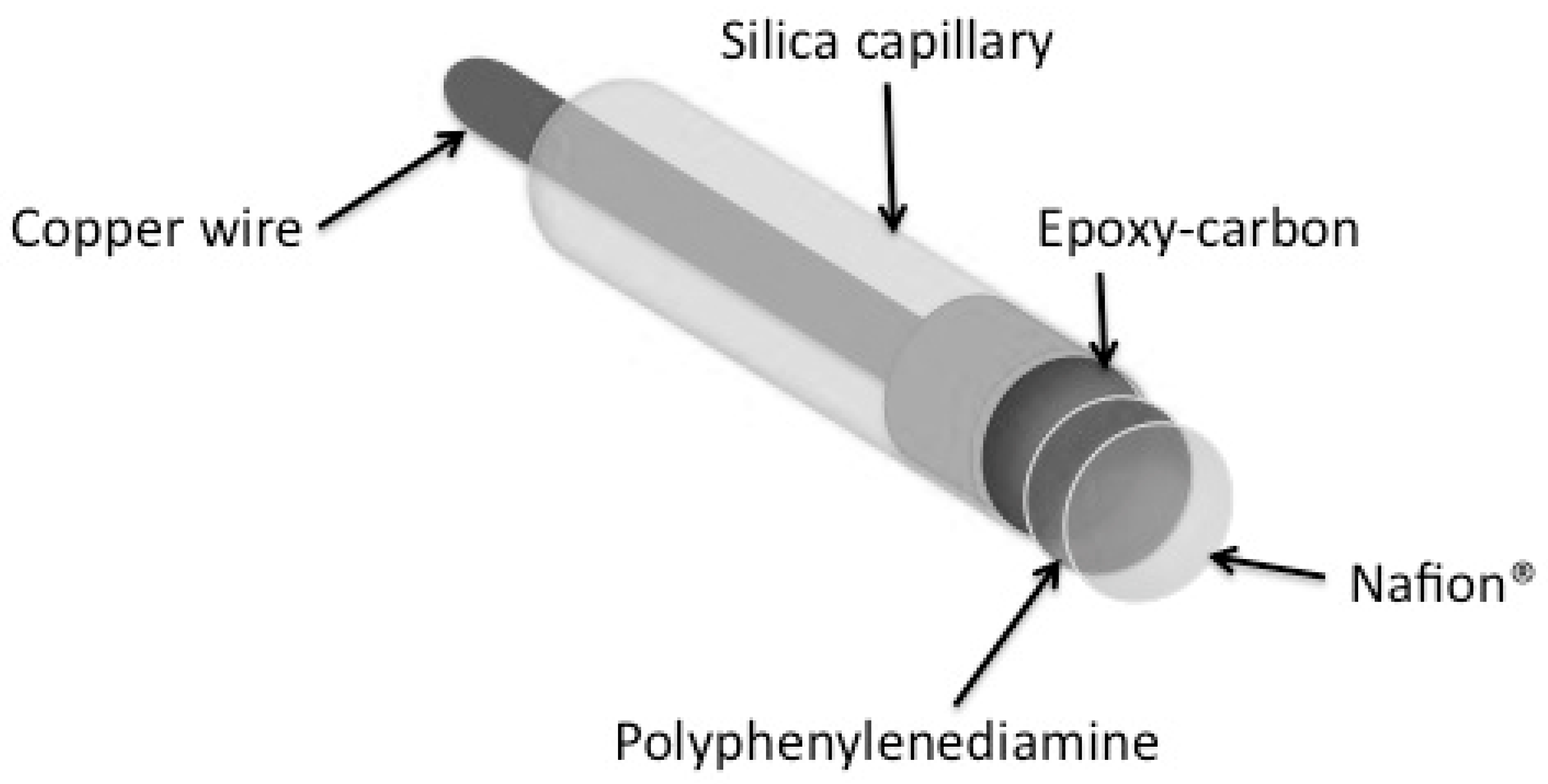
3.4.1. Preparation of NO Microsensors
3.4.2. Microsensor Characterization of NO Donors
3.5. Cell Culture and Capillary Tube Construction for In Vitro Microdialysis
3.6. Microdialysis Procedure
3.7. Chromatographic Analysis of Dialysates from PC12 Cell Suspension
3.8. Assessment of Cell Viability
3.9. Statistical Analysis
4. Conclusions
Supplementary Materials
Acknowledgments
Author Contributions
Conflicts of Interest
References
- Ignarro, L.J.; Buga, G.M.; Wood, K.S.; Byrns, R.E.; Chaudhuri, G. Endothelium-derived relaxing factor produced and released from artery and vein is nitric oxide. Proc. Natl. Acad. Sci. USA 1987, 84, 9265–9269. [Google Scholar] [CrossRef] [PubMed]
- Palmer, R.M.J.; Ferrige, A.G.; Moncada, S. Nitric oxide release accounts for the biological activity of endothelium-derived relaxing factor. Nature 1987, 327, 524–526. [Google Scholar] [CrossRef] [PubMed]
- Furchgott, R.F. Vasodilatation: Vascular Smooth Muscle, Peptides, Autonomic Nerves, and Endothelium; Vanhoutte, P.M., Ed.; Raven Press: New York, NY, USA, 1988; pp. 401–414. [Google Scholar]
- Ignarro, L.J. Nitric Oxide Biology and Pathobiology; Academic Press: San Diego, CA, USA, 2000. [Google Scholar]
- Alderton, W.K.; Cooper, C.E.; Knowles, R.G. Nitric oxide synthases: Structure, function and inhibition. Biochem. J. 2001, 357, 593–615. [Google Scholar] [CrossRef] [PubMed]
- Hirst, D.G.; Robson, T. Methods in molecular biology. In Nitric Oxide, Methods and Protocols; McCarthy, H.O., Coulter, J.A., Eds.; Humana Press: New York, NY, USA, 2011; Volume 704, pp. 1–13. [Google Scholar]
- Garthwaite, J. Nitric oxide signaling in the central nervous system. Annu. Rev. Physiol. 1995, 57, 683–706. [Google Scholar] [CrossRef] [PubMed]
- Wink, D.A.; Mitchell, J.B. Chemical biology of nitric oxide: Insights into regulatory, cytotoxic, and cytoprotective mechanisms of nitric oxide. Free Radic. Biol. Med. 1998, 25, 434–456. [Google Scholar] [CrossRef]
- Murphy, S. Production of nitric oxide by glial cells: Regulation and potential roles in the CNS. Glia 2000, 29, 1–13. [Google Scholar] [CrossRef]
- West, A.R.; Galloway, M.P.; Grace, A.A. Regulation of striatal dopamine neurotransmission by nitric oxide: Effector pathways and signaling mechanisms. Synapse 2002, 44, 227–245. [Google Scholar] [CrossRef] [PubMed]
- West, A.; Galloway, M. Nitric oxide and potassium chloride-facilitated striatal dopamine efflux in vivo: Role of calcium-dependent release mechanisms. Neurochem. Int. 1998, 33, 493–501. [Google Scholar] [CrossRef]
- Trabace, L.; Kendrick, K.M. Nitric oxide can differentially modulate striatal neurotransmitter concentrations via soluble guanylate cyclase and peroxynitrite formation. J. Neurochem. 2002, 75, 1664–1674. [Google Scholar] [CrossRef]
- Serra, P.A.; Rocchitta, G.; Esposito, G.; Delogu, M.R.; Migheli, R.; Miele, E.; Desole, M.S.; Miele, M. A study on the role of nitric oxide and iron in 3-morpholino-sydnonimine-induced increases in dopamine release in the striatum of freely moving rats. Br. J. Pharmacol. 2001, 134, 275–282. [Google Scholar] [CrossRef] [PubMed]
- Yamamoto, T.; Yuyama, K.; Nakamura, K.; Kato, T.; Yamamoto, H. Kinetic characterization of the nitric oxide toxicity for PC12 cells: Effect of half-life time of NO release. Eur. J. Pharmacol. 2000, 397, 25–33. [Google Scholar] [CrossRef]
- Stamler, J.S.; Singel, D.J.; Loscalzo, J. Biochemistry of nitric oxide and its redox-activated forms. Science 1992, 258, 1898–1902. [Google Scholar] [CrossRef] [PubMed]
- Millar, J. The nitric oxide/ascorbate cycle: How neurones may control their own oxygen supply. Med. Hypothese 1995, 45, 21–26. [Google Scholar] [CrossRef]
- Reiser, M.; Schild, L.; Keilhoff, G.; Wolf, G. Interaction of nitric oxide donors and ascorbic acid on D-[3H] aspartate efflux from rat striatal slices. Neurochem. Res. 1999, 24, 61–67. [Google Scholar] [CrossRef] [PubMed]
- Serra, P.A.; Esposito, G.; Delogu, M.R.; Migheli, R.; Rocchitta, G.; Grella, G.; Miele, E.; Miele, M.; Desole, M.S. Analysis of 3-morpholinosydnonimine and sodium nitroprusside effects on dopamine release in the striatum of freely moving rats: Role of nitric oxide, iron and ascorbic acid. Br. J. Pharmacol. 2000, 131, 836–842. [Google Scholar] [CrossRef] [PubMed]
- Serra, P.A.; Esposito, G.; Delogu, M.R.; Migheli, R.; Rocchitta, G.; Miele, E.; Desole, M.S.; Miele, M. Analysis of S-nitroso-N-acetylpenicillamine effects on dopamine release in the striatum of freely moving rats: Role of endogenous ascorbic acid and oxidative stress. Br. J. Pharmacol. 2001, 132, 941–949. [Google Scholar] [CrossRef] [PubMed]
- Nakashima, Y.; Yasui, H; Sakurai, H. A new determination method of nitric oxide (NO) with a NO-selective electrode. Chem. Lett. 2002, 31, 1214–1215. [Google Scholar] [CrossRef]
- Brown, M.D.; Schoenfish, M.H. Nitric oxide permselectivity in electropolymerized films for sensing. ACS Sens. 2016, 1, 1453–1461. [Google Scholar] [CrossRef]
- Hunter, R.A.; Storm, W.L.; Coneski, P.N.; Schoenfisch, M.H. Inaccuracies of nitric oxide measurement methods in biological media. Anal. Chem. 2013, 85, 1957–1963. [Google Scholar] [CrossRef] [PubMed]
- He, W.; Frost, C.M. Direct measurement of actual levels of nitric oxide (NO) in cell culture conditions using soluble NO donors. Redox Biol. 2016, 9, 1–14. [Google Scholar] [CrossRef] [PubMed]
- Bryan, N.S.; Grisham, M.M. Methods to detect nitric oxide and its metabolites in biological samples. Free Radic. Biol. Med. 2007, 43, 645–657. [Google Scholar] [CrossRef] [PubMed]
- Csonka, C.; Páli, T.; Bencsik, P.; Görbe, A.; Ferdinandy, P.; Csont, T. Measurement of NO in biological samples. Br. J. Pharmacol. 2015, 172, 1620–1632. [Google Scholar] [CrossRef] [PubMed]
- Liang, H.; Nacharaju, P.; Friedman, A.; Friedman, J.M. Nitric oxide generating/releasing materials. Future Sci. OA 2015, 1, FSO54. [Google Scholar] [CrossRef] [PubMed]
- Serra, P.A.; Migheli, R.; Rocchitta, G.; Taras, M.G.; Mura, M.P.; Delogu, M.R.; Esposito, G.; Desole, M.S.; Miele, E.; Miele, M. Role of the nitric oxide/cyclic GMP pathway and ascorbic acid in 3-morpholinosydnonimine (SIN-1)-induced increases in dopamine secretion from PC12 cells. A microdialysis in vitro study. Neurosci. Lett. 2003, 353, 5–8. [Google Scholar] [CrossRef] [PubMed]
- Kita, Y.; Ohkubo, K.; Hirasawa, Y.; Katayama, Y.; Ohno, M.; Nishino, S.; Kato, M.; Yoshida, K. FR144420, a novel, slow, nitric oxide-releasing agent. Eur. J. Pharmacol. 1995, 275, 125–130. [Google Scholar] [CrossRef]
- Amatore, C.; Arbault, S.; Ducrocq, C.; Hu, S.; Tapsoba, I. Angelis salt (Na2N2O3) is a precursor of HNO and NO: A voltammetric study of the reactive intermediates released by Angelis salt decomposition. Chem. Med. Chem. 2007, 2, 898–903. [Google Scholar] [CrossRef] [PubMed]
- Piloty, O. Ueber eine oxydation des hydroxylamins durch benzolsulfochlorid. Eur. J. Inorg. Chem. 1896, 29, 1559–1567. [Google Scholar] [CrossRef]
- Dumond, J.F.; King, S.B. The chemistry of nitroxyl-releasing compounds. Antioxid. Redox Signal. 2011, 14, 1637–1648. [Google Scholar] [CrossRef] [PubMed]
- Fukuto, J.M.; Cisneros, C.J.; Kinkade, R.L. A comparison of the chemistry associated with the biological signaling and actions of nitroxyl (HNO) and nitric oxide (NO). J. Inorg. Biochem. 2013, 118, 201–208. [Google Scholar] [CrossRef] [PubMed]
- Nakagawa, H. Controlled release of HNO from chemical donors for biological applications. J. Inorg. Biochem. 2013, 118, 187–190. [Google Scholar] [CrossRef] [PubMed]
- Subedi, H.; Brasch, N.E. Mechanistic studies of the reactions of the reduced vitamin B12 derivatives with the HNO donor Piloty’s acid: Further evidence for oxidation of cob(I)alamin by (H)NO. Dalton Trans. 2016, 45, 352–360. [Google Scholar] [CrossRef] [PubMed]
- Seel, F.B.C. Mechanism of the decomposition of sodium benzenesulfohydroxamate in aqueous solution. Z. Anorg. Allg. Chem. 1972, 394, 187–196. [Google Scholar] [CrossRef]
- Toscano, J.P.; Brookfield, F.A.; Cohen, A.D.; Courtney, S.M.; Frost, L.M.; Kalish, V.J. Preparation of N-Hydroxylsulfonamide Derivatives as Nitroxyl (HNO) Donors. U.S. Patent 11/724,792, 16 March 2007. [Google Scholar]
- Miranda, K.M.; Nagasawa, H.T.; Toscano, J.P. Donors of HNO. Curr. Top. Med. Chem. 2005, 5, 647–664. [Google Scholar] [CrossRef]
- Hughes, M.; Cammack, R. Synthesis, chemistry, and applications of nitroxyl ion releasers sodium trioxodinitrate or Angeli’s salt and Piloty’s acid. Methods Enzymol. 1999, 301, 279–287. [Google Scholar] [PubMed]
- Nagasawa, H.T.; Kawle, S.P.; Elberling, J.A.; DeMaster, E.G.; Fukuto, J.M. Prodrugs of nitroxyl as potential aldehyde dehydrogenase inhibitors vis-à-vis vascular smooth muscle relaxants. J. Med. Chem. 1995, 38, 1865–1871. [Google Scholar] [CrossRef] [PubMed]
- Doctorovich, F.; Farmer, P.J.; Marti, M.A. The Chemistry and Biology of Nitroxyl (HNO); Elsevier: Amsterdam, The Netherlands, 2016. [Google Scholar]
- Grzesiok, A.; Weber, H.; Pino, R.Z.; Feelisch, M. The Biology of Nitric Oxide Part 4: Enzymology, Biochemistry and Immunology; Moncada, S., Freelisch, M., Busse, R., Higgs, E.A., Eds.; Portland Press Ltd.: London, UK, 1994; pp. 238–241. [Google Scholar]
- Zamora, R.; Grzesiok, A.; Weber, H.; Feelisch, M. Oxidative release of nitric oxide accounts for guanylyl cyclase stimulating, vasodilator and anti-platelet activity of Pilotys acid: A comparison with Angelis salt. Biochem. J. 1995, 312, 333–339. [Google Scholar] [CrossRef] [PubMed]
- Miao, Z.; King, S.B. Recent advances in the chemical biology of nitroxyl (HNO) detection and generation. Nitric Oxide 2016, 57, 1–14. [Google Scholar] [CrossRef] [PubMed]
- Aizawa, K.; Nakagawa, H.; Matsuo, K.; Kawai, K.; Ieda, N.; Suzuki, T.; Miyata, N. Piloty’s acid derivative with improved nitroxyl-releasing characteristics. Bioorg. Med. Chem. Lett. 2013, 23, 2340–2343. [Google Scholar] [CrossRef] [PubMed]
- Sirsalmath, K.; Suárez, S.A.; Bikiel, D.E.; Doctorovich, F. The pH of HNO donation is modulated by ring substituents in Piloty’s acid derivatives: Azanone donors at biological pH. J. Inorg. Biochem. 2013, 118, 134–139. [Google Scholar] [CrossRef] [PubMed]
- Irvine, J.C.; Ritchie, R.H.; Favarolo, J.L.; Andrews, K.L.; Widdop, R.E.; Kemp-Harper, B.K. Nitroxyl (HNO): the Cinderella of the nitric oxide story. Trends Pharmacol. Sci. 2008, 29, 601–608. [Google Scholar] [CrossRef] [PubMed]
- Jorolan, J.H.; Buttitta, L.A.; Cheah, C.; Miranda, K.M. Comparison of the chemical reactivity of synthetic peroxynitrite with that of the autoxidation products of nitroxyl or its anion. Nitric Oxide 2015, 30, 39–46. [Google Scholar] [CrossRef] [PubMed]
- Marmion, C.J.; Murphy, T.; Docherty, J.R.; Nolan, K.B. Hydroxamic acids are nitric oxide donors. Facile formation of ruthenium(II)-nitrosyls and NO-mediated activation of guanylate cyclase by hydroxamic acids. Chem. Commun. 2000, 1153–1154. [Google Scholar] [CrossRef]
- Davies, S.; Evans, D.; Hughes, D.; Konkol, M.; Richards, R.; Sanders, J.; Sobota, P. Mononuclear, binuclear, trinuclear and tetranuclear iron complexes of the N(CH2CH2S)33− (NS3) ligand with nitrosyl co-ligands. J. Chem. Soc. Dalton Trans. 2002, 2473–2482. [Google Scholar] [CrossRef]
- Hogg, N. Detection of nitric oxide by electron paramagnetic resonance spectroscopy. Free Radic. Biol. Med. 2010, 49, 122–129. [Google Scholar] [CrossRef] [PubMed]
- Papapetropoulos, A.; Oresti, R.F.; Ferdinandy, P. Pharmacology of the ‘gasotransmitters’ NO, CO and H2S: Translational opportunities. Br. J. Pharmacol. 2015, 172, 1395–1396. [Google Scholar] [CrossRef] [PubMed]
- Maia, L.B.; Moura, J.J.G. Plant Nitric Oxide. In Methods in Molecular Biology; Springer: New York, NY, USA, 2016; Volume 1424, pp. 81–102. [Google Scholar]
- Bryukov, M.G.; Kachanov, A.A.; Timonnen, R.; Seetula, J.; Vandoren, J.; Sarkisov, O.M. Kinetics of HNO reactions with O2 and HNO. Chem. Phys. Lett. 1993, 208, 392–398. [Google Scholar] [CrossRef]
- Suárez, S.A.; Fonticelli, M.H.; Rubert, A.A.; De La Llave, E.; Scherlis, D.; Salvarezza, R.C.; Martí, M.A.; Doctorovich, F. A surface effect allows HNO/NO discrimination by a cobalt porphyrin bound to gold. Inorg. Chem. 2010, 49, 6955–6966. [Google Scholar] [CrossRef] [PubMed]
- Williams, D.L.H. Nitrosation Reaction and the Chemistry of Nitric Oxide, 1st ed.; Elsevier: Amsterdam, The Netherlands, 2004; pp. 222–231. [Google Scholar]
- Wang, P.G.; Cai, T.B.; Taniguchi, N. Nitric Oxide Donors; Wiley-VCH: Weinheim, Germany, 2006. [Google Scholar]
- Huang, Z.; Velázquez, C.A.; Abdellatif, K.R.A.; Chowdhury, M.A.; Reisz, J.A.; Dumond, J.F.; King, S.B.; Knaus, E.E. Ethanesulfohydroxamic acid ester prodrugs of nonsteroidal anti-inflammatory drugs (NSAIDs): Synthesis, nitric oxide and nitroxyl release, cyclooxygenase inhibition, anti-inflammatory, and ulcerogenicity index studies. J. Med. Chem. 2011, 54, 1356–1364. [Google Scholar] [CrossRef] [PubMed]
- Huang, Z.; Velázquez, C.; Abdellatif, K.; Chowdhury, M.; Jain, S.; Reisz, J.; Dumond, J.; King, S.B.; Knaus, E. Acyclic triaryl olefins possessing a sulfohydroxamic acid pharmacophore: Synthesis, nitric oxide/nitroxyl release, cyclooxygenase inhibition, and anti-inflammatory studies. Org. Biomol. Chem. 2010, 8, 4124. [Google Scholar] [CrossRef] [PubMed]
- Gattermann, L. Laboratory Methods of Organic Chemistry; The Macmillan Company: New York, NY, USA, 1937. [Google Scholar]
- Keasling, H.H.; Schumann, E.L.; Veldkamp, W. The relationship between structure and anticonvulsant activity in a series of benzenesulfonamides. J. Med. Chem. 1965, 8, 548–550. [Google Scholar] [CrossRef] [PubMed]
- Kim, H.K.; Park, Y.D.; Lee, M.H.; Chung, H.A.; Kweon, D.H.; Cho, S.D.; Yoon, Y.J. Chemoselective N-benzenesulfonylation of aliphatic amines. Bull. Korean Chem. Soc. 2003, 24, 1655–1658. [Google Scholar] [CrossRef]
- Katritzky, A.R.; Rodriguez-Garcia, V.; Nair, S.K. A general and efficient synthesis of sulfonylbenzotriazoles from N-chlorobenzotriazole and sulfinic acid salts. J. Org. Chem. 2004, 69, 1849–1852. [Google Scholar] [CrossRef] [PubMed]
- Caddick, S.; Wilden, J.D.; Judd, D.B. Observations on the reactivity of pentafluorophenyl sulfonate esters. Chem. Commun. 2005, 2727–2728. [Google Scholar] [CrossRef] [PubMed]
- Fitzmaurice, R.J.; Ahern, J.M.; Caddick, S. Synthesis of unsymmetrical ketonesvia simple C–H activation of aldehydes and concomitant hydroacylation of vinyl sulfonates. Org. Biomol. Chem. 2009, 7, 235–237. [Google Scholar] [CrossRef] [PubMed]
- Chantarasriwong, O.; Jang, D.O.; Chavasiri, W. A practical and efficient method for the preparation of sulfonamides utilizing Cl3CCN/PPh3. Tetrahedron Lett. 2006, 47, 7489–7492. [Google Scholar] [CrossRef]
- Harmata, M.; Zheng, P.; Huang, C.; Gomes, M.G.; Ying, W.; Ranyanil, K.O.; Balan, G.; Calkins, N.L. Expedient synthesis of sulfinamides from sulfonyl chlorides. J. Org. Chem. 2007, 72, 683–685. [Google Scholar] [CrossRef] [PubMed]
- Wilden, J.D.; Geldeard, L.; Lee, C.C.; Judd, D.B.; Caddick, S. Trichlorophenol (TCP) sulfonate esters: A selective alternative to pentafluorophenol (PFP) esters and sulfonyl chlorides for the preparation of sulfonamides. Chem. Commun. 2007, 1074–1076. [Google Scholar] [CrossRef] [PubMed]
- Porcheddu, A.; De Luca, L.; Giacomelli, G. A straightforward route to Piloty’s acid derivatives: A class of potential nitroxyl-generating prodrugs. Synlett 2009, 13, 2149–2153. [Google Scholar] [CrossRef]
- Mocci, R.; De Luca, L.; Delogu, F.; Porcheddu, A. An environmentally sustainable mechanochemical route to hydroxamic acid derivatives. Adv. Synth. Catal. 2016, 358, 3135–3144. [Google Scholar] [CrossRef]
- Xia, Y.; Cardounel, A.J.; Vanin, A.F.; Zweier, J.L. Electron paramagnetic resonance spectroscopy with N-methyl-d-glucamine dithiocarbamate iron complexes distinguishes nitric oxide and nitroxyl anion in a redoxdependent manner: Applications in identifying nitrogen monoxide products from nitric oxide synthase. Free Radic. Biol. Med. 2000, 29, 793–797. [Google Scholar] [PubMed]
- Serra, P.A.; Rocchitta, G.; Delogu, M.R.; Migheli, R.; Taras, M.G.; Mura, M.P.; Esposito, G.; Miele, E.; Desole, M.S.; Miele, M. Role of the nitric oxide/cyclic GMP pathway and extracellular environment in the nitric oxide donor-induced increase in dopamine secretion from PC12 cells: A microdialysis in vitro study. J. Neurochem. 2003, 86, 1403–1413. [Google Scholar] [CrossRef] [PubMed]
- Zhang, X.; Cardosa, L.; Broderick, M.; Fein, H.; Lin, J. An integrated nitric oxide sensor based on carbon fiber coated with selective membranes. Electroanalysis 2000, 12, 1113–1117. [Google Scholar] [CrossRef]
- Komiyama, T.; Fujimori, K. Kinetic studies of the reaction of S-nitroso-l-cysteine with l-cysteine. Bioorg. Med. Chem. Lett. 1997, 7, 175–180. [Google Scholar] [CrossRef]
- Xu, A.; Vita, J.A.; Keaney, J.F. Ascorbic acid and glutathione modulate the biological activity of S-nitrosoglutathione. Hypertension 2000, 36, 291–295. [Google Scholar] [CrossRef] [PubMed]
- Ford, P.C.; Wink, D.A.; Stanbury, D.M. Autoxidation kinetics of aqueous nitric oxide. FEBS Lett. 1993, 326, 1–3. [Google Scholar] [CrossRef]
- Ferrero, R.; Rodríguez-Pascual, F.; Miras-Portugal, M.T.; Torres, M. Comparative effects of several nitric oxide donors on intracellular cyclic GMP levels in bovine chromaffin cells: Correlation with nitric oxide production. Br. J. Pharmacol. 1999, 127, 779–787. [Google Scholar] [CrossRef] [PubMed]
- Dischia, M.; Costantini, C. Nitric oxide-induced nitration of catecholamine neurotransmitters: A key to neuronal degeneration? Bioorg. Med. Chem. 1995, 3, 923–927. [Google Scholar] [CrossRef]
- Daveu, C.; Servy, C.; Dendane, M.; Marin, P.; Ducrocq, C. Oxidation and nitration of catecholamines by nitrogen oxides derived from nitric oxide. Nitric Oxide 1997, 1, 234–243. [Google Scholar] [CrossRef] [PubMed]
- Shinobu, L.A.; Jones, S.G.; Jones, M.M. Sodium N-methyl-d-glucamine dithiocarbamate and cadmium intoxication. Acta Pharmacol. Toxicol. 2009, 54, 189–194. [Google Scholar] [CrossRef]
- Vassilev, V.P.; Liaw, W.C.; Lai, C.S. Method for Preparation of Pharmaceutical-Grade Dithiocarbamate. U.S. Pantent 6124349, 18 February 1999. Available online: http://patft.uspto.gov/netacgi/nph-Parser?Sect2=PTO1&Sect2=HITOFF&p=1&u=/netahtml/PTO/search-bool.html&r=1&f=G&l=50&d=PALL&RefSrch=yes&Query=PN/6124349 (accessed on 29 August 2017).
- Olson, D.E.; Bois, J.D. Catalytic C−H amination for the peparation of substituted 1,2-diamines. J. Am. Chem. Soc. 2008, 130, 11248–11249. [Google Scholar] [CrossRef] [PubMed]
- Yajima, H.; Kitagawa, K.; Kurobe, M. Application of a new arginine derivative, ng-mesitylene-2-sulfonylarginine, to the synthesis of substance p and neurotensin. Chem. Pharm. Bull. 1973, 21, 2566–2567. [Google Scholar] [CrossRef]
- Weil, J.A.; Bolton, J.R. Electron Paramagnetic Resonance: Elementary Theory and Practical Applications; John Wiley & Sons: Hoboken, NJ, USA, 2007. [Google Scholar]
- Westenberger, U.; Thanner, S.; Ruf, H.H.; Gersonde, K.; Sutter, G.; Trentz, O. Formation of free radicals and nitric oxide derivative of hemoglobin in rats during shock syndrome. Free Radic. Res. Commun. 1990, 11, 167–178. [Google Scholar] [CrossRef] [PubMed]
- Mordvintcev, P.; Mülsch, A.; Busse, R.; Vanin, A. On-line detection of nitric oxide formation in liquid aqueous phase by electron paramagnetic resonance spectroscopy. Anal. Biochem. 1991, 199, 142–146. [Google Scholar] [CrossRef]
- Komarov, A.; Mattson, D.; Jones, M.M.; Singh, P.K.; Lai, C.S. In vivo spin trapping of nitric oxide in mice. Biochem. Biophys. Res. Commun. 1993, 195, 1191–1198. [Google Scholar] [CrossRef] [PubMed]
- Bazzu, G.; Puggioni, G.G.M.; Dedola, S.; Calia, G.; Rocchitta, G.; Migheli, R.; Desole, M.S.; Lowry, J.P.; O’Neill, R.D.; Serra, P.A. Real-time monitoring of brain tissue oxygen using a miniaturized biotelemetric device implanted in freely moving rats. Anal. Chem. 2009, 81, 2235–2241. [Google Scholar] [CrossRef] [PubMed]
- Calia, G.; Rocchitta, G.; Migheli, R.; Puggioni, G.; Spissu, Y.; Bazzu, G.; Mazzarello, V.; Lowry, J.P.; O’Neill, R.D.; Desole, M.S.; et al. Biotelemetric monitoring of brain neurochemistry in conscious rats using microsensors and biosensors. Sensors 2009, 9, 2511–2523. [Google Scholar] [CrossRef] [PubMed]
- Cahill, P.S.; Wightman, R.M. Simultaneous amperometric measurement of ascorbate and catecholamine secretion from individual bovine adrenal medullary cells. Anal. Chem. 1995, 67, 2599–2605. [Google Scholar] [CrossRef] [PubMed]
- Friedemann, M.N.; Robinson, S.W.; Gerhardt, G.A. O-phenylenediamine-modified carbon fiber electrodes for the detection of nitric oxide. Anal. Chem. 1996, 68, 2621–2628. [Google Scholar] [CrossRef] [PubMed]
- Zhang, X.; Kislyak, Y.; Lin, J.; Dickson, A.; Cardosa, L.; Broderick, M.; Fein, H. Nanometer size electrode for nitric oxide and S-nitrosothiols measurement. Electrochem. Commun. 2002, 4, 11–16. [Google Scholar] [CrossRef]
- Rocchitta, G.; Migheli, R.; Dedola, S.; Calia, G.; Desole, M.S.; Miele, E.; Lowry, J.P.; O’Neill, R.D.; Serra, P.A. Development of a distributed, fully automated, bidirectional telemetry system for amperometric microsensor and biosensor applications. Sens. Actuators B Chem. 2007, 126, 700–709. [Google Scholar] [CrossRef]
- Serra, P.A.; Rocchitta, G.; Bazzu, G.; Manca, A.; Puggioni, G.M.; Lowry, J.P.; O’Neill, R.D. Design and construction of a low cost single-supply embedded telemetry system for amperometric biosensor applications. Sens. Actuators B Chem. 2007, 122, 118–126. [Google Scholar] [CrossRef]
- Rocchitta, G.; Migheli, R.; Mura, M.P.; Esposito, G.; Marchetti, B.; Desole, M.S.; Miele, E.; Serra, P.A. Role of endogenous melatonin in the oxidative homeostasis of the extracellular striatal compartment: A microdialysis study in PC12 cells in vitro and in the striatum of freely moving rats. J. Pineal Res. 2005, 39, 409–418. [Google Scholar] [CrossRef] [PubMed]
- Migheli, R.; Giudice, M.G.D.; Spissu, Y.; Sanna, G.; Xiong, Y.; Dawson, T.M.; Dawson, V.L.; Galioto, M.; Rocchitta, G.; Biosa, A.; et al. LRRK2 affects vesicle trafficking, neurotransmitter extracellular level and membrane receptor localization. PLoS ONE 2013, 8, e77198. [Google Scholar] [CrossRef] [PubMed] [Green Version]
- ICH Q2 (R1). Validation of analytical procedures: Text and methodology. In Proceedings of the International Conference on Harmonization of Technical Requirements for Registration of Pharmaceuticals for Human Use, Geneva, Switzerland, 10 November 2005; pp. 11–12. [Google Scholar]
- Martí, M.M.; Bari, S.E.; Estrin, D.E.; Doctorovich, F. Discrimination of nitroxyl and nitric oxide by water-soluble Mn(III) porphyrins. J. Am. Chem. Soc. 2005, 127, 4680–4684. [Google Scholar] [CrossRef] [PubMed]
- Karthikeyan, M.; Deepa, K.J. Therapeutic applications of nitric oxide releasing nonsteroidal anti-inflammatory drugs. Chem. Pharm. Res. 2009, 1, 134–147. [Google Scholar]
- Wallace, J.L. Gastric Tolerability and prolonged prostaglandin inhibition in the brain with a nitric oxide-releasing flurbiprofen derivative, NCX-2216 [3-[4-(2-Fluoro-α-methyl-[1,1-biphenyl]-4-acetyloxy)-3-methoxyphenyl]-2-propenoic acid 4-nitrooxy butyl ester]. J. Pharmacol. Exp. Ther. 2004, 309, 626–633. [Google Scholar] [CrossRef] [PubMed]


| NO-Donor | Slope (nA μM−1) ± SEM | R2 | NO Released (μM mM−1donor) b |
|---|---|---|---|
| SNAP | 0.180 ± 0.005 | 0.997 | 14.0 |
| PI | 0.086 ± 0.004 | 0.987 | 7.7 |
| 4-MeO-PI | 0.079 ± 0.003 | 0.992 | 6.9 |
| 4-NO2-PI | 0.096 ± 0.005 | 0.993 | 8.4 |
© 2017 by the authors. Licensee MDPI, Basel, Switzerland. This article is an open access article distributed under the terms and conditions of the Creative Commons Attribution (CC BY) license (http://creativecommons.org/licenses/by/4.0/).
Share and Cite
Sanna, D.; Rocchitta, G.; Serra, M.; Abbondio, M.; Serra, P.A.; Migheli, R.; De Luca, L.; Garribba, E.; Porcheddu, A. Synthesis of Nitric Oxide Donors Derived from Piloty’s Acid and Study of Their Effects on Dopamine Secretion from PC12 Cells. Pharmaceuticals 2017, 10, 74. https://doi.org/10.3390/ph10030074
Sanna D, Rocchitta G, Serra M, Abbondio M, Serra PA, Migheli R, De Luca L, Garribba E, Porcheddu A. Synthesis of Nitric Oxide Donors Derived from Piloty’s Acid and Study of Their Effects on Dopamine Secretion from PC12 Cells. Pharmaceuticals. 2017; 10(3):74. https://doi.org/10.3390/ph10030074
Chicago/Turabian StyleSanna, Daniele, Gaia Rocchitta, Maria Serra, Marcello Abbondio, Pier Andrea Serra, Rossana Migheli, Lidia De Luca, Eugenio Garribba, and Andrea Porcheddu. 2017. "Synthesis of Nitric Oxide Donors Derived from Piloty’s Acid and Study of Their Effects on Dopamine Secretion from PC12 Cells" Pharmaceuticals 10, no. 3: 74. https://doi.org/10.3390/ph10030074