Oral Administration of Shark Type II Collagen Suppresses Complete Freund’s Adjuvant-Induced Rheumatoid Arthritis in Rats

Abstract
:1. Introduction
2. Results
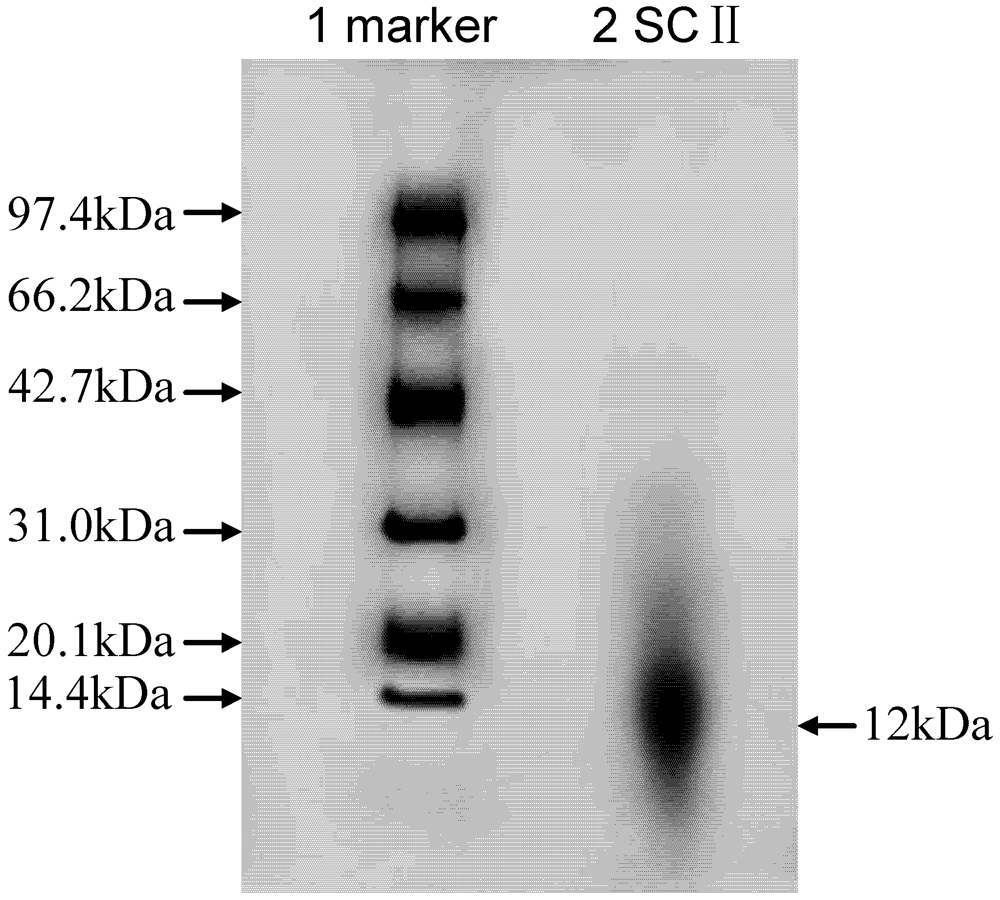
2.1. Peptide Mapping
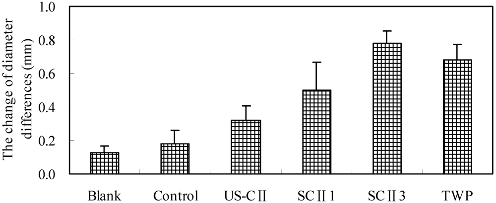
2.2. Clinical Assessment

| Group | Blank | Control | US-CII | SCII1 | SCII3 | TWP |
|---|---|---|---|---|---|---|
| Body weight(g) | 160 ± 11 | 169 ± 24 | 166 ± 15 | 151 ± 16 | 176 ± 14 | 167 ± 17 |
| scores | 0.0 | 2.8 | 2.7 | 2.8 | 2.8 | 3.0 |
| Diameter of right ankle joint (mm) | 7.92 ± 0.29 | 7.94 ± 0.43 | 7.78 ± 0.43 | 7.86 ± 0.53 | 7.81 ± 0.21 | 7.47 ± 0.30 |
| Diameter of left ankle joint (mm) | 8.07 ± 0.71 | 8.62 ± 0.46 ** | 8.35 ± 0.38 ** | 8.58 ± 0.66 ** | 8.61 ± 0.29 ** | 8.38 ± 0.42 ** |
2.3. Appearance Characteristic of Ankle
| Group | Blank | Control | US-CII | SCII1 | SCII3 | TWP |
|---|---|---|---|---|---|---|
| Body weight (g) | 340 ± 10 | 288 ± 39 | 271 ± 26 | 245 ± 35 | 282 ± 13 | 284 ± 29 |
| Scores | 0.0 | 3.4 | 1.3 | 1.3 | 1.2 | 1.2 |
| Diameter of right ankle joint (mm) | 8.08 ± 0.52 | 8.53 ± 0.44 | 8.00 ± 0.41 | 8.30 ± 0.43 | 8.50 ± 0.38 | 7.92 ± 0.23 |
| Diameter of left ankle joint (mm) | 8.10 ± 0.39 | 9.03 ± 0.90 * | 8.25 ± 0.39 ** | 8.52 ± 0.39 ** | 8.52 ± 0.38 ** | 8.16 ± 0.24 ** |

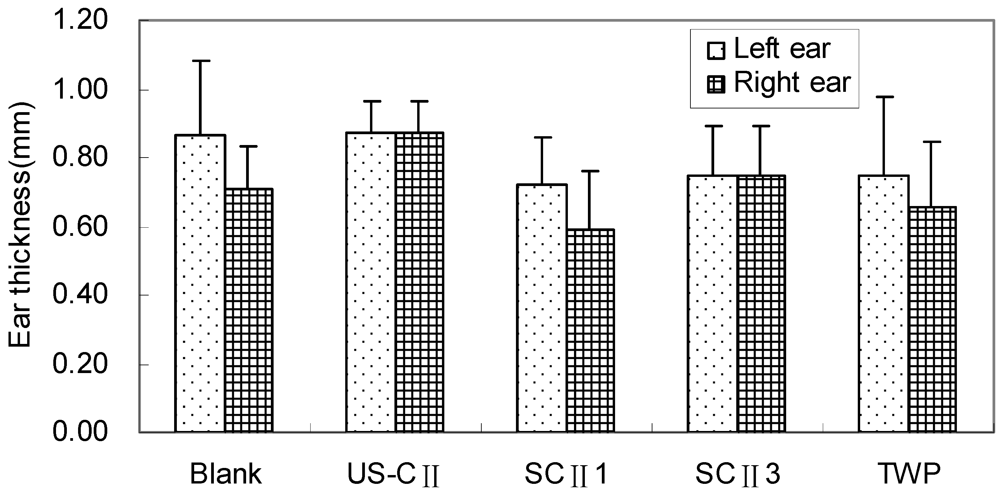
2.4. DTH Responses

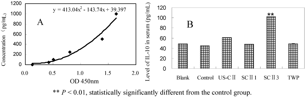
2.5. IL-10 Level in Serum

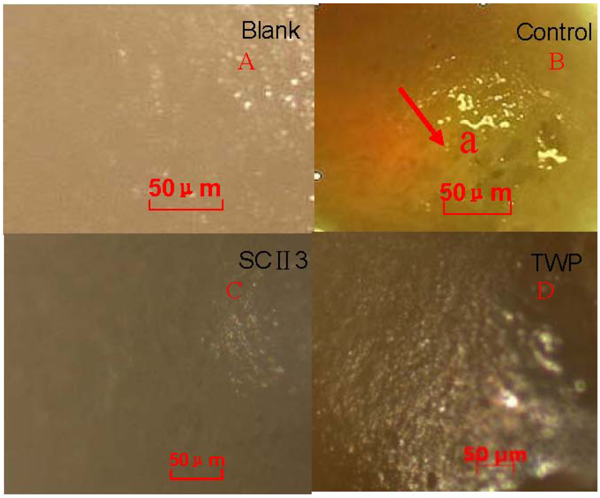
2.7. Histopathology of Arthritis

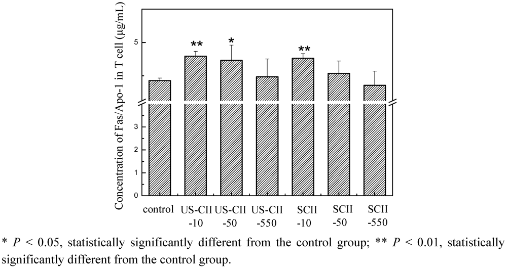
2.8. Fas/Apo-1 Characteristic in T Cell Culture with US-CII and SCII
3. Experimental
3.1. Experimental Animals
3.2. Reagents
3.3. SDS-Polyacrylamide Gel Electrophoresis (SDS-PAGE) for SCII
3.4. Induction of CFA and Clinical Assessment of Arthritis
3.5. Administration of Type II Collagen
3.6. Cell-Mediated Immunity to Type II Collagen
3.7. Detection of IL-10 Production by ELISA
3.8. Histomorphometry
3.9. Measurement of Tolerance in T cell induced by US-CII and SCII
3.10. Statistical Analysis
4. Discussion
5. Conclusions
Acknowledgments
References
- Lydyard, P.M.; Whelan, A.; Fanger, M.W. Immunology; Science Press: Beijing, China, 2009; p. 215, Section L. [Google Scholar]
- Borchers, A.T.; Keen, C.L.; Cheema, G.S.; Gershwin, M.E. The use of methotrexate in rheumatoid arthritis. Semin. Arthritis Rheum. 2004, 1, 465–483. [Google Scholar]
- Caporali, R.; Caprioli, M.; Bobbi-Pallavicini, F.; Montecucco, C. DMARDS and infections in rheumatoid arthritis. Autoimmun. Rev. 2008, 8, 139–143. [Google Scholar]
- Iván, A.F.; Machín, S.; Carmona, L.; Isidoro, G.A.; Federico, D.G. Pattern of use and safety of non-steroidal anti-inflammatory drugs in rheumatoid arthritis patients. A prospective analysis from clinical practice. Reumatol. Clin. 2009, 6, 252–258. [Google Scholar]
- Cremer, M. Type II Collagen-induced Arthritis in Rats. In Handbook of Animal Models for the Rheumatic Diseases; Greenwald, R.A., Diamond, H.S., Eds.; CRC Press: Boca Raton, FL, USA, 1988; pp. 17–27. [Google Scholar]
- Banerjee, S.; Wei, B.Y.; Hillman, K.; Luthra, H.S.; David, C.S. Immunosuppression of collagen-induced arthritis in mice with an anti-IL-2 receptor antibody. J. Immunol. 1988, 141, 1150–1154. [Google Scholar]
- Strobel, S.; Mowat, A.M. Immune responses to dietary antigens: oral tolerance. Immunol. Today 1998, 19, 173–181. [Google Scholar]
- Weiner, H.L. Oral tolerance: Immune mechanisms and treatment of autoimmune diseases. Immunol. Today 1997, 18, 335–343. [Google Scholar]
- Thompson, S.J.; Thompson, H.S.; Harper, N.; Day, M.J.; Coad, A.J.; Elson, C.J.; Staines, N.A. Prevention of pristine-induced arthritis by the oral administration of type II collagen. Immunology 1993, 79, 152–157. [Google Scholar]
- Khare, S.D.; Krco, C.J.; Griffiths, M.M.; Luthra, H.S.; David, C.S. Oral administration of an immunodominant human collagen peptide modulates collagen-induced arthritis. J. Immunol. 1995, 155, 3653–3659. [Google Scholar]
- Meyer, O. L’immunomodulation par voie orale dans la polyarthrite. Rev. Rhum. 2000, 67, 593–603. [Google Scholar]
- Zhao, W.; Tong, T.; Wang, L.; Li, P.P.; Chang, Y.; Zhang, L.L.; Wei, W. Chicken type II collagen induced immune tolerance of mesenteric lymph node lymphocytes by enhancing beta2-adrenergic receptor desensitization in rats with collagen-induced arthritis. Int. Immunopharmacol. 2011, 11, 12–18. [Google Scholar]
- Trentham, D.E.; Dynesius-Trentham, R.A.; Orav, E.J.; Combitchi, D.; Lorenzo, C.; Sewell, K.L.; Hafler, D.A. Effects of oral administration of type II collagen on rheumatoid arthritis. Science 1993, 261, 1727–1730. [Google Scholar]
- Barnett, M.L.; Kremer, J.M.; William, E.; Clegg, D.O.; Furst, D.; Weisman, M. Treatment of rheumatoid arthritis with oral type II collagen: Results of a multicenter, double-blind, placebo-controlled trial. Arthritis Rheum. 1998, 290–297. [Google Scholar]
- Poole, A.R. Cartilage in Health and Disease. In Arthritis and Allied Conditions: A Textbook of Rheumatology, 15th; Koopman, W.J., Moreland, L.W., Eds.; Williams and Wilkins: Baltimore, MD, USA, 2005; pp. 223–269. [Google Scholar]
- Nomura, Y. Properties and Utilization of Shark Collagen. In More Efficient Utilization of Fish and Fisheries Products; Elsevier: New York, NY, USA, 2004; Volume 42, pp. 147–158. [Google Scholar]
- Kittiphattanabawon, P.; Benjakul, S.; Visessanguan, W.; Nagai, T.; Tanaka, M. Characterisation of acid-soluble collagen from skin and bone of bigeye snapper (Priacanthus tayenus). Food Chem. 2005, 89, 363–372. [Google Scholar] [CrossRef]
- Nalinanon, S.; Benjakul, S.; Visessanguan, W.; Kishimura, H. Tuna pepsin: Characteristics and its use for collagen extraction from the skin of threadfin bream (Nemipterus spp.). J. Food Sci. 2008, 73, C413–C419. [Google Scholar] [CrossRef]
- Laemmli, U.K. Cleavage of structural proteins during the assembly of the head of bacteriophage T4. Nature 1970, 277, 680–685. [Google Scholar]
- Pincus, T.; Marcum, S.B.; Callahan, L.F. Long-term drug therapy for rheumatoid arthritis in seven rheumatology private practices: II. Second line drugs and prednisone. J. Rheumatol. 1992, 19, 1885–1894. [Google Scholar]
- Newbould, B.B. Chemotherapy of arthritis induced in rats by mycobacterium adjuvant. Br. J. Pharmacol. 1963, 21, 127–136. [Google Scholar]
- Omura, K.; Imai, S.; Maeda, T.; Hukuda, S. Prolonged and increasing expression of Fos related antigens in the hippocampus of adjuvant arthritic rats. J. Rheumatol. 1998, 25, 936–944. [Google Scholar]
- Hughes, C.; Wolos, J.A.; Giannini, E.H.; Hirsch, R. Induction of T helper cell hyporesponsiveness in an experimental model of autoimmunity by using nonmitogenic anti-CD3 monoclonal antibody. J. Immunol. 1994, 153, 3319–3325. [Google Scholar]
- Banerjee, S.; Wei, B.Y.; Hillman, K.; Luthra, H.S.; David, C.S. Immunosuppression of collagen-induced arthritis in mice with an anti-IL-2 receptor antibody. J. Immunol. 1988, 141, 1150–1154. [Google Scholar]
- Thorbecke, G.J.; Schwarcz, R.; Leu, J.; Huang, C.; Simmons, W.J. Modulation by cytokines of induction of oral tolerance to type II collagen. Arthritis Rheum. 1999, 42, 110–118. [Google Scholar] [CrossRef]
- Zhu, P.; Li, X.Y.; Wang, H.K.; Jia, J.F.; Zheng, Z.H.; Ding, J.; Fan, C.M. Oral administration of type-II collagen peptide 250-270 suppresses specific cellular and humoral immune response in collagen-induced arthritis. Clin. Immunol. 2007, 122, 75–84. [Google Scholar]
- Garcia, G.; Komagata, Y.; Slavin, A.J.; Maron, R.; Weiner, H.L. Suppression of collagen-induced arthritis by oral or nasal administration of type II collagen. J. Autoimmun. 1999, 13, 315–324. [Google Scholar]
- Weiner, H.L. Oral tolerance, an active immunologic process mediated by multiple mechanisms. J. Clin. Invest. 2000, 106, 935–937. [Google Scholar]
- Illum, L.; Farraj, N.F.; Davis, S.S. Chitosan as a novel nasal delivery system for peptide drugs. Pharm. Res. 1994, 11, 1186–1189. [Google Scholar]
- Hall, G.; Lund, L.; Lamb, J.R.; Jarman, E.R. Kinetics and mode of peptide delivery via the respiratory mucosa determine the outcome of activation versus TH2 immunity in allergic inflammation of the airways. J. Allergy Clin. Immunol. 2002, 110, 883–890. [Google Scholar] [CrossRef]
- Toussirot, E.A. Oral tolerance in the treatment of rheumatoid arthritis. Curr. Drug Targets 2002, 1, 45–52. [Google Scholar]
© 2012 by the authors; licensee MDPI, Basel, Switzerland. This article is an open-access article distributed under the terms and conditions of the Creative Commons Attribution license (http://creativecommons.org/licenses/by/3.0/).
Share and Cite
Chen, L.; Bao, B.; Wang, N.; Xie, J.; Wu, W. Oral Administration of Shark Type II Collagen Suppresses Complete Freund’s Adjuvant-Induced Rheumatoid Arthritis in Rats. Pharmaceuticals 2012, 5, 339-352. https://doi.org/10.3390/ph5040339
Chen L, Bao B, Wang N, Xie J, Wu W. Oral Administration of Shark Type II Collagen Suppresses Complete Freund’s Adjuvant-Induced Rheumatoid Arthritis in Rats. Pharmaceuticals. 2012; 5(4):339-352. https://doi.org/10.3390/ph5040339
Chicago/Turabian StyleChen, Lijuan, Bin Bao, Nanping Wang, Jing Xie, and Wenhui Wu. 2012. "Oral Administration of Shark Type II Collagen Suppresses Complete Freund’s Adjuvant-Induced Rheumatoid Arthritis in Rats" Pharmaceuticals 5, no. 4: 339-352. https://doi.org/10.3390/ph5040339