Article

Isolation, Structure Elucidation and Total Synthesis of Lajollamide A from the Marine Fungus Asteromyces cruciatus

1
Kekulé-Institute of Organic Chemistry and Biochemistry, University of Bonn, Gerhard-Domagk Str. 1, Bonn 53121, Germany
2
Institute for Pharmaceutical Biology, University of Bonn, Nussallee 6, Bonn 53115, Germany
3
Kieler Wirkstoff-Zentrum (KiWiZ) at the Helmholtz-Zentrum für Ozeanforschung GEOMAR, Am Kiel-Kanal 44, Kiel 24106, Germany
4
Institute for Pharmaceutical Biology, University of Tübingen, Auf der Morgenstelle 8, Tübingen 72076, Germany
*
Authors to whom correspondence should be addressed.
These authors contributed equally to this work.
Current address: Institute for Microbiology and Biotechnology, University of Bonn, Meckenheimer Allee 168, Bonn 53115, Germany.
Mar. Drugs 2012, 10(12), 2912-2935; https://doi.org/10.3390/md10122912
Submission received: 9 November 2012 / Revised: 29 November 2012 / Accepted: 5 December 2012 / Published: 19 December 2012
(This article belongs to the Special Issue Bioactive Compounds from Marine Fungi)

Abstract

:
The marine-derived filamentous fungus Asteromyces cruciatus 763, obtained off the coast of La Jolla, San Diego, USA, yielded the new pentapeptide lajollamide A (1), along with the known compounds regiolone (2), hyalodendrin (3), gliovictin (4), 1N-norgliovicitin (5), and bis-N-norgliovictin (6). The planar structure of lajollamide A (1) was determined by Nuclear Magnetic Resonance (NMR) spectroscopy in combination with mass spectrometry. The absolute configuration of lajollamide A (1) was unambiguously solved by total synthesis which provided three additional diastereomers of 1 and also revealed that an unexpected acid-mediated partial racemization (2:1) of the L-leucine and L-N-Me-leucine residues occurred during the chemical degradation process. The biological activities of the isolated metabolites, in particular their antimicrobial properties, were investigated in a series of assay systems.

Graphical Abstract

1. Introduction

Filamentous fungi, particularly marine-derived fungi, are widely recognized as an emerging source for the production of novel and bioactive secondary metabolites [1,2,3,4]. This empirical fact is corroborated by the insights gained from recent fungal genome sequencing projects. These genomic studies attribute filamentous fungi an overall tremendous biosynthetic capacity because they commonly carry about 30 to 40 biosynthetic gene clusters coding for secondary metabolites [5,6]. However, the majority of these gene clusters have not yet been correlated to their corresponding natural products. Considering the given therapeutic value of fungal metabolites [1,2,3,4] and the huge amount of hitherto unexplored orphan and silent gene clusters, the activation and discovery of such biosynthetic loci represents a valuable strategy to obtain new bioactive products [7].
Filamentous fungi of the genus Asteromyces comprise currently only one valid species and belong to the division of mitosporic fungi [8]. To date, aside from triglycerides and sterols [9,10], only the compounds gliovictin (4) [9,11], 2,4-dimethyl-4,5-dihydrofuran-3-carbaldehyde, 3S,5R-dihydrodimethylfuran-2-one, and cyclo-phenylalanine-serine [11] have been reported from different Asteromyces cruciatus samples. Fueled by the significantly high metabolic capacity of filamentous fungi and the fact that Asteromyces species are chemically underexplored, we performed a comprehensive chemical investigation with the strain Asteromyces cruciatus 763, obtained off La Jolla shore, San Diego, USA. In order to stimulate the production of secondary metabolites encoded by silent biosynthesis gene clusters and to enhance the production of constitutively produced secondary metabolites in our fungal isolate, we integrated the “One Strain Many Compounds” (OSMAC) approach [12,13,14,15] into our study. In this paper, we report the isolation of a new pentapeptide designated lajollamide A (1), and four known 3R,6R configured epipolythiopiperazinedione antibiotics hyalodendrin (3) [16], gliovictin (4) [9,11,16,17,18,19], 1N-norgliovictin (5) [18] and bis-N-norgliovictin (6) [18,19,20], in addition to the phytotoxic naphthalenone regiolone (2) [21,22] (See Figure 1). The elucidation of the absolute configuration of 1 has been a challenging problem and we report here the results of these efforts.
Figure 1. Secondary metabolites isolated from Asteromyces cruciatus 763.
Marinedrugs 10 02912 g001

2. Results and Discussion

2.1. OSMAC Studies

A panel of extracts was prepared under 14 different culture conditions (different media composition, changing cultivation periods, extraction procedures, UV-light exposure, and temperature) and screened by LC-MS. Two whole broth extracts of A. cruciatus 763, that were grown either in a Czapek-Dox medium supplemented with cofactors or in a Czapek-Dox medium with an altered nitrogen source, afforded a good variety of known metabolites (e.g., cyclo-phenylalanine-serine and gliovictin [9,11]) and previously unknown compounds (e.g., m/z [M + H]+ = 566). The HPLC profiles of these two extracts indicated that they contained almost the same metabolites but with significant differences in their relative concentrations. Thus, the two cultivation conditions were separately scaled up and processed to isolate and characterize the major metabolites present in each of these extracts.

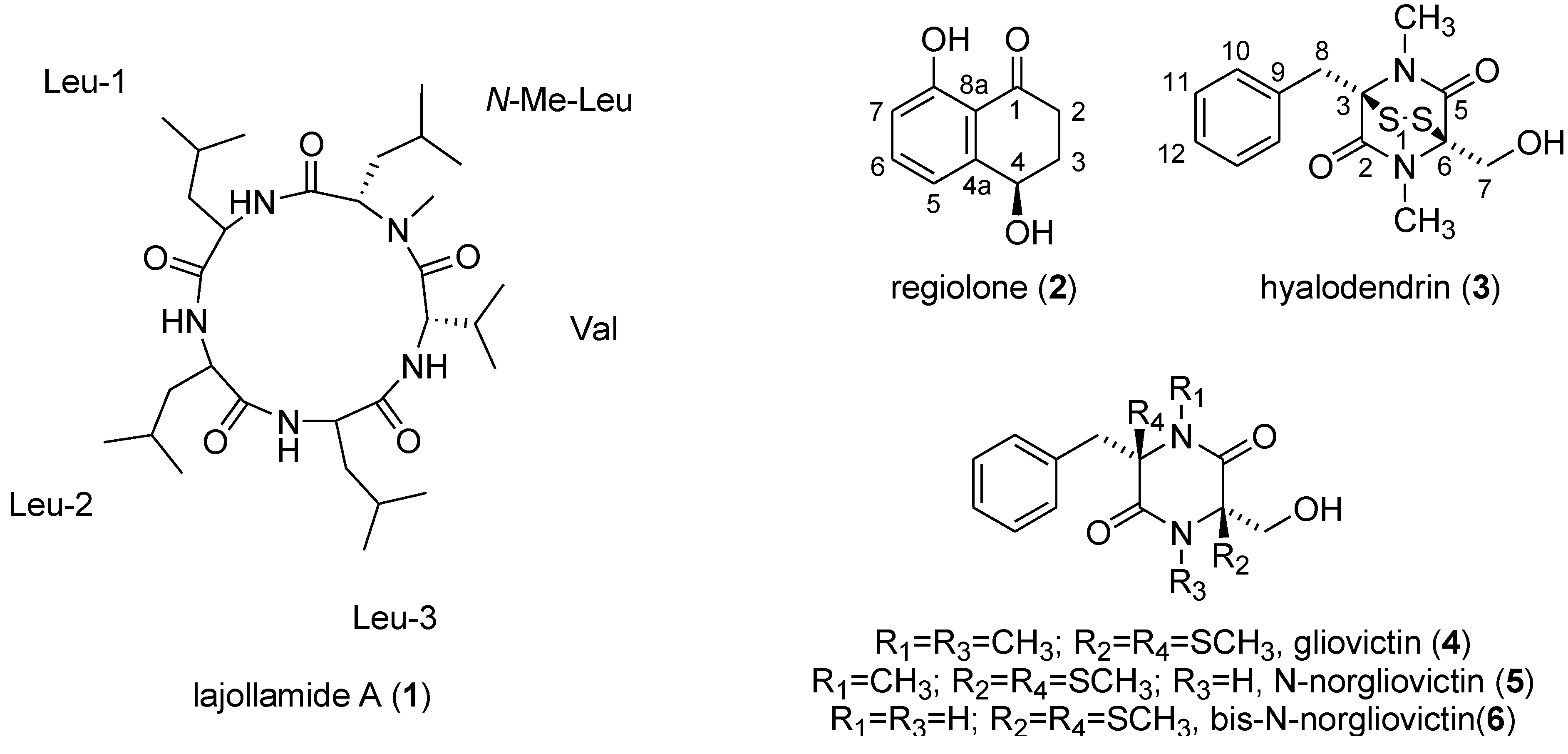
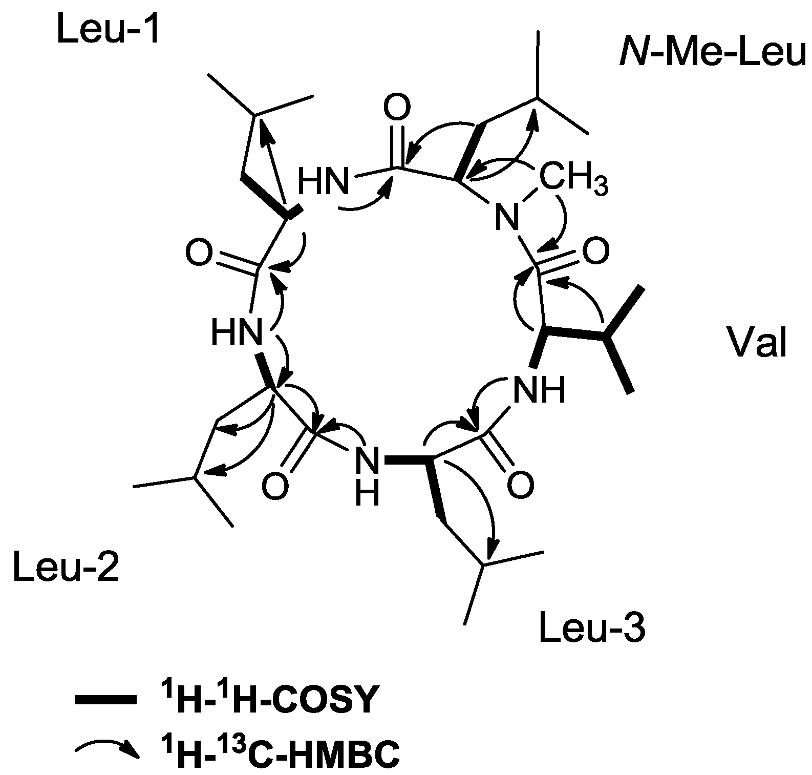
2.2. Isolation and Structure Elucidation

The ethyl acetate extracts of the scaled-up fungal cultures were filtered through an RP-SPE cartridge and direct separation by RP-HPLC led to the isolation of compounds 16. While the identity of the known metabolites 26 was established by direct comparison of the [α]D, MS, 1H and 13C NMR data with literature data, for the new compound 1 a complete structure determination study was performed.
Compound 1 was assigned the molecular formula C30H55N5O5 via HR-ESI-MS data (m/z 566.4280 for [M + H]+), indicating a structure with six degrees of unsaturation. The IR spectrum showed absorption bands at 1633 cm−1, indicating the presence of amide functionalities. The 1H and 13C NMR spectra suggested that 1 was a peptide-type compound on the basis of chemical shifts and multiplicities typical for α-protons and carbons. In the 1H NMR spectrum, signals for five α-protons (δH 3.49, 4.23, 4.47, 4.48, and 4.58), 4 NH protons (δH 6.21, 6.92, 7.27, and 7.42) and an N-methyl group (δH 3.29) were observed. Most other resonances were found in the upfield region, including those for both methylene and methine protons (δH 1.37–2.33) and ten doublet methyl resonances (δH 0.85–1.02). The 13C and DEPT135 NMR spectra indicated 30 carbon resonances, consistent with the molecular formula derived from HR-ESI-MS, including five amide carbonyl carbons, ten methine carbons (five nitrogen-bearing), four methylene groups and eleven methyl groups (one nitrogen-bearing thereof) (Table 1). Interpretation of 1D and 2D NMR spectroscopic data of 1 allowed construction of five partial structures assigned as valine (Val), N-methylleucine (N-Me-Leu), and three leucine residues (Leu-1, Leu-2 and Leu-3). Resonances of each amino acid residue were identified by starting with an α carbon resonance in the region δC 50–65, locating the resonance of the α proton via the HSQC (Heteronuclear Single Quantum Coherence) spectrum, and using the COSY (Correlation Spectroscopy) spectrum to delineate the five independent spin systems formed by the respective amide and side chain protons of each amino acid. The resulting substructures were extended and corroborated by HMBC (Heteronuclear Multiple Bond Correlation) correlations (See Figure 2).
Table 1. 1H and 13C NMR spectroscopic data for lajollamide A (1) a.
UnitPositionδCbδH (mult, J in Hz)
Leu-1C=O171.7, C
α53.6, CH4.23, m
β40.7, CH21.56, m; 1.77, m
γ24.9, CH1.54 m
δ23.0, CH3c0.94, d c
21.3, CH3c0.90, d c
NH 6.21, brs
Leu-2C=O172.5, C
α51.2, CH4.48, m
β39.9, CH21.49, m; 1.86, m
γ25.2, CH1.59, m
δ22.1, CH3c0.86, d c
22.8, CH3c0.90, d c
NH 7.42, d (8.3)
Leu-3C=O173.1, C
α50.1, CH4.58, m
β37.2, CH21.48, m; 1.69, m
γ24.6, CH1.51, m
δ23.2, CH3c0.92, d c
21.9, CH3c0.88, d c
NH 7.27, m
ValC=O173.6, C
α55.3, C4.48, m
β30.3, CH1.89, m
γ18.5, CH30.94
19.2, CH30.92
NH 6.92, d (8.9)
N-Me-LeuC=O171.2, C
α65.2, CH3.49, dd (4.1, 3.6)
β37.6, CH21.44, m; 2.24, m
γ25.3, CH1.59, m
δ23.5, CH3c0.96, d c
21.7, CH3c0.97, d c
N-Me40.9, CH33.29, s
a Measured at 300 (1H) and 75 (13C) MHz in CDCl3; b Multiplicities were deduced from DEPT135 and HSQC experiments; c Assignments within a column may be interchanged.
Figure 2. Key 1H–1H-COSY and 1H–13C-HMBC correlations leading to the identification of lajollamide A (1).
Marinedrugs 10 02912 g002
The latter were key in the assignment of corresponding carbonyl groups, the γ-atoms of the leucine residues and the location of the N-methyl group. In this way, all resonances for each amino acid residue could be assigned apart from those for the Leu methyl groups, which could not be distinguished unambiguously from each other due to a heavy overlap of the methyl proton resonances. HMBC correlations from each NH/NCH3 resonance to one carbonyl carbon established the connectivity in the core of the structure (Figure 2): the NH signal of Leu-1 (δH 6.21) showed a long-range correlation to δC 171.2 which suggested that Leu-1 is connected to N-Me-Leu via its carbonyl group; further HMBC crosspeaks, this time between the signals of NH(Leu-2)/δC 171.7, NH(Leu-3)/δC 172.5, NH(Val)/δC 173.1 established the primary peptide sequence to be N-Me-Leu/Leu-1/Leu-2/Leu-3/Val. The ring closure was evident from 1H–13C couplings between the resonance of N-CH3 of N-Me-Leu and δC 173.6. These connectivities confirmed the cyclic pentapeptidic nature of 1, thereby satisfying the required six elements of unsaturation. Based on the origin of the source microorganism and on its peptidic structure, 1 was given the name lajollamide A.

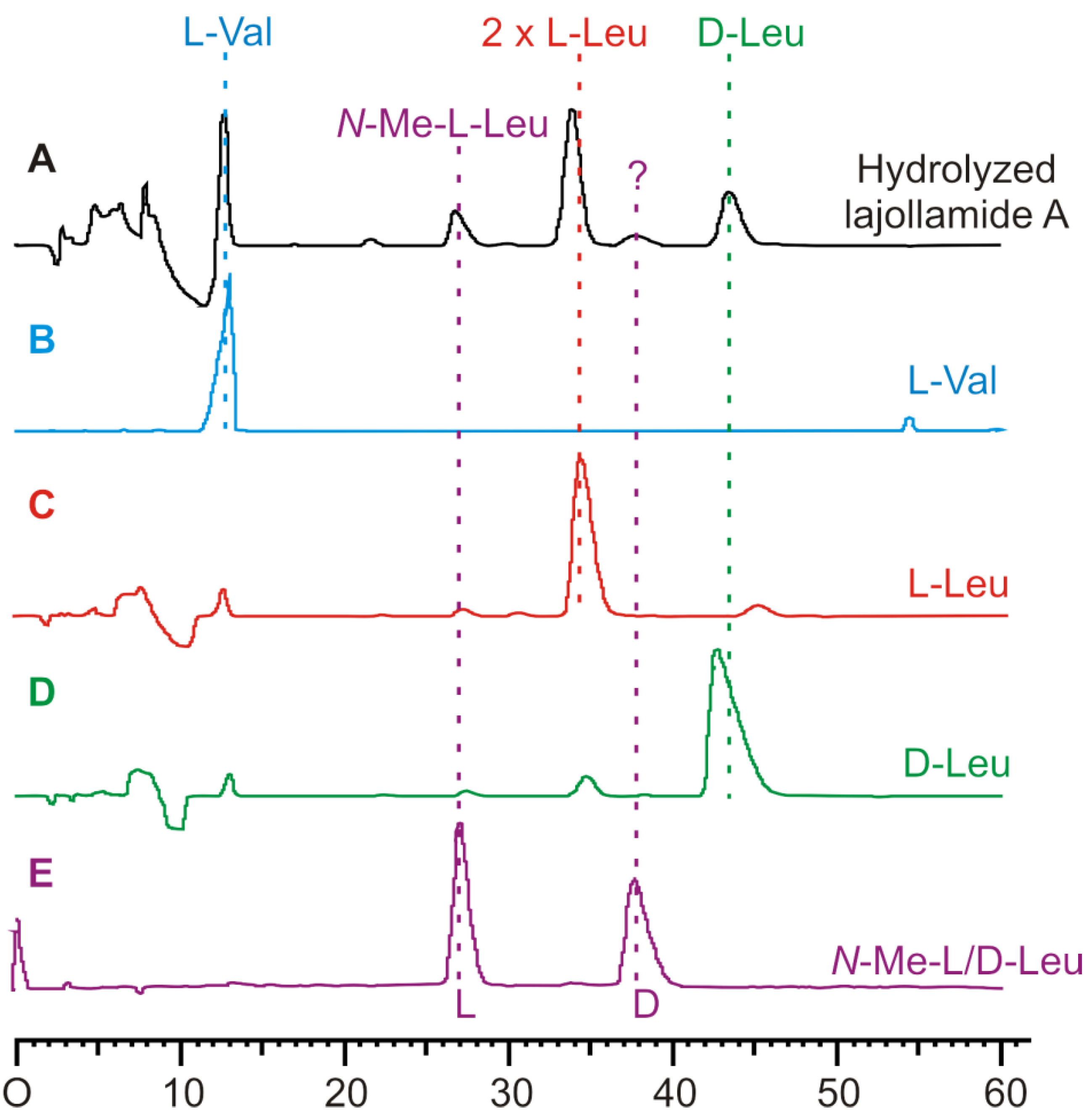
2.3. Initial Stereochemical Investigations

To elucidate the configurations at the stereogenic centers of the amino acid moieties in 1, the natural product was fully hydrolyzed under acidic conditions. The hydrolysate obtained was analyzed by chiral HPLC. The stereochemical identity of the hydrolyzed amino acids observed in the HPLC chromatogram (Figure 3) was established by comparison with the respective commercial authentic standards of known absolute configuration. This led to the identification of N-Me-L-Leu, L-Val and three Leu residues, two of which are bearing L- and one D-configuration. The position of the expected D-Leu residue within the peptide backbone, however, was not evident from this, nor from the NMR spectroscopic data. To clarify this unsolved question and thus to fully stereochemically characterize 1, we embarked on the total synthesis of the three possible diastereomers of this cyclic peptide.
Figure 3. Stereochemical analysis of the amino acid building blocks in 1 by chiral HPLC after its acid-mediated hydrolysis. Selection of HPLC-UV traces used for assignments: (A) hydrolyzed lajollamide A (1, black), (B) L-Val (blue), (C) L-Leu (red), (D) D-Leu (green), and (E) N-Me-L/D-Leu (purple).
Marinedrugs 10 02912 g003

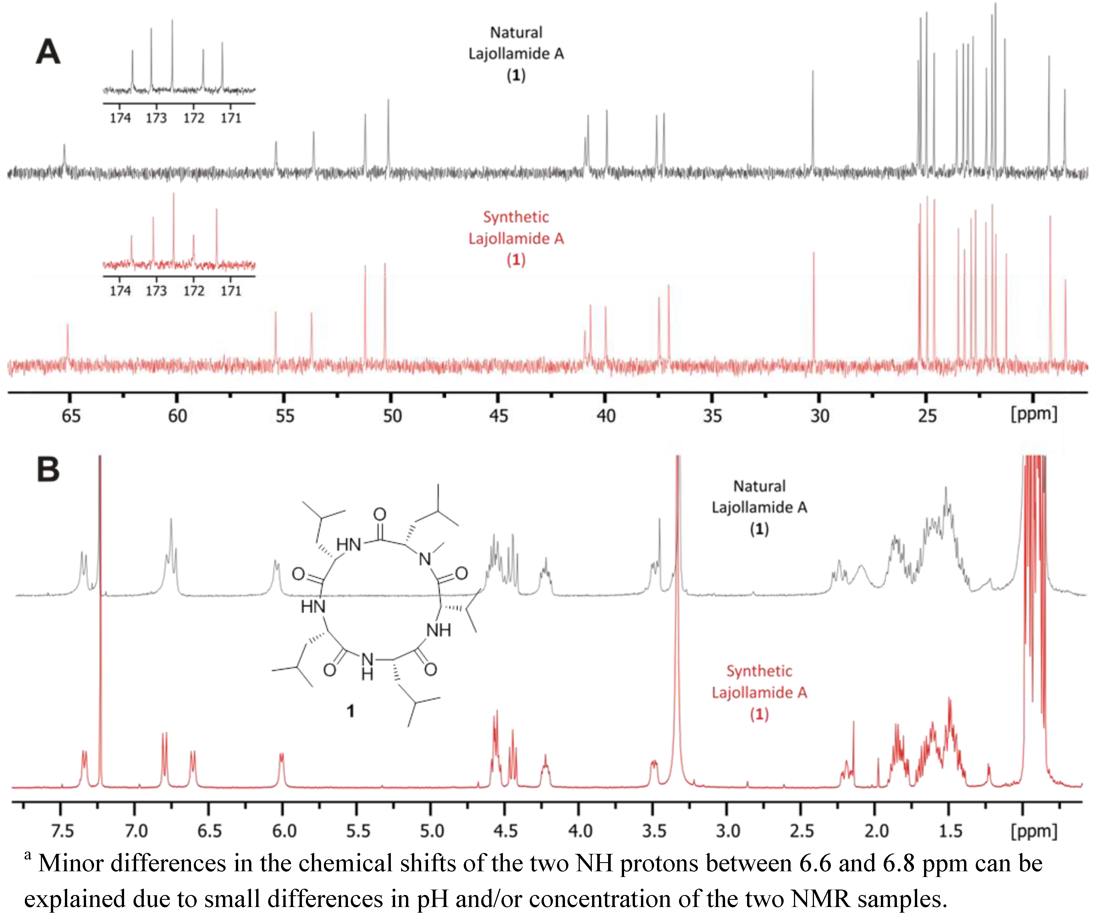
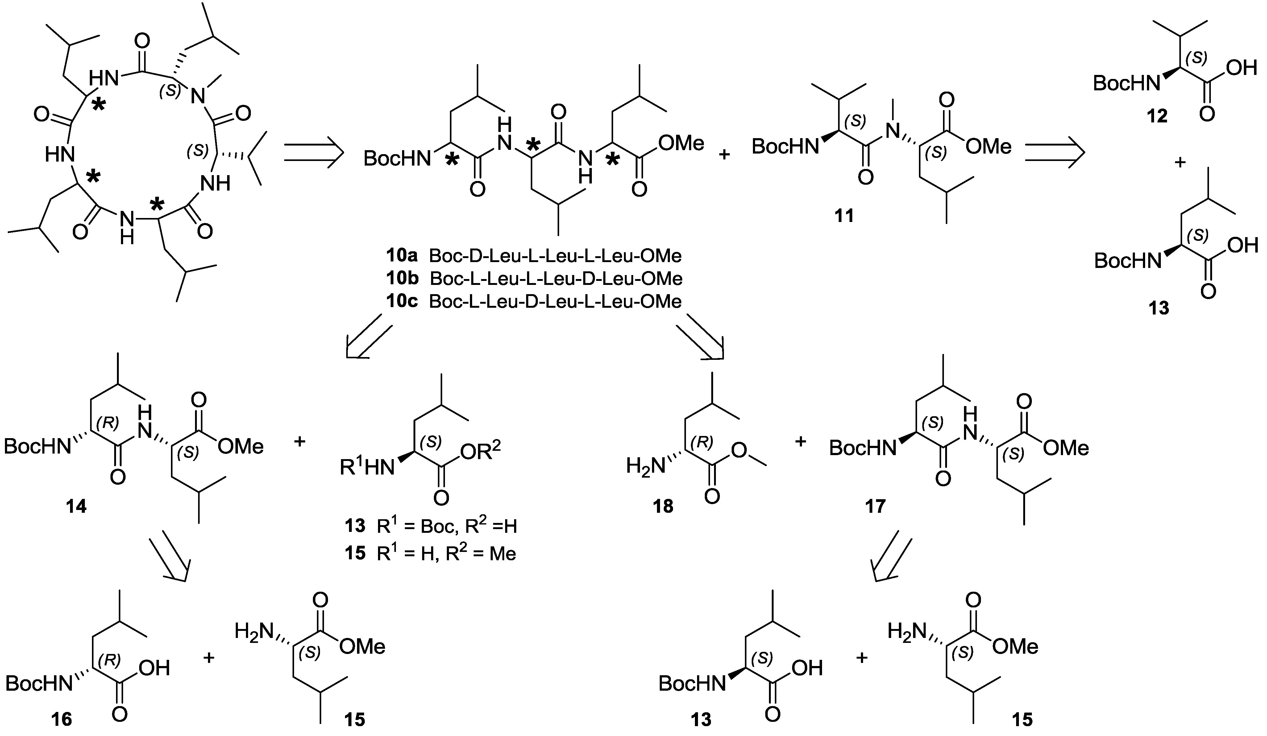
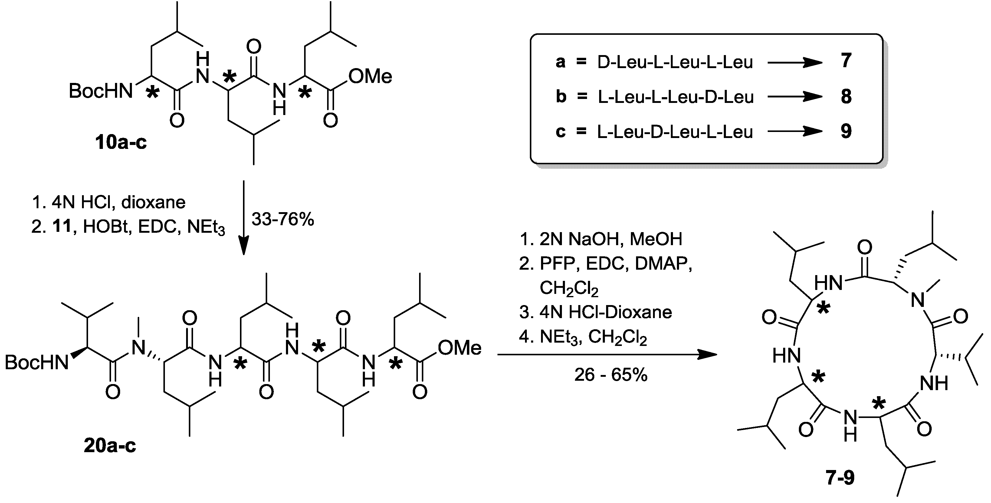
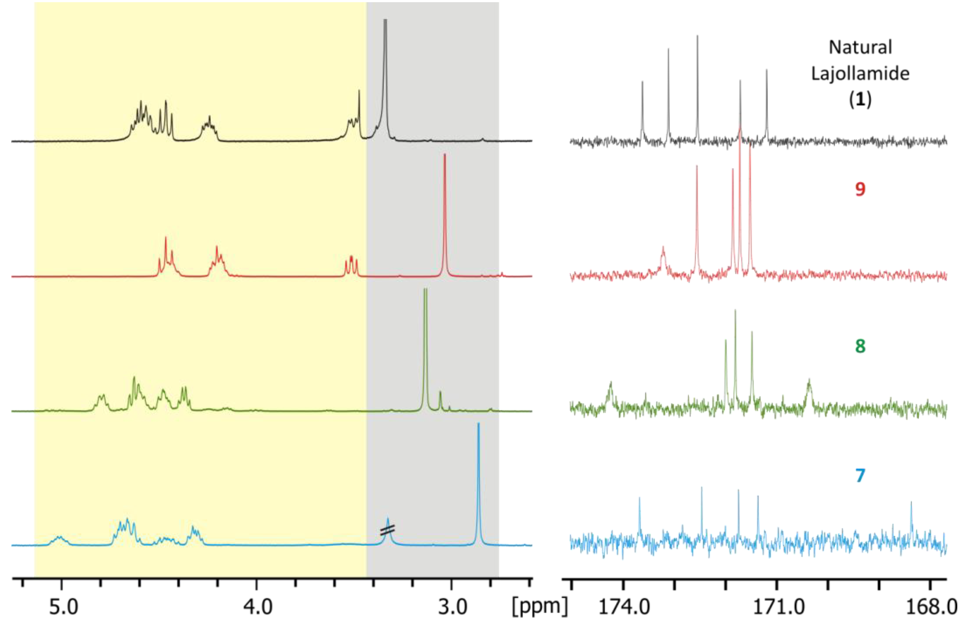
2.4. Total Synthesis of Natural Lajollamide A (1) and of Its Diastereomeric Analogs B–D (7–9)

In order to most efficiently access the three diastereomeric forms of lajollamide feasible based on the results of the peptide hydrolysis experiments, we chose a highly convergent synthetic approach applying solution phase peptide coupling chemistry. Retrosynthetically, the cyclic pentapeptide was divided into the three diastereomeric tripeptides 10ac and the stereochemically defined dipeptide building block 11 common to all target structures (Scheme 1). The latter can be derived of N-Boc-L-Val (12) and N-Boc-L-Leu (13). For the preparation of 10a and 10c, again a joint precursor dipeptide 14 could be defined, which is divergently transformable into the desired tripeptides by attaching N-Boc-L-Leu (13) or L-Leu-OMe (15) to its N- or C-terminus, respectively. The third fragment, 10b, is accessible from D-Leu-OMe (18) and N-Boc-L-Leu-L-Leu-OMe (17). Dipeptides 14 and 17 are easily affordable from the commercially available amino acid precursors 13, 15 and 16.
Scheme 1. Retrosynthetic analysis for the preparation of all possible lajollamide diastereomers bearing one D-Leu unit.
Marinedrugs 10 02912 g006
The synthesis of the tri- (10ac) and dipeptide (11) building blocks proceeded smoothly using standard peptide deprotection and coupling chemistry (Scheme 2): all peptide coupling steps were conducted using EDC and HOBt in CH2Cl2 with NEt3 as the base. Mild N-deprotection was achieved employing in-situ generation of HCl in MeOH by addition of acetylchloride. Saponification of the ester protective groups was generally carried out by applying 2 N NaOH in MeOH. Using these conditions, peptides 10a and 10c were prepared in three steps each, starting with 16 via dipeptide 14 in an overall yield of 62% and 54%, respectively. Analogously, 10b was accessible from 13 in 71% overall yield. For the preparation of the universal dipeptide precursor 11, N-Boc-L-Leu (13) was first dimethylated using MeI and NaH, with subsequent N-Boc removal to give 19. This compound was coupled to 12 by using EDC and HOBt in CH2Cl2 with DIPEA as the base and saponified to furnish 11 in 55% overall yield.
Having all desired building blocks in hands, we started the assembly of the three lajollamide diastereomers (Scheme 3). After removal of the N-Boc group in 10ac using 4N HCl in dioxane, coupling to 11 was again achieved with HOBt/EDC. The resulting fully protected pentapeptides 20ac were saponified, activated by PFP-ester formation and directly cyclized by N-deprotection followed by in-situ addition of NEt3, leading to compounds 79.
Scheme 2. Synthesis of the tri- (10ac) and dipeptide (11) building blocks of lajollamide A (1). a: HOBt, EDC, NEt3, CH2Cl2; b: AcCl, MeOH; c: 2N NaOH, MeOH; d: MeI, NaH, THF/DMF (10:1), 80 °C, 20h; e: HOBt, EDC, DIPEA, CH2Cl2.
Marinedrugs 10 02912 g007
Scheme 3. Assembly of the di- (11) and tripeptide (10ac) building blocks to the target cyclopentapeptide structures 79.
Marinedrugs 10 02912 g008
With the finalization of the synthesis of all anticipated lajollamide diastereomers 79, we started to compare the spectroscopic data of the authentic natural product with that of the synthetic material. To our surprise, however, none of the peptides 79 was identical to the natural compound. This was clearly evident from substantial differences in chemical shifts in both 1H and 13C NMR spectra. For example, the chemical shifts of the amino acid α protons or the N-methyl groups differed significantly, as did all carbon chemical shifts, which was particularly apparent for the amide carbonyl signals (Figure 4).
Figure 4. Comparison of selected signals in the 1H (left) and 13C (right) NMR spectra of natural lajollamide A (1, black), and compounds 7 (blue), 8 (green) and 9 (red). Shaded areas in the 1H spectra show α protons (yellow) and N-methyl groups (grey).
Marinedrugs 10 02912 g004
This unexpected outcome of our synthetic endeavors led us to revisit the initial analytical data of the fungal metabolite. While no ambiguities were found in the interpretation of the NMR data, a small peak with a retention time of ca. 38 min. in the HPLC chromatogram of the full hydrolysate of the natural product drew our attention (see Figure 3). In fact, based on comparison with authentic standards, this signal turned out to correspond to N-Me-D-Leu. As the planar structure of lajollamide only contains one N-Me-Leu moiety, which was assigned to be L-configured due to the large peak at ca. 27 min in our stereochemical analysis, the occurrence of a fraction of N-Me-D-Leu clearly indicated a partial epimerization of this unit during peptide hydrolysis. This led to the assumption that a similar racemization process can also be expected for the three other Leu building blocks present in the molecule. This opened the possibility of three L-Leu units initially being present in the natural product, which, assuming an epimerization rate of approx. 30% for each of these residues, would result in a 2:1 ratio of L- to D-Leu in the analysis of the peptide hydrolysate, thus potentially explaining the spectroscopic differences of the natural product 1 when compared to the synthetic analogs 79. With these thoughts in mind, the preparation of a lajollamide diastereomer bearing L-configuration at all amino acids residues was undertaken. The synthesis of the respective all L-tripeptide precursor 10d followed the route depicted for the preparation of 10b as shown in Scheme 2, altered only by simply attaching L-Leu-OMe to the C-terminus of 17 in the second peptide coupling step. Compound 10d was obtained in overall 67% yield and further elaborated into the desired cyclic pentapeptide via all L-configured 20d according to the sequence depicted in Scheme 3. Due to the convergent nature of our synthetic approach, the preparation of the all L-configured lajollamide-type target molecule was thus easily achieved with relatively little additional experimental work. To our great relief, this time, the analytical data for the synthetic material perfectly matched those of the natural product 1 (Figure 5). We thus unambiguously established the absolute configuration in natural 1, henceforth named lajollamide A, to be all L. The three stereochmically altered, purely synthetic analogs of 1 were consequently defined as lajollamides B–D (79).
Figure 5. Comparison of the 13C (A) and 1H (B) NMR data of the isolated lajollamide A (1, black) with that of the identical, synthetically derived, all-L-configured material (red) a.
Marinedrugs 10 02912 g005

2.5. Biological Activities of Compounds 1–9

No cytotoxic or enzyme inhibitory activity was observed for compounds 19. In antibacterial assays, metabolites 25 were found to be inactive. However compounds 1, 7, 8 and 9 exhibited a weak antibacterial activity against Gram-positive bacteria at a concentration of 100 μM. The growth inhibition of Bacillus subtilis was 61%, 51%, 67%, and 41%, respectively. Pathogenic bacterial strains causing infectious diseases were also inhibited. Inhibition of Staphylococcus epidermidis was shown for compound 1 (30%), 7 (43%) and 8 (32%). Only compound 7 was active against methicillin-resistant Staphylococcus aureus (MRSA). Furthermore, in agar diffusion assays at the 50 μg level, potent antibacterial and antifungal properties towards Escherichia coli (2.5 mm total inhibition), Bacillus megaterium (7 mm total inhibition), Mycotypha microspora (13.5 mm total inhibition), Eurotium rubrum (4 mm total inhibition), and Microbotryum violaceum (13 mm total inhibition) were demonstrated by 6, which in some cases surpassed the activity of the positive controls streptomycin, benzylpenicillin and miconazole.

3. Experimental Section

3.1. General Experimental Procedures

Optical rotations were measured on a Jasco DIP 140 polarimeter. UV and IR spectra were obtained using Perkin-Elmer Lambda and Perkin-Elmer spectrum BX instruments, respectively. All NMR spectra of the isolated material were recorded on a Bruker Avance 300 DPX spectrometer. Spectra of the synthetic compounds were acquired on Bruker AM 300 and AM 400 spectrometers. Spectra were referenced to residual protonated solvent signals with resonances at δH/C 3.35/49.0 (CD3OD) or 7.26/77.0 (CDCl3), respectively. HR-EI and HR-ESI-MS data was recorded on Kratos MS50, a Bruker Daltonics micrOTOF-Q and a Thermo Finnigan MAT 95 XL. Semipreparative HPLC was carried out using a Waters system consisting of a degasser, a 600 pump, a 996 photodiode array detector, and a 717 plus autosampler in combination with a Waters fraction collector. Chiral HPLC was performed on a Merck-Hitachi system consisting of a L-6200 A pump, a L-4500 A photodiode array detector (PDA), and a D-6000 A interface. LC-MS measurements were obtained by employing an Applied Biosystems LC/MS system consisting of an Agilent 1100 HPLC system and an MDS Sciex API 2000 mass spectrometer equipped with an API-ESI source. All solvents used were distilled under argon and dried over 3 Å molecular sieves prior to use. All amino acids, 1-(3-dimethylaminopropyl)-3-ethylcarbodiimide (EDC), pentafluorophenol (PFP) were purchased from Carbolution Chemicals. 1-Hydroxybenzotriazole hydrate (HOBt∙H2O) was purchased from Aldrich, triethylamine (NEt3) from Grüssing GmbH and methyl iodide from Merck. Sodium hydride and 4N HCl/dioxane were delivered from Acros Organics. All chemicals were used without further purification. For thin layer chromatography TLC Silica gel 60 F254 from Merck was used. The dyeing reagent consisted of ninhydrin in ethanol. For column chromatography, silica gel was purchased from Merck.

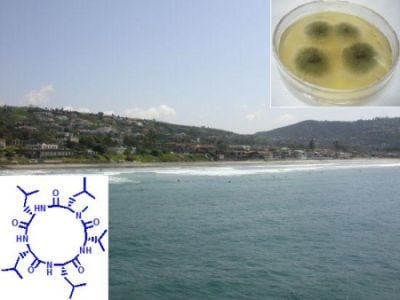
3.2. Biological Material

A piece of an unidentified decaying green alga, floating in the water column at a depth of 20 feet was collected by scuba in 2005 at La Jolla shore, San Diego, USA. Following transport in sterile seawater to Germany, the fungal strain #763 was isolated among other fungi from the algal sample, freed from competing microorganisms and other contaminants as previously described [23,24,25] and deposited at the fungal culture collection of the Institute for Pharmaceutical Biology, University of Bonn, Germany. The fungus was identified as Asteromyces cruciatus Moreau et Moreau ex Hennebert 1962.

3.3. Screening of Secondary Metabolites Biosynthesized under Different Conditions (OSMAC Aproach)

The OSMAC approach was based on cultures of the fungus in media derived from the basic components of the Czapek-Dox broth [26,27]: NaNO3 (3 g/L), FeSO4 (0.01 g/L), KCl (0.5 g/L), MgSO4 (0.5 g/mL), K2HPO4 (1.0 g/L), carbon source (30 g/L, originally sucrose, however, when not indicated, glucose was used) in artificial sea water (ASW). For the preparation of the culture medium, all components except the carbon source were mixed in 250 mL (1 L capacity, baffled Erlenmeyer flasks) of ASW and the mixtures were autoclaved (121 °C, 20 min). Subsequently, 20 mL of 40% (m/V) carbon source were added by sterile filtration to the medium, followed by inoculation with small pieces of seed culture. Small agar pieces (1 × 1 cm) of a 12–25-day-old culture of A. cruciatus grown on biomalt salt agar medium served as seed cultures. In order to find culture conditions conducive to production of new metabolites, two replicate cultures of A. cruciatus were grown in each of 14 different culture conditions: (#1) Czapek-Dox medium with shaking (#2) Czapek-Dox medium without shaking for generation of low O2 levels [28], (#3) dixenic cultivation [29] with 10 mL of the bacterium Pseudomonas fluorescens Pf-5 [30], added on day 6, (#4) Czapek-Dox medium containing 20 g/L Amberlite XAD-16, (#5) Czapek-Dox medium supplemented with 1 mL/L cofactor solution consisting of MnCl2·4H2O (100 mg), CoCl2 (20 mg), CuSO4 (10 mg), Na2MoO4·2H2O (10 mg), ZnCl2 (20 mg), LiCl (5 mg), SnCl2·2H2O (5 mg), H3BO3 (10 mg), K3BO3 (10 mg), KBr (20 mg), KI (20 mg), EDTA-Na+ (5.8 mg) in 1 L of distilled water [28], (#6) Czapek-Dox supplemented with 4.6 mg/L CdCl2 as polyketide inducer [31], (#7) Czapek-Dox medium, cultivated from day 10 to 13 under elevated UV-B light exposition, (#8) Czapek-Dox medium, cultivated from day 8–10 at 35 °C [28], (#9) Czapek-Dox medium, cultivated with sub-inhibitory concentrations of 0.4 mL/L cycloheximide [32,33], (#10) Czapek-Dox medium, supplemented with 8 mL/L toluene, (#11) Czapek-Dox without NaNO3, containing 1.0 g/L Arg, 2.5 g/L Asn and 1.5 g/L Glu [28], (#12) Czapek-Dox with reduced levels of K2HPO4 (228.2 mg/L) for phosphate repression [28], (#13) Czapek-Dox medium without FeSO4 for induction of siderophores, (#14) Czapek-Dox medium with sucrose instead of glucose as carbon source [28]. If not stated otherwise, the cultures were grown with shaking (120 rpm) at 24 °C. The broth was harvested at 10 days after inoculation. The culture broths of each fermentation were extracted separately with EtOAc (2 × 250 mL), and evaporated under reduced pressure to afford dark brown semisolids, ranging from 0.3 to 47.2 mg. In the case of culture condition #4, additionally the medium polarity compounds absorbed by the XAD resin material were eluted by shaking, first for 1 h in 200 mL EtOAc, and subsequently for 1 h in 200 mL acetone. All resulting three organic crude extracts of cultivation experiment #4 were combined. The resultant crude extracts of each experiment were dissolved in methanol to a final concentration of 5 mg/mL, and 10 μL was profiled by LC/MS using a 2 mM NH4OAc buffered MeOH/H2O gradient, increasing the MeOH portion from 10% to 100% over 20 min and holding it at 100% MeOH for additional 10 min (Macherey-Nagel C18 Nucleodur 100-5 column, 2 × 125 mm, 5 μm column; 0.25 mL/min flow rate, with total ion current and photodiode array monitoring at 200–400 nm).

3.4. Scaled-Up Cultivation, Extraction and Isolation of Metabolites of A. cruciatus

The fungus was cultivated for ten days in five 5 L-Erlenmeyer flasks, each containing 1.5 L of liquid media (=7.5 L). The liquid medium consisted of Czapek-Dox medium with glucose (30 g/L) as carbon source which was either supplemented with co-factors or contained solely the amino acids Arg, Asn and Glu as nitrogen source instead of NaNO3. Flasks were shaken on a rotary shaker incubator at 24 °C and 120 rpm. On day 10, the cultivation medium and mycelium of each experiment was extracted with EtOAc (2 × 7.5 L). After evaporation of the organic solvent, both crude extracts (medium with trace elements: 126 mg; medium with an altered nitrogen source: 98 mg) were re-suspended separately in MeOH and filtered through reversed phase silica gel (RP-SPE cartridge, 100 mg). Separation of each filtrate by RP-HPLC analysis using a linear gradient of 40:60–100:0 MeOH-H2O over a period of 54 min (column: Knauer Eurospher-100-C18; 8 × 250 mm, 5 μm; 2 mL/min; DAD detection) yielded collectively a compound (amount from trace element medium/amount from altered nitrogen source) 1 (18.6/7.1 mg), 2 (0.9/0.7 mg), 3 (1.6/3.3 mg), 4 (19.8/19.4 mg), 5 (1.8/0.9 mg) and 6 (1.0/3.2 mg).
lajollamide A (1): yellowish glass; [α]D24 −80 (c 0.17, MeOH); UV/VIS (MeOH) λmax (ε): 204 (12,600); IR (ATR): 3284, 2956, 1633, 1530 cm−1; HR-ESI-MS m/z 566.4280 [M + H]+ (calc. for C30H56N5O5, 566.4276, Δ +0.7 ppm); 1H and 13C NMR data in CDCl3, see Table 1.
regiolone, syn. (−)-isosclerone (2): Colorless film; [α] D25 −16 (c 0.02, MeOH), {lit. [21] [α]D −68 (c 0.05, MeOH)}; UV/VIS (MeOH) λmax (ε): 212 (15,800), 262 (9900), 324 (4200); 1H NMR (300 MHz, CDCl3) δH ppm: 1.87 (1H, brs, OH-4), 2.19 (1H, m, H-3a), 2.34 (1H, m, H-3b), 2.65 (1H, m, H-2a), 3.01 (1H, m, H-2b), 4.92 (1H, m, H-4), 6.93 (1H, d, 8.5 Hz, H-7), 7.02 (1H, d, 7.4 Hz, H-5), 7.50 (1H, dd, 7.4, 8.5 Hz, H-6), 12.43 (1H, s, OH-8); 13C NMR (75 MHz, CDCl3) δC ppm: 31.2 (CH2, C-3), 34.6 (CH2, C-2), 67.7 (CH, C-4), 115.2 (qC, C-8a), 117.3 (CH, C-5), 117.8 (CH, C-7), 137.0 (CH, C-6), 145.8 (qC, C-4a), 162.7 (qC, C-8), 204.2 (qC, C-1); LR-ESI-MS m/z 177.1 [M − H].
(3R,6R)-hyalodendrin, syn. A26771A (3): Yellowish film; [α] D24 −71 (c 0.15, CHCl3) {lit. [16] [α]D27 −88 (c 0.15, MeOH)}; 1H NMR (300 MHz, CDCl3) δH ppm: 2.97 (3H, s, 4NCH3), 3.21 (3H, s, 1NCH3), 3.61 (1H, d, 16 Hz, H-8a), 4.08 (1H, d, 16 Hz, H-8b), 4.35 (2H, m, H2-7), 7.30 (5H, m, H-10, H-11, H-12, H-13, H-14); 13C NMR (75 MHz, CDCl3) δC ppm: 27.5 (4NCH3), 28.6 (1NCH3), 36.8 (CH2, C-8), 61.2 (CH2, C-7), 75.2 (qC, C-6), 75.7 (qC, C-3), 127.4 (CH, C-12), 128.7 (2 × CH, H-10, H-14), 129.0 (2 × CH, H-11, H-13), 134.0 (qC, C-9), 165.5 (qC, C-2), 166.9 (qC, C-5); HR-ESI-MS m/z 347.0491 [M + Na]+ (calc. for C14H16N2NaO3S2, 347.0495, Δ −1.2 ppm).
(3R,6R)-gliovictin, syn. A26771E, (3R,6R)-bisdethiodi(methylthio)hyalodendrin (4): Colorless film; [α]D24 −58 (c 0.88, CHCl3) {lit. [17] [α]D25 −65 (CHCl3); [9] [α]D −62 (c 1.88, CHCl3); [16] [α]D27 −47 (c 0.13, CH3OH); [18] [α]D24 −43 (c 0.24, CHCl3)}; 1H NMR (300 MHz, CDCl3) δH ppm: 1.24 (1H, t, 6.9 Hz, OH-7), 2.13 (3H, s, 6SCH3), 2.30 (3H, s, 3SCH3), 3.02 (3H, s, 4NCH3), 3.10 (1H, dd, 6.9 and 11.8 Hz, H-7a), 3.13 (1H, d, 13.8 Hz, H-8a), 3.31 (3H, s, 1NCH3), 3.71 (1H, d, 13.8 Hz, H-8b), 3.85 (1H, dd, 6.9 and 11.8 Hz, H-7b), 7.09–7.27 (5H, m, H-10, H-11, H-12, H-13, H-14). 13C NMR (75 MHz, CDCl3) δC ppm: 13.4 (6SCH3), 14.3 (3SCH3), 29.3 (1NCH3), 31.0 (4NCH3), 42.3 (CH2,C-8), 64.3 (CH2, C-7), 71.5 (qC, C-6), 73.7 (qC, C-3), 127.9 (CH-12), 128.7 (2 × CH, C-11 and C-13), 130.1 (2 × CH, C-10 and C-14), 134.1 (qC, C-9), 165.3 (qC, C-2), 165.5 (qC, C-5); HR-ESI-MS m/z 377.0965 [M + Na]+ (calc. for C16H22N2NaO3S2, 377.0970, Δ −1.3 ppm).
(3R,6R)-1N-norgliovictin, syn. bisdethiodi(methylthio)-1-demethylhyalodendrin (5): Colorless film; [α]D24 −37 (c 0.05, CH3OH) {lit. [18] [α] D24 −63 (c 0.30, CHCl3)}; 1H NMR (300 MHz, CDCl3) δH ppm: 1.71 (1H, brs, OH-7); 2.19 (3H, s, 3SCH3), 2.22 (3H, s, 6SCH3), 2.71 (1H, d, 11.6 Hz, H-7a), 3.15 (1H, d, 13.8 Hz, H-8a), 3.28 (3H, s, 4NCH3) , 3.41 (1H, d, 11.6 Hz, H-7b), 3.53 (1H, d, 13.8 Hz, H-8b), 6.36 (1H, brs, NH-1), 7.09–7.32 (5H, m, H-10, H-11, H-12, H-13, H-14); 13C NMR (75 MHz, CDCl3) δC ppm: 13.5 (6SCH3), 14.4 (3SCH3), 30.3 (4NCH3), 42.3 (CH2, C-8), 64.8 (qC, C-6), 65.2 (CH2, C-7), 75.7 (qC, C-3), 128.0 (CH, C-12), 128.8 (2 × CH, C-10, C-14), 130.0 (2 × CH, C-11, C-13), 133.7 (qC, C-9), 164.9 (2 × qC, C-2, C-5); HR-ESI-MS m/z 363.0818 [M + Na]+ (calc. for C15H20N2NaO3S2, 363.0813, Δ +1.4 ppm).
(3R,6R)-bis-N-norgliovictin (6): [α] D24 −8 (c 0.1, CH3OH), {lit. [20] [α]D −32 (c 0.1, CH3OH); [18] [α]D25 −34 (c 0.30, dioxane)}; 1H NMR (300 MHz, CH3OD) δH ppm: 2.23 (3H, s, 6SCH3), 2.37 (3H, s, 3SCH3), 3.05 (H, d, 11.5 Hz, H-7a), 3.07 (1H, d, 13.3 Hz, H-8a), 3.40 (H, d, 11.5 Hz, H-7b), 3.62 (1H, d, 13.3 Hz, H-8b), 7.29 (5H, m, H-10, H-11, H-12, H-13, H-14); 13C NMR (75 MHz, CH3OD) δC ppm: 13.5 (3SCH3 or 6SCH3), 13.9 (3SCH3 or 6SCH3), 46.0 (CH2, C-8), 66.5 (CH2, C-7), 67.8 (qC, C-3 or C-6), 68.8 (qC, C-3 or C-6), 128.4 (CH, C-12), 129.4 (2 × CH, C-11, C-13), 131.9 (2 × CH, C-10, C-14), 135.7 (qC, C-9), 167.8 (qC, C2 or C5), 168.2 (qC, C-2 or C-5); HR-ESI-MS m/z 349.0651 [M + Na]+ (calc. for C14H18N2NaO3S2, 349.0657, Δ −1.7 ppm).

3.5. Amino Acid Analysis by Chiral HPLC

Lajollamide A (1.3 mg) was hydrolyzed with 6 N HCl in a sealed vial at 108 °C for 19 h. Excess aqueous HCl was removed under vacuum and the resulting hydrolysate was analyzed by chiral HPLC employing a Phenomenex Chirex 3126 (D) column (4.6 × 250 mm) eluting with 2 mM CuSO4/MeCN (95:5) at a flow of 0.8 mL/min, monitoring at 240 nm. Comparison with commercially available standards suggested the presence of L-Val, N-Me-L-Leu, 2 × L-Leu and 1 × D-Leu. The hydrolysate was chromatographed alone and co-injected with standards to confirm assignments.

3.6. General Synthetic Protocols for the Preparation of Lajollamides A–D (1, 7–9)

3.6.1. Peptide Coupling (Protocol A)

The amine (1 equivalent) was dissolved in CH2Cl2. After cooling to 0 °C, the acid (1.1 equivalent), HOBt (1 equivalent), EDC (1.25 equivalent) and NEt3 (5 equivalent) were added successively to the solution. The reaction mixture was stirred at room temperature overnight. The solvent was removed under vacuum. The crude product was taken up in water and extracted with CH2Cl2. The organic layers were combined and washed successively with saturated aqueous NaHCO3 solution, 40% citric acid solution, brine and dried over MgSO4. The solvent was removed under reduced pressure and the crude product purified by column chromatography.

3.6.2. N-Deprotection (Protocol B)

3.6.2.1. Deprotection with Acetyl Chloride in Methanol

N-Boc-protected peptide ester (1 equivalent) was dissolved in MeOH (24 equivalent). After cooling to 0 °C, acetyl chloride (2.2 equivalent) was added dropwise to the reaction solution which was further stirred at room temperature until completion of N-Boc removal as indicated by TLC. After solvent evaporation the crude product was taken up in CH2Cl2 and neutralized with saturated NaHCO3 solution. The aqueous layer was extracted with CH2Cl2. The organic layers were combined and dried over anhydrous MgSO4. After filtration the solvent was removed under high vacuum.

3.6.2.2. Deprotection with 4 M HCl∙Dioxane

N-Boc-protected peptide ester (1 equivalent) was treated with 4 M HCl/dioxane (100 equivalent) under argon and stirred at room temperature for 1 h. The resulting reaction mixture was concentrated under high vacuum.

3.6.3. Saponification (Protocol C)

N-Boc-protected peptide ester (1 equivalent) was dissolved in MeOH (60 equivalent) und treated with 2 N NaOH (65 equivalent) solution. The reaction mixture was stirred at room temperature until the saponification finished as indicated by TLC. After solvent evaporation the crude product was taken up in CH2Cl2 and washed with H2O. The aqueous phase was acidified with 1 N HCl to pH 2 and extracted with CH2Cl2. The combined organic layers were dried over anhydrous MgSO4 and the solvent was removed under high vacuum.

3.6.4. Macrolactonization (Protocol D)

A fully protected pentapeptide (1 equivalent) was treated with 2 N NaOH following general protocol c. The resulting product was dissolved in CH2Cl2. After cooling to −10 °C EDC (1.3 equivalent), PFP (3 equivalent) and DMAP (0.1 equivalent) were added. The reaction mixture was stirred at room temperature overnight. After solvent evaporation and purification by column chromatography the resulting activated PFP ester was treated with 4 N HCl/dioxane (100 equivalent) following general protocol b2 to give the precursor for macrocyclisation. A highly dilute solution of the PFP ester (1 equivalent) in CH2Cl2 (0.003–0.01 M) was added dropwise to a solution of NEt3 (20 equivalent) in CH2Cl2. The resulting solution was stirred at room temperature overnight. After solvent evaporation the crude product was taken up in H2O and extracted with CH2Cl2. The combined organic layers were washed with 40% citric acid solution and brine and dried over anhydrous MgSO4. After filtration the solvent was removed under high vacuum.

3.7. Total Synthesis of Lajollamides A–D (1, 7–9)

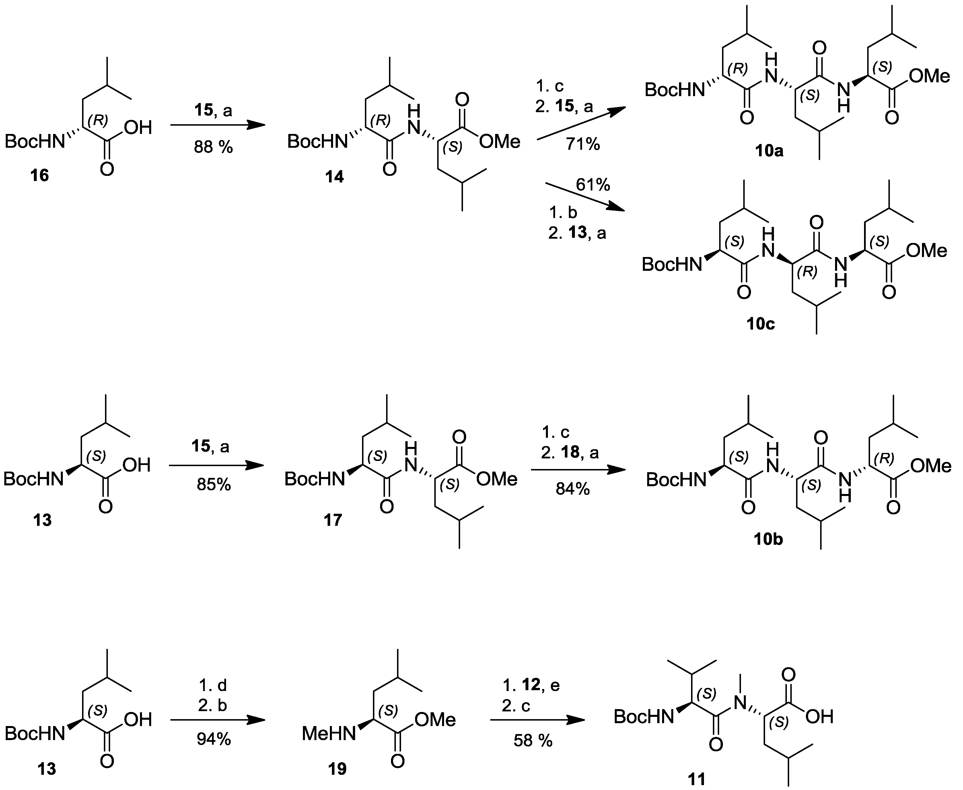
3.7.1. N-Boc-L-Leu-L-Leu-OMe (17)

Peptide coupling of 10.0 g (40.1 mmol) N-Boc-L-Leu (13) with 6.4 g (44.1 mmol) L-Leu-OMe (15) was conducted following general protocol a, yielding 12.2 g (34.1 mmol, 85%) of the desired product 17 after chromatography on silica gel using ethylacetate and cyclohexane in 1:1 ratio as the eluent. 1H NMR (400 MHz, CDCl3) δH ppm: 0.85–0.98 (12H, m), 1.42 (9H, s), 1.51–1.65 (6H, m), 3.71 (3H, s), 4.09 (1H, brs), 4.59 (1H, m), 4.92 (1H, brs), 6.49 (1H, d, 6.5 Hz); 13C NMR (100 MHz, CDCl3) δC ppm: 21.8, 22.8, 22.8, 24.6, 24.6, 24.7, 28.2, 40.8, 41.5, 50.6, 52.2, 52.9, 80.2, 155.6, 172.2, 173.1; ESI-MS m/z 381.2 [M + Na]+, 281.2 [M + Na − C5H9NO2]+.

3.7.2. N-Boc-D-Leu-L-Leu-OMe (14)

Peptide coupling of 4.99 g (20.0 mmol) N-Boc-D-Leu (16) with 3.2 g (22.0 mmol) L-Leu-OMe (15) was conducted following general protocol a, yielding 6.3 g (17.6 mmol, 88%) of the desired product 14 after chromatography on silica gel using ethyl acetate and cylcohexane in 1:1 ratio as the eluent. 1H NMR (400 MHz, CDCl3) δH ppm: 0.87–0.95 (12H, m), 1.43 (9H, s), 1.51–1.65 (6H, m), 3.70 (3H, s), 4.13 (1H, m), 4.57 (1H, m), 4.94 (1H, brs), 6.67 (1H, brs); 13C NMR (100 MHz, CDCl3) δC ppm: 21.9, 22.9, 23.0, 24.8, 24.9, 24.9, 28.4, 41.1, 41.5, 50.8, 52.4, 53.1, 80.2, 155.8, 172.6, 173.4; ESI-MS m/z 381.2 [M + Na]+, 281.2 [M + Na − C5H10N]+.

3.7.3. N-Boc-D-Leu-L-Leu-L-Leu-OMe (10a)

6 g (16.7 mmol) N-Boc-D-Leu-L-Leu-OMe (14) were saponified following general protocol c. The success of the deprotection step was monitored by 1H NMR of the resulting crude product (1H data for N-Boc-D-Leu-L-Leu: (400 MHz, CDCl3) δH ppm: 0.88–0.97 (12H, m), 1.41 (9H, s), 1.57–1.67 (6H, m), 4.41 (1H, m), 4.60 (1H, m), 5.39 (1H, m), 7.11 (1H, brs), 10.55 (1H, brs)). This material was used in the peptide coupling step according to general protocol a with 2.1 g (14.8 mmol) L-Leu-OMe (15), yielding 5.6 g (11.9 mmol, 71% overall) of the desired product 10a after chromatography on silica gel using ethylacetate and cyclohexane in 1:1 ratio as the eluent. 1H NMR (400 MHz, CDCl3) δH ppm: 0.85–0.95 (18H, m), 1.42 (9H, s), 1.51–1.72 (9H, m), 3.70 (3H, s), 4.09 (1H, m), 4.47 (1H, m), 4.54 (1H, m), 4.97 (1H, brs), 6.70 (1H, brs), 6.78 (1H, brs); 13C NMR (75 MHz, CDCl3) δC ppm: 21.9, 22.1, 22.1, 22.9, 23.0, 24.7, 24.7, 24.8, 24.8, 28.4, 40.7, 41.1, 41.7, 50.8, 51.5, 52.2, 53.4, 80.0, 155.6, 171.9, 172.9, 173.1; EI-MS m/z 456.3 [M − CH3], 440.3 [M − OCH3]+, 398.3 [M − C4H9O]+, 372.3 [M − Boc]+, 285.2 [M − C10H20NO2]+, 259.2 [M − C13H27N2O3]+, 186.2 [M − C14H25N2O4]+.

3.7.4. N-Boc-L-Leu-L-Leu-D-Leu-OMe (10b)

2.6 g (7.3 mmol) N-Boc-L-Leu-L-Leu-OMe (17) were saponified following general protocol c. The success of the deprotection step was monitored by 1H NMR of the resulting crude product (1H data for N-Boc-L-Leu-L-Leu: (300 MHz, CDCl3) δH ppm: 0.86–0.99 (12H, m), 1.43 (9H, s), 1.58–1.73 (6H, m), 3.93 (1H, m), 4.62 (1H, m), 5.40 (1H, d, 5.4 Hz), 6.79 (1H, d, 6.8 Hz), 9.03 (1H, brs)). This material was used in the peptide coupling step according to general protocol a with 1.11 g (7.7 mmol) D-Leu-OMe (18), yielding 2.9 g (6.15 mmol, 84% overall) of the desired product 10b after chromatography on silica gel using ethylacetate and cyclohexane in 1:1 ratio as the eluent. 1H NMR (400 MHz, CDCl3) δH ppm: 0.88–0.97 (18H, m), 1.44 (9H, s), 1.49–1.78 (9H, m), 3.69 (3H, s), 4.04 (1H, m), 4.44–4.56 (2H, m), 4.89 (1H, d, 6.7 Hz), 6.45 (1H, d, 6.5 Hz), 6.77 (1H, d, 6.8 Hz); 13C NMR (75 MHz, CDCl3) δC ppm: 21.7, 21.8, 21.9, 22.1, 22.9, 22.9, 23.0, 24.8, 26.9, 28.3, 40.8, 40.9, 41.0, 50.8. 51.6, 52.1, 53.5, 80.2, 156.0, 172.0, 172.8, 173.2; ESI-MS m/z 494.3 [M + Na]+, 438.3 [M + Na − C4H9]+, 394.3 [M + Na − C4H9O]+.

3.7.5. N-Boc-L-Leu-D-Leu-L-Leu-OMe (10c)

3.09 g (8.62 mmol) N-Boc-D-Leu-L-Leu-OMe (14) were N-deprotected following general protocol b1. The success of the deprotection step was monitored by 1H NMR of the resulting crude product (1H data for D-Leu-L-Leu-OMe: (400 MHz, CDCl3) δH ppm: 0.86–0.95 (12H, m), 1.37 (2H, m), 1.52–1.69 (6H, m), 3.38 (1H, m), 3.69 (3H, s), 4.57 (1H, m) 7.57 (1H, d, 7.6 Hz)). This material was used in the peptide coupling step according to general protocol a with 1.35 g (5.42 mmol) N-Boc-L-Leu (13), yielding 2.5 g (5.30 mmol, 61% overall) of the desired product 10c after chromatography on silica gel using ethylacetate and cyclohexane in 1:1 ratio as the eluent. 1H NMR (400 MHz, CDCl3) δH ppm: 0.88–0.98 (18H, m), 1.44 (9H, s), 1.49–1.76 (9H, m), 3.70 (3H, s), 4.07 (1H, m), 4.48 (1H, m), 4.54 (1H, m), 4.86 (1H, m), 6.46 (1H, brs), 6.75 (1H, brs); 13C NMR NMR (75 MHz, CDCl3) δC ppm: 21.5, 22.4, 22.6, 22.7, 23.0, 24.4, 24.4, 27.9, 29.3, 38.9, 39.9, 40.8, 41.5, 50.4, 51.8, 52.9, 55.6, 79.9, 155.3, 171.4, 172.7, 175.6; ESI-MS m/z 494.3 [M + Na]+, 438.2 [M + Na − C4H9]+, 394.3 [M + Na − C4H9O]+.

3.7.6. N-Boc-L-Leu-L-Leu-l-Leu-OMe (10d)

6.51 g (18.17 mmol) N-Boc-L-Leu-L-Leu-OMe (17) were saponified following general protocol c. The success of the deprotection step was monitored by 1H NMR of the resulting crude product (1H data for N-Boc-L-Leu-L-Leu: see section 3.7.4. This material was used in the peptide coupling step according to general protocol a with 2.67 g (18.3 mmol) L-Leu-OMe (15), yielding 6.79 g (14.4 mmol, 79% overall) of the desired product 10d after chromatography on silica gel using ethylacetate and cyclohexane in 1:1 ratio as the eluent. 1H NMR (400 MHz, CDCl3) δH ppm = 0.82–0.92 (18H, m), 1.39 (9H, s), 1.46–1.62 (9H, m), 3.68 (3H,s), 4.11 (1H, m), 4.52 (2H, m), 5.24 (1H, m), 6.82 (1H, m), 7.00 (1H, m); 13C NMR (100 MHz, CDCl3) δC ppm: 21.9, 22.0, 22.3, 22.8, 22.9, 24.6, 24.7, 24.8, 24.8, 28.3, 41.01, 41.1, 41.3, 50.7, 51.6, 52.2, 53.0, 79.9, 155.8, 171.9, 172.9, 173.2; ESI-MS m/z 965.5 [2M + Na]+, 494.3 [M + Na]+, 394.3 [M + Na − Boc]+.

3.7.7. N-Me-L-Leu-OMe (19)

6 g (24.06 mmol, 1 equivalent) N-Boc-L-Leu (13) were dissolved in a mixture of THF and DMF in a 10:1 ratio (198 mL) and treated with MeI (10 mL, 192.54 mmol, 8 equivalent). After cooling down to 0 °C NaH (2.89 g, 72.20 mmol, 3 equivalent, 60% dispersion in oil) was added slowly to the reaction solution. The resulting mixture was stirred at 80 °C under reflux for 20 h. After removing the solvent in vacuo, the crude product was taken up in water (150 mL) and extracted with ethyl acetate (3 × 100 mL). The combined organic layers were washed with saturated NaHCO3 (2 × 100 mL) and H2O (3 × 100 mL) and dried over MgSO4. After solvent evaporation the desired product was purified via chromatography on silica gel using ethyl acetate and cyclohexane in 1:1 ratio as the eluent yielding 6.09 g (23.5 mmol, 98%). Treatment of the resulting N-Boc-N-Me-L-Leu-OMe under conditions described in protocol B1 furnished 3.61 g (22.7 mmol, 94% overall) of product 19. 1H NMR (300 MHz, CDCl3) δH ppm: 0.83 (6H, m), 1.36–1.39 (3H, m), 1.57–1.64 (1H, m), 2.27 (3H, s), 3.11 (1H, m), 3.64 (3H, s); 13C NMR (75 MHz, CDCl3) δC ppm: 22.3, 22.6, 24.9, 34.6, 42.5, 51.5, 61.8, 176.2; ESI-MS m/z 182.1[M + Na]+, 160.1 [M + H]+, 100.1 [M − CO2Me]+.

3.7.8. N-Boc-L-Val-N-Me-L-Leu (11)

1.40 g (8.80 mmol) N-Me-L-Leu-OMe (19)were dissolved in CH2Cl2 (40 mL) and HOBt, EDC and DIPEA were added successively to the solution which was stirred at 0 °C for 20 min. 1.95 g (9.96 mmol) N-Boc-L-Val (12) in CH2Cl2 (40 mL) was added to the reaction mixture and it was stirred at room temperature overnight. After quenching with saturated NH4Cl solution, the aqueous layer was extracted with CH2Cl2. The combined organic layers were washed with saturated NH4Cl solution and dried over MgSO4. After solvent evaporation the product was purified via chromatography on silica gel using ethyl acetate and cyclohexane in 1:1 ratio, yielding 2.14 g (5.97 mmol, 68%) of the desired coupling product N-Boc-L-Val-N-Me-L-Leu-OMe. 1H NMR (300 MHz, CDCl3) δH ppm: 0.88–0.95 (12H, m), 1.41 (9H, s), 1.62–1.71 (3H, m), 1.99 (1H, m), 3.00 (3H, s), 3.68 (3H, s), 4.41 (1H, m), 5.21 (1H, m), 5.36 (1H, m); 13C NMR (75 MHz, CDCl3) δC ppm: 17.7, 19.4, 21.5, 23.4, 24.8, 28,4, 29.8, 31.2, 37.1, 52.2, 54.5, 55.5, 79.6, 156.1, 172.3, 173.6; ESI-MS m/z (%) = 381.3 [M + Na]+, 367.2 [M + Na − CH3]+, 281.2 [M − C4H9O2]+. Saponification of the latter following protocol c delivered 1.8 g (5.13 mmol, 58% overall) of the dipeptide building block 11. 1H NMR (300 MHz, CDCl3) δH ppm: 0.88–0.98 (12H, m), 1.41 (9H, s), 1.74 (3H, m), 1.99 (1H, m), 3.05 (3H, s), 4.41 (1H, m), 5.36 (1H, d, 5.2 Hz), 5.48 (1H, m), 8.54 (1H, brs); 13C NMR (75 MHz, CDCl3) δC ppm: 17.9, 19.4, 21.5, 23.4, 24.8, 28.4, 29.8, 31.7, 37.0, 54.8, 55.7, 79.7, 156.3, 174.3, 176.1; ESI-MS m/z 367.2 [M + Na]+, 353.2 [M + Na − CH3]+.

3.7.9. N-Boc-L-Val-N-Me-L-Leu-D-Leu-L-Leu-L-Leu-OMe (20a)

3.00 g (6.36 mmol) N-Boc-D-Leu-L-Leu-L-Leu-OMe (10a) were N-deprotected following general protocol b1. The success of the deprotection step was monitored by 1H NMR of the resulting crude product (1H data for D-Leu-L-Leu-L-Leu-OMe: (400 MHz, DMSO-d6) δH ppm: 0.82–0.89 (18H, m), 1.23 (1H, m), 1.44–1.63 (8H, m), 3.22 (2H, m), 3.59 (3H, s), 3.22 (1H, m), 4.26 (1H, m), 4.45 (1H, m), 8.01 (1H, m), 8.26 (1H, d, 8.3 Hz)). This material was used in the peptide coupling step according to general protocol a with 1.02 g (4.55 mmol) N-Boc-L-Val-N-Me-L-Leu (11), yielding 2.17 g (3.11 mmol, 49% overall) of the desired product 20a after chromatography on silica gel using ethyl acetate and cyclohexane in 1:1 ratio as the eluent. ESI-MS m/z 720.4 [M + Na]+, 706.4 [M + Na − CH3]+.

3.7.10. N-Boc-L-Val-N-Me-L-Leu-L-Leu-L-Leu-D-Leu-OMe (20b)

3.51 g (7.44 mmol) N-Boc-L-Leu-L-Leu-D-Leu-OMe (10b) were N-deprotected following general protocol b1. The success of the deprotection step was monitored by 1H NMR of the resulting crude product (1H data for L-Leu-L-Leu-D-Leu-OMe: (400 MHz, CDCl3-d6) δH ppm: 0.88–0.98 (18H, m), 1.34 (1H, m), 1.50–1.77 (8H, m), 3.46 (1H, m), 3.69 (3H, s), 4.44 (1H, m), 4.54 (1H, m), 6.80 (1H, d, 8.0 Hz), 7.66 (1H, d, 8.4 Hz)). This material was used in the peptide coupling step according to general protocol a with 1.26 g (6.57 mmol) N-Boc-L-Val-N-Me-L-Leu (11), yielding 2.94 g (4.21 mmol, 57% overall) of the desired product 20b after chromatography on silica gel using ethyl acetate and cyclohexane in 1:1 ratio as the eluent. ESI-MS m/z 720.5 [M + Na]+, 706.5 [M + Na − CH3]+, 370.2 [C19H36N3O4], 327.2 [C17H32N2O4]+.

3.7.11. N-Boc-L-Val-N-Me-L-Leu-L-Leu-d-Leu-L-Leu-OMe (20c)

1.91 g (4.04 mmol) N-Boc-L-Leu-D-Leu-L-Leu-OMe (10c) were N-deprotected following general protocol b2. The success of the deprotection step was monitored by 1H NMR of the resulting crude product (1H data for L-Leu-D-Leu-L-Leu-OMe: (400 MHz, DMSO-d6) δH ppm: 0.81–0.90 (18H, m), 1.45–1.71 (9H, m), 3.60 (3H, s), 3.82 (1H, m), 4.30 (1H, m), 4.37 (1H, m), 8.35 (2H, m), 8.52 (1H, d, 8.5 Hz), 8.89 (1H, d, 8.9 Hz)). This material was used in the peptide coupling step according to general protocol a with 0.41 g (1.81 mmol) N-Boc-L-Val-N-Me-L-Leu (11), yielding 0.94 g (1.35 mmol, 33% overall) of the desired product 20c after chromatography on silica gel using ethyl acetate and cyclohexane in 1:1 ratio as the eluent. ESI-MS m/z 1418.0 [2M + Na]+, 720.5 [M + Na]+, 706.5 [M + Na − CH3]+, 698.5 [M + H]+, 598.5 [M − Boc − H]+, 553.4 [M − C7H14NO2]+, 440.3 [M − C13H25N2O3]+, 327.2 [M − C19H36N3O4], 370.2 [C19H36N3O4].

3.7.12. N-Boc-L-Val-N-Me-L-Leu-L-Leu-L-Leu-L-Leu-OMe (20d)

3.99 g (8.46 mmol) N-Boc-L-Leu-L-Leu-L-Leu-OMe (10d) were N-deprotected following general protocol b2. The success of the deprotection step was monitored by 1H NMR of the resulting crude product (1H data for L-Leu-L-Leu-L-Leu-OMe: (400 MHz, DMSO-d6) δH ppm: 0.81–0.93 (18H, m), 1.46–1.66 (9H, m), 3.60 (3H, s), 3.77 (1H, m), 4.30 (1H, m), 4.41 (1H, m), 8.28 (2H, brs), 8.45 (1H, d, 7.8 Hz), 8.67 (1H, d, 8.3 Hz)). This material was used in the peptide coupling step according to general protocol a with 2.75 g (12.26 mmol) N-Boc-L-Val-N-Me-L-Leu (11), yielding 4.5 g (6.45 mmol, 76% overall) of the desired product 20d after chromatography on silica gel using ethyl acetate and cyclohexane in 1:1 ratio as the eluent. ESI-MS m/z 720.6 [M + Na]+, 706.5 [M + Na − CH3]+.

3.7.13. Lajollamide B (7)

1.79 mg (2.56 mmol) N-Boc-l-Val-N-Me-L-Leu-D-Leu-L-Leu-L-Leu-OMe (20a) were saponified following general protocol c. The resulting product was dissolved in CH2Cl2 and PFP, EDC and DMAP were added following general protocol d. After the N-deprotection following general protocol b2 the resulting crude product was further used without further purification. A solution of 1.62 g (2.06 mmol) of the linear precursor in CH2Cl2 (350 mL) and a solution of NEt3 (4.7 mL, 33.6 mmol) in CH2Cl2 (240 mL) were conducted following general protocol d, yielding 0.94 g (1.66 mmol, 65% overall) of lajollamide B. 1H NMR (300 MHz, CDCl3) δH ppm: 0.90–0.98 (30H, m), 1.42–1.63 (6H, m), 1.70 (6H, m), 2.28 (1H, m), 2.86 (3H, s), 3.33 (1H, brs), 4.31 (1H, m), 4.47 (1H, m), 4.63 (1H, m), 4.70 (1H, m), 5.00 (1H, m), 7.08 (1H, brs), 8.00 (1H, brs), 8.30 (1H, brs); 13C NMR (300 MHz, CDCl3) δC ppm: 18.8, 19.7, 21.8, 22.4, 22.6, 22.7, 22.9, 23.1, 23.3, 23.4, 24.9, 25.1, 25.2, 25.5, 29.8, 36.7, 37.7, 39.7, 39.9, 43.2, 50.8, 51.4, 54.0, 54.1, 58.2, 168.5, 171.5, 171.9, 172.6, 173.9; ESI-MS m/z 588.4 [M + Na]+, 574.4 [M + Na − CH3]+, 560.4 [M + Na − C4H16]+.

3.7.14. Lajollamide C (8)

270 mg (0.39 mmol) of N-Boc-L-Val-N-Me-L-Leu-L-Leu-L-Leu-d-Leu-OMe (20b) were saponified following general protocol c. The resulting product was dissolved in CH2Cl2 and PFP, EDC and DMAP were conducted following general protocol d. After N-deprotection following protocol b2 the resulting crude product was used without further purification. A solution of 180 mg (0.23 mmol) of the linear precursor in CH2Cl2 (80 mL) and a solution of NEt3 (0.6 mL, 4.58 mmol) in CH2Cl2 (25 mL) were conducted following general protocol d, yielding 120 mg (0.012 mmol, 54% overall) of lajollamide C. 1H NMR (400 MHz, CDCl3) δH ppm: 0.88–0.97 (30H, m), 1.46–1.76 (12H, m), 2.00 (1H, m), 3.13 (3H, brs), 4.36 (1H, m), 4.47 (1H, m), 4.58–4.63 (2H, m), 4.79 (1H, m), 6.72 (1H, brs), 7.01 (2H, brs), 7.43 (1H, brs); 13C NMR (400 MHz, CDCl3) δC ppm: 18.7, 19.2, 21.8, 22.4, 22.4, 22.5, 22.8, 22.8, 23.0, 23.1, 25.0, 25.1, 25.2, 25.3, 31.0, 31.5, 37.1, 38.2, 40.3, 41.4, 51.0, 52.2, 53.0, 55.3, 55.6, 170.5, 171.7, 172.0, 172.2, 174.4; ESI-MS m/z 588.4 [M + Na]+, 566.4 [M + H]+.

3.7.15. Lajollamide D (9)

0.19 g (0.27 mmol) of N-Boc-L-Val-N-Me-L-Leu-L-Leu-d-Leu-L-Leu-OMe (20c) were saponified following general protocol c, yielding 0.14 g (0.20 mmol, 75%) of the desired product. This was dissolved in CH2Cl2 and PFP, EDC and DMAP were conducted following general protocol d. After N-deprotection the following general protocol b2 resulting crude product was used without further purification. 80 mg (0.11 mmol) of the linear precursor in CH2Cl2 (37 mL) and a solution of NEt3 (0.3 mL, 2.13 mmol) in CH2Cl2 (12.5 mL) were conducted following general protocol d, yielding 40 mg (0.07 mmol, 26% overall) of lajollamide D. 1H NMR (300 MHz, CDCl3) δH ppm: 0.86–0.94 (30H, m), 1.50–1.86 (12H, m), 2.13 (1H, m), 3.03 (3H, s), 3.51 (1H, m), 4.20 (2H, m), 4.47 (2H, m), 6.42 (1H, brs), 6.58 (1H, brs), 7.10 (1H, brs), 8.19 (1H, d, 8.2 Hz); 13C NMR (300 MHz, CDCl3) δC ppm: 18.1, 19.9, 21.3, 22.0, 22.4, 22.4, 22.7, 22.8, 23.0, 23.3, 24.9, 25.0, 25.4, 25.8, 30.5, 38.4, 39.6, 40.0, 40.1, 41.0, 51.1, 52.8, 54.2, 55.9, 70.3, 171.7, 171.9, 172.0, 172.7, 173.4; ESI-MS m/z 588.4 [M + Na]+, 574.4 [M + Na − CH3]+, 560.4 [M + Na − C4H16]+.

3.7.16. Lajollamide A (1)

4.5 g (6.45 mmol) of N-Boc-L-Val-N-Me-L-Leu-L-Leu-L-Leu-L-Leu-OMe (20d) was saponified following the general protocol c. The resulting product was dissolved in CH2Cl2 and PFP, EDC and DMAP were added following the general procedure d. After N-deprotection following protocol b2 the resulting crude product was used without further purification. A solution of 4g (5.09 mmol) of the linear precursor in CH2Cl2 (500 mL) and a solution of NEt3 (14 mL, 101.74 mmol) in CH2Cl2 (200 mL) were conducted following general protocol d, yielding 1.73 g (3.06 mmol, 47% overall) of lajollamide A. 1H NMR (400 MHz, CDCl3) δH ppm: 0.88–1.00 (30H, m), 1.51–1.64 (9H, m), 1.89 (3H, m), 2.22 (1H, m), 3.36 (3H, s), 3.51 (1H, brs), 4.25 (1H, m), 4.47 (1H, m), 4.58 (2H, m), 6.04 (1H, brs), 6.65 (1H, brs), 6.84 (1H, brs), 7.38 (1H, brs); 13C NMR (300 MHz, CDCl3) δC ppm: 18.6, 19.3, 21.4, 21.9, 22.1, 22.4, 22.8, 23.0, 23.4, 23.6, 24.8, 25.1, 25.4, 25.5, 30.4, 37.2, 37.6, 40.1, 40.8, 41.1, 50.4, 51.4, 53.9, 55.5, 65.3, 171.6, 172.2, 172.7, 173.3, 173.9; ESI-MS m/z 588.4 [M + Na]+, 566.4 [M + H]+.

3.8. Biological Activity

The antimicrobial activities of compounds 1, 7, 8 and 9 were determined as previously described [34] using the following indicator strains: Candida albicans (DSM 1386), Bacillus subtilis (DSM 347), E. coli K12 (DSM 498), Xanthomonas campestris (DSM 2405), Staphylococcus epidermidis (DSM 20044) and methicillin-resistant Staphylococcusaureus (MRSA) (DSM 18827). Furthermore, the compounds were tested for inhibition of phosphodiesterase (PDE4-4B2) and cytotoxicity to HepG2(human hepatocellular carcinoma) cells. The determination of the acetylcholinesterase inhibitory activity was performed according to Ohlendorf et al. (2012) [35]. Glycogen synthase kinase-3β inhibition was determined as described by Baki et al. (2007) [36]. The activity of compounds 4 and 6 was tested in agar diffusion assays against the bacteria Escherichiacoli and Bacillus megaterium, the fungi Microbotryum violoaceum, Eurotium repens and Mycothypha microsporum, and the green microalga Chlorella fusca, as previously described [37]. Benzylpenicillin (6 mm growth inhibition towards Bacillus megaterium) and streptomycin (5 mm and 10 mm total inhibition towards Escherichia coli and Bacillus megaterium, respectively) were used as positive control for the antibacterial agar diffusion assays. In antifungal and antialgal assays, miconazole served as a positive control, resulting in total inhibition zones of 6–10 mm.

4. Conclusions

In this paper we described the isolation and full structural characterization of the antibacterial marine fungus-derived cyclopentapeptide lajollamide A (1). Stereochemical investigations were successfully completed by total synthesis of 1, along with three unnatural congeners, lajollamides B–D (79). The unexpected epimerization of Leu units in the lajollamide framework observed during our studies clearly demonstrates the danger in misassigning natural product structures elucidated by spectroscopic methods and relatively harsh analytical techniques alone. This demonstrates the importance of total synthesis for the unambiguous structural assignment of new molecules.
It is interesting to note that cyclopentapeptides structurally related to lajollamide A (1) seem to be widespread among fungal strains. Examples include the cytotoxic sansalvamide [cyclo-(O-Leu-Val-Leu-Phe-Leu)] from a marine Fusarium strain [38], a cyclic pentapeptide recently isolated by Laatsch and coworkers [cyclo-(Ile-Leu-Leu-Leu-Leu)] from an endophytic fungus Cryptosporiopsis sp. [39], and a compound described by Li et al. [cyclo-(Phe-Leu-Leu-Leu-Leu)] [40] These metabolites share a Leu-rich backbone incorporating one or two differing amino acids with likewise hydrophobic (Val, Ile) or aromatic (Phe) side-chains. In addition, similar compounds can also be found in bacteria, e.g., the game-X-peptides [e.g., cyclo-(L-Leu-D-Phe-D-Leu-Leu-D-Val)] from Photorhabdus luminescens, albeit with D-configured amino acids being incorporated into these compounds via their NRPS-mediated biosynthesis [41]. The broad occurrence of such peptides clearly raises as yet unanswered questions about their true function and importance in ecological systems.
Besides the characterization of lajollamide A (1), this work provided further insights into the secondary metabolome of an underexplored marine fungal isolate Asteromyces cruciatus. This organism proved capable of producing a series of other biomedically interesting natural products, compounds 2–6. The biological properties of all isolated compounds were investigated in a series of assay systems, revealing the lack of any cytotoxic or enzyme inhibitory activities in the biological tests used, but antibiotic properties of 1 and 6–9. The work presented here thus expands the small number of biomedically interesting metabolites known from A. cruciatus and further demonstrates the high potential of marine fungi for the production of diverse biologically active small molecules.

Acknowledgments

H.H. thanks the Evonik Foundation for her PhD scholarship. T.A.M.G. thanks Evonik Industries and Bayer Crop Science for generous material support. Funding of the work in the T.A.M.G. laboratory by the North Rhine-Westphalian Academy of Sciences, Humanities and the Arts (Junges Kolleg), the Fonds of the Chemical Industry and the Emmy Noether Program of the DFG (GU 1233/1-1) are gratefully acknowledged. Funding of the work in the H.G. laboratory was supported by the Deutsche Forschungsgemeinschaft (DFG) grant GR 2673/2-1 within the Research Unit FOR854. We wish to thank also E. Neu, Institute for Pharmaceutical Biology, University of Bonn, for performing agar diffusion assays. J.C. thanks the Panamanian government for his IFARHU-SENACYT M.Sc. scholarship.

Supplementary Files

Supplementary File 1

References

  1. Bugni, T.S.; Ireland, C.M. Marine-derived fungi: A chemically and biologically diverse group of microorganisms. Nat. Prod. Rep. 2004, 21, 143–163. [Google Scholar] [CrossRef]
  2. Greve, H.; Mohamed, I.E.; Pontius, A.; Kehraus, S.; Gross, H.; König, G.M. Fungal metabolites: Structural diversity as incentive for anticancer drug development. Phytochem. Rev. 2010, 9, 537–545. [Google Scholar] [CrossRef]
  3. Saleem, M.; Ali, M.S.; Hussain, S.; Jabbar, A.; Ashraf, M.; Lee, Y.S. Marine natural products of fungal origin. Nat. Prod. Rep. 2007, 24, 1142–1152. [Google Scholar] [CrossRef]
  4. Bhadury, P.; Mohammad, B.T.; Wright, P.C. The current status of natural products from marine fungi and their potential as anti-infective agents. J. Ind. Microbiol. Biotechnol. 2006, 33, 325–337. [Google Scholar]
  5. Van den Berg, M.A.; Albang, R.; Albermann, K.; Badger, J.H.; Daran, J.-M.; Driessen, A.J.M.; Garcia-Estrada, C.; Fedorova, N.D.; Harris, D.M.; Heijne, W.H.M.; et al. Genome sequencing and analysis of the filamentous fungus Penicillium chrysogenum. Nat. Biotechnol. 2008, 26, 1161–1168. [Google Scholar] [CrossRef]
  6. Sanchez, J.F.; Somoza, A.D.; Keller, N.P.; Wang, C.C.C. Advances in Aspergillus secondary metabolite research in the post-genomic era. Nat. Prod. Rep. 2012, 29, 351–371. [Google Scholar] [CrossRef]
  7. Gross, H. Genomic mining—A concept for the discovery of new bioactive natural products. Curr. Opin. Drug Discov. Dev. 2009, 12, 207–219. [Google Scholar]
  8. Hennebert, G.L. Wardomyces and Asteromyces. Can. J. Bot. 1962, 40, 1203–1216. [Google Scholar] [CrossRef]
  9. Shin, J.; Fenical, W. Isolation of gliovictin from the marine deuteromycete Asteromyces cruciatus. Phytochemistry 1987, 26, 3347. [Google Scholar] [CrossRef]
  10. Höller, U.; König, G.M.; Wright, A.D. A new tyrosine kinase inhibitor from a marine isolate of Ulocladium botrytis and new metabolites from the marine fungi Asteromyces cruciatus and Varicosporina ramulosa. Eur. J. Org. Chem. 1999, 1999, 2949–2955. [Google Scholar] [CrossRef]
  11. Mesry, R. Isolierung und Strukturaufklärung von Sekundärmetaboliten aus den Marinen Pilzen Asteromyces cruciatus und Stagonospora sp. Diploma Thesis, University of Bonn, Germany, 2008. [Google Scholar]
  12. Bode, H.B.; Bethe, B.; Höfs, A.; Zeeck, A. Big effects from small changes: Possible ways to explore nature’s chemical diversity. ChemBioChem 2002, 3, 619–627. [Google Scholar] [CrossRef]
  13. Pimenta, E.F.; Vita-Marques, A.M.; Tininis, A.; Seleghim, M.H.R.; Sette, L.D.; Veloso, K.; Ferreira, A.G.; Williams, D.E.; Patrick, B.O.; Dalisay, D.S.; et al. Use of experimental design for the optimization of the production of new secondary metabolites by two Penicillium species. J. Nat. Prod. 2010, 73, 1821–1832. [Google Scholar] [CrossRef]
  14. Paranagama, P.A.; Wijeratne, E.M.K.; Gunatilaka, A.A.L. Uncovering biosynthetic potential of plant-associated fungi: Effect of culture conditions on metabolite production by Paraphaeosphaeria quadriseptata and Chaetomium chiversii. J. Nat. Prod. 2007, 70, 1939–1945. [Google Scholar] [CrossRef]
  15. Williams, R.B.; Henrikson, J.C.; Hoover, A.R.; Lee, A.E.; Cichewicz, R.H. Epigenetic remodeling of the fungal secondary metabolome. Org. Biomol. Chem. 2008, 6, 1895–1897. [Google Scholar] [CrossRef]
  16. Michel, K.H.; Chaney, M.O.; Jones, N.D.; Hoehn, M.M.; Nagarajan, R. Epipolythiopiperazinedione antibiotics from Penicillium turbatum. J. Antibiot. 1974, 27, 57–64. [Google Scholar] [CrossRef]
  17. Dorn, F.; Arigoni, D. Gliovictin, ein neuer Metabolit von Helminthosporium victoriae. Experientia 1974, 30, 134–135. [Google Scholar] [CrossRef]
  18. Isaka, M.; Palasarn, S.; Rachtawee, P.; Vimuttipong, S.; Kongsaeree, P. Unique diketopiperazine dimers from the insect pathogenic fungus Verticillium hemipterigenum BCC 1449. Org. Lett. 2005, 7, 2257–2260. [Google Scholar] [CrossRef]
  19. Prachyawarakorn, V.; Mahidol, C.; Sureram, S.; Sangpetsiripan, S.; Wiyakrutta, S.; Ruchirawat, S.; Kittakoop, P. Diketopiperazines and phthalides from a marine-derived fungus of the order Pleosporales. Planta Med. 2008, 74, 69–72. [Google Scholar] [CrossRef]
  20. Kirby, G.W.; Rao, G.V.; Robins, D.J. New co-metabolites of gliotoxin in Gliocladium virens. J. Chem. Soc. Perkin Trans. I 1988, 301–304. [Google Scholar] [CrossRef]
  21. Venkatasubbaiah, P.; Chilton, W.S. Toxins produced by the dogwood anthracnose fungus Discula sp. J. Nat. Prod. 1991, 54, 1293–1297. [Google Scholar] [CrossRef]
  22. Evidente, A.; Superchi, S.; Cimmino, A.; Mazzeo, G.; Mugnai, L.; Rubiales, D.; Andolfi, A.; Villegas-Fernández, A.M. Regiolone and isosclerone, two enantiomeric phytotoxic naphthalenone pentaketides: Computational assignment of absolute configuration and its relationship with phytotoxic activity. Eur. J. Org. Chem. 2011, 2011, 5564–5570. [Google Scholar]
  23. Höller, U. Isolation, Biological Activity, and Secondary Metabolite Investigations of Marine-Derived Fungi and Selected Host Sponges. Ph.D. Thesis, Braunschweig University of Technology, Germany, 1999. [Google Scholar]
  24. Höller, U.; König, G.M.; Wright, A.D. Three new metabolites from marine-derived fungi of the genera Coniothyrium and Microsphaeropsis. J. Nat. Prod. 1999, 62, 114–118. [Google Scholar] [CrossRef]
  25. Höller, U.; Wright, A.D.; Matthée, G.F.; König, G.M.; Draeger, S.; Aust, H.-J.; Schulz, B. Fungi from marine sponges: Diversity, biological activity and secondary metabolites. Mycol. Res. 2000, 104, 1354–1365. [Google Scholar] [CrossRef]
  26. Thom, C.; Raper, K.B. A Manual of the Aspergilli; Williams and Wilkins Co.: Baltimore, MD, USA, 1945. [Google Scholar]
  27. Smith, G. An Introduction to Industrial Mycology, 5th ed; Edward Arnold Ltd.: London, UK, 1960. [Google Scholar]
  28. Demain, A.L. Regulation of secondary metabolism in fungi. Pure Appl. Chem. 1986, 58, 219–226. [Google Scholar] [CrossRef]
  29. Pettit, R.K. Mixed fermentation for natural product drug discovery. Appl. Microbiol. Biotechnol. 2009, 83, 19–25. [Google Scholar] [CrossRef]
  30. Howell, C.R.; Stipanovic, R.D. Control of Rhizoctonia solani on cotton seedlings with Pseudomonas fluorescens and with an antibiotic produced by the bacterium. Phytopathology 1979, 69, 480–482. [Google Scholar] [CrossRef]
  31. Serafimidis, I.; Kay, R.R. New prestalk and prespore inducing signals in Dictyostelium. Dev. Biol. 2005, 282, 432–441. [Google Scholar] [CrossRef]
  32. Mitova, M.I.; Lang, G.; Wiese, J.; Imhoff, J.F. Subinhibitory concentractions of antibiotics induce phenazine production in a marine Streptomyces sp. J. Nat. Prod. 2008, 71, 824–827. [Google Scholar] [CrossRef]
  33. Cummins, J.; Reen, F.J.; Baysse, C.; Mooij, M.J.; O’Gara, F. Subinhibitory concentrations of the cationic antimicrobial peptide colistin induce the pseudomonas quinolone signal in Pseudomonas aeruginosa. Microbiology 2009, 155, 2826–2837. [Google Scholar] [CrossRef]
  34. Schulz, D.; Beese, P.; Ohlendorf, B.; Erhard, A.; Zinecker, H.; Dorador, C.; Imhoff, J.F. Abenquines A–D: Aminoquinone derivatives produced by Streptomyces sp. strain DB634. J. Antibiot. 2011, 64, 763–768. [Google Scholar] [CrossRef]
  35. Ohlendorf, B.; Schulz, D.; Erhard, A.; Nagel, K.; Imhoff, J.F. Geranylphenazinediol, an acetylcholinesterase inhibitor produced by a Streptomyces species. J. Nat. Prod. 2012, 75, 1400–1404. [Google Scholar] [CrossRef]
  36. Baki, A.; Bielik, A.; Molnar, L.; Szendrei, G.; Keseru, G.M.A. A high throughput luminescent assay for glycogen synthase kinase-3beta inhibitors. Assay Drug Dev. Technol. 2007, 5, 75–83. [Google Scholar] [CrossRef]
  37. Schulz, B.; Sucker, J.; Aust, H.J.; Krohn, K.; Ludewig, K.; Jones, P.G.; Döring, D. Biologically active secondary metabolites of the endophytic Pezicula species. Mycol. Res. 1995, 99, 1007–1015. [Google Scholar] [CrossRef]
  38. Belofsky, B.N.; Jensen, P.R.; Fenical, W. Sansalvamide: A new cytotoxic cyclic depsipeptide produced by a marine fungus of the genus Fusarium. Tetrahedron Lett. 1999, 40, 2913–2916. [Google Scholar] [CrossRef]
  39. Mouafo Talontsi, F.; Facey, P.; Kongue Tatong, M.D.; Tofazzal Islam, M.; Frauendorf, H.; Draeger, S.; von Tiedemann, A.; Laatsch, H. Zoosporicidal metabolites from an endophytic fungus Cryptosporiopsis sp. of Zanthoxylum leprieurii. Phytochemistry 2012, 83, 87–94. [Google Scholar]
  40. Li, H.J.; Lin, Y.C.; Yao, J.H.; Vrijmoed, L.; Jones, G. Two new metabolites from the mangrove endophytic fungus No. 2524. J. Asian Nat. Prod. Res. 2004, 6, 185–191. [Google Scholar] [CrossRef]
  41. Bode, H.B.; Reimer, D.; Fuchs, S.W.; Kirchner, F.; Dauth, C.; Kegler, C.; Lorenzen, W.; Brachmann, A.O.; Grün, P. Determination of the absolute configuration of peptide natural products by using stable isotope labeling and mass spectrometry. Chem. Eur. J. 2012, 18, 2342–2348. [Google Scholar]
  • Samples Availability: Available from the authors.

