Article

Chondrosterins A–E, Triquinane-Type Sesquiterpenoids from Soft Coral-Associated Fungus Chondrostereum sp.

1
School of Chemistry and Chemical Engineering, Sun Yat-sen University, Guangzhou 510275, China
2
School of Pharmaceutical Sciences, Sun Yat-sen University, Guangzhou 510006, China
*
Author to whom correspondence should be addressed.
Mar. Drugs 2012, 10(3), 627-638; https://doi.org/10.3390/md10030627
Submission received: 6 February 2012 / Revised: 5 March 2012 / Accepted: 5 March 2012 / Published: 13 March 2012

Abstract

:
The marine fungus Chondrostereum sp. was collected from a soft coral Sarcophyton tortuosum from the South China Sea. This fungus was cultured in potato dextrose broth medium and the culture broth was extracted with EtOAc. Five new triquinane-type sesquiterpenoids, chondrosterins A–E (15), and the known sesquiterpenoid hirsutanol C (6), were isolated. The structures were elucidated mainly on the basis of NMR, MS, and X-ray single-crystal diffraction data. Chondrosterin A (1) showed significant cytotoxic activities against cancer lines A549, CNE2, and LoVo with IC50 values of 2.45, 4.95, and 5.47 μM, respectively.

1. Introduction

Sarcophyton tortuosum is the most abundant soft coral in the shallow water regions of Hainan Sanya National Coral Reef Reserve, China. Our previous metabolites isolation work on this soft coral afforded six novel tetraterpenoids, methyl sartortuoate [1,2,3], methyl isosartortuoate [4], and methyl tortuoate A–D [5,6,7]. Our current studies concentrate on microorganisms, e.g., bacteria and fungi, associated with Sarcophyton tortuosum, with the main goal to discover novel metabolites with potent pharmacological properties. Forty-nine fungal strains were purified in our primary isolation. These fungi purifications were conducted using small-scale fermentation. The EtOAc extracts of their culture broth were screened for their cytotoxicity. The fungal strain, Chondrostereum sp. nov. (collection No. SF002), was cultured in glucose-peptone-yeast extract (GPY) medium and potato dextrose broth (PDB) medium. Both of the metabolites extracts showed significant cytotoxic activities with a 90% inhibitor ratio against the CNE2 cell line at 20 μg/mL. Investigation of the metabolites of Chondrostereum sp. cultured in GPY medium led to the isolation and structural determination of a new hirsutane sesquiterpenoid hirsutanol E, together with the known compounds hirsutanol A and gloeosteretriol. Hirsutanol A exhibited potent cytotoxic activities against various cancer cell lines, and can induce autophagic cell death by increasing ROS production [8,9]. The HPLC-MS data of GPY and PDB culture extracts showed distinct differences. This indicated that some metabolites in PDB medium were not present in GPY medium. Furthermore, the 13C NMR spectrum of the semi-pure EtOAc extract of Chondrostereum sp. in PDB medium showed many carbonyl carbon resonance signals (δC > 200). To the best of our knowledge, the carbonyl group is common in the naturally occurring hirsutane sesquiterpenoids. Therefore, the crude extract was assumed to be rich in hirsutane-type compounds, especially containing carbonyl groups. The isolation work on the extract from PDB medium resulted in the characterization of five new triquinane-type sesquiterpenoids, chondrosterins A–E (15), and the known sesquiterpenoid hirsutanol C (6). The structures were elucidated mainly based on the NMR, MS, and X-ray single-crystal diffraction experiments data. Chondrosterin A (1) showed significant cytotoxic activities. Herein we describe the structure elucidation and biological evaluation of these compounds (16, Figure 1).
Figure 1. Chemical structures of compounds 16.
Marinedrugs 10 00627 g001

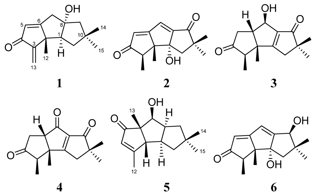
2. Results and Discussion

Chondrosterin A (1) was obtained as yellowish oil. The molecular formula of 1 was established as C15H20O2, based on the HREIMS peak at m/z 232.1456 [M]+ and 13C NMR data (Table 1). The strong IR absorptions at 3433 and 1693 cm−1 indicated the presence of hydroxyl and conjugated carbonyl groups, respectively. The 13C NMR and DEPT spectra displayed three methyls, four methylenes, two methines and six quaternary carbons. One carbonyl carbon (δC 197.3), one trisubstituted double bond (δC 186.9; δC 125.0, δH 6.02, d, J = 1.5 Hz), and one terminal double bond (δC 154.0; δC 113.4, δH 5.89, s, and 5.16, s) represented three double bond equivalents. Thus, 1 must be tricyclic to account for the six double bond equivalents required by the molecular formula. The hydroxyl group was attached to quaternary carbon C-8 (δC 92.5), based on the large chemical shift. Two methyl groups with singlets at δH 1.12 and 1.19 were connected to quaternary carbon C-10 (δC 43.4), the other methyl group with singlet at δH 1.14 was connected to C-2 (δ
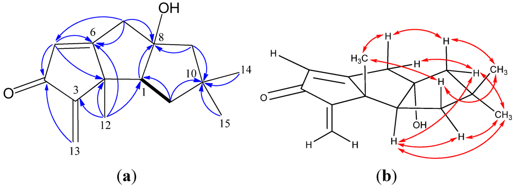
C 52.6), on the basis of their HMBC correlations (Figure 2). The cross-peaks of H-1/H-11 in 1H–1H COSY showed a partial structure –CHCH
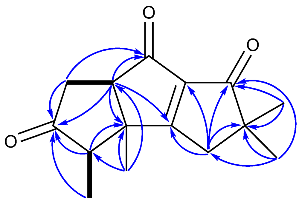
2– in this molecule. The HMBC correlations of H-5/C-4 and H-13/C-4 revealed a cross-conjugated dienone fragment. The HMBC correlations of H-1/C-8, H-5/C-2, H-7/C-5, H-7/C-6, H-7/C-8, H-9/C-8, H-9/C-10, H-11/C-10, H-12/C-1, H-12/C-2, H-12/C-3 and H-12/C-6 established the planar structure of compound 1. To the best of our knowledge, the methyl group C-12 of natural hirsutane sesquiterpenoids always seems to be in β-orientation. The ROESY correlations of H-12/H-7β (δH 2.71), H-12/H-11β (δH 1.60) (Figure 2) established all these protons as β-oriented. In addition, ROESY correlations between H-1/H-9α (δH 1.90), H-1/H-11αH 1.76), H-1/H-15 allowed assignment of H-1 in α-orientation.
Table 1. 13CNMR data of compounds 16(125 MHz).
Position1 a2 a3 a4 a5 a6 b
160.6, CH84.0, C191.8, C197.6, C46.2, CH82.1, C
252.6, C62.9, C53.1, C49.5, C63.8, CH61.6, C
3154.0, C48.5, CH53.1, CH52.4, CH182.6, C47.0, CH
4197.3, C210.5, C218.5, C214.3, C128.4, CH210.4, C
5125.0, CH126.9, CH34.4, CH236.4, CH2211.1, C116.8, CH
6186.9, C187.5, C54.3, CH59.5, CH61.8, C192.3, C
743.6, CH2126.2, CH67.4, CH212.8, C76.9, CH116.0, CH
892.5, C158.0, C145.2, C139.8, C49.8, CH173.9, C
955.9, CH2207.6, C209.1, C202.6, C39.9, CH275.8, CH
1043.4, C51.0, C50.6, C51.7, C41.3, C42.3, C
1141.2, CH241.7, CH240.1, CH241.0, CH249.1, CH244.5, CH2
1222.8, CH322.9, CH317.6, CH317.5, CH315.0, CH323.9, CH3
13113.4, CH29.5, CH39.3, CH39.4, CH319.7, CH39.4, CH3
1430.2, CH327.7, CH325.4, CH325.2, CH329.3, CH329.6, CH3
1528.1, CH326.2, CH325.0, CH325.0, CH329. 2, CH323.0, CH3
a Measured in CDCl3, and CDCl3 was used as an internal standard (δC 77.0); b Measured in DMSO-d6, and DMSO-d6 was used as an internal standard (δC 39.43).
Figure 2. (a) 1H–1H COSY (bold line), main HMBC (arrow); and (b) selected key ROESY correlations of 1.
Marinedrugs 10 00627 g002
Chondrosterin B (2) was isolated as yellowish oil. The HREIMS displays a molecular ion peak at m/z 246.1250 corresponding to the molecular formula C15H18O3. The UV λmax 301 nm indicated the presence of a long conjugated system. Two carbonyl carbons (δC 210.5 and 207.6) and two trisubstituted double bonds (δC 126.9, δH 6.20, s, and δC 187.5; δC 126.2, δH 7.12, s, and δC 158.0) suggested that 2 also possessed a tricyclic system. Three methyl groups with singlets (δH 1.04, 1.18 and 1.40) and one methyl group with doublet (δH 1.16, d, J = 7.0 Hz) which connected with methine carbon C-3 (δC 48.5, δH 2.99, q, J = 7.0 Hz) are diagnostic resonance signals of hirsutane sesquiterpenoids. The hydroxyl group was placed at quaternary carbon C-1 (δC 84.0). The methylene at δ
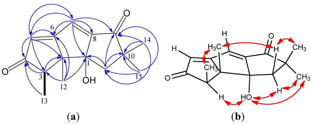
C 41.7 was assigned to 11-position due to the HMBC correlations between H-11 and C-1, C-10, C-14, C-15. The HMBC correlations (Figure 3) of H-5/C-4, H-5/C-6, H-7/C-5, H-7/C-6, H-7/C-8, and H-7/C-9 allowed to establish the large conjugated system.
Figure 3. (a) 1H–1H COSY (bold line), main HMBC (arrow); and (b) selected key ROESY correlations of 2.
Marinedrugs 10 00627 g003
The proton resonance signal of 1-OH showed a broad singlet at δ 1.94 in CDCl3 (500 MHz), whereas a sharp singlet signal at δ 5.52 in the solvent DMSO-d6 (400 MHz). ROESY data acquired in DMSO-d6 showed correlations of 1-OH/H-3, 1-OH/H-11 (δH 1.89), 1-OH/H-15, H-12/H-13, H-12/H-11 (δH 2.01), H-15/H-11 (δH 1.89), H-14/H-11 (δH 2.01). Based on these observations, 1-OH was assigned an α position, whereas H-12 (CH3) and H-13 (CH3) were assigned β positions.
The molecular formula of chondrosterin C (3) was determined to be C15H20O3 due to its molecular ion peak at m/z 248.1405 [M]+ in the HREIMS spectrum. The IR spectrum displayed the characteristic absorptions of hydroxyl group (3355 cm−1), ketone carbonyl (1734 cm−1), and α,β-unsaturated carbonyl group (1684 cm−1). The 13C NMR data showed signals for one tetrasubstituted double bond (δC 191.8 and 145.2), two carbonyl groups (δC 218.5 and 209.1), and one tertiary carbon bearing one hydroxyl group (δC 67.4, C-7). The 1H NMR spectrum indicated the presence of three methyl singlets (δH 1.08, H-12; 1.17, H-14; and 1.16, H-15), and one methyl doublet (δH 1.05, d, J = 7.0 Hz, H-13). The 1H–
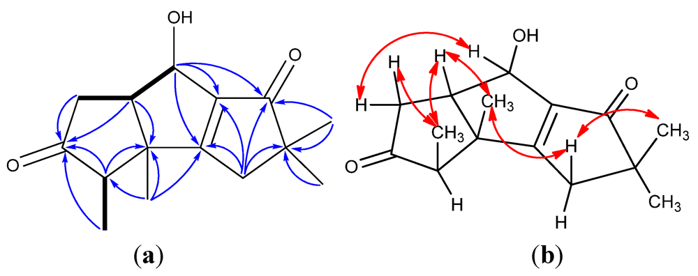
1H COSY correlations of H-5/H-6, H-6/H-7 and H-3/H-13 revealed the presence of two fragments –CH2–CH–CHOH– and –CH–CH3 (Figure 4). Two carbonyl groups at δC 218.5 and 209.1 were placed at C-4 and C-9, respectively, based on the HMBC correlations of H-3/C-4, H-5/C-4, H-7/C-9, H-11/C-9, and H-14/C-9. The tetrasubstituted double bond was placed between C-1 and C-8, which was supported by the HMBC correlations of H-7/C-1, H-7/C-8, H-11/C-1, H-11/C-8, and H-12/C-1. By a combination of the 1H–1H COSY and HMBC spectra, the structure of 
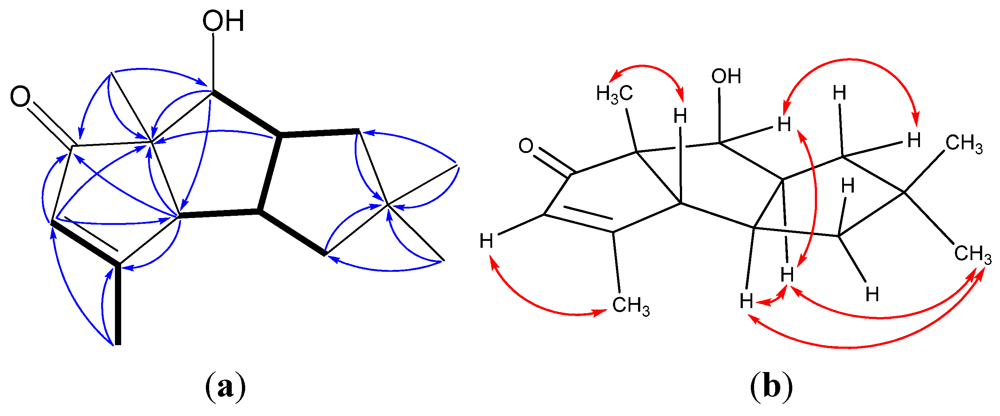
3 could be established. A NOE experiment showed that selective irradiation at δ 1.05 (H-13) gave a clear signal enhancement of H-5 (δ 2.32) and H-6 (δ 2.83). Likewise, irradiation of the H-12 resonance at δ 1.08 gave NOE’s at the signals of H-5 (δ 2.32), H-6, and H-11 (δ 2.48). Irradiation of H-14 at δ 1.17 caused an NOE enhancement of H-11 (δ 2.48). Irradiation of the H-7 at δ 4.80 gave NOE’s to H-5 at δ 2.91. So, H-5 (δ 2.32), H-6, H-11 (δ 2.48), and two CH3 (H-12 and H-13) were determined as β-orientation, H-3 and H-7 were assigned as α-orientation.
Figure 4. (a) 1H–1H COSY (bold line), main HMBC (arrow); and (b) selected key NOE correlations of 3.
Marinedrugs 10 00627 g004
Chondrosterin D (4) was obtained as colorless crystals. The molecular formula was deduced as C15H18O3, based on the HREIMS, which showed a molecular ion peak at m/z 246.1255 [M]+ (calculated for C15H18O3, 246.1250). The 13C NMR and DEPT spectra showed signals corresponding to four methyls, two methylenes, two methines, and seven quaternary carbons. Its NMR spectra showed the following functionalities: three carbonyl groups (δC 214.3, C-4; 212.8, C-7; 202.6, C-9), one tetrasubstituted double bond (δC 197.6, C-1; 139.8, C-8), four methyl groups, giving three singlets (δH 1.20, 1.25 and 1.28) and one doublet (δH 1.15, d, J = 7.0 Hz). Its IR spectra exhibited strong absorptions at 1737, 1687, and 1610 cm−1and supported the presence of the separate ketone and α,β-unsaturated carbonyl group. 1H–1H COSY indicated two partial structures, –CHCH3 and –CHCH2–, in this molecule (Figure 5). The planar structure of 4 could be established, based on its HMBC correlations of H-3/C-2, H-3/C-4, H-5/C-4, H-6/C-1, H-6/C-2, H-6/C-7, H-11/C-1, H-11/C-8, H-11/C-9, H-11/C-10. Compound 
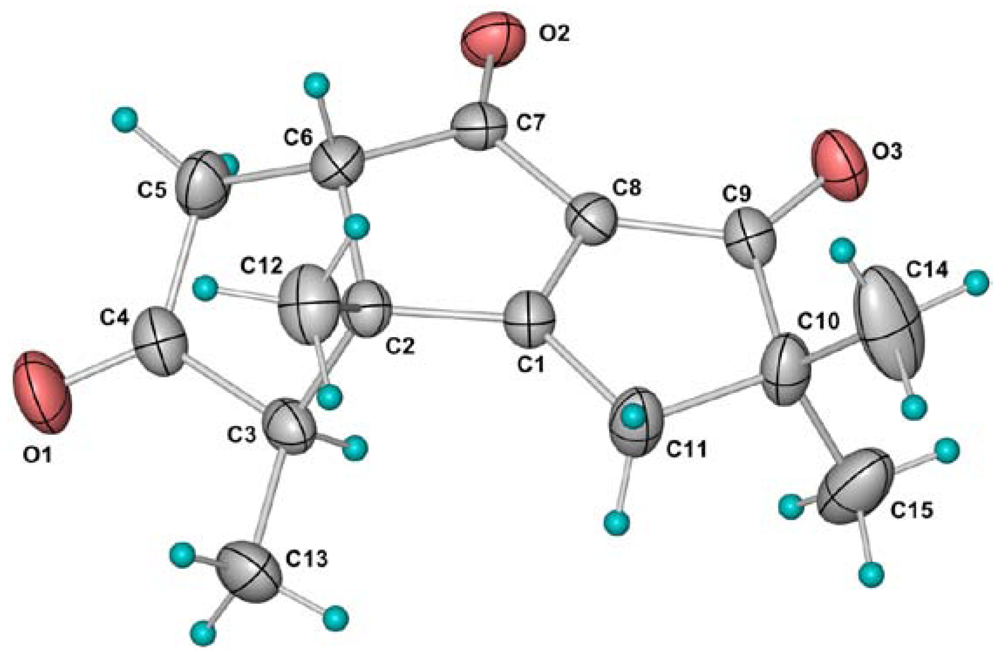
4 is an unprecedented hirsutane sesquiterpenoid which bears three ketone carbonyl groups in the molecule. Finally, the structure and relative configuration of 4 was also confirmed by X-ray crystallography (Figure 6). The molecules related by the two-fold screw axis along the b-axis are joined by the weak C11–H…O1 hydrogen bonds to form a helical ribbon. Adjacent ribbons related by simple translation along the a-axis are further connected by pairs of weak C3–H…O2 hydrogen bonds to form a three-dimensional network.
Figure 5. 1H–1H COSY (bold line) and main HMBC (arrow)correlations of 4.
Marinedrugs 10 00627 g005
Figure 6. Molecular structure of 4 in the crystal. Thermal ellipsoids are plotted at 30% probability level.
Marinedrugs 10 00627 g006
Chondrosterin E (5) was obtained as white solid. The molecular formula of 5 was deduced as C15H22O2 by HREIMS and NMR data. This molecule contained the following diagnostic functional groups: one carbonyl carbon (δC 211.1), one trisubstituted double bond (δC 128.4, δH 5.73, q, J = 1.0 Hz; δC 182.6), three methyl groups with singlets (δH 0.93, 1.09 and 1.35), and one methyl group with doublet (δH 2.03, d, J = 1.0 Hz). Further 1H–1H COSY and HMBC analysis revealed the methyl group with doublet was connected to the trisubstituted double bond (Figure 7), and one hydroxyl group was connected to the methine group C-7 (δC 76.9, δH 3.92, d, J = 6.5 Hz). The 1H–1H COSY spectra displayed the following cross-peaks: H-7/H-8 (CH, δH 2.64, dddd, J = 8.5, 8.5, 6.5, 6.5 Hz); H-8/H-9 (CH2, δH 1.45, dd, J = 13.5, 8.5 Hz; 1.66, dd, J = 13.5, 6.5 Hz), H-8/H-1 (CH, δH 2.58, dddd, J = 10.5, 8.5, 7.5, 2.5 Hz), H-1/H-2 (CH, δH 2.34, d, J = 2.5 Hz), and H-1/H-11 (CH2, δH 1.48, dd, J = 12.0, 10.5 Hz; 1.76, dd, J = 12.0, 7.5 Hz), so the fragment –CH(–OH) –CH(–CH2–)–CH–CH(–CH2–)– was established (Figure 7). The HMBC correlations of H-2/C-3, H-2/C-6, H-4/C-5, H-7/C-6, H-9/C-10, H-11/C-10, H-14/C-10, H-15/C-10, H-13/C-5, H-13/C-6, and H-13/C-7, established the planar structure of 5. The ROESY correlations of H-1/H-8, H-1/H-15, H-2/H-13, H-7/H-8, and H-8/H-15 revealed H-2 and H-13 (CH3) have a β-orientation, whereas, H-1, H-7, H-8 and H-15 (CH3) have an α-orientation.
Figure 7. (a) 1H–1H COSY (bold line), main HMBC (arrow); and (b) selected key ROESY correlations of 5.
Marinedrugs 10 00627 g007
Compound 6 was identified as hirsutanol C, which was firstly isolated by Crews and co-workers from an unidentified fungus [10]. Its NMR data recorded in DMSO-d6 (Table 1 and Table 2) were slightly different from the reference data recorded in CD3OD and dioxane-
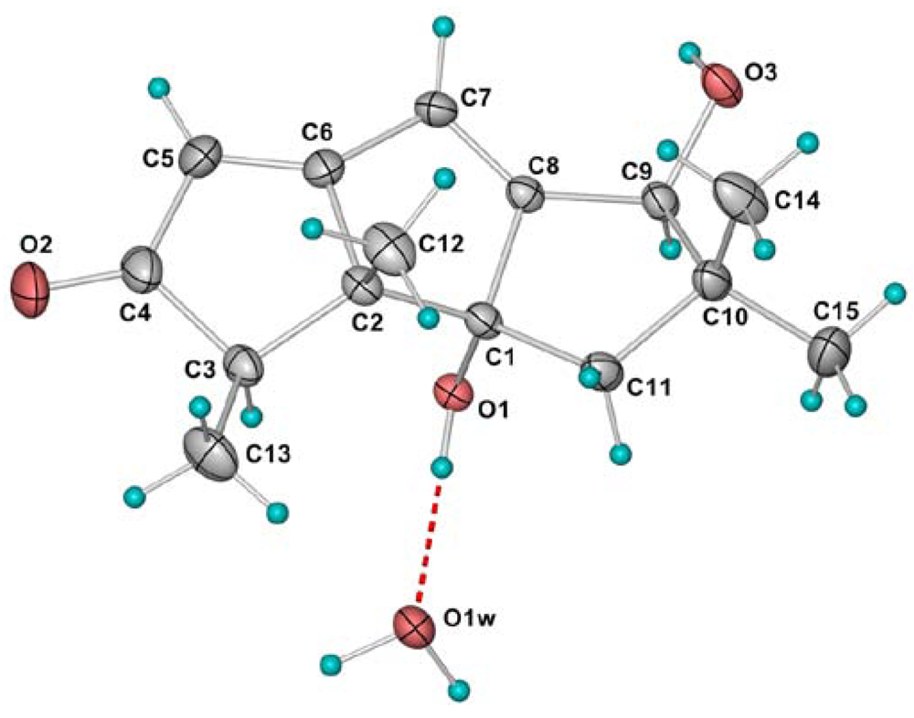
d8. The relative configuration was established by single-crystal X-ray diffraction. In the crystal structure of 6, the molecules related by a simple translation along the b-axis are connected by pairs of O3–H…O1 hydrogen bonds to form a ribbon, which is further consolidated by the bridging water molecules with pairs of O1w–H…O3 and O1–H…O1w hydrogen bonds. The composite ribbons are further joined together with O1w–H…O2 hydrogen bonds to form a double layer parallel to the (001) family of planes. Adjacent layers are stacked together with the hydrophobic hydrocarbon skeleton pointing outwards (Figure 8).
Figure 8. Molecular structure of 6. Thermal ellipsoids are plotted at 30% probability level.
Marinedrugs 10 00627 g008
Table 2. 1H NMR data of compounds 16 (500 MHz, mult., J in [Hz]).
Position1 a2 a3 a4 a5 a6 b
12.33, dd (10.0, 9.0) 2.58, dddd (10.5, 8.5, 7.5, 2.5)
2 2.34, d (2.5)
3 2.99, q (7.0)2.41, qd (7.0, 1.5)2.13, qd (7.0, 1.5) 2.77, q (7.0)
4 5.73, q (1.0)
56.02, d (1.5)6.20, sα: 2.91, ddd (19.0, 3.5, 1.5);
β: 2.32, dd (19.0, 10.0)
α: 2.79, ddd (19.5, 4.5, 1.5);
β: 2.62, dd (19.5, 12.0)
5.69, s
6 2.83, ddd (10.0, 7.0, 3.5)3.12, dd (12.0, 4.5)
7α: 2.76, d (15.5);
β: 2.71, dd (15.5, 1.5)
7.12, s4.80, dd (7.0, 1.0) 3.92, d (6.5)6.32, d (2.5)
8 2.64, dddd (8.5, 8.5, 6.5, 6.5)
9α: 1.90, d (14.0);
β: 1.65, d (14.0)
α: 1.66, dd (13.5, 6.5);
β: 1.45, dd (13.5, 8.5)
4.55 dd (6.0, 2.5)
10
11α: 1.76, dd (13.5, 9.0);
β: 1.60, dd (13.5, 10.0)
α: 1.97, d(14.0);
β: 2.10, d (14.0)
α: 2.38, d (19.0);
β: 2.48, dd (19.0, 1.0)
2.78, d (21.0);
2.58, d (21.0)
α: 1.76, dd (12.0, 7.5);
β: 1.48, dd (12.0, 10.5)
1.98, d (14.5);
1.52, d (14.5)
121.14, s1.04, s1.08, s1.28, s2.03, d (1.0)0.89, s
135.89, s; 5.16, s1.16,d (7.0)1.05, d (7.0)1.15, d (7.0)1.35, s0.93, d (7.0)
141.12, s1.18, s1.17, s1.25, s1.09, s1.19, s
151.19, s1.40, s1.16, s1.20, s0.93, s0.80, s
1α-OH 1.94, brs 5.15, s
7β-OH 3.42, brs
8α-OH2.05, brs
9β-OH 5.250, d(6.0)
a Measured in CDCl3, and CDCl3 was used as an internal standard (δH 7.26); b Measured in DMSO-d6, and DMSO-d6 was used as an internal standard (δH 2.50).
Three cancer cell lines: human lung cancer cell line A549, human nasopharyngeal carcinoma cell line CNE2, and human colon cancer cell line LoVo, were used to evaluate the cytotoxic activities of 16 in vitro. As a result, 1 showed potent cytotoxicity against these cancer cell lines with the IC50 values of 2.45, 4.95, and 5.47 μM, respectively. In contrast, 26 were apparently inactive in this assay (IC50 > 200 μM).

3. Experimental Section

3.1. General Experimental Procedures

Preparative HPLC was conducted on a Shimadzu LC-20AT HPLC pump equipped with a SPD-20A dual λ absorbance detector and Shim-pack PRC-ODS HPLC column (250 × 20 mm). Melting points were measured on an X-6 micro-melting-point apparatus (Beijing Fukai Science and Technology Development, Beijing, China) and were uncorrected. Optical rotations were acquired using a Schmidt and Haensch polartronic HNQW5 optical rotation spectrometer. IR spectra were recorded on a Nicolet Avatar 330 FT-IR spectrophotometer. UV spectra were recorded on a Shimadzu UV-Vis-NIR spectrophotometer. 1D and 2D NMR spectra were recorded on a Varian Inova-500 spectrometer and Bruker Avancell-400 spectrometer. The chemical shifts were referenced to the residual solvent signal (CDCl3: δH 7.26 and δC 77.0; DMSO-d6: δH 2.50 and δC 39.43). HPLC-MS analyses were performed with Thermo Finnigan LCQTM DECA XP liquid chromatography-mass spectrometry. Mass spectra were obtained on Thermo DSQ EI low resolution mass spectrometer and Thermo MAT95XP EI high resolution mass spectrometer. X-ray diffraction data were acquired on a Bruker SMART 1000 CCD X-ray single crystal diffractometer.

3.2. Fungal Strain and Culture Method

Chondrostereum sp. was isolated from the inner tissue of soft coral Sarcophyton tortuosum collected from Hainan Sanya National Coral Reef Reserve, China. This fungal strain was maintained on potato dextrose agar (PDA) slants. Fermentation medium was potato dextrose broth (PDB, potatoes 200 g, dextrose 20 g, seawater 1 L). Plugs of agar supporting mycelia growth were cut and transferred aseptically to a 500 mL Erlenmeyer flask containing 200 mL PDB liquid medium. The liquid medium was sterilized at 120 °C for 30 min. The flask was incubated at 28 °C on a rotary shaker (120 rpm) for 5 days. The mycelia were aseptically transferred to 500 mL Erlenmeyer flasks containing 200 mL of the same liquid medium. The flasks were incubated at 28 °C on a rotary shaker (120 rpm) for 20 days.

3.3. Extraction and Isolation

60 L of grown culture broth was filtered through cheesecloth. The culture broth was successively extracted three times with EtOAc. The EtOAc extract was concentrated by low-temperature rotary evaporation. The extract (24.6 g) was chromatographed on a silica gel column with petroleum ether-EtOAc (100:0–0:100) and then EtOAc-MeOH (100:0–0:100) as the eluent to afford 18 fractions (code Fr. 1–Fr. 18). Fr. 9 was further purified by RP-HPLC with a gradient of H2O-MeCN (40:60 up to 0:100, v/v) to afford compound 1 (48 mg). Fr. 10–12 was further purified by Sephadex LH-20 gel column chromatography and repeated RP-HPLC, eluted with H2O-MeCN (60:40, v/v) to yield compounds 2 (39 mg) and 5 (21 mg). Compounds 3 (59 mg) and 4 (32 mg) were obtained from Fr. 13 by repeated RP-HPLC using H2O-MeCN (70:30, v/v) as eluent. Fr. 14–15 was purified by repeated silica gel column chromatography, followed by recrystallization from EtOAc solution, to afford compound 6 (46 mg).
Chondrosterin A (1): Yellowish oil; [α]20D +112 (c 0.024, MeOH); UV (MeOH) λmax (log ε) 249 nm (4.06); IR (KBr) vmax 3433, 2951, 2867, 1693, 1622, 1462, 1377, 1312, 1259, 1211, 1156, 1123, 1037, 941, 864 cm−1; 1H and 13C NMR data, see Table 1; EIMS m/z 232, 217, 176, 122, 91, 92, 77, 55; HREIMS m/z 232.1456 [M]+ (calcd for C15H20O2, 232.1458).
Chondrosterin B (2): Yellowish oil; [α]20D +154 (c 0.022, MeOH); UV (MeOH) λmax (log ε) 301 nm (4.07); IR (KBr) vmax 3423, 2969, 2872, 1698, 1616, 1590, 1463, 1379, 1312, 1256, 1191, 1142, 1115, 1069, 998, 903 cm–1; 1H and 13C NMR data in CDCl3, see Table 1; 1H NMR (400 MHz, DMSO-d6) δ: 7.25 (s, H-7), 6.20 (s, H-5), 5.52 (s, 1α-OH), 2.87 (q, J = 7.0 Hz, H-3), 2.01 (d, J = 14.0 Hz, H-11β), 1.89 (d, J = 14.0 Hz, H-11α), 1.30 (s, H-15), 1.09 (s, H-14), 1.03 (d, J = 7.0 Hz, H-13), 0.94 (s, H-12). EIMS m/z 246, 231, 218, 203, 190, 175, 162, 147, 134, 119, 105, 91, 77; HREIMS m/z 246.1250 [M]+ (calcd for C15H18O3, 246.1250).
Chondrosterin C (3): White solid; mp 130–131 °C; [α]20D −177 (c 0.034, MeOH); UV (MeOH) λmax (log ε) 236 nm (3.83); IR (KBr) vmax 3355, 2968, 2958, 2938, 2910, 1734, 1684, 1635, 1460, 1429, 1388, 1370, 1315, 1288, 1224, 1174, 1096, 1075, 1047, 995 cm−1; 1H and 13C NMR data, see Table 1; EIMS m/z 248, 233, 230, 215, 205, 189, 177, 173, 163, 159, 145, 135, 119, 105, 91, 77, 55; HREIMS m/z 248.1405 [M]+(calcd for C15H20O3, 248.1407).
Chondrosterin D (4): Colorless crystals (ethyl acetate); mp 263–264 °C; [α]20D −300 (c = 0.025, MeOH); UV (MeOH) λmax (log ε) 305 nm (2.34), 261 nm (3.36), 222 nm (3.38); IR (KBr) vmax 2975, 2927, 1737, 1687, 1610, 1454, 1420, 1391, 1336, 1322, 1047, 1027 cm–1; 1H and 13C NMR data, see Table 1; EIMS m/z 246, 231, 218, 203, 189, 175, 161, 147, 135, 119, 105, 91, 83, 77, 65, 55; HREIMS m/z 246.1255 [M]+ (calcd for C15H18O3, 246.1250).
Chondrosterin E (5): White solid; mp 84–85 °C; [α]20D +66 (c 0.028, MeOH); UV (MeOH) λmax (log ε) 235 nm (3.81); IR (KBr) vmax 3463, 2950, 2927, 2865, 1678, 1613, 1440, 1378, 1361, 1314, 1259, 1170, 1096, 1065, 1011, 954, 876, 852 cm−1; 1H and 13C NMR data, see Table 1; EIMS m/z 234, 216, 206, 201, 173, 135, 110, 95, 91, 79, 69, 55; HREIMS m/z 234.1613 [M]+ (calcd for C15H22O2, 234.1614).

3.4. Crystal Structure Determination of 4 and 6

Crystals of 4 and 6 were obtained from EtOAc solution. Chondrosterin D (4): C15H18O3, M = 246.29, colorless block, orthorhombic system, space group P212121, a = 6.5259(11), b = 12.735(2), c = 16.396(3) Å, V = 1362.6(4) Å3, Z = 4, d = 1.201 g/cm3, crystal size 0.40 × 0.38 × 0.36 mm3. Hirsutanol C (6): C15H20O3·H2O, M = 266.34, colorless block, monoclinic system, space group P21, a = 8.0825(19), b = 8.912(2), c = 10.695(3) Å, V = 734.1(3) Å3, Z = 2, d = 1.205 g/cm3, crystal size 0.42 × 0.40 × 0.38 mm3. X-ray diffraction data were collected on a Bruker SMART 1000 CCD diffractometer with Mo Kα radiation (λ = 0.71073Å) at room temperature. The data were processed using CrysAlis. The structures were solved by direct method. H-atoms were added in ideal positions and refined as riding models. The structures were refined using full-matrix least-squares based on F2 with program SHELXL [11,12].
CCDC 847843 and 847844 contain the supplementary crystallographic data of compounds 4 and 6 respectively [13].

3.5. Cytotoxicity Assay

The in vitro cytotoxicity of 16 was determined by means of the colorimetric MTT (3-(4,5-dimethylthiazol-2-yl)-2,5-diphenyl-2H-tetrazolium bromide) assay. The tested human cancer cell lines were seeded in 96-well plates at a density of 3 × 10⁷ cells/L, and the compounds were added at various concentrations (0.125–50 mg/L). After 48 h, MTT was added to the culture medium at a final concentration of 0.5 mg/mL, and the plates were incubated for 4 h at 37 °C. The supernatant was removed. The formazan crystals were dissolved in DMSO (150 μL) with gentle shaking at room temperature. The absorbance at 570 nm was recorded with a microplate reader (Bio-Rad, USA), and the data were analyzed with the SPSS [14].

4. Conclusions

The marine fungus Chondrostereum sp. was cultured in PDB medium and afforded five new sesquiterpenoids, chondrosterins A–E (15), and the known compound hirsutanol C (6). 14 and 6 are hirsutane-type sesquiterpenoids, 5 has a novel rearranged hirsutane skeleton, which could be derived by migration of a methyl group from C-2 to C-6. Chondrosterin A (1), with the typical α-methylene ketone group, showed significant cytotoxic activities. These results indicated the metabolites produced by Chondrostereum sp. in PDB medium were different from those in GPY medium. By altering the fermentation conditions, e.g. carbon and nitrogen source, inorganic salts, Chondrostereum sp. can produce highly functionalized hirsutane derivatives with a surprising chemodiversity. Furthermore, the metabolites isolation work based on 13C NMR screening seems effectively to obtain the novel hirsutane-type compounds containing carbonyl groups.

Acknowledgments

This work was supported by the National Natural Science Foundation of China (No. 20502036, 20602044 and 30973633), Guangdong Provincial Science and Technology Research Program (No. 2009B030801190, 2010B030800002, and 2010B030600011), Guangzhou Science and Technology Research Program (No. 2011308 and 2010U1-E00531-5), and the Fundamental Research Funds for the Central Universities (No. 11lgpy09).

References

  1. Su, J.Y.; Long, K.H.; Peng, T.S.; Zheng, Q.T.; Lin, X.Y. Chemical constituents of Chinese soft coral. 12. The structure of methyl sartortuoate, a unique new tetraterpenoid. Acta Chim. Sinica 1985, 43, 796–797. [Google Scholar]
  2. Su, J.Y.; Long, K.H.; Peng, T.S.; Zeng, L.M.; Zheng, Q.T.; Lin, X.Y. Determination of molecular and crystal structures of a unique new tetraterpenoid-methyl sartortuoate. Sci. China Ser. B 1988, 31, 1172–1184. [Google Scholar]
  3. Lan, W.J.; Su, J.Y.; Zeng, L.M. Isolation and 1H, 13C NMR assignments formethyl sartortuoate. Chinese J. Org. Chem. 2005, 25, 1465–1468. [Google Scholar]
  4. Su, J.Y.; Long, K.H.; Peng, T.S.; He, C.H.; Clardy, J. The structure ofmethylisosartortuoate, a novel tetracyclic tetraterpenoid from the soft coral Sarcophyton tortuosum. J. Am. Chem. Soc. 1986, 108, 177–178. [Google Scholar]
  5. Zeng, L.M.; Lan, W.J.; Su, J.Y.; Zhang, G.W.; Feng, X.L.; Liang, Y.J.; Yang, X.P. Two new cytotoxic tetracyclic tetraterpenoids from the soft coral Sarcophyton tortuosum. J. Nat. Prod. 2004, 67, 1915–1918. [Google Scholar] [CrossRef]
  6. Lan, W.J.; Li, H.J.; Yan, S.J.; Su, J.Y.; Zeng, L.M. New tetraterpenoidfrom the soft coral Sarcophyton tortuosum. J. Asian Nat. Prod. Res. 2007, 9, 267–271. [Google Scholar] [CrossRef]
  7. Lan, W.J.; Wang, S.L.; Li, H.J. Additional new tetracyclic tetraterpenoid:methyltortuoateD from soft coral Sarcophyton tortuosum. Nat. Prod. Commun. 2009, 4, 1193–1196. [Google Scholar]
  8. Li, H.J.; Lan, W.J.; Lam, C.K.; Yang, F.; Zhu, X.F. Hirsutane sesquiterpenoids from the marine-derived fungus Chondrostereum sp. Chem. Biodivers. 2011, 8, 317–324. [Google Scholar] [CrossRef]
  9. Yang, F.; Gao, Y.H.; Wu, K.W.; Deng, R.; Li, D.D.; Wei, Z.X.; Jiang, S.; Wu, X.Q.; Feng, G.K.; Li, H.J.; Zhu, X.F. A novel sesquiterpene hirsutanolAinduces autophagical cell death in human hepatocellular carcinoma cells by increasing reactive oxygen species. Chin. J. Cancer 2010, 29, 655–660. [Google Scholar]
  10. Wang, G.Y.S.; Abrell, L.M.; Avelar, A.; Borgeson, B.M.; Crews, P. New hirsutane based sesquiterpenes from salt water cultures of a marine sponge-derived fungus and the terrestrial fungus Coriolus consors. Tetrahedron 1998, 54, 7335–7342. [Google Scholar]
  11. SADABS; University of Göttingen: Göttingen, Germany, 1996; Program for empirical absorption correction of area detector data.
  12. SHELXTL, 5.10 for Windows NT, Bruker Analytical X-Ray Systems, Inc.: Madison, WI, USA, 1998; Structure determination software programs.
  13. Available online: http://www.ccdc.cam.ac.uk/cgi-bin/catreq.cgi (accessed on 12 March 2012). These data can be obtained free of charge.
  14. SPSS, student version 13.0 for Windows, Statistical Program for Social Sciences, IBM Company: New York, NY, USA, 2001.
  • Sample Availability: Available from the authors.

Supplementary Files

  • Supplementary File 1::

    PDF-Document (PDF, 1401 KB)

  • Share and Cite

    MDPI and ACS Style

    Li, H.-J.; Xie, Y.-L.; Xie, Z.-L.; Chen, Y.; Lam, C.-K.; Lan, W.-J. Chondrosterins A–E, Triquinane-Type Sesquiterpenoids from Soft Coral-Associated Fungus Chondrostereum sp. Mar. Drugs 2012, 10, 627-638. https://doi.org/10.3390/md10030627

    AMA Style

    Li H-J, Xie Y-L, Xie Z-L, Chen Y, Lam C-K, Lan W-J. Chondrosterins A–E, Triquinane-Type Sesquiterpenoids from Soft Coral-Associated Fungus Chondrostereum sp. Marine Drugs. 2012; 10(3):627-638. https://doi.org/10.3390/md10030627

    Chicago/Turabian Style

    Li, Hou-Jin, Ying-Lu Xie, Zhong-Liang Xie, Ying Chen, Chi-Keung Lam, and Wen-Jian Lan. 2012. "Chondrosterins A–E, Triquinane-Type Sesquiterpenoids from Soft Coral-Associated Fungus Chondrostereum sp." Marine Drugs 10, no. 3: 627-638. https://doi.org/10.3390/md10030627

    Article Metrics

    Back to TopTop