Article

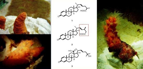
Steroidal Carboxylic Acids from Soft Coral Paraminabea acronocephala

1
Department of Marine Biotechnology and Resources, National Sun Yat-sen University, Kaohsiung 80424, Taiwan
2
Chinese Medicine Research and Development Center, China Medical University Hospital, Taichung 40402, Taiwan
3
China Medical University, Taichung 40402, Taiwan
4
Graduate Institute of Integrated Medicine, College of Chinese Medicine, China Medical University, Taichung 40402, Taiwan
5
Asia-Pacific Ocean Research Center, National Sun Yat-sen University, Kaohsiung 80424, Taiwan
*
Author to whom correspondence should be addressed.
Mar. Drugs 2013, 11(1), 136-145; https://doi.org/10.3390/md11010136
Submission received: 28 November 2012 / Revised: 28 December 2012 / Accepted: 31 December 2012 / Published: 11 January 2013

Abstract

:
Three new steroidal carboxylic acids, paraminabic acids A–C (13) were isolated from a Formosan soft coral Paraminabea acronocephala. The structures of these compounds were established by extensive spectroscopic analysis and chemical methods. Application of the PGME method allowed the establishment of the absolute configurations at C-25 and C-24 for 1 and 2, respectively. Compound 3 showed potent cytotoxicity toward Hep3B, MDA-MB-231, MCF-7, and A-549 cancer cell lines, with IC50 values ranging from 2.05 to 2.83 μg/mL. Compounds 2 and 3 were found to inhibit the accumulation of the pro-inflammatory iNOS protein.

Graphical Abstract

1. Introduction

Marine withanolides, with potent pro-inflammatory inducible nitric oxide synthase (iNOS) inhibitory activity, have previously been reported from two species of soft corals, Paraminabea acronocephala [1] and Minabea sp. [2]. These compounds possess a different A-ring structure (1,4-dien-3-one or 4-en-3-one) from those of plant origin [1,2,3]. Our previous chemical investigation of the soft coral P. acronocephala led to the isolation of novel withanolides with a 24β,25β-dimethyl-γ-lactone or a 24β,25α-dimethyl-γ-lactone in the steroidal side chain moiety [1]. As part of our continuing search for bioactive, structurally interesting metabolites from this coral, three steroidal carboxylic acids (13) were isolated and their structures were elucidated (Figure 1). The cytotoxicity of compounds 13 against human liver carcinoma (HepG2 and HepG3), human breast carcinoma (MCF-7 and MDA-MB-231), and human lung carcinoma (A-549) cell lines and the ability of 13 to inhibit up-regulation of the pro-inflammatory iNOS and COX-2 (cyclooxygenase-2) proteins in LPS (lipopolysaccharide)-stimulated RAW264.7 macrophage cells were also evaluated.
Figure 1. The structures of paraminabic acids A–C (13).
Marinedrugs 11 00136 g001

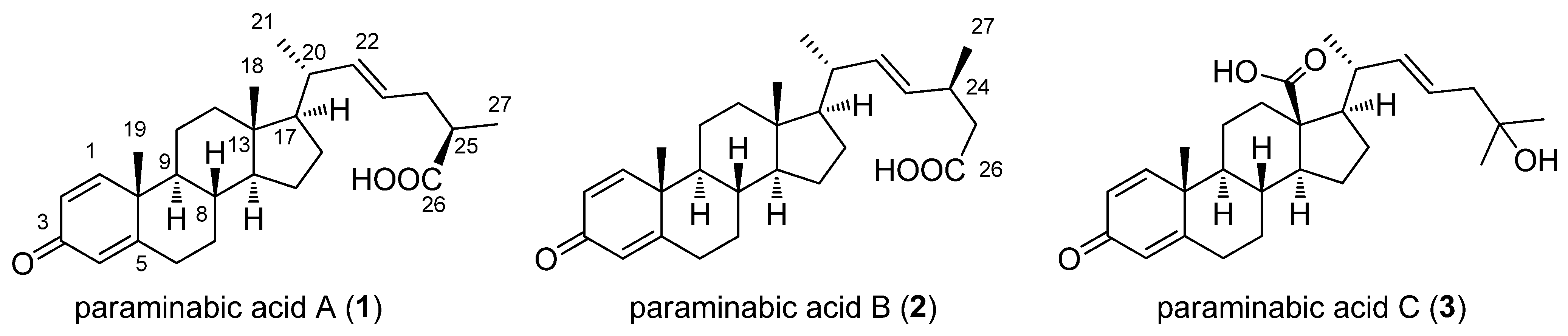
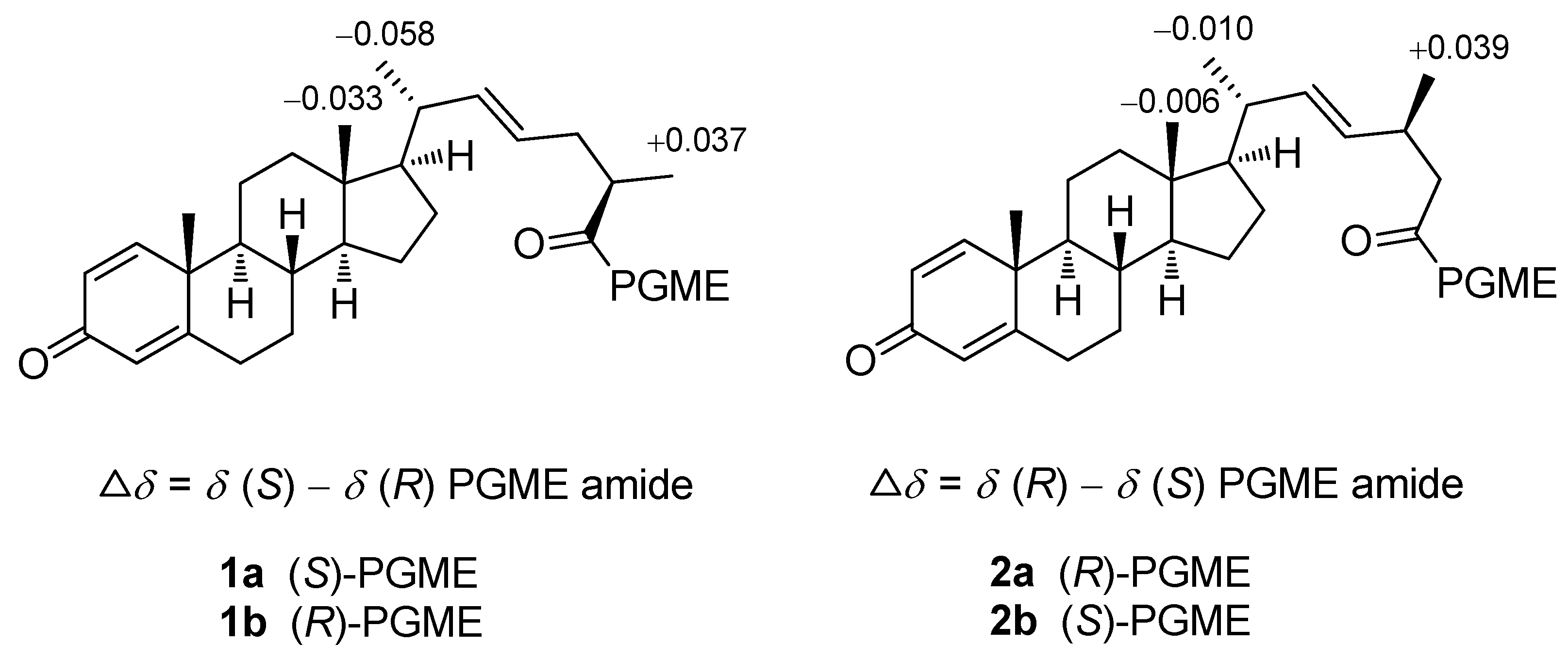
2. Results and Discussion

The ethanolic extract of the soft coral P. acronocephala was partitioned between EtOAc and H2O to afford the EtOAc-soluble fraction. It was then subjected to silica gel column chromatography. The fractions containing steroids were selected for further study, based on characteristic methyl signals in the 1H NMR spectrum. These fractions were subsequently subjected to a series of chromatographic separations to afford three new steroidal carboxylic acids, paraminabic acids A–C (13).
The HRESIMS and 13C NMR spectroscopic data of paraminabic acid A (1) suggested a molecular formula of C27H38O3, appropriate for nine degrees of unsaturation. The 13C NMR and DEPT spectroscopic data (Table 1) displayed 27 carbon signals, including 4 methyls, 7 methylenes, 11 methines, and 5 quaternary carbons. A broad O–H stretching absorption in the region of 3400–2600 cm−1 is ascribable to a carboxylic acid, which was evidenced by the carbon resonance at δC 180.6 (C). The same steroidal nucleus as that of paraminabeolides D and E was deduced for 1 by detailed comparison of their NMR spectroscopic data [1]. The side chain moiety of 1 resembles that of a known steroidal carboxylic acid, (25S)-3-oxocholesta-1,4-dien-26-oic acid [4], isolated from the Indonesian soft coral Minabea sp. However, 1 varied from (25S)-3-oxocholesta-1,4-dien-26-oic acid in the respective side chain. The proton resonances at δH 5.50 (1H, dt, J = 15.6, 6.4 Hz, H-23) and 5.44 (1H, dd, J = 15.6, 8.8 Hz, H-22), measured in C5D5N, were due to the presence of a trans C-22/C-23 double bond, which was confirmed by the HMBC correlations from H3-21 to C-17, C-20, and C-22. The absolute configuration at C-25 was determined by the application of Kusumi’s method (PGME method) [5,6,7]. The chemical shift differences of (S)-PGME amide (1a) and (R)-PGME amide (1b) (Δδ = δ(S) − δ(R)) were summarized in Figure 2 and established the R configuration at C-25.
Table 1. 13C NMR spectroscopic data of compounds 13.
Position1 a, δC, mult.2 a, δC, mult.3 a, δC, mult.
1156.1, CH156.1, CH155.9, CH
2127.4, CH127.4, CH127.5, CH
3186.5, C186.5, C186.5, C
4123.8, CH123.7, CH123.8, CH
5169.5, C169.6, C169.3, C
632.9, CH232.9, CH232.7, CH2
733.7, CH233.7, CH233.4, CH2
835.5, CH35.5, CH37.1, CH
952.4, CH52.4, CH52.4, CH
1043.6, C43.6, C43.6, C
1122.8, CH222.8, CH224.6, CH2
1239.3, CH239.3, CH235.1, CH2
1342.6, C42.5, C55.8, C
1455.6, CH255.5, CH255.7, CH2
1524.4, CH224.3, CH225.0, CH2
1628.4, CH228.3, CH225.3, CH2
1755.5, CH55.5, CH55.3, CH
1812.2, CH312.2, CH3176.8, C b
1918.7, CH318.7, CH318.7, CH3
2040.0, CH39.9, CH38.4, CH
2120.6, CH320.6, CH322.0, CH3
22139.6, CH136.1, CH136.7, CH
23123.8, CH131.3, CH124.8, CH
2436.4, CH233.7, CH46.8, CH2
2539.3, CH41.6, CH2 b71.7, C
26180.6, C b176.9, C b27.6, CH3
2716.3, CH3 b20.6, CH330.5, CH3
a Spectra were measured in CDCl3 (100 MHz); b values obtained from the relevant HMBC or HSQC correlation peaks.
Paraminabic acid B (2) gave the same molecular formula, C27H38O3, as that of 1, based on the analysis of the HRESIMS and 13C NMR spectroscopic data (Table 1). The NMR spectroscopic data of 2 are similar to those of 1, but differences were observed in their side chains. The HMBC correlations from H3-21 to C-17, C-20, and C-22 allowed the assignment of a C-22/C-23 double bond. A large coupling constant (15.2 Hz, C5D5N) between H-22 and H-23 suggested the E geometry of this double bond. The H-23 signal appeared as a doublet of doublets, revealing that the adjacent carbon (C-24) should be a methine. This might be due to the attachment of a methyl group (δH 1.03, 3H, d, J = 6.4 Hz, H3-27) at C-24 (Table 2). This was confirmed by the HMBC correlations from H3-27 to C-23, C-24, and C-25 as well as from H2-25 to the carboxyl carbon (C-26). The absolute configuration at C-24 of 2 was determined by the application of Kusumi’s method developed for chiral β,β-disubstituted propionic acid derivatives [6,7]. The 1H NMR shift differences (Δδ = δ(R) − δ(S)) between the diastereomeric (R)- and (S)-PGME amides, 2a and 2b, respectively, are summarized in Figure 2 and establish the 24S configuration for 2.
Table 2. 1H NMR spectroscopic data of compounds 13.
#1, δH (J in Hz) a2, δH (J in Hz) a3, δH (J in Hz) a
17.06, d (10.0)7.06, d (10.0)7.03, d (10.0)
26.23, dd (10.0, 1.6)6.24, d (10.0)6.23, d (10.0)
46.07, s6.07, s6.07, s
6a: 2.45, ma: 2.46, td (13.6, 4.4)a: 2.46, td (13.4, 4.4)
b: 2.35, mb: 2.35, mb: 2.35, m
7a: 1.93, ma: 1.93, ma: 2.04, m
b: 1.02, mb: 1.02, mb: 1.07, m
81.60, m1.59, m1.69, m
91.04, m1.03, m1.10, m
111.67, m1.67, m1.85, m
1.69, m
12a: 2.00, ma: 1.99, ma: 2.66, br d (14.0)
b: 1.18, mb: 1.17, mb: 1.03, m
141.12, m1.11, m1.30, m
15a: 1.55, ma: 1.52, ma: 1.91, m
b: 1.08, mb: 1.02, mb: 1.66, m
16a: 1.65, ma: 1.62, ma: 1.78, m
b: 1.22, mb: 1.20, mb: 1.70, m
170.99, m0.99, m1.62, m
180.74, s0.74, s
191.23, s1.23, s1.15, s
202.03, m2.00, m2.36, m
210.99, d (6.8)0.98, d (6.4)1.05, d (6.4)
225.27–5.30 b5.23–5.26 b5.39 dd (15.2, 8.8)
235.27–5.30 b5.23–5.26 b5.48, ddd (15.2, 8.8, 5.2)
24a: 2.33, m2.59, m2.18, dd (14.0, 5.2)
b: 2.10, m 2.11, dd (14.0, 8.8)
252.49, m2.30, d (7.2)
26 1.25, s
271.15, d (7.2)1.03, d (6.4)1.25, s
a Spectra were measured in CDCl3 (400 MHz); b overlapped signals.
The HRESIMS and 13C NMR spectroscopic data of paraminabic acid C (3) established a molecular formula of C27H38O4 and nine degrees of unsaturation. The IR absorptions at 3419 and 1714 cm−1 suggested the presence of hydroxy and carbonyl groups, respectively. Both 1H and 13C NMR spectra of 3 lacked signals of the angular methyl group, which might be replaced by a carboxylic acid according to the carbon signal at δC 176.8 (C) (Table 1). The carboxylic acid attached at C-13 was further confirmed by the HMBC correlations from both H2-12 and H-17 to C-18. The trans C-22/C-23 double bond was deduced by the HMBC correlations from H3-21 to C-17, C-20, and C-22 as well as J value (15.2 Hz) (Table 2) between H-22 and H-23. In addition, the downfield-shifted quaternary carbon at δC 69.9 was ascribable to a hydroxy group attached at C-25, which was correlated by H2-24, H3-26, and H3-27 in the HMBC spectrum.
Figure 2. 1H NMR chemical shift differences of PGME amides of 1 and 2.
Marinedrugs 11 00136 g002
The cytotoxicity of compounds 13 against HepG2, Hep3B, MDA-MB-231, MCF-7, and A-549 cancer cells was studied and shown in Table 3. Compound 3 showed potent cytotoxicity toward Hep3B, MDA-MB-231, MCF-7, and A-549 cancer cell lines, with IC50 values ranging from 2.05 to 2.83 μg/mL. We also investigated the inhibition of these compounds toward LPS-induced pro-inflammatory protein (iNOS and COX-2) expression in RAW264.7 macrophage cells by Western blot analysis. At a concentration of 10 μM, compounds 2 and 3 reduced the levels of iNOS to 63.9 ± 6.3% and 53.5 ± 8.6%, respectively; whereas, compound 2 enhanced the expression of COX-2 (130.5 ± 9.8%) in comparison with those of control cells stimulated with LPS only (100% for both iNOS and COX-2). Also, compound 3 could inhibit the expression of iNOS protein but did not induce cytotoxicity in macrophage cells as determined through internal control β-actin expression, as shown in Figure 3. These results indicate that 3 possesses moderate anti-inflammatory activity and potent cytotoxicity, and might be useful for further medicinal study.
Table 3. Cytotoxicity data of compounds 13.
Compound Cell lines IC50 (μg/mL)
Hep G2Hep 3BMDA-MB-231MCF-7A549
115.2119.66
219.77
313.572.832.252.232.05
doxorubicin0.310.401.320.681.33
(–): Compound is considered inactive with IC50 > 20 μg/mL.
Figure 3. Effect of compounds 13 at 10 μM on the LPS-induced pro-inflammatory iNOS and on COX-2 protein expression of RAW264.7 macrophage cells by immunoblot analysis (A) Quantification of immunoblots of iNOS; (B) Quantification of immunoblots of COX-2. The values are means ± SEM (n = 6). The relative intensity of the LPS alone stimulated group was taken as 100%. * Significantly different from LPS alone stimulated group (* P < 0.05). a Stimulated with LPS. b Stimulated with LPS in the presence of 13 (10 μM); (C) Quantification of immunoblots of β-actin.
Marinedrugs 11 00136 g003

3. Experimental Section

3.1. General Experimental Procedures

Optical rotations were determined with a JASCO P1020 digital polarimeter. The IR spectra were obtained on a JASCO FT/IR-4100 spectrophotometer. The NMR spectra were recorded on a Bruker AVANCE 300 FT-NMR (or Varian MR-400 NMR) instrument at 300 MHz (or 400 MHz) for 1H (referenced to TMS for both CDCl3 and C5D5N) and 75 MHz (or 100/125 MHz) for 13C (referenced to δC 77.0 for CDCl3 and to internal TMS for C5D5N). ESIMS were recorded by ESI FT-MS on a Bruker APEX II mass spectrometer. Silica gel 60 (230−400 mesh, Merck, Darmstadt, Germany) and LiChroprep RP-18 (Merck, 40–63 μm) were used for column chromatography. Precoated silica gel plates (Kieselgel 60 F254, 0.25 mm, Merck, Darmstadt, Germany) and precoated RP-18 F254S plates (Merck, Darmstadt, Germany) were used for TLC analyses. High-performance liquid chromatography was performed on a Hitachi L-7100 pump equipped with a Hitachi L-7400 UV detector at 210 nm and a semi-preparative reversed-phase column (Hibar Purospher RP-18e, 5 μm, 250 × 10 mm, Merck, Darmstadt, Germany).

3.2. Animal Material

The soft coral P. acronocephala was collected by scuba divers, off the western coast of Pingtung county, in May 2009, at a depth of 10 m, and was stored in a freezer until being extracted. This soft coral was identified by Prof. Chang-Fong Dai, Institute of Oceanography, National Taiwan University. A voucher specimen (specimen No. 200905PA) was deposited in the Department of Marine Biotechnology and Resources, National Sun Yat-sen University.

3.3. Extraction and Isolation

The soft coral P. acronocephala (3.8 kg fresh wt) was collected and freeze-dried. The freeze-dried material was minced and extracted exhaustively with EtOH (6 × 2 L). The organic extract was concentrated to an aqueous suspension and was further partitioned between EtOAc and water. The EtOAc extract (30 g) was fractionated by open column chromatography on silica gel using n-hexane–EtOAc and EtOAc–MeOH mixtures of increasing polarity to yield 28 fractions. Fraction 21 (3.6 g), eluted with n-hexane–EtOAc (1:6), was further separated by silica gel column chromatography with gradient elution (n-hexane-acetone, 5:1 to 2:1) to yield eight subfractions (21A to 21H). Subfraction 21D was fractionated by RP-18 open column (MeOH–H2O, 50% to 100%) to afford six subfractions (21D1 to 21D6). Compounds 1 (1.9 mg) and 2 (2.6 mg) were obtained from subfraction 21D5 using RP-18 HPLC (MeOH–H2O, gradient 75% to 85%). Compound 3 (5.1 mg) was obtained from fraction 25 (0.85 g) using repeatedly column chromatography over silica gel (n-hexane–EtOAc, 1:3 to 0:1) and RP-18 gel (MeOH–H2O, 50% to 100%), and subsequently separated by RP-18 HPLC (MeOH–H2O, gradient, 65%–75%).
Paraminabic acid A (1): amorphous solid; [α]24D +13 (c 0.09, CHCl3); IR (KBr) νmax 3400–2600 (br), 2933, 2868, 2853, 1718, 1662, 1615, 1602, 1457, 1406, 1375, 1292, 1241 cm−1; 13C NMR and 1H NMR data, see Table 1 and Table 2; Selected 1H NMR (C5D5N, 400 MHz) of 1: δ 7.01 (1H, d, J = 10.0 Hz, H-1), 6.42 (1H, dd, J = 10.0, 2.0 Hz, H-2), 6.26 (1H, s, H-4), 5.50 (1H, dt, J = 15.6, 6.4 Hz, H-23), 5.44 (1H, dd, J = 15.6, 8.8 Hz, H-22), 2.78 (1H, m, H-25), 2.65 (1H, m, H-24a), 2.40 (1H, m, H-24b), 1.38 (3H, d, J = 6.8 Hz, H3-27), 1.10 (3H, s, H3-19), 1.03 (3H, d, J = 6.4 Hz, H3-21), 0.67 (3H, s, H3-18); ESIMS m/z 433 [M + Na]+; HRESIMS m/z 433.2715 [M + Na]+ (calcd for C27H38O3Na, 433.2718).
Paraminabic acid B (2): amorphous solid; [α]24D +60 (c 0.16, CHCl3); IR (KBr) νmax 3400–2600 (br), 2934, 2868, 1718, 1662, 1617, 1601, 1455, 1405, 1375, 1292, 1241 cm−1; 13C NMR and 1H NMR data, see Table 1 and Table 2; 1H NMR (C5D5N, 400 MHz) of 2: δ 7.01 (1H, d, J = 10.0 Hz, H-1), 6.42 (1H, dd, J = 10.0, 1.6 Hz, H-2), 6.27 (1H, s, H-4), 5.51 (1H, dd, J = 15.2, 7.2 Hz, H-23), 5.41 (1H, dd, J = 15.2, 8.4 Hz, H-22), 2.97 (1H, m, H-24), 2.63 (1H, dd, J = 14.8, 7.6 Hz, H-25a), 2.55 (1H, dd, J = 14.8, 7.2 Hz, H-25b), 2.31 (1H, td, J = 13.6, 4.4 Hz, H-6a), 2.18 (1H, dt, J = 13.6, 2.4 Hz, H-6b), 2.04 (1H, m, H-20), 1.90 (1H, dt, J = 12.4, 3.2 Hz, H-12a), 1.71 (1H, m, H-16a), 1.70 (1H, m, H-7a), 1.54 (2H, m, H2-11), 1.42 (1H, m, H-8), 1.39 (1H, m, H-15a), 1.25 (1H, m, H-16b), 1.21 (3H, d, J = 6.8 Hz, H3-27), 1.09 (3H, s, H3-19), 1.08 (1H, m, H-17), 1.06 (1H, m, H-12b), 1.04 (3H, d, J = 6.0 Hz, H3-21), 0.99 (1H, m, H-15b), 0.88 (1H, m, H-9), 0.83 (1H, m, H-14), 0.82 (1H, m, H-7b), 0.67 (3H, s, H3-18); 13C NMR (C5D5N, 100 MHz) of 2: δ 185.9 (C, C-3), 175.2 (C, C-26), 169.2 (C, C-5), 156.0 (CH, C-1), 135.5 (CH, C-22), 132.7 (CH, C-23), 127.7 (CH, C-2), 124.0 (CH, C-4), 55.9 (CH, C-17), 55.7 (CH, C-14), 52.6 (CH, C-9), 43.7 (C, C-10), 43.1 (CH2, C-25), 42.7 (C, C-13), 40.3 (CH, C-20), 39.6 (CH2, C-12), 35.4 (CH, C-8), 34.1 (CH, C-24), 33.8 (CH2, C-7), 32.9 (CH2, C-6), 28.7 (CH2, C-16), 24.4 (CH2, C-15), 22.9 (CH2, C-11), 20.9 (CH3, C-21), 20.8 (CH3, C-27), 18.7 (CH3, C-19), 12.3 (CH3, C-18); ESIMS m/z 433 [M + Na]+; HRESIMS m/z 433.2715 [M + Na]+ (calcd for C27H38O3Na, 433.2718).
Paraminabic acid C (3): amorphous solid; [α]24D +43 (c 0.09, CHCl3); IR (KBr) νmax 3400–2600 (br), 3419, 2967, 2936, 2870, 1714, 1660, 1616, 1599, 1456, 1442, 1375, 1295, 1240, 1161cm−1; 13C NMR and 1H NMR data, see Table 1 and Table 2; 1H NMR (C5D5N, 400 MHz) of 3: δ 7.01 (1H, d, J = 10.0 Hz, H-1), 6.41 (1H, dd, J = 10.0, 2.0 Hz, H-2), 6.25 (1H, s, H-4), 5.73 (1H, dt, J = 15.2, 7.4 Hz, H-23), 5.49 (1H, dd, J = 15.2, 8.4 Hz, H-22), 3.00 (1H, br d, J = 12.4 Hz, H-12a), 2.47 (1H, m, H-20), 2.41 (2H, m, H2-24), 2.25 (1H, m, H-15a), 2.22 (1H, m, H-6a), 2.17 (1H, m, H-6b), 2.02 (1H, m, H-8), 1.98 (1H, m, H-16a), 1.92 (2H, m, H-7a and H-11a), 1.90 (1H, m, H-16b), 1.84 (1H, m, H-11b), 1.64 (1H, m, H-15b), 1.60 (1H, m, H-17), 1.40 (3H, s, H3-26), 1.39 (3H, d, J = 6.4 Hz, H3-21), 1.39 (3H, s, H3-27), 1.38 (1H, m, H-14), 1.22 (1H, m, H-12b), 1.06 (1H, m, H-9), 1.00 (3H, s, H3-19), 0.80 (1H, m, H-7b); 13C NMR (C5D5N, 100 MHz) of 3: δ 185.9 (C, C-3), 176.9 (C, C-18), 169.1 (C, C-5), 155.9 (CH, C-1), 139.3 (CH, C-22), 127.7 (CH, C-2), 125.3 (CH, C-23), 124.2 (CH, C-4), 69.9 (CH, C-25), 57.1 (C, C-13), 56.4 (CH, C-14), 56.0 (CH, C-17), 52.6 (CH, C-9), 48.2 (CH2, C-24), 43.7 (C, C-10), 42.0 (CH, C-20), 37.5 (CH, C-8), 37.2 (CH2, C-12), 33.8 (CH2, C-7), 32.8 (CH2, C-6), 30.5 (CH2, C-16), 29.9 (CH3, C-27), 29.7 (CH3, C-26), 25.5 (CH2, C-15), 25.2 (CH2, C-11), 21.0 (CH3, C-21), 18.7 (CH3, C-19); ESIMS m/z 449 [M + Na]+; HRESIMS m/z 449.2666 [M + Na]+ (calcd for C27H38O4Na, 449.2668).

3.4. Preparation of (S) and (R)-PGME amides of 1 and 2

To a stirred solution of compound 1 (0.5 mg) and (S)-PGME (2 mg) in a 1 mL mixture of CHCl3–DMF (10:1) were successively added DMAP (2 mg) and 4-DMAP·HCl (2 mg) [5]. After the mixture was stirred at 0 °C for 5 min, EDC·HCl (2 mg) was added. The reaction mixture was then moved to a refrigerator at 4 °C for overnight. The mixture was then stirred at room temperature for 3 h. Subsequently, ethyl acetate was added, and the resulting solution was successively washed with 5% HCl, saturated NaHCO3 (aq), and brine. The organic layer was dried over anhydrous Na2SO4 and concentrated to give a residue, which was chromatographed on silica gel using n-hexane–EtOAc (5:1) as eluent to afford the (S)-PGME amide (1a) (0.3 mg). The same procedure was used to prepare the (R)-PGME amide (1b) (0.3 mg from 0.5 mg of 1) with (R)-PGME. Selective 1H NMR (CDCl3, 300 MHz) of 1a: δ 7.343 (5H, br s, Ph), 7.049 (1H, d, J = 10.2 Hz, H-1), 6.384 (1H, d, J = 7.0 Hz, NH), 6.228 (1H, d, J = 10.2 Hz, H-2), 6.068 (1H, s, H-4), 5.586 (1H, d, J = 7.0 Hz, CH-N), 5.207 (2H, overlapped, H-22 and H-23), 3.730 (3H, s, OMe), 1.225 (3H, s, H3-19), 1.143 (3H, d, J = 6.3 Hz, H3-27), 0.911 (3H, d, J = 6.4 Hz, H3-21), 0.711 (3H, s, H3-18); selective 1H NMR (CDCl3, 300 MHz) of 1b: δ 7.345 (5H, br s, Ph), 7.052 (1H, d, J = 9.7 Hz, H-1), 6.405 (1H, d, J = 7.4 Hz, NH), 6.226 (1H, d, J = 9.7 Hz, H-2), 6.066 (1H, s, H-4), 5.568 (1H, d, J = 7.4 Hz, CH-N), 5.286 (2H, overlapped, H-22 and H-23), 3.726 (3H, s, OMe), 1.229 (3H, s, H3-19), 1.106 (3H, d, J = 6.0 Hz, H3-27), 0.969 (3H, d, J = 6.5 Hz, H3-21), 0.744 (3H, s, H3-18). The same procedure was applied on 2 (0.5 mg) to prepare the (R)-PGME amide 2a (0.4 mg) and the (S)-PGME amide 2a (0.4 mg from 0.5 mg of 2). Selective 1H NMR (CDCl3, 300 MHz) of 2b: δ 7.357 (5H, br s, Ph), 7.050 (1H, d, J = 10.1 Hz, H-1), 6.362 (1H, d, J = 7.4 Hz, NH), 6.224 (1H, d, J = 10.1 Hz, H-2), 6.067 (1H, s, H-4), 5.597 (1H, d, J = 7.4 Hz, CH-N), 5.241 (2H, overlapped, H-22 and H-23), 3.727 (3H, s, OMe), 1.225 (3H, s, H3-19), 1.016 (3H, d, J = 6.6 Hz, H3-27), 0.947 (3H, d, J = 6.5 Hz, H3-21), 0.716 (3H, s, H3-18); selective 1H NMR (CDCl3, 300 MHz) of 2a: δ 7.341 (5H, br s, Ph), 7.051 (1H, d, J = 10.2 Hz, H-1), 6.455 (1H, d, J = 6.9 Hz, NH), 6.223 (1H, d, J = 10.2 Hz, H-2), 6.066 (1H, s, H-4), 5.579 (1H, d, J = 6.9 Hz, CH-N), 5.238 (2H, overlapped, H-22 and H-23), 3.722 (3H, s, OMe), 1.226 (3H, s, H3-19), 0.977 (3H, d, J = 6.2 Hz, H3-27), 0.957 (3H, d, J = 6.0 Hz, H3-21), 0.722 (3H, s, H3-18). It has to be noted that the chemical shifts of H-22 and H-23 in both PGME amides of 1 and 2 were overlapped seriously, that might interfere the correct assignment of the corresponding protons. Fortunately, we afford the Δδ values of the H3-21 and H3-18 of (S) and (R)-PGME amides of both 1 and 2 which could be used for configuration assignment of C-25 in 1 and C-24 in 2, respectively.

3.5. Cytotoxicity Testing

Cell lines were purchased from the American Type Culture Collection (ATCC). Compounds were assayed for cytotoxicity against human liver carcinoma (HepG2 and HepG3), human breast carcinoma (MCF-7 and MDA-MB-231), and human lung carcinoma (A-549) cells using the 3-(4,5-dimethylthiazol-2-yl)-2,5-diphenyltetrazolium bromide (MTT) method [8]. Freshly trypsinized cell suspensions were seeded in 96-well microtiter plates at densities of 5000–10,000 cells per well with tested compounds added from DMSO-diluted stock. After 3 days in culture, attached cells were incubated with MTT (0.5 mg/mL, 1 h) and subsequently dissolved in DMSO. The absorbency at 550 nm was then measured using a microplate reader. The IC50 is the concentration of agent that reduced cell growth by 50% under the experimental conditions.

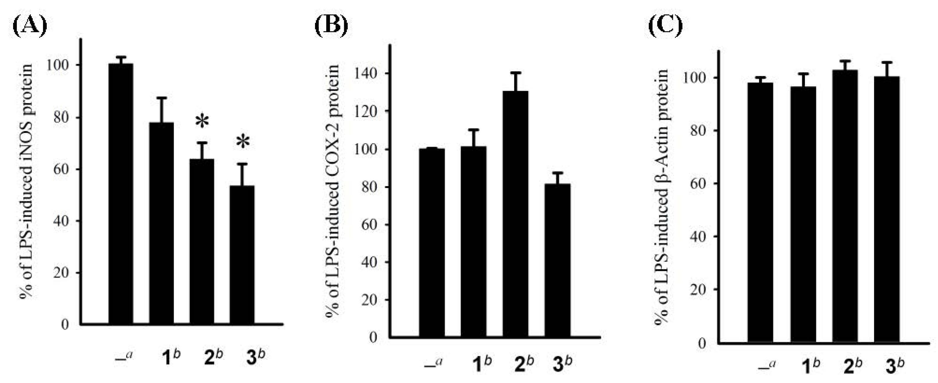
3.6. In Vitro Anti-Inflammatory Assay

Macrophage (RAW264.7) cell was purchased from ATCC. In vitro anti-inflammatory activities of tested compounds were measured by examining the inhibition of lipopolysaccharide (LPS) induced upregulation of iNOS and COX-2 proteins in macrophage cells using Western blotting analysis [9,10].

4. Conclusions

Our previous investigation on P. acronocephala has successfully discovered marine withanolides with potent anti-inflammatory activity. In this study, we reported three steroidal carboxylic acids, of which 3 exhibited potent cytotoxicity toward Hep3B, MDA-MB-231, MCF-7, and A-549 cancer cell lines. Compound 2, the second member of 27-norergostan-26-oic acid obtained from nature [11,12], was isolated from the soft coral for the first time. Our present investigation demonstrated that the soft coral, P. acronocephala, is a useful source for the discovery of bioactive substances.

Acknowledgments

This work was supported by grants from the National Science Council of Taiwan (NSC100-2320-B-110-001-MY2) and Ministry of Education (00C030205) awarded to J.-H. Sheu.

References

  1. Chao, C.-H.; Chou, K.-J.; Wen, Z.-H.; Wang, G.-H.; Wu, Y.-C.; Dai, C.-F.; Sheu, J.-H. Paraminabeolides A–F, cytotoxic and anti-inflammatory marine withanolides from the soft coral Paraminabea acronocephala. J. Nat. Prod. 2011, 74, 1132–1141. [Google Scholar]
  2. Ksebati, M.B.; Schmitz, F.J. Minabeolides: A group of withanolides from a soft coral, Minabea sp. J. Org. Chem. 1988, 53, 3926–3929. [Google Scholar] [CrossRef]
  3. Su, B.-N.; Park, E.K.; Nikolic, D.; Santarsiero, B.D.; Mesecar, A.D.; Vigo, J.S.; Graham, J.G.; Cabieses, F.; van Breemen, R.B.; Fong, H.H.S.; et al. Activity-guided isolation of novel norwithanolides from Deprea subtriflora with potential cancer chemopreventive activity. J. Org. Chem. 2003, 68, 2350–2361. [Google Scholar]
  4. Wang, W.; Lee, J.S.; Nakazawa, T.; Ukai, K.; Mangindaan, R.E.P.; Wewengkang, D.S.; Rotinsulu, H.; Kobayashi, H.; Tsukamoto, S.; Namikoshi, M. (25S)-Cholesten-26-oic acid derivatives from an Indonesian soft coral Minabea sp. Steroids 2009, 74, 758–760. [Google Scholar] [CrossRef]
  5. Chao, C.-H.; Wen, Z.-H.; Chen, I.-M.; Su, J.-H.; Huang, H.-C.; Chiang, M.Y.; Sheu, J.-H. Anti-inflammatory steroids from the octocoral Dendronephthya griffini. Tetrahedron 2008, 64, 3554–3560. [Google Scholar] [CrossRef]
  6. Nagai, Y.; Kusumi, T. New chiral anisotropic reagents for determining the absolute configurationof carboxylic acids. Tetrahedron Lett. 1995, 36, 1853–1856. [Google Scholar] [CrossRef]
  7. Yabuuchi, T.; Kusumi, T. Phenylglycine methyl ester, a useful tool for absolute configuration determination of various chiral carboxylic acids. J. Org. Chem. 2000, 65, 397–404. [Google Scholar] [CrossRef]
  8. Alley, M.C.; Scudiero, D.A.; Monks, A.; Hursey, M.L.; Czerwinski, M.J.; Fine, D.L.; Abbott, B.J.; Mayo, J.G.; Shoemaker, R.H.; Boyd, M.R. Feasibility of drug screening with panels of human tumor cell lines using a microculture tetrazolium assay. Cancer Res. 1988, 48, 589–601. [Google Scholar]
  9. Jean, Y.-H.; Chen, W.-F.; Sung, C.-S.; Duh, C.-Y.; Huang, S.-Y.; Lin, C.-S.; Tai, M.-H.; Tzeng, S.-F.; Wen, Z.-H. Capnellene, a natural marine compound derived from soft coral, attenuates chronic constriction injury-induced neuropathic pain in rats. Br. J. Pharmacol. 2009, 158, 713–725. [Google Scholar] [CrossRef]
  10. Wen, Z.-H.; Chao, C.-H.; Sheu, J.-H. A neuroprotective sulfone of marine origin and the in vivo anti-inflammatory activity of an analogue. Eur. J. Med. Chem. 2010, 45, 5998–6004. [Google Scholar] [CrossRef]
  11. Finamore, E.; Minale, L.; Riccio, R.; Rinaldo, G.; Zollo, F. Novel marine polyhydroxylated steroids from the starfish Myxoderma platyacanthum. J. Org. Chem. 1991, 56, 1146–1153. [Google Scholar]
  12. Sarma, N.S.; Krishna, M.S.; Pasha, S.G.; Prakasa Rao, T.S.; Venkateswarlu, Y.; Parameswaran, P.S. Marine metabolites: The sterols of soft coral. Chem. Rev. 2009, 109, 2803–2828. [Google Scholar] [CrossRef]
  • Samples Availability: Not available.

