Article

Fucoxanthin Enhances Cisplatin-Induced Cytotoxicity via NFκB-Mediated Pathway and Downregulates DNA Repair Gene Expression in Human Hepatoma HepG2 Cells

1
Department of Food Science and Biotechnology, National Chung Hsing University, Taichung 402, Taiwan
2
Department of Pharmacy, College of Pharmacy, China Medical University, Taichung 404, Taiwan
3
Department of Emergency, Toxicology Center, China Medical University Hospital, Taichung 404, Taiwan
4
Agricultural Biotechnology Center, National Chung Hsing University, Taichung 402, Taiwan
*
Author to whom correspondence should be addressed.
Mar. Drugs 2013, 11(1), 50-66; https://doi.org/10.3390/md11010050
Submission received: 10 October 2012 / Revised: 14 November 2012 / Accepted: 13 December 2012 / Published: 8 January 2013

Abstract

:
Cisplain, a platinum-containing anticancer drug, has been shown to enhance DNA repair and to inhibit cell apoptosis, leading to drug resistance. Thus, the combination of anticancer drugs with nutritional factors is a potential strategy for improving the efficacy of cisplatin chemotherapy. In this study, we investigated the anti-proliferative effects of a combination of fucoxanthin, the major non-provitamin A carotenoid found in Undaria Pinnatifida, and cisplatin in human hepatoma HepG2 cells. We found that fucoxanthin (1–10 μΜ) pretreatment for 24 h followed by cisplatin (10 μΜ) for 24 h significantly decreased cell proliferation, as compared with cisplatin treatment alone. Mechanistically, we showed that fucoxanthin attenuated cisplatin-induced NFκB expression and enhanced the NFκB-regulated Bax/Bcl-2 mRNA ratio. Cisplatin alone induced mRNA expression of excision repair cross complementation 1 (ERCC1) and thymidine phosphorylase (TP) through phosphorylation of ERK, p38 and PI3K/AKT pathways. However, fucoxanthin pretreatment significantly attenuated cisplatin-induced ERCC1 and TP mRNA expression, leading to improvement of chemotherapeutic efficacy of cisplatin. The results suggest that a combined treatment with fucoxanthin and cisplatin could lead to a potentially important new therapeutic strategy against human hepatoma cells.

1. Introduction

Hepatocellular carcinoma (HCC) is the major cause of cancer death in Taiwan and one of the most common cancers in the world, accounting for approximately 6% of all human cancers [1,2]. For most patients with unresectable HCC, chemotherapy plays an important role in the treatment of HCC [3]. Unfortunately, chemotherapy has limited effect on survival owing to multiple drug resistance (MDR) [4]. Much evidence indicates that the MDR is involved in drug efflux, DNA repair activity, and altered survival and apoptotic signaling pathways [5]. Thus, the new therapeutic strategies for malignant HCC using combined therapies or combined agents with distinct molecular mechanisms are considered more promising for higher therapy efficacy, resulting in better survival.
Platinum antitumor compounds, such as cisplatin (cis-diamminedichloroplatinum (II)) and its analogs, have been used widely as a chemotherapeutic drug for a variety of malignancies including hepatocellular carcinoma [6]. Platinum can directly bind to intra- and inter-strand DNA molecules to form predominantly platinum-DNA adducts that ultimately interfere with DNA transcription and replication and result in cell death [7,8,9]. Although cisplatin is extensively used in chemotherapy, its effectiveness is limited by acquired or intrinsic resistance [10]. A number of mechanisms of cisplatin resistance in cancer cells has been recognized: (1) increased reflux; (2) increased inactivation by sulfhydryl molecules such as glutathione; (3) altered expression of proteins in signal transduction pathways that control apoptosis; and (4) increased DNA repair [11]. DNA repair pathways that may result in platinum-based chemotherapeutic resistance include mismatch repair (MMR) and nucleotide excision repair (NER) [12,13]. Excision repair cross-complementation group 1 (ERCC1) is the initial enzyme in the NER pathway of DNA repair, and reports have shown that increased mRNA levels of ERCC1 are associated with clinical resistance to platinum-based chemotherapy in human lung, gastric, ovarian, cervical, and colorectal carcinomas and impact with the survival rate of cancer patients [14,15,16,17,18]. In addition, thymidine phosphorylase (TP), a key enzyme in the pyrimidine nucleoside salvage pathway, is known to catalyze the reversible conversion of thymidine to thymine and 2-deoxy-D-ribose-1-phosphate [19]. TP expression in various kinds of tumors is higher than that in the adjacent non-neoplastic tissues [20], and TP-overexpressed cell lines are more resistant to various apoptosis-inducing stimuli such as cisplatin and microtubule-interfering agents, hypoxia, and Fas ligands [21,22,23].
Nuclear transcription factor kappa B (NFκB), a heterodimeric protein composed of different combinations of members of the Rel family of transcription factors, is inactivated in the cytoplasm by IκBs, a class of inhibitor proteins. Phosphorylation of IκB by upstream kinases promotes its ubiquitination-dependent degradation, allowing NFκB to translocate to the nucleus and induce target genes, which is associated with cell proliferation [24], angiogenesis [25], metastasis [26], suppression of apoptosis [27], promotion of oncogenesis [28], and cancer therapy resistance [24]. NFκB is known to inhibit apoptosis through induction of anti-apoptotic proteins or suppression of pro-apoptotic genes, and the ratio of pro- and antiapoptotic Bcl-2 family members is critical to determine cell susceptibility to apoptotic insults [29,30]. Much evidence indicates that several anticancer drugs such as cisplatin, docetaxel, gemcitabine induce NFκB nuclear translocation and activation of its target genes, thereby potentially leading to chemoresistance [31]. For example, it has been reported that increased resistance of human cervical carcinoma cells to cisplatin is partly mediated via enhancement of cisplatin-induced NFκB activation [32]. Therefore, agents capable of inhibiting NFκB function might be considered as an adjuvant approach in combination with chemotherapeutic agents for a variety of cancers.
Fucoxanthin is one of the most abundant carotenoids and contributes more than 10% of the estimated total production of carotenoids in nature [33]. This carotenoid has been shown to have several biological functions, such as antioxidant activity [34,35,36,37], anti-obese effect [38,39], antidiabetic activity [40], antimutagenicity [41], anti-inflammation [42,43], and anticancer effects [44,45]. A previous report revealed that inhibition of proliferation of human hepatoma HepG2 cells by fucoxanthin is related to cell cycle arrest by downregulation of cyclin D and induction of GADD45A gene expression [45,46,47]. We recently reported that fucoxanthin exhibits anti-drug resistance potential and that the effect is likely associated with attenuated interaction between pregnane X receptor (PXR) and coactivator (SRC-1), thereby potentially preventing activation of PXR-mediated CYP3A4 and MDR1 expression [48]. As the combination of anticancer drugs with nutritional factors is a potential strategy for improving the efficacy of chemotherapy, we herein employed human hepatoma HepG2 cells to determine whether a combination of fucoxanthin and cisplatin may enhance the inhibition of cell proliferation.

2. Results

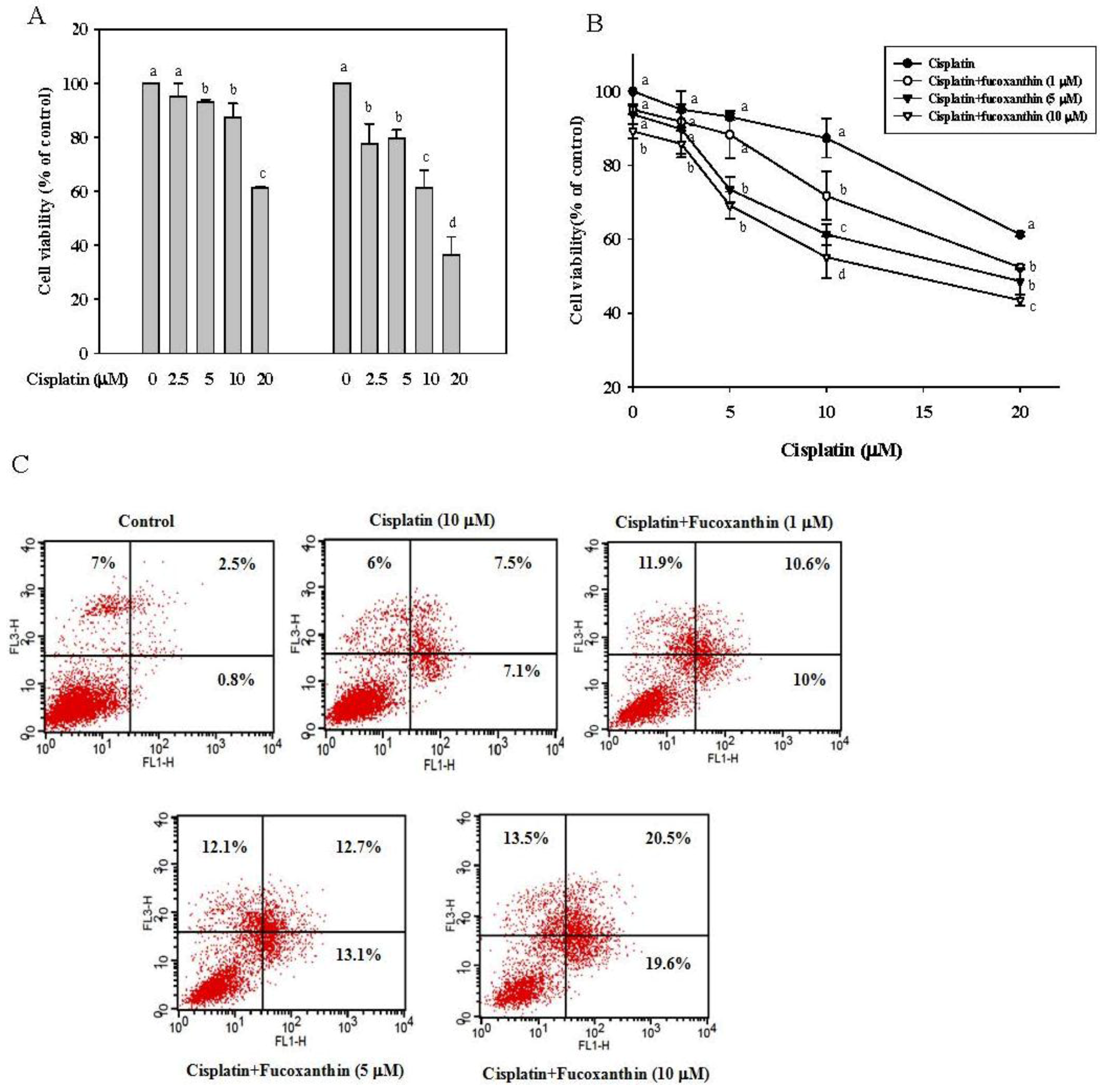
2.1. Fucoxanthin Increases the Sensitivity of Cisplatin in HepG2 Cells

InHepG2 cells treated with cisplatin (2.5–20 μM) for 24 and 48 h, (Figure 1A), we found that cisplatin significantly inhibited the cell viability (13% at 24 h and 39% at 48 h, P < 0.05, respectively, at 10 μM cisplatin). In addition, fucoxanthin significantly inhibited the cell proliferation of HepG2 by 17% and 28% after incubation with 10 μM fucoxanthin for 24 h and 48 h, respectively (Figure S1). To investigate whether fucoxanthin increases the sensitivity of cisplatin in HepG2 cells, we pre-incubated HepG2 cells with fucothanxin (1–10 μM) for 24 h followed by incubation with cisplatin (2.5–20 μM) for 24 h. Results reveal that the cell viability of HepG2 cells was significantly and concentration-dependently inhibited (Figure 1B), with an inhibition of 37% at 10 μM fucoxanthin and 10 μM cisplatin, as compared with cisplatin treatment alone. In addition, the combination of fucoxanthin with cisplatin increased early apoptotic cells (PI negative, Annexin V-FITC positive) and late apoptotic cells (PI positive, Annexin V-FITC positive) (Figure 1C). The results indicate that fucoxanthin enhances the anti-proliferative effect of cisplatin in human hepatoma HepG2 cells.
Figure 1. Effects of cisplatin (2.5–20 μM) alone or in combination with fucoxanthin (1–10 μM) on cell viability of human hepatoma HepG2 cells. (A) Cell viability of HepG2 cells incubated with cisplatin for 24 and 48 h. (B) Cell viability of HepG2 cells incubated with cisplatin (2.5–10 μM) for 24 h after pretreatment with fucoxanthin (1–10 μM) for 24 h. (C) Apoptotic cells in HepG2 cells incubated with cisplatin (2.5–10 μM) for 24 h after pretreatment with fucoxanthin (1–10 μM) for 24 h. Values are means ± SD, n = 3; means without a common letter differ significantly, P < 0.05.
Marinedrugs 11 00050 g001

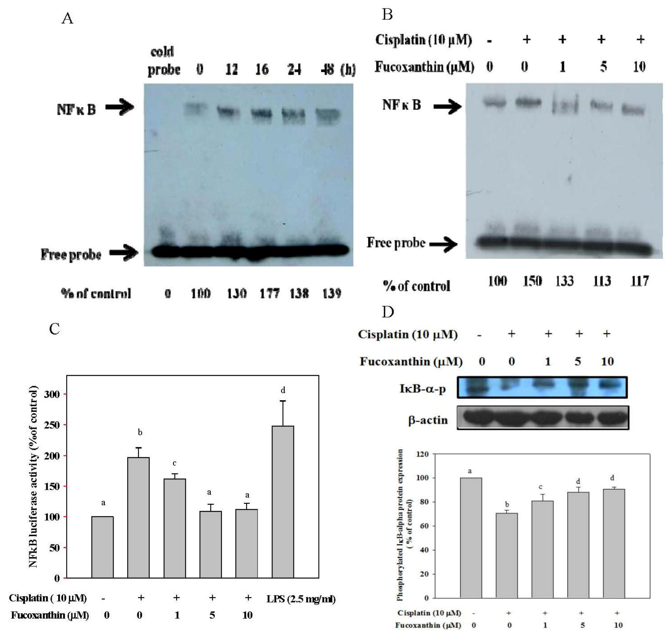
2.2. Fucoxanthin Attenuates the NFκB Expression Induced by Cisplatin and Restores the Phosphorylation of IκB-α Inhibited by Cisplatin

We also evaluated the effect of fucoxanthin on NFκB expression induced by cisplatin in HepG2 cells, as determined by the EMSA and NFκB reporter gene assays. As shown as in Figure 2A, cisplatin μM) most strongly induced NFκB binding activity at 16 h of incubation (by 77%, as compared with untreated cells). However, fucoxanthin concentration-dependently attenuated cisplatin-induced NFκB binding activity, with a 37% inhibition at 5 μM fucoxanthin (Figure 2B). We also showed that fucoxanthin significantly and concentration-dependently attenuated cisplatin-induced NFκB luciferase activity in a similar pattern to that of NFκB binding activity (Figure 2C). In addition, fucoxanthin significantly and concentration-dependently restored cisplatin-inhibited IκB-α-phosphorylation in HepG2 cells at 24 h of incubation, as compared with cisplatin treatment alone (Figure 2D).
Figure 2. NFκB expression in HepG2 cells pretreated with fucoxanthin (0–10 μM) followed by incubation with cisplatin (10 μM). (A) NFκB expression in HepG2 cells incubated with cisplatin (10 μM) for 0–48 h; (B) NFκB expression in HepG2 cells incubated with cisplatin (10 μM) for 16 h after pretreatment with fucoxanthin (1–10 μM) for 24 h; (C) NFκB luciferase activity expression in HepG2 cells incubated with cisplatin (10 μM) for 16 h after pretreatment with fucoxanthin (1–10 μM) for 24 h; (D) Phosphorylation of IκBin HepG2 cells incubated with cisplatin (10 μM) for 16 h after pretreatment with fucoxanthin (1–10 μM) for 24 h. Values are means ± SD, n = 3; means without a common letter differ significantly, P < 0.05.
Marinedrugs 11 00050 g002

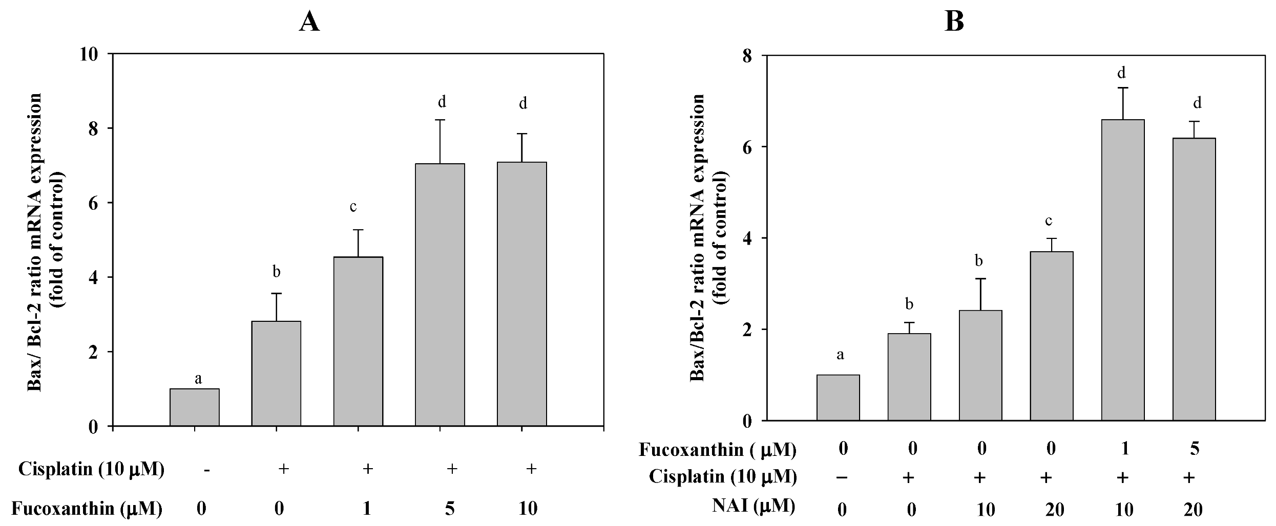
2.3. Fucoxanthin Combined with Cisplatin Increases the Ratio of Bax/Bcl-2 mRNA Expression in HepG2 Cells

Treatment of HepG2 cells with cisplatin (10 μM) for 24 h significantly increased the ratio of Bax/Bcl-2 mRNA expression (by 1.8-fold, P < 0.001, as compared with untreated cells). However, pretreatment of HepG2 cells with fucoxanthin for 24 h followed by incubation with cisplatin for 24 h significantly and concentration-dependently increased the ratio of Bax/Bcl-2 mRNA expression (by 4.3 fold, P < 0.001, as compared with cisplatin treatment alone) (Figure 3A). To further determine whether fucoxanthin in combination with cisplatin enhances the ratio of Bax/Bcl-2 mRNA primarily through NFκB-regulated pathways, we pretreated HepG2 cells with fucoxanthin for 24 h followed by incubation with an NFκB activation inhibitor (NAI) (10 and 20 μM) for 2 h and then by incubation with cisplatin (10 μM) for 24 h. We found that the combination of fucoxanthin, NAI and cisplatin synergistically or additively increased the ratio of Bax/Bcl-2 mRNA expression, as compared with the NFκB activation inhibitor alone (Figure 3B). Thus, the data indicate that fucoxanthin increases the ratio of Bax/Bcl-2 mRNA expression and that this effect is likely associated with inhibition of the NFκB pathway.
Figure 3. (A) The ratio of Bax/Bcl-2 mRNA in HepG2 cells pretreated with fucoxanthin (1–10 μM) for 24 h followed by incubation with cisplatin (10 μM) for 24 h. (B) The ratio of Bax/Bcl-2 mRNA in HepG2 cells pretreated with fucoxanthin (5 μM) for 24 h followed by incubation with NFκB activation inhibitor (NAI, 20 μΜ) for 2 h and then treated cisplatin (10 μM) for 24 h. Values are means ± SD, n = 3; means without a common letter differ significantly, P < 0.05.
Marinedrugs 11 00050 g003

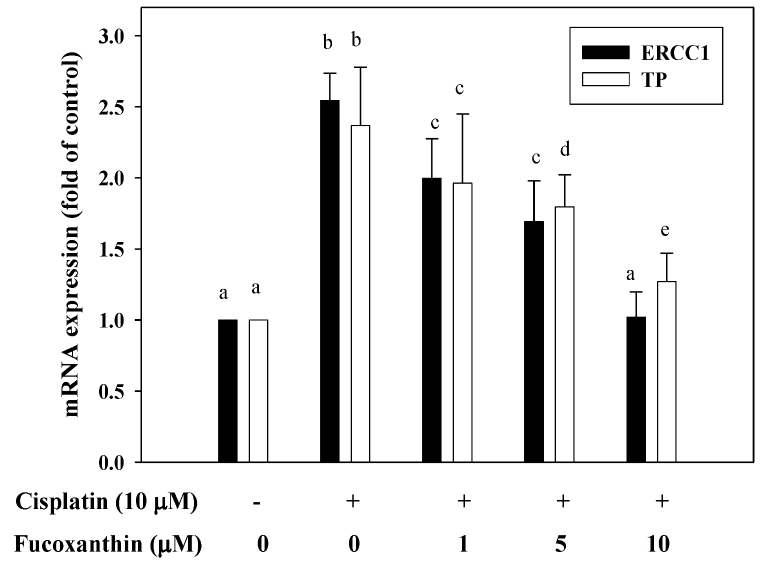
2.4. Fucoxanthin Attenuates mRNA Expression of ERCC1 and TP Induced by Cisplatin

Real-time PCR was performed to evaluate the mRNA levels of ERCC1 and TP. As shown in Figure 4, cisplatin (10 μM) treatment alone significantly increased the mRNA expression of ERCC1 and TP in HepG2 cells. However, the induced mRNA expression of ERCC1 and TP in HepG2 cells by cisplatin (10 μM) was attenuated by pretreatment with fucoxanthin (1–10 μM) for 24 h, with a 1.5-fold and a 1.2-fold inhibition, respectively, at 10 μM fucoxanthin, as compared with cisplatin treatment alone.
Figure 4. The level of ERCC1 and TP mRNA in HepG2 cells pretreated with fucoxanthin (1–10 μM) for 24 h followed by incubation with cisplatin (10 μM) for 24 h. Values are means ± SD, n = 3; means without a common letter differ significantly, P < 0.05.
Marinedrugs 11 00050 g004

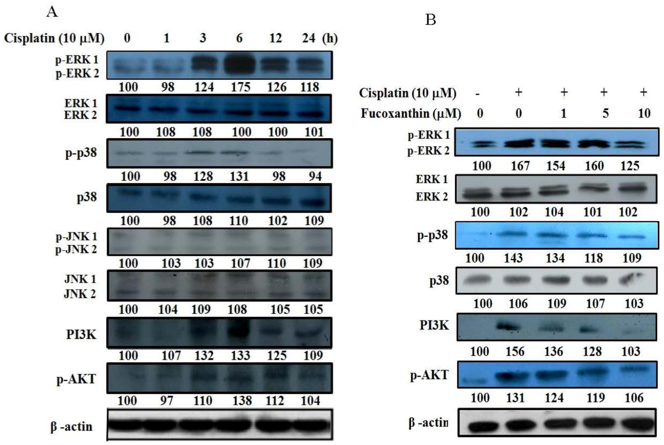
2.5. Fucoxanthin Attenuates the Phosphorylation of ERK1/2, p38, AKT and PI3K in HepG2 Cells

The time effect of cisplatin on protein expression of the mitogen-activated protein kinase (MAPK) family (p38, ERK, and JNK) and phosphatidylinositol 3-kinase (PI3K)/AKT in HepG2 cells were determined by Western blotting. Results reveal that cisplatin (10 μM) markedly increased the phosphorylation of ERK, p38 and PI3K/AKT at 6 h of incubation, but it did not affect the phosphorylation of JNK or the protein expression of ERK, p38 and JNK (Figure 5A). We then determined whether pretreatment of HepG2 cells with fucoxanthin (1–10 μM) for 24 h attenuates the induction of MAPK family and PI3K/AKT protein expression by cisplatin (10 μM). We found that fucoxanthin concentration-dependently attenuated cisplatin-induced phosphorylation of ERK, p38 and PI3K/AKT (Figure 5B).
Figure 5. The protein expression of MAPK family and PI3K/AKT in HepG2 cells pretreated with fucoxanthin (0–10 μM) followed by incubation with cisplatin (10 μM). (A) The protein expression of MAPK family and PI3K/AKT in HepG2 cells incubated with cisplatin (10 μM) for 0–24 h. (B) The protein expression of MAPK family and PI3K/AKT in HepG2 cells incubated with cisplatin (10 μM) for 6 h after pretreatment with fucoxanthin (1–10μM) for 24 h.
Marinedrugs 11 00050 g005

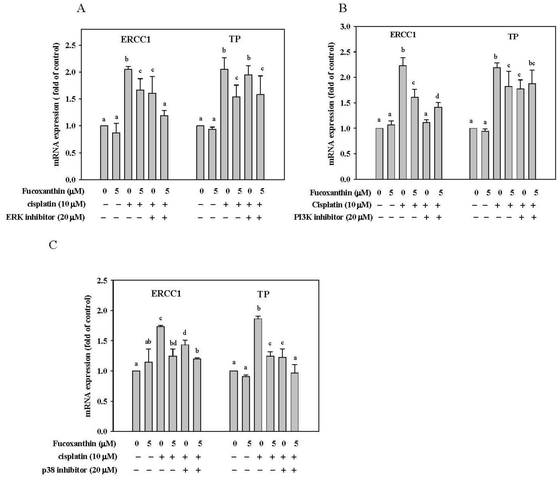
2.6. Effect of Fucoxanthin in Combination with ERK, p38 and PI3K Inhibitor on ERCC1 and TP mRNA Expression in HepG2 Cells

We then determined whether the attenuation of fucoxanthin on ERCC1 and TP mRNA expression induced by cisplatin occur primarily through the inhibition of ERK, p38 and PI3K/AKT pathway. HepG2 cells were pre-incubated with fucoxanthin (5 μM) for 24 h followed by incubation with ERK inhibitor (PD98059; 20 μM), p38 inhibitor (SB203580; 20 μM) or PI3K inhibitor (LY294002; 20 μM) for 1 h and then with cisplatin (10 μM) for 24 h. The concentration of 5 μM fucoxanthin was chosen because it only produced a slight inhibition of mRNA expression for ERCC1 and TP (see Figure 4). We found that the combination of fucoxanthin, cisplatin and the ERK inhibitor or PI3K inhibitor synergistically inhibited ERCC1 mRNA expression but not TP mRNA expression (Figure 6A,B). In contrast, the combination of fucoxanthin with p38 inhibitor enhanced the inhibition of mRNA expression of TP but not ERCC1 (Figure 6C). Thus, the results reveal that fucoxanthin may inhibit ERCC1 mRNA expression through ERK and PI3K/AKT pathway but may inhibit TP mRNA expression through p38 pathway.
Figure 6. The level of ERCC1 and TP mRNA in HepG2 cells pretreated with fucoxanthin (5 μM) for 24 h followed by incubation with a specific inhibitor (20 μM) for 1 h and then treated cisplatin (10 μM) for 24 h. (A) ERK inhibitor (PD98059); (B) PI3K inhibitor (LY294002); (C) p38 inhibitor (SB203580). Values are means ± SD, n = 3; means without a common letter differ significantly, P < 0.05.
Marinedrugs 11 00050 g006

3. Discussion

The main question addressed by this study was whether a combination of fucoxanthin and cisplatin improves the efficacy of chemotherapy and enhances the inhibition of cell proliferation. Our data demonstrate for the first time that fucoxanthin in combination with cisplatin inhibits the cell proliferation of human hepatoma HepG2 cells and that this combined effect involves NFκB mediated-pathways and attenuation of the DNA repair gene expression induced by cisplatin.
A probable mechanism by which fucoxanthin improves the efficacy of cisplatin chemotherapy is through the inhibition of NFκB expression and the increase in NFκB-regulated Bax/Bcl-2 mRNA expression. NFκB is known to suppress apoptosis through the loss of pro-apoptotic factors (e.g., functional p53 or Bax) or the activation of anti-apoptotic factors such as Bcl-2, Bcl-XL or IAPs (Inhibitors of Apoptosis Proteins) to block caspase-8 activation [24]. Moreover, NFκB binding sites were found in the promoter of Bcl-2, Bcl-xL, and survivin [49], suggesting that these apoptotic factors may be regulated by NFκB. Overexpression of the antiapoptotic molecules Bcl-2 or Bcl-xL can cause resistance to anticancer drugs [50]. We found that fucoxanthin pretreatment attenuated cisplatin-induced DNA-binding activity of NFκB, restored cisplatin-inhibited IκB-α-phosphorylation, and increased the ratio of Bax/Bcl-2 mRNA expression, rendering cancer cells sensitive to apoptosis induced by cisplatin. Many NFκB inhibitors have been identified in vitro such as proteasome inhibitors, genistein, parthenolide, flavopiridol and gliotoxin, enhance the cytotoxic effect of anticancer agents [29]. In the present study, we found that fucoxanthin strongly attenuated cisplatin-induced NFκB activation, suggesting that fucoxanthin may be an NFκB inhibitor.
Another probable mechanism by which fucoxanthin improves the efficacy of cisplatin chemotherapy is through the inhibition of mRNA expression of DNA repair genes ERCC1 and TP. Tumor DNA repair capacity is frequently increased as an inherent cellular mechanism for evading cell death as a result of administration of chemotherapeutic drugs. Increased expression of ERCC1 in several cancers has been associated with more efficient removal of DNA adducts induced by platinum, leading to clinical resistance to cisplatin-based chemotherapy [51]. Depletion of endogenous ERCC1 expression by si-ERCC1 RNA transfection was shown to significantly enhance the cytotoxicity of cisplatin [52]. In the present study, we found that fucoxanthin pretreatment significantly attenuated cisplatin-induced ERCC1 and TP mRNA expression. Recently, Tsai et al. [53] have shown that curcumin downregulates the expression levels of TP and ERCC1, which helps overcome platinum resistance in cancer cells.
MAPK family and PI3K/AKT pathways play important roles in the regulation of cell proliferation, differentiation, apoptosis and DNA repair pathways [54]. Protein kinase C and Ras have been suggested to be involved in the activation of ERK1/2 by cisplatin [55]. AKT may promote cell survival by phosphorylating and inactivating the pro-apoptotic proteins BAD (Bcl-2-associated death protein) and caspase-9 [56]. It has been reported that the inactivation of NFκB-binding activity sensitizes human ovarian cancer cells to cisplatin in vitro [57] and that the combined therapy of cisplatin with a PI3K inhibitor enhances the apoptotic effect of cisplatin in vivo [58]. In this study, we found that cisplatin activated the phosphorylation of ERK1/2, p38, AKT and PI3K in human hepatoma HepG2 cells and that fucoxanthin pretreatment attenuated cisplatin-induced phosphorylation of all these signaling molecules to sensitize cancer cells to apoptosis induced by cisplatin. With regard to the DNA repair system, it has been shown that specific MERK/ERK and PI3K inhibitors prevent ERCC1 induction, whereas JNK and p38 inhibitors are without effects in human hepatoma cells [59]. Interestingly, curcumin and emodin, which are natural anthraquinone derivatives found in the roots and rhizomes of numerous plants, were found to enhance cisplatin-induced cytotoxicity via downregulation of ERCC1 and inactivation of ERK1/2 in non-small cell lung cancer [52]. The inhibition of the PI3K pathway on ERCC1 basal expression has been confirmed by using shRNA against FRAP/mTOR, a key kinase involved in nucleotide excision repair pathway [60]. TP, a key enzyme in the pyrimidine nucleoside salvage pathway, was also found to be regulated by NFκB- and p38 MAPK-mediated signaling in patients with nasopharyngeal carcinoma [61]. In the present study, we found that fucoxanthin inhibited ERCC1 mRNA expression through the ERK and PI3K/AKT pathways, whereas this carotenoid inhibited TP expression through the p38 pathway, as evidenced by using specific inhibitors. The results indicate that the improved chemotherapeutic efficacy of cisplatin by fucoxanthin may also involve inhibition of mRNA expression of some DNA repair genes through downregulation of ERK, p38, and PI3K/AKT pathways.

4. Experimental Section

4.1. Materials

Dulbecco’s modified eagle medium (DMEM), fetal bovine serum (FBS), trypsin, penicillin, sodium pyruvate, and non-essential amino acids (NEAA) were purchased from GIBCO/BRL (Maryland, MD, USA). MAPK/extracellular signal-regulated kinase (ERK) 1/2, c-Jun NH2-terminal kinase (JNK)/stress-activated protein kinase and p38 MAPK proteins and phosphorylated proteins, phosphatidylinositol 3-kinase (PI3K)/AKT, ERK inhibitor (PD98059), p38 inhibitor (SB203580) and PI3K inhibitor (LY294002) were purchased from Cell Signaling Technology (Beverly, MA). NFκB activation inhibitor was purchased from Merck Millipore (Billerica, MA, USA). Fucoxanthin was extracted from Undaria pinnatifida and purified, as we reported previously [62]. The purified fucoxanthin was dissolved in ethanol to a final concentration of 10 mM as the stock solution. Before the experiment, fucoxanthin solutions were prepared freshly in a mixture of ethanol and FBS (1:9), as adopted from the preparation of lycopene solution [63].

4.2. Cell Cultures

The human hepatoblastoma HepG2 cell line was obtained from Food Industry Research and Development Institute (FIRDI, Hsinchu, Taiwan) and maintained in DMEM supplemented with 10% fetal bovine serum without antibiotics under 5% CO2 at 37 °C.

4.3. Assessment of Cell Viability

Cell viability was evaluated using the modified acid-phosphatase (ACP) assay, with p-nitrophenyl phosphate (PNPP) disodium salt as a substrate. The cell culture media were aspirated, and the cells were washed with phosphate-buffered saline (PBS). Following the wash, 100 μL of the ACP reagent (0.1 M sodium acetate (pH 5.5), 0.1% Triton X-100, and 10 mM PNPP) was added. After 1 h of incubation at 37 °C, the enzyme activity was stopped by adding 10 μL of 1 N NaOH, and the enzyme activity was determined photometrically at a wavelength of 405 nm [64].

4.4. Real-Time Polymerase Chain Reaction

Total RNA in cell cultures was extracted with REzol reagent (PROtech Technologies, Inc., Placentia, CA, USA), and 1 μg of total RNA was reverse-transcribed by using oligo-dT as a primer in 20 μL reverse-transcription solutions containing 1 μL reverse transcriptase (Promega, Sunnyvale, CA, USA). Real-time PCR performed with a Corbett instrument (Applied Biosystems, Carlsbad, CA, USA) using SYBR Green Master Mix (ProTech, Placentia, CA, USA) according to the manufacturer’s instructions. In all real-time PCR experiments, both a non-template control (NTC) and a standard curve were amplified, as well. The RNA abundance was normalized to β-actin RNA in each sample. The primers used in this study were as follows: ERCC1 forward 5′-CCCTGGGAATTTGGCGACGTAA-3′, reverse 5′-CTCCAGGTACCGCCCAGCTTCC-3′; TP forward 5′-AGCTGGAGTCTATTCCTGGATT-3′, reverse 5′-GGCTGCATATAGGATTCCGTC-3′; β-actin forward 5′-GTGGGGCGCCCCAGGCACCA-3′, reverse 3′-CACCCCGCGGGGTCCGTGGT-5′.

4.5. Western Blotting

Protein expression of MAPK family (ERK, p38, JNK, p-JNK, p-ERK and p-p38) and PI3K/AKT (PI3K, AKT and p-AKT) was measured by Western blotting. In cell culture experiments, the medium was removed and cells were rinsed with PBS twice. After the addition of 0.5 mL of cold RIPA buffer and protease inhibitors, cells were scraped and followed by a mild vortexing at 0 °C for 20 min. The cell lysates were then subjected to a centrifugation of 10,000 rpm for 30 min at 4 °C. Total protein (50 μg) from the supernatant was resolved on SDS-PAGE and transferred onto a PVDF membrane. After blocking with TBS buffer (20 mmol/L Tris-HCl, 150 mmol/L NaCl, pH 7.4) containing 5% nonfat milk, the membrane was incubated with monoclonal antibody followed by incubation with horseradish peroxidase-conjugated anti-goat IgG, and then visualized using an ECL chemiluminescent detection kit (Amersham, Sweden). The relative density of the immunoreactive bands was quantitated by densitometry (Gel Pro Analyzer TM, version 3.0, Media Cybernetics, Rockville, MD, USA).

4.6. Preparation of Nuclear Extracts and Electrophoretic Mobility Shift Assay (EMSA)

Nuclear protein extracts (5 μg) were prepared according to the modified method of a previous study [65]. Binding activities of transcription factors including NFκB were analyzed by EMSA. Electrophoretic mobility shift assay (EMSA) was performed with LightShift Chemiluminescent EMSA Kit (Pierce Biotechnology, Rockford, IL, Country), as described previously [66]. The NFκB consensus oligonucleotide probe (5′-AGTTGAGGGGACTTTCCCAGGC-3′) was end-labeled with biotin (Sangon, Shanghai, China). Briefly, nuclear extract (5 μg) was incubated with 10 ng NFκB (p65) probe. For the cold probe assay, 40 ng of unlabeled (cold) NFκB probe was mixed with sample 5 min before adding 10 ng biotin-labeled NFκB probe. Protein-DNA complexes were then resolved by non-denaturing polyacrylamide gel electrophoresis (PAGE). After blocking, avidin-HRP was applied and detected by enhanced chemiluminescence (ECL, Amersham). The relative NFκB levels were quantitated by Matrox Inspector 2.1 software.

4.7. Transfection and Luciferase Reporter Gene Assays

HepG2 cells (1.8 × 10⁴ cells/well) were plated in 96-microwell-white-plates (Nalge Nunc, Rochester, New York, NY, USA) before transfection. The NFκB plasmid vector (pGL4.32 (luc2P/NF-κB-RE/Hygro)) contains five copies of an NFκB response element was purchased from Promega (Sunnyvale, CA, USA). Transfection of NFκB plasmid vector (0.15 μg) into HepG2 cells was performed using TransIL-LT1 Transfection Reagent (Mirus, Madison, WI, USA), and in all experiments, the pRL-TK Renilla reporter vector (0.02 μg) (Promega, Sunnyvale, CA, USA) was used as an internal control. The cells were then incubated with cisplatin (10 μM) for 16 h after pretreatment with fucoxanthin (1–10 μM) for 24 h. Renilla and firefly luciferase activities were measured using the Dual-Luciferase Reporter Assay System (Promega, Sunnyvale, CA, USA).

4.8. Statistical Analysis

All experiments were repeated at least thrice. Values are expressed as means ± SD and analyzed using one way ANOVA followed by LSD test for comparisons of group means, when the F ratios were significant. All statistical analyses were performed using SPSS for Windows, version 10 (SPSS, Inc., Armonk, NY, USA); a P value < 0.05 is considered statistically significant.

5. Conclusions

In conclusion, the present study demonstrates that pretreatment with fucoxanthin improves the chemotherapeutic efficacy of cisplatin by enhancing the inhibition of cell proliferation of human hepatoma HepG2. These effects of fucoxanthin may involve the inhibition of NFκB expression and the increase in Bax/Bcl-2 mRNA ratios regulated by NFκB, as well as the decrease of DNA repair systems regulated by ERK, p38 and PI3K/AKT, leading to sensitized cancer cells to apoptosis induced by cisplatin. The results suggest that the combined treatment of fucoxanthin and cisplatin may provide a novel therapeutic approach to decrease cisplatin-induced drug resistance.

Acknowledgments

This work was supported in part by the Ministry of Education, Taiwan, ROC, under the ATU plan and NSC101-2320-B-039-007-MY3 from the National Science Council, Executive Yuan, Taiwan.

Supplementary Files

  • Supplementary File 1:

    Supplementary Information (PDF, 42 KB)

  • References

    1. Di Bisceglie, A.M. Epidemiology and clinical presentation of hepatocellular carcinoma. J. Vasc. Interv. Radiol. 2002, 13, S169–S171. [Google Scholar] [CrossRef]
    2. Montalto, G.; Cervello, M.; Giannitrapani, L.; Dantona, F.; Terranova, A.; Castagnetta, L.A. Epidemiology, risk factors, and natural history of hepatocellular carcinoma. Ann. N. Y. Acad. Sci. 2002, 963, 13–20. [Google Scholar]
    3. Chau, G.Y.; Lui, W.Y.; Tsay, S.H.; Chao, Y.; King, K.L.; Wu, C.W. Postresectional adjuvant intraportal chemotherapy in patients with hepatocellular carcinoma: A case-control study. Ann. Surg. Oncol. 2006, 13, 1329–1337. [Google Scholar] [CrossRef]
    4. Tong, S.W.; Yang, Y.X.; Hu, H.D.; An, X.; Ye, F.; Hu, P.; Ren, H.; Li, S.L.; Zhang, D.Z. Proteomic investigation of 5-fluorouracil resistance in a human hepatocellular carcinoma cell line. J. Cell. Biochem. 2012, 113, 1671–1680. [Google Scholar]
    5. Dietel, M. Molecular mechanisms and possibilities of overcoming drug resistance in gastrointestinal tumors. Recent Results Cancer Res. 1996, 142, 89–101. [Google Scholar] [CrossRef]
    6. Go, R.S.; Adjei, A.A. Review of the comparative pharmacology and clinical activity of cisplatin and carboplatin. J. Clin. Oncol. 1999, 17, 409–422. [Google Scholar]
    7. Cepeda, V.; Fuertes, M.A.; Castilla, J.; Alonso, C.; Quevedo, C.; Perez, J.M. Biochemical mechanisms of cisplatin cytotoxicity. Anticancer Agents Med. Chem. 2007, 7, 3–18. [Google Scholar] [CrossRef]
    8. Zorbas, H.; Keppler, B.K. Cisplatin damage: Are DNA repair proteins saviors or traitors to the cell? ChemBioChem 2005, 6, 1157–1166. [Google Scholar] [CrossRef]
    9. Van de Vaart, P.J.; van der Vange, N.; Zoetmulder, F.A.; van Goethem, A.R.; van Tellingen, O.; ten Bokkel Huinink, W.W.; Beijnen, J.H.; Bartelink, H.; Begg, A.C. Intraperitoneal cisplatin with regional hyperthermia in advanced ovarian cancer: Pharmacokinetics and cisplatin-DNA adduct formation in patients and ovarian cancer cell lines. Eur. J. Cancer 1998, 34, 148–154. [Google Scholar]
    10. Kartalou, M.; Essigmann, J.M. Mechanisms of resistance to cisplatin. Mutat. Res. 2001, 478, 23–43. [Google Scholar] [CrossRef]
    11. Shahzad, M.M.; Lopez-Berestein, G.; Sood, A.K. Novel strategies for reversing platinum resistance. Drug Resist. Updat. 2009, 12, 148–152. [Google Scholar] [CrossRef]
    12. Helleday, T.; Petermann, E.; Lundin, C.; Hodgson, B.; Sharma, R.A. DNA repair pathways as targets for cancer therapy. Nat. Rev. Cancer 2008, 8, 193–204. [Google Scholar] [CrossRef]
    13. Rabik, C.A.; Dolan, M.E. Molecular mechanisms of resistance and toxicity associated with platinating agents. Cancer Treat. Rev. 2007, 33, 9–23. [Google Scholar] [CrossRef]
    14. Shirota, Y.; Stoehlmacher, J.; Brabender, J.; Xiong, Y.P.; Uetake, H.; Danenberg, K.D.; Groshen, S.; Tsao-Wei, D.D.; Danenberg, P.V.; Lenz, H.J. ERCC1 and thymidylate synthase mRNA levels predict survival for colorectal cancer patients receiving combination oxaliplatin and fluorouracil chemotherapy. J. Clin. Oncol. 2001, 19, 4298–4304. [Google Scholar]
    15. Lord, R.V.; Brabender, J.; Gandara, D.; Alberola, V.; Camps, C.; Domine, M.; Cardenal, F.; Sanchez, J.M.; Gumerlock, P.H.; Taron, M.; et al. Low ERCC1 expression correlates with prolonged survival after cisplatin plus gemcitabine chemotherapy in non-small cell lung cancer. Clin. Cancer Res. 2002, 8, 2286–2291. [Google Scholar]
    16. Zhou, W.; Gurubhagavatula, S.; Liu, G.; Park, S.; Neuberg, D.S.; Wain, J.C.; Lynch, T.J.; Su, L.; Christiani, D.C. Excision repair cross-complementation group 1 polymorphism predicts overall survival in advanced non-small cell lung cancer patients treated with platinum-based chemotherapy. Clin. Cancer Res. 2004, 10, 4939–4943. [Google Scholar]
    17. Selvakumaran, M.; Pisarcik, D.A.; Bao, R.; Yeung, A.T.; Hamilton, T.C. Enhanced cisplatin cytotoxicity by disturbing the nucleotide excision repair pathway in ovarian cancer cell lines. Cancer Res. 2003, 63, 1311–1316. [Google Scholar]
    18. Gossage, L.; Madhusudan, S. Current status of excision repair cross complementing-group 1 (ERCC1) in cancer. Cancer Treat. Rev. 2007, 33, 565–577. [Google Scholar] [CrossRef]
    19. Bijnsdorp, I.V.; Azijli, K.; Jansen, E.E.; Wamelink, M.M.; Jakobs, C.; Struys, E.A.; Fukushima, M.; Kruyt, F.A.; Peters, G.J. Accumulation of thymidine-derived sugars in thymidine phosphorylase overexpressing cells. Biochem. Pharmacol. 2010, 80, 786–792. [Google Scholar] [CrossRef]
    20. Nakayama, Y.; Inoue, Y.; Nagashima, N.; Katsuki, T.; Matsumoto, K.; Kadowaki, K.; Shibao, K.; Tsurudome, Y.; Hirata, K.; Sako, T.; et al. Expression levels of thymidine phosphorylase (TP) and dihydropyrimidine dehydrogenase (DPD) in patients with gastrointestinal cancer. Anticancer Res. 2005, 25, 3755–3761. [Google Scholar]
    21. Mori, S.; Takao, S.; Ikeda, R.; Noma, H.; Mataki, Y.; Wang, X.; Akiyama, S.; Aikou, T. Thymidine phosphorylase suppresses Fas-induced apoptotic signal transduction independent of its enzymatic activity. Biochem. Biophys. Res. Commun. 2002, 295, 300–305. [Google Scholar] [CrossRef]
    22. Ikeda, R.; Furukawa, T.; Mitsuo, R.; Noguchi, T.; Kitazono, M.; Okumura, H.; Sumizawa, T.; Haraguchi, M.; Che, X.F.; Uchimiya, H.; et al. Thymidine phosphorylase inhibits apoptosis induced by cisplatin. Biochem. Biophys. Res. Commun. 2003, 301, 358–363. [Google Scholar] [CrossRef]
    23. Jeung, H.C.; Che, X.F.; Haraguchi, M.; Furukawa, T.; Zheng, C.L.; Sumizawa, T.; Rha, S.Y.; Roh, J.K.; Akiyama, S. Thymidine phosphorylase suppresses apoptosis induced by microtubule-interfering agents. Biochem. Pharmacol. 2005, 70, 13–21. [Google Scholar]
    24. Baldwin, A.S. Control of oncogenesis and cancer therapy resistance by the transcription factor NF-κB. J. Clin. Investig. 2001, 107, 241–246. [Google Scholar] [CrossRef]
    25. Tabruyn, S.P.; Griffioen, A.W. A new role for NF-κB in angiogenesis inhibition. Cell. Death Differ. 2007, 14, 1393–1397. [Google Scholar] [CrossRef]
    26. Andela, V.B.; Gordon, A.H.; Zotalis, G.; Rosier, R.N.; Goater, J.J.; Lewis, G.D.; Schwarz, E.M.; Puzas, J.E.; O’Keefe, R.J. NFκB: A pivotal transcription factor in prostate cancer metastasis to bone. Clin. Orthop. Relat. Res. 2003, 415 (Suppl.), S75–S85. [Google Scholar]
    27. Tomita, M.; Kawakami, H.; Uchihara, J.N.; Okudaira, T.; Masuda, M.; Takasu, N.; Matsuda, T.; Ohta, T.; Tanaka, Y.; Ohshiro, K.; et al. Curcumin (diferuloylmethane) inhibits constitutive active NF-κB, leading to suppression of cell growth of human T-cell leukemia virus type I-infected T-cell lines and primary adult T-cell leukemia cells. Int. J. Cancer 2006, 118, 765–772. [Google Scholar] [CrossRef]
    28. Sharma, H.W.; Narayanan, R. The NF-κB transcription factor in oncogenesis. Anticancer Res. 1996, 16, 589–596. [Google Scholar]
    29. Nakanishi, C.; Toi, M. Nuclear factor-κB inhibitors as sensitizers to anticancer drugs. Nat. Rev. Cancer 2005, 5, 297–309. [Google Scholar] [CrossRef]
    30. Shou, Y.; Li, N.; Li, L.; Borowitz, J.L.; Isom, G.E. NF-κB-mediated up-regulation of Bcl-XS and Bax contributes to cytochrome c release in cyanide-induced apoptosis. J. Neurochem. 2002, 81, 842–852. [Google Scholar] [CrossRef]
    31. Sarkar, F.H.; Li, Y. Using chemopreventive agents to enhance the efficacy of cancer therapy. Cancer Res. 2006, 66, 3347–3350. [Google Scholar] [CrossRef]
    32. Yeh, P.Y.; Chuang, S.E.; Yeh, K.H.; Song, Y.C.; Cheng, A.L. Involvement of nuclear transcription factor-κB in low-dose doxorubicin-induced drug resistance of cervical carcinoma cells. Biochem. Pharmacol. 2003, 66, 25–33. [Google Scholar]
    33. Dembitsky, V.M.; Maoka, T. Allenic and cumulenic lipids. Prog. Lipid Res. 2007, 46, 328–375. [Google Scholar] [CrossRef]
    34. Sachindra, N.M.; Sato, E.; Maeda, H.; Hosokawa, M.; Niwano, Y.; Kohno, M.; Miyashita, K. Radical scavenging and singlet oxygen quenching activity of marine carotenoid fucoxanthin and its metabolites. J. Agric. Food Chem. 2007, 55, 8516–8522. [Google Scholar] [CrossRef]
    35. Heo, S.J.; Jeon, Y.J. Protective effect of fucoxanthin isolated from Sargassum siliquastrum on UV-B induced cell damage. J. Photochem. Photobiol. B 2009, 95, 101–107. [Google Scholar] [CrossRef]
    36. Liu, C.L.; Liang, A.L.; Hu, M.L. Protective effects of fucoxanthin against ferric nitrilotriacetate-induced oxidative stress in murine hepatic BNL CL.2 cells. Toxicol. In Vitro 2011, 25, 1314–1319. [Google Scholar] [CrossRef]
    37. Liu, C.L.; Chiu, Y.T.; Hu, M.L. Fucoxanthin enhances HO-1 and NQO1 expression in murine hepatic BNL CL.2 cells through activation of the Nrf2/ARE system partially by its pro-oxidant activity. J. Agric. Food Chem. 2011, 59, 11344–11351. [Google Scholar]
    38. Maeda, H.; Hosokawa, M.; Sashima, T.; Takahashi, N.; Kawada, T.; Miyashita, K. Fucoxanthin and its metabolite, fucoxanthinol, suppress adipocyte differentiation in 3T3-L1 cells. Int. J. Mol. Med. 2006, 18, 147–152. [Google Scholar]
    39. Jeon, S.M.; Kim, H.J.; Woo, M.N.; Lee, M.K.; Shin, Y.C.; Park, Y.B.; Choi, M.S. Fucoxanthin-rich seaweed extract suppresses body weight gain and improves lipid metabolism in high-fat-fed C57BL/6J mice. Biotechnol. J. 2010, 5, 961–969. [Google Scholar] [CrossRef]
    40. Maeda, H.; Hosokawa, M.; Sashima, T.; Miyashita, K. Dietary combination of fucoxanthin and fish oil attenuates the weight gain of white adipose tissue and decreases blood glucose in obese/diabetic KK-Ay mice. J. Agric. Food Chem. 2007, 55, 7701–7706. [Google Scholar] [CrossRef]
    41. Nishino, H.; Tokuda, H.; Murakoshi, M.; Satomi, Y.; Masuda, M.; Onozuka, M.; Yamaguchi, S.; Takayasu, J.; Tsuruta, J.; Okuda, M.; et al. Cancer prevention by natural carotenoids. Biofactors 2000, 13, 89–94. [Google Scholar] [CrossRef]
    42. Kim, K.N.; Heo, S.J.; Yoon, W.J.; Kang, S.M.; Ahn, G.; Yi, T.H.; Jeon, Y.J. Fucoxanthin inhibits the inflammatory response by suppressing the activation of NF-κB and MAPKs in lipopolysaccharide-induced RAW 264.7 macrophages. Eur. J. Pharmacol. 2010, 649, 369–375. [Google Scholar] [CrossRef]
    43. Zaragoza, M.C.; Lopez, D.; Sáiz, M.P.; Poquet, M.; Perez, J.; Puig-Parellada, P.; Marmol, F.; Simonetti, P.; Gardana, C.; Lerat, Y.; et al. Toxicity and antioxidant activity in vitro and in vivo of two Fucus vesiculosus extracts. J. Agric. Food Chem. 2008, 56, 7773–7780. [Google Scholar]
    44. Yu, R.X.; Hu, X.M.; Xu, S.Q.; Jiang, Z.J.; Yang, W. Effects of fucoxanthin on proliferation and apoptosis in human gastric adenocarcinoma MGC-803 cells via JAK/STAT signal pathway. Eur. J. Pharmacol. 2011, 657, 10–19. [Google Scholar] [CrossRef]
    45. Satomi, Y.; Nishino, H. Implication of mitogen-activated protein kinase in the induction of G1 cell cycle arrest and gadd45 expression by the carotenoid fucoxanthin in human cancer cells. Biochim. Biophys. Acta 2009, 1790, 260–266. [Google Scholar] [CrossRef]
    46. Das, S.K.; Hashimoto, T.; Kanazawa, K. Growth inhibition of human hepatic carcinoma HepG2 cells by fucoxanthin is associated with down-regulation of cyclin D. Biochim. Biophys. Acta 2008, 1780, 743–749. [Google Scholar] [CrossRef]
    47. Yoshiko, S.; Hoyoku, N. Fucoxanthin, a natural carotenoid, induces G1 arrest and GADD45 gene expression in human cancer cells. In Vivo 2007, 21, 305–309. [Google Scholar]
    48. Liu, C.L.; Lim, Y.P.; Hu, M.L. Fucoxanthin attenuates rifampin-induced cytochrome P450 3A4 (CYP3A4) and multiple drug resistance 1 (MDR1) gene expression through pregnane X receptor (PXR)-mediated pathways in human hepatoma HepG2 and colon adenocarcinoma LS174T cells. Mar. Drugs 2012, 10, 242–257. [Google Scholar] [CrossRef] [Green Version]
    49. Viatour, P.; Bentires-Alj, M.; Chariot, A.; Deregowski, V.; de Leval, L.; Merville, M.P.; Bours, V. NF-κB2/p100 induces Bcl-2 expression. Leukemia 2003, 17, 1349–1356. [Google Scholar] [CrossRef]
    50. Minn, A.J.; Rudin, C.M.; Boise, L.H.; Thompson, C.B. Expression of bcl-xL can confer a multidrug resistance phenotype. Blood 1995, 86, 1903–1910. [Google Scholar]
    51. Altaha, R.; Liang, X.; Yu, J.J.; Reed, E. Excision repair cross complementing-group 1: Gene expression and platinum resistance. Int. J. Mol. Med. 2004, 14, 959–970. [Google Scholar]
    52. Ko, J.C.; Su, Y.J.; Lin, S.T.; Jhan, J.Y.; Ciou, S.C.; Cheng, C.M.; Chiu, Y.F.; Kuo, Y.H.; Tsai, M.S.; Lin, Y.W. Emodin enhances cisplatin-induced cytotoxicity via down-regulation of ERCC1 and inactivation of ERK1/2. Lung Cancer 2010, 69, 155–164. [Google Scholar] [CrossRef]
    53. Tsai, M.S.; Weng, S.H.; Kuo, Y.H.; Chiu, Y.F.; Lin, Y.W. Synergistic effect of curcumin and cisplatin via down-regulation of thymidine phosphorylase and excision repair cross-complementary 1 (ERCC1). Mol. Pharmacol. 2011, 80, 136–146. [Google Scholar] [CrossRef]
    54. Zhang, W.; Liu, H.T. MAPK signal pathways in the regulation of cell proliferation in mammalian cells. Cell Res. 2002, 12, 9–18. [Google Scholar] [CrossRef]
    55. Basu, A.; Tu, H. Activation of ERK during DNA damage-induced apoptosis involves protein kinase Cδ. Biochem. Biophys. Res. Commun. 2005, 334, 1068–1073. [Google Scholar] [CrossRef]
    56. Datta, S.R.; Dudek, H.; Tao, X.; Masters, S.; Fu, H.; Gotoh, Y.; Greenberg, M.E. Akt phosphorylation of BAD couples survival signals to the cell-intrinsic death machinery. Cell 1997, 91, 231–241. [Google Scholar] [CrossRef]
    57. Mabuchi, S.; Ohmichi, M.; Nishio, Y.; Hayasaka, T.; Kimura, A.; Ohta, T.; Saito, M.; Kawagoe, J.; Takahashi, K.; Yada-Hashimoto, N.; et al. Inhibition of NFκB increases the efficacy of cisplatin in in vitro and in vivo ovarian cancer models. J. Biol. Chem. 2004, 279, 23477–23485. [Google Scholar]
    58. Ohta, T.; Ohmichi, M.; Hayasaka, T.; Mabuchi, S.; Saitoh, M.; Kawagoe, J.; Takahashi, K.; Igarashi, H.; Du, B.; Doshida, M.; et al. Inhibition of phosphatidylinositol 3-kinase increases efficacy of cisplatin in in vivo ovarian cancer models. Endocrinology 2006, 147, 1761–1769. [Google Scholar]
    59. Andrieux, L.O.; Fautrel, A.; Bessard, A.; Guillouzo, A.; Baffet, G.; Langouet, S. GATA-1 is essential in EGF-mediated induction of nucleotide excision repair activity and ERCC1 expression through ERK2 in human hepatoma cells. Cancer Res. 2007, 67, 2114–2123. [Google Scholar]
    60. Bessard, A.; Coutant, A.; Rescan, C.; Ezan, F.; Fremin, C.; Courselaud, B.; Ilyin, G.; Baffet, G. An MLCK-dependent window in late G1 controls S phase entry of proliferating rodent hepatocytes via ERK-p70S6K pathway. Hepatology 2006, 44, 152–163. [Google Scholar] [CrossRef]
    61. Chen, C.C.; Chen, L.C.; Liang, Y.; Tsang, N.M.; Chang, Y.S. Epstein-Barr virus latent membrane protein 1 induces the chemotherapeutic target, thymidine phosphorylase, via NF-κB and p38 MAPK pathways. Cell. Signal. 2010, 22, 1132–1142. [Google Scholar] [CrossRef]
    62. Liu, C.L.; Huang, Y.S.; Hosokawa, M.; Miyashita, K.; Hu, M.L. Inhibition of proliferation of a hepatoma cell line by fucoxanthin in relation to cell cycle arrest and enhanced gap junctional intercellular communication. Chem. Biol. Interact. 2009, 182, 165–172. [Google Scholar] [CrossRef]
    63. Lin, C.Y.; Huang, C.S.; Hu, M.L. The use of fetal bovine serum as delivery vehicle to improve the uptake and stability of lycopene in cell culture studies. Br. J. Nutr. 2007, 98, 226–232. [Google Scholar] [CrossRef]
    64. Lim, Y.P.; Kuo, S.C.; Lai, M.L.; Huang, J.D. Inhibition of CYP3A4 expression by ketoconazole is mediated by the disruption of pregnane X receptor, steroid receptor coactivator-1, and hepatocyte nuclear factor 4alpha interaction. Pharmacogenetics Genomics 2009, 19, 11–24. [Google Scholar] [CrossRef]
    65. Chen, Y.C.; Kuo, T.C.; Lin-Shiau, S.Y.; Lin, J.K. Induction of HSP70 gene expression by modulation of Ca(+2) ion and cellular p53 protein by curcumin in colorectal carcinoma cells. Mol. Carcinog. 1996, 17, 224–234. [Google Scholar] [CrossRef]
    66. Shukla, S.; Maclennan, G.T.; Marengo, S.R.; Resnick, M.I.; Gupta, S. Constitutive activation of P I3 K-Akt and NF-κB during prostate cancer progression in autochthonous transgenic mouse model. Prostate 2005, 64, 224–239. [Google Scholar] [CrossRef]
    • Samples Availability: Available from the authors.

    Share and Cite

    MDPI and ACS Style

    Liu, C.-L.; Lim, Y.-P.; Hu, M.-L. Fucoxanthin Enhances Cisplatin-Induced Cytotoxicity via NFκB-Mediated Pathway and Downregulates DNA Repair Gene Expression in Human Hepatoma HepG2 Cells. Mar. Drugs 2013, 11, 50-66. https://doi.org/10.3390/md11010050

    AMA Style

    Liu C-L, Lim Y-P, Hu M-L. Fucoxanthin Enhances Cisplatin-Induced Cytotoxicity via NFκB-Mediated Pathway and Downregulates DNA Repair Gene Expression in Human Hepatoma HepG2 Cells. Marine Drugs. 2013; 11(1):50-66. https://doi.org/10.3390/md11010050

    Chicago/Turabian Style

    Liu, Cheng-Ling, Yun-Ping Lim, and Miao-Lin Hu. 2013. "Fucoxanthin Enhances Cisplatin-Induced Cytotoxicity via NFκB-Mediated Pathway and Downregulates DNA Repair Gene Expression in Human Hepatoma HepG2 Cells" Marine Drugs 11, no. 1: 50-66. https://doi.org/10.3390/md11010050

    Article Metrics

    Back to TopTop