Article

New Azalomycin F Analogs from Mangrove Streptomyces sp. 211726 with Activity against Microbes and Cancer Cells

1
College of Bioscience and Engineering, Jiangxi Agricultural University, 1101 Zhimin Road, Nanchang 330045, China
2
Key Laboratory of Combinatorial Biosynthesis and Drug Discovery (Ministry of Education), School of Pharmaceutical Sciences, Wuhan University, Wuhan 430071, China
3
Institute of Tropical Bioscience and Biotechnology, Chinese Academy of Tropical Agriculture Sciences, Haikou 571101, China
4
School of Chemistry and Chemical Engineering, Sun Yat-Sen University, Guangzhou 510275, China
5
National Center for Drug Screening, Shanghai Institute of Materia Medica, Shanghai 201203, China
*
Author to whom correspondence should be addressed.
Mar. Drugs 2013, 11(3), 817-829; https://doi.org/10.3390/md11030817
Submission received: 7 November 2012 / Revised: 30 January 2013 / Accepted: 26 February 2013 / Published: 12 March 2013

Abstract

:
Seven new azalomycin F analogs (17) were isolated from the broth of mangrove Streptomyces sp. 211726, and respectively identified as 25-malonyl demalonylazalomycin F5a monoester (1), 23-valine demalonylazalomycin F5a ester (2), 23-(6-methyl)heptanoic acid demalonylazalomycins F3a ester (3), F4a ester (4) and F5a ester (5), 23-(9-methyl)decanoic acid demalonylazalomycin F4a ester (6) and 23-(10-methyl)undecanoic acid demalonylazalomycin F4a ester (7). Their structures were established by their spectroscopic data and by comparing with those of azalomycins F3a, F4a and F5a. Biological assays exhibited that 17 showed broad-spectrum antimicrobial and anti HCT-116 activities.

1. Introduction

Mangroves are woody plants located in tropical and subtropical intertidal coastal regions, which are high productive ecosystems [1,2]. Novel bioactive compounds have been reported from the plant materials [3,4,5]. Mangrove streptomycetes are also potential resources for the discovery of anti-infection, anti-tumor and hypoglycemic compounds [6,7,8,9,10]. Streptomyces sp. 211726, a remarkable producer of macrocyclic lactones, was selected from 288 strains when we carried on the chemical screening for macrolide-producing mangrove actinomycetes. Five azalomycin F analogs including azalomycins F3a, F4a, F5a, azalomycin F4a 2-ethylpentyl ester and azalomycin F5a 2-ethylpentyl ester were identified from the culture broth of this strain in our previous work [11], while the HPLC profiles of the methanol extract and several macrolide constituents indicated that many azalomycin F analogs were produced by this strain. After the relative configurations of azalomycins F3a, F4a and F5a were assigned [12], further research on minor azalomycin F analogs produced by this strain led to seven new compounds (Figure 1) which were respectively identified as 25-malonyl demalonylazalomycin F5a monoester (1), 23-valine demalonylazalomycin F5a ester (2), 23-(6-methyl)heptanoic acid demalonylazalomycins F3a (3), F4a (4) and F5a (5) esters, 23-(9-methyl)decanoic acid demalonylazalomycin F4a ester (6) and 23-(10-methyl)undecanoic acid demalonylazalomycin F4a ester (7). Their structures were established by their spectroscopic data (IR, UV, NMR, MS) and by comparing with those of azalomycins F3a, F4a and F5a which were reported in our previous paper [11], and their complete 1H and 13C assignments were achieved by using 1H, 13C, DEPT, HSQC, 1H-1H COSY and HMBC spectra in MeOH-d4. Moreover, biological assays of 17 showed broad-spectrum antimicrobial activity as well as anti HCT-116 activity.
Figure 1. Structure of compounds 17 from Mangrove Streptomyces sp. 211726.
Marinedrugs 11 00817 g001

2. Results and Discussion

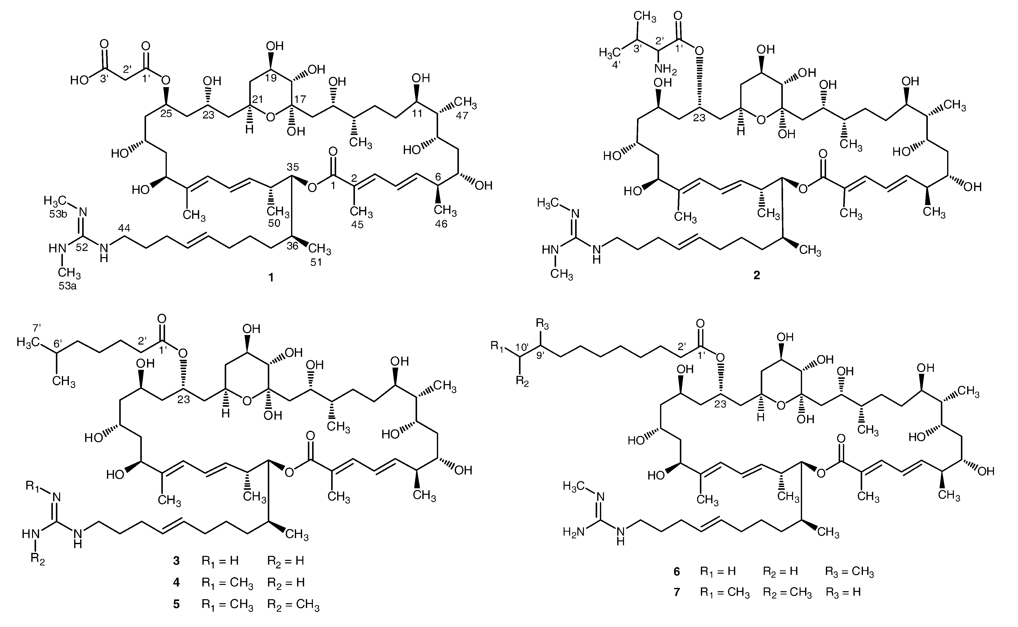
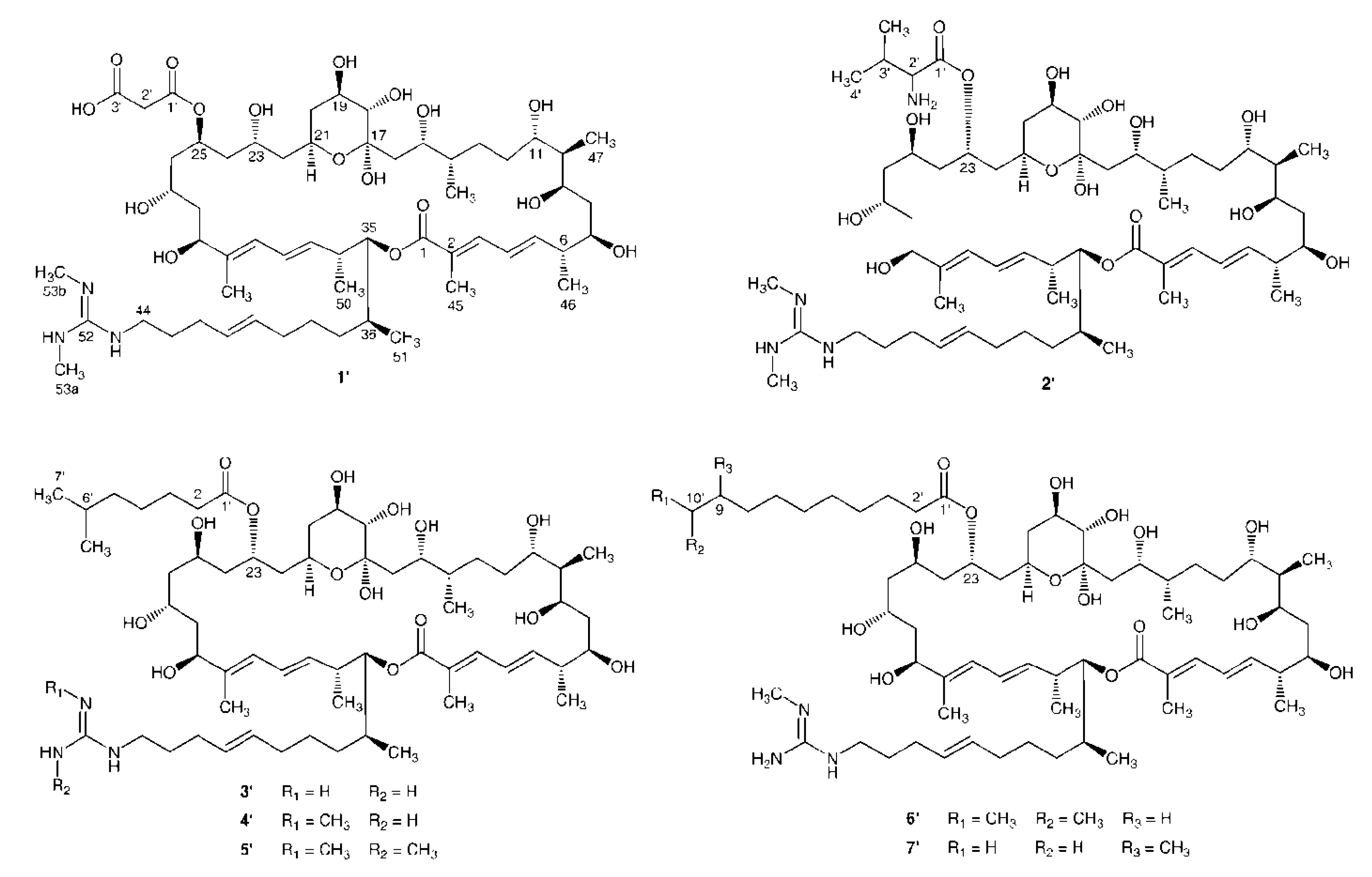
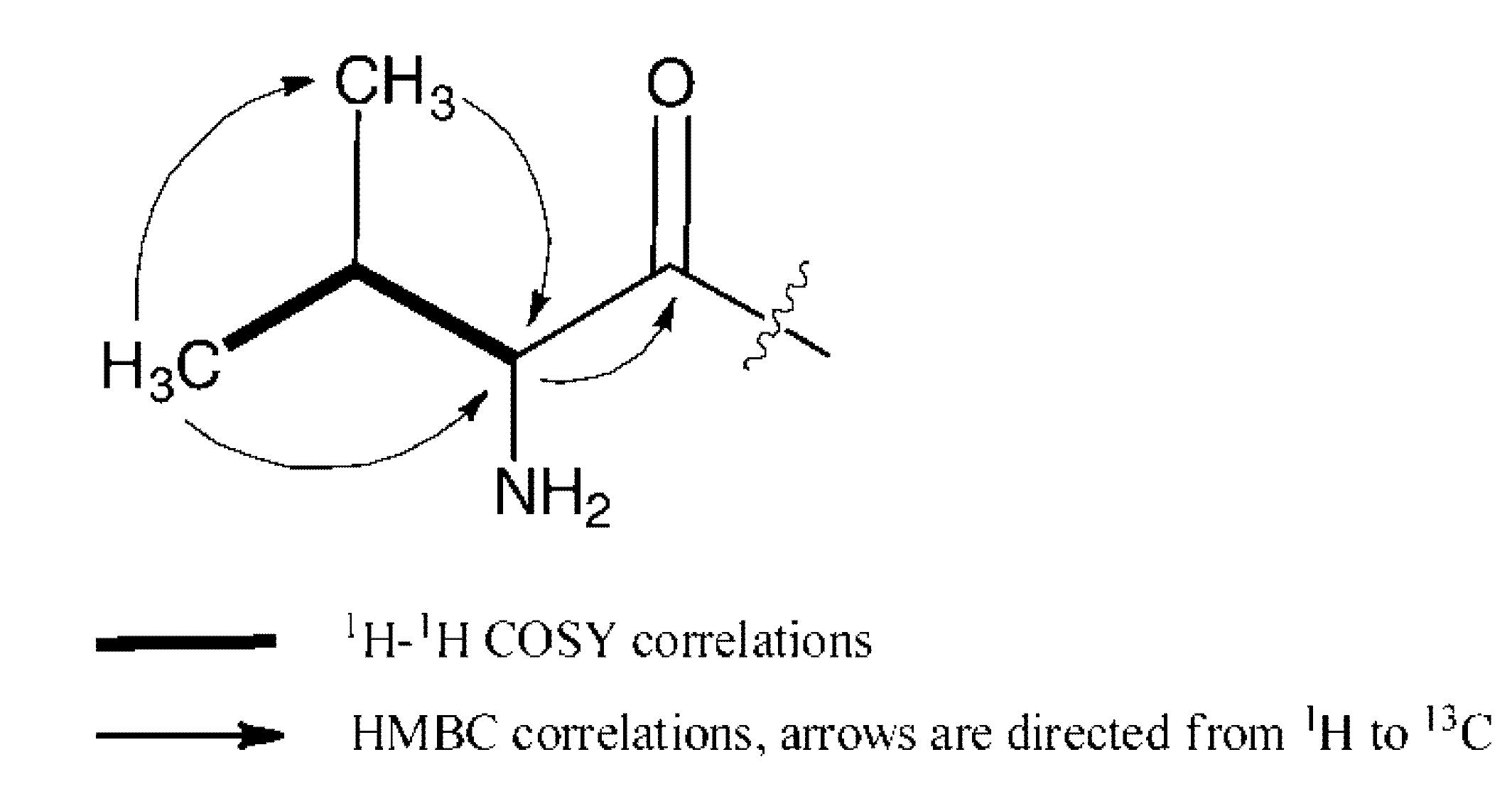
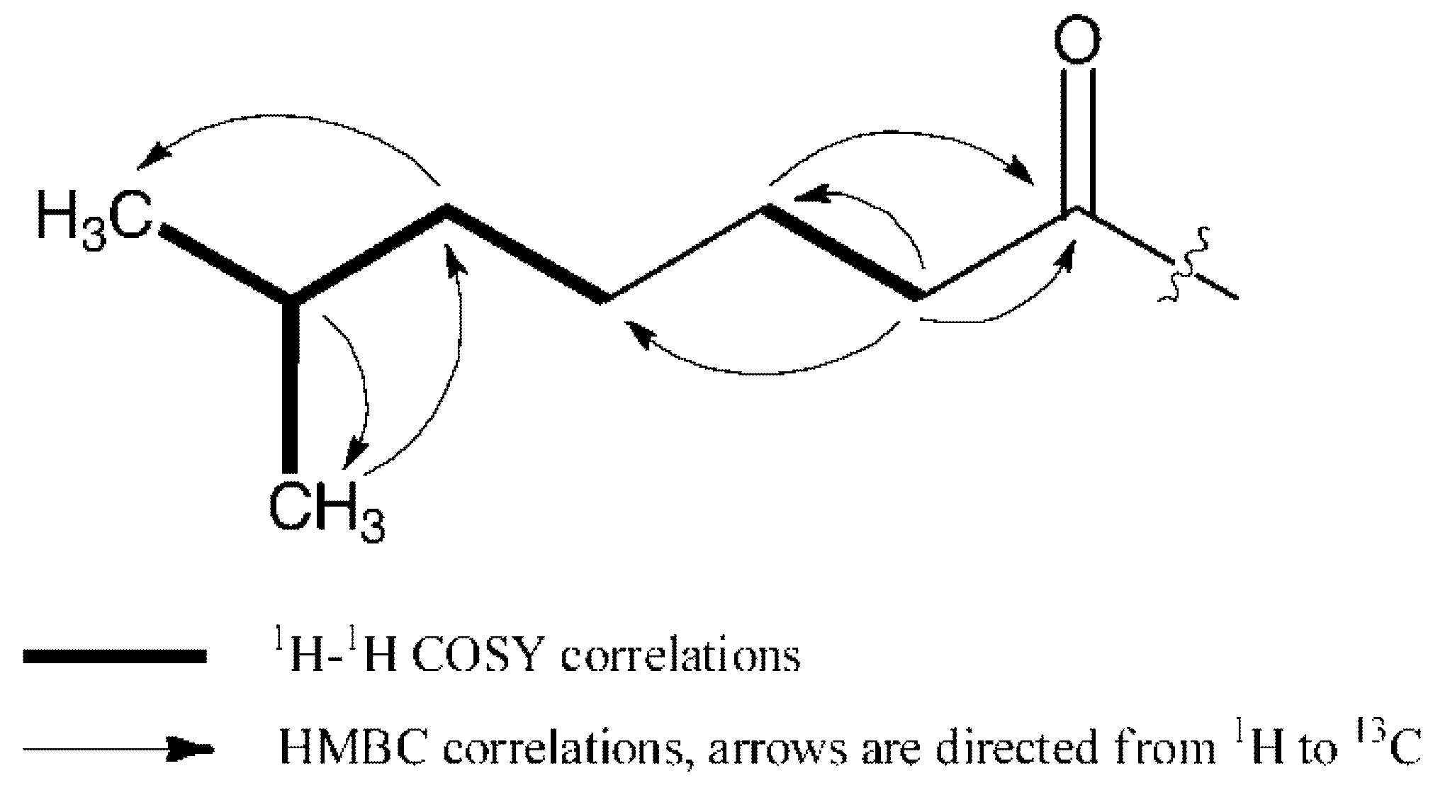
2.1. Structural Elucidation

25-Malonyl demalonylazalomycin F5a monoester (1) was obtained as a white, amorphous powder with [α]D29 +6.7° (c 0.1, MeOH). Its molecular formula C57H97N3O17 was established by the HRESIMS spectrometric data at m/z 1096.6914 [M + H]+ (calcd. for C57H98N3O17, 1096.6896), which showed that its molecular formula was identical to that of azalomycin F5a. Like azalomycin F5a, its UV absorption maxima at 241 nm (log ε, 4.6) and 269 nm (log ε, 4.3) also indicated the presence of a conjugated diene and an α,β,γ,δ-unsaturated acid (or ester) group. The 13C, DEPT and HSQC spectra of 1 (Table 1) showed one guanidino carbon signal at δC 157.42, three carbonyl carbon signals at δC 170.2, 171.9 and 173.9, ten olefinic carbon signals at δC 125.3, 126.8, 127.6, 128.6, 130.3, 132.6, 136.3, 140.2, 140.3 and 146.2, one quaternary hemiacetical carbon at δC 99.9 and one methine carbon at δC 70.8. So, 1 was deduced as an isomer of azalomycin F5a. When we compared the 13C and DEPT spectra of 1 with those of azalomycin F5a [13], the signal at δC 46.6 (C-26) in the 13C NMR spectrum of azalomycin F5a, was not observed while a signal at δC 44.0 appeared in that of 1. Based on the HSQC, 1H-1H COSY and HMBC spectra of 1, the linking position of the malonyl group was assigned to C-25 in 1, and the signal at δC 44.0 was assigned to C-26. It is interesting that 1 was found to be convertible to azalomycin F5a. HPLC analysis showed that the ratio of 1 to azalomycin F5a was about 15:85 after 1 stood in MeOH-d4 at room temperature for 30 days. This phenomenon was also observed by Iwasaki S. et al. [14]. The compound convertible to azalomycin F5a was named as azalomycin F5b, although spectroscopic information and structure of azalomycin F5b was not given in the paper [14]. There is not enough evidence to confirm that 1 and azalomycin F5bare the same compound. So, 1 was identified as 25-malonyl demalonylazalomycin F5a monoester.
Table 1. NMR spectroscopic data (400 MHz for 1H, 100 MHz for 13C) of 1, 2, 3 and 6 in MeOH-d4 (δ in ppm).
Position1236
δCδH (J in Hz)δCδH (J in Hz)δCδH (J in Hz)δCδH (J in Hz)
C-1170.2-170.1-170.1-170.1-
C-2126.8-126.7-126.8-126.8-
C-3140.37.09 d (11.2)140.37.10 d (11.0)140.27.10 d (11.2)140.37.10 d (11.0)
C-4127.66.43 dd (11.5, 14.9)127.66.43 dd (11.5, 14.7)127.66.43 dd (11.9, 14.3)127.66.43 dd (11.5, 14.8)
C-5146.26.07 dd (15.1, 9.0)146.16.08 dd (14.8, 9.0)146.16.08 dd (14.0, 9.0)146.16.08 dd (14.9, 9.0)
C-644.82.43 m44.72.44 m44.62.43 m44.72.44 m
C-775.93.80 m75.83.77 m75.83.77 m75.83.77 m
C-839.31.50 m, 1.78 m39.31.50 m, 1.78 m39.31.50 m, 1.77 m39.31.50 m, 1.78 m
C-975.43.80 m75.23.80 m75.23.80 m75.23.80 m
C-1044.71.54 m44.61.53 m44.51.54 m44.61.53 m
C-1172.23.91 m72.23.87 m72.23.91 m72.23.87 m
C-1233.51.62 m, 1.38 m33.51.60 m, 1.38 m33.51.62 m, 1.37 m33.41.62 m, 1.37 m
C-1330.71.30 m, 1.45 m30.61.30 m, 1.42 m30.61.30 m, 1.43 m30.61.30 m, 1.43 m
C-1440.61.60 m40.51.61 m40.71.61 m40.51.61 m
C-1572.73.86 m72.73.87 m72.43.86 m72.73.87 m
C-1641.91.80 m41.81.81 m41.91.82 m41.81.81 m
C-1799.9-100.0-99.8-99.9-
C-1877.53.34 d (9.2)77.43.35 d (9.1)77.23.35 d (9.2)77.53.35 d (9.1)
C-1969.93.87 m69.93.88 m69.73.87 m69.83.88 m
C-2041.41.89 m, 1.30 m41.31.89 m, 1.30 m41.21.90 m, 1.31 m41.31.89 m, 1.31 m
C-2165.74.17 m66.24.16 m66.34.16 m66.34.16 m
C-2244.51.52 m41.91.82 m41.81.88 m41.91.81 m
C-2366.34.03 m70.95.29 m70.75.27 m70.95.29 m
C-2444.61.69 m44.01.70 m44.01.72 m44.11.76 m, 1.66 m
C-2570.85.28 m65.73.86 m65.63.87 m65.73.86 m
C-2644.01.61 m, 1.83 m46.31.51 m46.41.51 m46.31.51 m
C-2765.73.88 m65.84.04 m65.64.04 m65.84.04 m
C-2844.21.78 m44.11.54 m44.11.53 m44.11.63 m
C-2974.24.18 m74.24.18 m74.24.18 m74.24.18 m
C-30140.2-140.1-140.1-140.1-
C-31125.35.98 d (10.4)125.35.98 d (10.7)125.25.98 d (10.5)125.35.98 d (10.7)
C-32128.66.22 dd (10.9, 14.5)128.56.23 dd (10.9, 14.9)128.66.22 dd (10.9, 14.9)128.56.23 dd (10.9, 14.8)
C-33136.35.43 m136.35.44 m136.25.45 m136.35.44 m
C-3441.02.57 m40.72.57 m41.02.57 m40.92.57 m
C-3580.94.78 dd (7.6, 4.0)80.94.79 dd (7.7, 4.1)80.84.78 dd (7.8, 3.9)80.94.79 dd (7.6, 4.1)
C-3635.31.82 m35.31.82 m35.21.81 m35.31.82 m
C-3734.41.15 m, 1.35 m34.41.15 m, 1.35 m34.51.15 m, 1.35 m34.41.15 m, 1.35 m
C-3827.91.42 m27.81.42 m27.91.41 m27.91.42 m
C-3933.61.99 m33.61.99 m33.61.99 m33.61.99 m
C-40132.65.44 m132.55.44 m132.65.44 m132.65.44 m
C-41130.35.44 m130.35.44 m130.15.50 m130.25.44 m
C-4230.72.07 m30.42.07 m30.82.07 m30.62.07 m
C-4329.91.67 m29.81.64 m29.81.64 m29.81.64 m
C-4442.23.17 t (7.3)42.03.15 t (7.1)42.03.15 t (7.0)42.03.15 t (7.1)
C-4512.91.92 s12.91.92 s12.91.92 s12.91.92 s
C-4617.11.11 d (6.8)17.11.12 d (6.8)17.11.11 d (6.8)17.11.12 d (6.8)
C-4710.50.89 d (6.9)10.50.89 d (6.9)10.50.89 d (6.9)10.50.89 d (6.9)
C-4815.20.91 d (6.7)15.30.92 d (6.7)15.30.91 d (6.7)15.30.92 d (6.7)
C-4913.11.65 s13.11.65 s13.31.65 s13.11.65 s
C-5017.81.01 d (6.7)17.91.00 d (6.7)17.91.00 d (6.6)17.91.00 d (6.8)
C-5114.40.94 d (6.7)14.50.95 d (6.7)14.40.94 d (6.7)14.50.94 d (6.7)
C-52157.4-157.4-158.7-158.3-
C-53a28.42.85 s28.42.85 s 28.42.84 s
C-53b28.42.85 s28.42.85 s
C-1′171.9-174.1-175.5-175.4-
C-2′46.03.22 m61.73.44 d (4.0)35.02.36 t (7.4)35.02.36 t (7.5)
C-3′173.9-30.82.28 m26.01.62 m26.01.61 m
3′-CH3 17.91.02 d (6.8)
C-4′ 19.21.07 d (6.8)30.41.42 m30.31.35 m
C-5′ 40.31.18 m30.41.31 m
C-6′ 29.21.29 m30.81.30 m
6′-CH3 23.80.88 d (6.6)
C-7′ 23.70.88 d (6.6)28.51.29 m
C-8′ 40.31.17 m
C-9′ 29.21.31 m
9′-CH3 23.10.89 d (6.8)
C-10′ 23.10.89 d (6.8)
23-Valine demalonylazalomycin F5a ester (2) was obtained as a white, amorphous powder with [α]D29 +4.4° (c 0.1, MeOH). Its molecular formula C59H104N4O15 was established by the HRESIMS spectrometric data at m/z 1109.7580 [M + H]+ (calcd. for C59H105N4O15, 1109.7576). Its UV absorption maxima at 241 nm (log ε, 4.6) and 268 nm (log ε, 4.4) indicated the presence of a conjugated diene and an α,β,γ,δ-unsaturated acid (or ester) group. The 13C, DEPT and HSQC spectra of 2 (Table 1) showed one guanidino carbon signal at δC 157.4, one carbonyl carbon signal at δC 170.1, ten olefinic carbon signals at δC 125.3, 126.7, 127.6, 128.5, 130.3, 132.5, 136.3, 140.1, 140.3 and 146.1, one quaternary hemiacetical carbon signal at δC 100.0, one methine carbon signal at δC 70.9 and two N-methyl carbon signals at δC 28.4. These spectroscopic data were very similar to azalomycin F5a reported in our previous paper [11], while there was no carbonyl carbon signal at δC 171.6 and methylene carbon signal at δC 46.1. Comparing the 13C, DEPT and HSQC spectra of 2 and those of azalomycin F5a, two additional methyl carbon signals at δC 17.9 and 19.2 and two methylene carbon signals at δC 30.81 and 61.7 were present. Based on the correlations (Figure 2) observed in the 1H-1H COSY and HMBC spectra of 2, a valyl group was established. Moreover, the correlation between H-23 (δH 5.29) and C-1′ (δC 174.1) observed in the HMBC spectrum of 2 indicated that the valyl group was linked to the lactonic ring at C-23 with an ester bond. So, 2 was identified as 23-valine demalonylazalomycin F5a ester.
Figure 2. Key 1H-1H COSY and HMBC correlations of the valyl moiety in 2.
Marinedrugs 11 00817 g002
23-(6-Methyl)heptanoic acid demalonylazalomycin F3a ester (3) was obtained as a white, amorphous powder with [α]D20 +6.8° (c 0.1, MeOH). Its molecular formula C60H105N3O15 was established by the HRESIMS spectrometric data at m/z 1108.7638 [M + H]+ (calcd. for C60H106N3O15, 1108.7624). Its UV absorption maxima at 238 nm (log ε, 4.6) and 269 nm (log ε, 4.3) indicated the presence of a conjugated diene and an α,β,γ,δ-unsaturated acid (or ester) group. The 13C, DEPT and HSQC spectra of 3 (Table 1) showed one guanidino carbon signal at δC 158.7, one carbonyl carbon signal at δC 170.1, ten olefinic carbon signals at δC 125.2, 126.8, 127.6, 128.6, 130.1, 132.6, 136.2, 140.1, 140.2 and 146.1, one quaternary hemiacetical carbon signal at δC 99.8 and one methine carbon signal at δC 70.7. These spectroscopic data were very similar to those of azalomycin F3a [15], which were reported as supporting information in our previous paper [11], while there were no carbonyl carbon signals at δC 171.8 and 174.0 and methylene carbon signal at δC 45.8 in the 13C NMR spectrum of 3. Comparing the 1H, 13C, DEPT and HSQC spectra of 3 with those of azalomycin F3a, two additional methyl carbon signals at δC 23.8 and 23.7, four methylene carbon signals at δC 40.3, 35.0, 30.4 and 26.0, one methine carbon signal at δC 29.2 and a carbonyl carbon signal at δC 175.5 were observed in the 13C NMR spectrum of 3. Based on the correlations observed in the 1H-1H COSY and HMBC spectra (Figure 3), a 6-methyl heptanoyl group was deduced. Moreover, the correlations between H-23 (δH 5.27) and C-1′ (δC 175.5) observed in the HMBC spectrum of 3 indicated that the 6-methyl heptanoyl group was linked to the lactonic ring at C-23 with an ester bond. So, 3 was identified as 23-(6-methyl)heptanoic acid demalonylazalomycin F3a ester.
Figure 3. Key 1H-1H COSY and HMBC correlations of the 6-methyl heptanoyl moiety in 3.
Marinedrugs 11 00817 g003
23-(6-Methyl)heptanoic acid demalonylazalomycin F4a ester (4) was obtained as a white, amorphous powder with [α]D20 +6.4° (c 0.1, MeOH). Its molecular formula C61H107N3O15 was established by the HRESIMS spectrometric data at m/z 1122.7788 [M + H]+ (calcd. for C61H108N3O15, 1122.7780). The difference of 14 mass units between 4 and 3 indicated that 4has one methylene unit more than 3. Similar 1H, 13C, DEPT spectra and UV absorption data allowed identification of these two compounds as analogs. Comparing the 13C and DEPT spectra of 4 with those of azalomycin F4a [16], the guanidino carbon signal at δC 158.3 (C-52) indicated that one methyl group was linked to a guanidino nitrogen [11], which was also corroborated by a proton signal at δH 2.84 (3H, s, H-53a), a carbon signal at δC 28.4 (C-53a) and the correlation between H-53a and C-52 observed in the HMBC spectrum of 4. So, 4 was identified as 23-(6-methyl)heptanoic acid demalonylazalomycin F4a ester.
23-(6-Methyl)heptanoic acid demalonylazalomycin F5a ester (5) was obtained as a white, amorphous powder with [α]D20 +6.1° (c 0.1, MeOH). Its molecular formula C62H109N3O15 was established by the HRESIMS spectrometric data at m/z 1136.7956 [M + H]+ (calcd. for C62H110N3O15, 1136.7937). The difference of 14 mass units between 5 and 4 indicated that 5 has one methylene unit more than 4. Similar 1H, 13C, DEPT spectra and UV absorption data allowed identification of these two compounds as analogs. Comparing their 1H, 13C and DEPT spectra, the guanidino carbon signal at δC 157.4 indicated two methyl groups were linked to two guanidino nitrogens, which was also corroborated by proton signals at δH 2.84 (6H, s, H-53a and H-53b) and carbon signals at δC 28.4 (C-53a and C-53b) in the 1H and 13C NMR spectrum of 5, respectively. So, 5 was identified as 23-(6-methyl)heptanoic acid demalonylazalomycin F5a ester.
23-(9-Methyl)decanoic acid demalonylazalomycin F4a ester (6) was obtained as a white, amorphous powder with [α]D20 +6.0° (c 0.1, MeOH). Its molecular formula C64H113N3O15 was established by the HRESIMS spectrometric data at m/z 1164.8269 [M + H]+ (calcd. for C64H114N3O15, 1164.8250). The difference of 42 mass units between 6 and 4 indicated that 6 has three methylene units more than 4. Similar 1H, 13C, DEPT spectra (Table 1) and UV absorption data of 6 and 4 also allowed identification of these two compounds as analogs. Comparing their spectroscopic data indicated the fatty acyl side chain of 6 has three methylenes more than 4, which was deduced by the 1H, 13C, DEPT, HSQC and HMBC spectra of 6. The 13C and 1H assignments of the fatty acyl side chain of 6 were achieved by its 1H-1H COSY, HSQC and HMBC spectra and ACD/Lab 6.0 software. 6 was identified as 23-(9-methyl)undecanoic acid demalonylazalomycin F4a ester.
23-(10-Methyl)undecanoic acid demalonylazalomycin F4a ester (7) was obtained as a white, amorphous powder with [α]D20 +6.0° (c 0.1, MeOH). Its molecular formula C65H115N3O15 was established by the HRESIMS spectrometric data at m/z 1178.8426 [M + H]+ (calcd. for C65H116N3O15, 1178.8406). The difference of 14 mass units between 7 and 6 indicated that 7 has one methylene unit more than 6. Similar 1H, 13C, DEPT spectra and UV absorption data of them allowed identification of these two compounds as analogs. Comparing their 1H, 13C and DEPT spectra, there was no obvious difference between the 13C NMR spectrum of 7 and 6 except that one methylene carbon signal at about δC 31.0 was presented in the 13C NMR spectrum of 7. So, 7 was identified as 23-(10-methyl)decanoic acid demalonylazalomycin F4a ester.
Figure 4. Structures of 1′–7′.
Marinedrugs 11 00817 g004
After the planar structures of 17 were established, we focused on their stereochemistries. As the core macrolide planar structures of 17 were accordingly identical to those of azalomycins F5a, F4a or F3a, the relative configurations of the core macrolide structures of 17 except the structural fragment from C-23 to C-27 of 1 could be directly established by comparing their 13C and 1H NMR spectra with those of azalomycins F5a, F4a or F3a [11,12,13,15,16]. Similar spectroscopic data of their core macrolide structures deduced that the relative configurations of 17 except that at C-23/C-25/C-27 of 1 were accordingly identical to those of azalomycins F5a, F4a or F3a. Like that of azalomycin F5a, the chemical shifts for C-21 (65.7 ppm), C-23 (66.3 ppm) and C-27 (65.7 ppm) lower than 68.0 ppm in MeOH-d4 deduced that the relavitve configuration at C-23/C-25/C-27 of 1 was also anti/anti according to the universal NMR Database 4 [12,17], which was further confirmed by two facts that 1 could be convertible to azalomycin F5a in MeOH-d4 at room temperature and that the chemical shift for C-23 was upfield by about 5.0 ppm when the malonyl group of azalomycin F4a was removed [12]. Because the relative stereochemistries of C6–C11 to C14–C36stereogenic centers of azalomycins F5a remain undefined in our previous work [12], azalomycin F5a was one of two possible stereoisomers which the relative configuration at C11/C14 was anti or syn. Similarly, azalomycins F4a and F3a were respectively one of two possible stereoisomers like that of azalomycin F5a. So, each compound of 17 was one of two possible stereoisomers numbered 17 and 1′–7′ presented in Figure 1, Figure 4, respectively.

2.2. Biological Assays

Biological assays indicated that 17 had broad-spectrum antimicrobial activity. Their minimal inhibitory concentrations (MICs) against Candida albicans ATCC 10231, Staphylococcus aureus S014, Bacillus subtilis S028 and Escherichia coli S002 were respectively 1.56–6.25, 0.39–1.56, 0.20–0.78 and 3.13–25.00 μg/mL (Table 2). Moreover, they also showed moderate cytotoxicity against human colon tumor cell HCT-116 in vitro with IC50 values of 1.81–5.00 μg/mL (Table 2).
Table 2. Minimal inhibitory concentrations (MICs) against test microbes and IC50 value against HCT-116 in vitro.
CompoundsMICs (μg/mL)IC50 (μg/mL)
Candida albicans ATCC 10231Staphylococcus aureus S014Bacillus subtilis S028Eschzerichia coli S002HCT-116
13.130.390.203.135.00
26.251.560.396.251.95
33.130.780.393.132.46
41.561.560.206.252.45
51.560.780.7812.51.81
63.130.390.3925.001.54
73.130.390.393.132.46
Positive controls *2.00.500.202.00.18
* Amphotericin B for C. albicans, oxacillin sodium for S. aureus and B. subtilis, kalamycin for E. coli and doxorubicin for HCT-116 were respectively used as positive controls.

2.3. Discussion

Azalomycin F complex, including azalomycins F3a, F4a, F5a and several minor analogs, was first isolated from the broth of Streptomyces hygroscopicus var. azalomyceticus by Arai in 1959 [18,19]. The structures of azalomycins F3a, F4a and F5a were progressively elucidated from 1982 to 2012 [12,14,20,21,22], while others minor analogs were not identified. Streptomyces sp. 211726, isolated from mangrove rhizosphere soil, showed a remarkable productivity of macrocyclic lactones, and five main components azalomycins F3a, F4a, F5aazalomycin F4a 2-ethylpentyl ester and azalomycin F5a 2-ethylpentyl ester produced by this strain were identified [11]. During our research on minor analogs produced by this strain, seven new compounds 17 were isolated and identified in this paper. Similar to these azalomycin F analogs, many 36-membered polyhydroxyl macrolides such as RS-22, guanidylfungins, amycins, niphimycin, malonylniphimycin, dihydroniphimycin, malonyl-4,5-dihydroniphimycin, shurimycins and others were identified [23,24,25,26]. There are about thirty 36-membered polyhydroxyl macrolides identified so far, and almost all of them were produced by Streptomyces. These compounds possess broad-spectrum antimicrobial activity, and azalomycin F was also an inhibitor of type-I interleukin-1 receptor [27]. In our research on biological activity of azalomycin F analogs produced by Streptomyces sp. 211726, these twelve compounds also showed remarkable broad-spectrum antimicrobial activity. Moreover, they had moderate cytotoxicity, and the acute toxicity (LD50) of a mixture of twelve azalomycin F analogs produced by this strain was 97.9 mg/kg in mice by intraperitoneal injection.

3. Experimental Section

3.1. General Experimental Procedures

Optical rotations were measured in methanol using an Autopol Ш digital polarimeter. UV spectra were determined by DU-800 UV/Visible spectrophotometer. IR spectra were obtained with Thermo Nicolet 380 FTIR spectrometer. All NMR experiments were recorded on a Bruker AV-400 NMR spectrometer equipped with a 5 mm PABBO BB-probe head (400 MHz for 1H shifts relative to MeOH-d4 at 3.31 ppm and 100 MHz for 13C shifts relative to MeOH-d4 at 49.05 ppm) at 30 °C. HR-ESI-MS spectra were carried on the API QSTAR Pulsar I MS System. Silica gel (Qingdao Haiyang Chemical Co. Ltd., Qingdao, China, 10–40 μm), octadecylsilyl silica gel (Silicycle, Quebec, Canada; 40–63 μm) and Sephadex LH-20 (Amersham Pharmacia Biotech AB, Uppsala, Sweden) were used for column chromatography. Precoated silica gel GF254 plates (Qingdao Haiyang Chemical Co., Ltd.) were used for thin layer chromatography. All compounds was prepared by Dionex Summit HPLC system (Dionex, Sunnyvale, CA, USA) consisting of Dionex P680 HPLC pumps (P680A HPG-4) with a UV detector (170 U), and a Shimadzu Shim-pack VP-ODS reversed-phase column (250 mm × 4.6 mm i.d., 5-μm particle size) was used.

3.2. Actinomycetes Material and Fermentation

The strain of Streptomyces sp. 211726 was isolated from mangrove rhizosphere soil of Heritiera globosa collected in Wenchang, China. Voucher specimens are stored in Key Laboratory of Combinatorial Biosynthesis and Drug Discovery (Wuhan University), Ministry of Education, Wuhan, China. The fermentation of strainStreptomyces sp. 211726 was reported in our previous paper [11]. In short, the strain of Streptomyces sp. 211726 was cultured with liquid medium containing glucose 1.0%, soluble starch 3.5%, yeast 0.2%, casein 0.4% and NaCl 1.8% (pH 7.2 before autoclaving), and incubated at 30 °C for 10 days on a rotary shaker at 190 rpm until 100 L of broth was obtained.

3.3. Extract and Isolation

After the 100 L broth of Streptomyces sp. 211726 was centrifuged, the mycelia was extracted with methanol, concentrated under vacuum and freeze-dried to obtain lyophilized powder. The powder was dissolve in CHCl3:MeOH (80:20), and separated into eight fractions (1–8) on a silica gel column using gradient elution of CHCl3:MeOH (3:1, 2:1 and 1:1). Fraction 2 was purified by reversed-phase C18 column eluted with MeOH:H2O (60:40), semi-preparative reversed-phase C18 high performance liquid chromatography eluted with MeOH:H2O (58:42) to give three pure fraction, and which were respectively concentrated under vacuum to obtain three extracts. These extracts were respectively dissolved in MeOH, purified by sephadex LH-20 column eluted with MeOH, concentrated under vacuum at 35 °C, and dried to give 1 (41 mg), 2 (22 mg) and 3 (31 mg). Similarly, fraction 3 was purified by reversed-phase C18 column eluted with MeOH:H2O (65:35), semi-preparative reversed-phase C18 high performance liquid chromatography eluted with MeOH:H2O (63:37) and sephadex LH-20 column eluted with MeOH to give 4 (15 mg) and 6 (27 mg); Fraction 4 was purified by reversed-phase C18 column eluted with MeOH:H2O (70:30), semi-preparative reversed-phase C18 high performance liquid chromatography eluted with MeOH:H2O (68:32) and sephadex LH-20 column eluted with MeOH to give 5 (10 mg) and 7 (24 mg).
25-Malonyl demalonylazalomycin F5a monoester (1): White amorphous powder; [α]D29 +6.7° (c 0.1, MeOH); UV λMeOHmax nm (log ε): 241(4.6), 269(4.3); IR υKBrmax (cm−1): 3385, 2964, 2936, 1701, 1636, 1597, 1459, 1243, 1089, 1066, 969; 13C NMR (MeOH-d4, 100 MHz) and 1H NMR (MeOH-d4, 400 MHz) data were showed in Table 1; HRESIMS m/z 1096.6914 [M + H]+ (calcd. for C57H98N3O17, 1096.6896).
23-Valine demalonylazalomycin F5a ester (2): White amorphous powder; [α]D29 +4.4° (c 0.1, MeOH); UV λMeOHmax nm (log ε): 241(4.6), 268(4.4); IR υKBrmax (cm−1): 3414, 3137, 2965, 2928, 1726, 1635, 1597, 1507, 1261, 1092, 968; 13C NMR (MeOH-d4, 100 MHz) and 1H NMR (MeOH-d4, 400 MHz) data were showed in Table 1; HRESIMS m/z 1109.7580 [M + H]+ (calcd. for C59H105N4O15, 1109.7576).
23-(6-Methyl)heptanoic acid demalonylazalomycin F3a ester (3): White amorphous powder; [α]D20 +6.8° (c 0.1, MeOH); UV λMeOHmax nm (log ε): 238(4.6), 269(4.3); IR υKBrmax (cm−1): 3423, 2962, 2935, 1736, 1707, 1637, 1184, 1045, 970, 721; 13C NMR (MeOH-d4, 100 MHz) and 1H NMR (MeOH-d4, 400 MHz) data were showed in Table 1; HRESIMS m/z 1108.7638 [M + H]+ (calcd. for C60H106N3O15, 1108.7624).
23-(6-Methyl)heptanoic acid demalonylazalomycin F4a ester (4): White amorphous powder; [α]D20 +6.4° (c 0.1, MeOH); UV λMeOHmax nm (log ε): 238(4.5), 269(4.4); IR υKBrmax (cm−1): 3423, 2965, 2936, 1734, 1708, 1635, 1181, 1049, 972, 722; 1H NMR (MeOH-d4, 400 MHz) and 13C NMR (MeOH-d4, 100 MHz) data were showed in supplementary file; HRESIMS m/z 1122.7788 [M + H]+ (calcd. for C61H108N3O15, 1122.7780).
23-(6-Methyl)heptanoic acid demalonylazalomycin F5a ester (5): White amorphous powder; [α]D20 +6.1° (c 0.1, MeOH); UV λMeOHmax nm (log ε): 238(4.5), 269(4.4); IR υKBrmax (cm−1): 3425, 2963, 2935, 1734, 1708, 1636, 1185, 1047, 972, 721; 1H NMR (MeOH-d4, 400 MHz) and 13C NMR (MeOH-d4, 100 MHz) data were showed in supplementary file; HRESIMS m/z 1136.7956 [M + H]+ (calcd. for C62H110N3O15, 1136.7937).
23-(9-Methyl)decanoic acid demalonylazalomycin F4a ester (6): White amorphous powder; [α]D20 +6.0° (c 0.1, MeOH); UV λMeOHmax nm (log ε): 238(4.5), 269(4.4); IR υKBrmax (cm−1): 3425, 2968, 2935, 1734, 1708, 1636, 1185, 1047, 972, 721; 1H NMR (MeOH-d4, 400 MHz) and 13C NMR (MeOH-d4, 100 MHz) data were showed in Table 1; HRESIMS m/z 1164.8269 [M + H]+ (calcd. for C64H114N3O15, 1164.8250).
23-(10-Methyl)undecanoic acid demalonylazalomycin F4a ester (7): White amorphous powder; [α]D20 +6.0° (c 0.1, MeOH); UV λMeOHmax nm (log ε): 238(4.5), 269(4.4); IR υ (cm−1): 3425, 2968, 2936, 1736, 1707, 1636, 1185, 1047, 972, 721; 13C NMR (MeOH-d4, 100 MHz) and 1H NMR (MeOH-d4, 400 MHz) data were showed in supplementary file; HRESIMS m/z 1178.8426 [M + H]+ (calcd. for C65H116N3O15, 1178.8406).

3.4. Biological Assays

The MICs of all compounds against C. albicans ATCC 10231, S. aureus S014, B. subtilis S028 and E. coli S002 were determined by agar dilution method. Amphotericin B for C. albicans, oxacillin sodiumfor S. aureus and B. subtilis and kalamycin for E. coli were respectively used as positive controls. Yeast extract-peptone-dextrose (YPD) medium was used for C. albicans, beef extract-peptone medium was used for S. aureus and B. subtilis, and Luria-Bertani (LB) medium was used for E. coli. Their cytotoxicities were evaluated by the MTT (3-(4,5-dimethylthiazol-2-yl)-2,5-diphenyl-tetrazolium bromide) method using human colon tumor cell HCT-116, and doxorubicin was used as a positive control.

4. Conclusions

Proceed with research on minor azalomycin F analogs produced by Streptomyces sp. 211726, seven new compounds were isolated and identified. Biological assays of 17 showed remarkable antifungal and antibacterial activity and moderate cytotoxicity against human colon tumor cell HCT-116 in vitro.

Acknowledgments

This work is financially supported by the National High Technology Development Project (863) (2007AA09Z415 & 2011AA09070106). Authors are thankful to Zhufen Gao and Xiaohui Li for their technical help on fermentation and extraction, and to Fanglu Du worked in Hunan College of Traditional Medicine for his help on the structural elucidation.

References and Notes

  1. Costanza, R.; d’Arge, R.; Groot, R.; Farberk, S.; Grasso, M.; Hannon, B.; Limburg, K.; Naeem, S.; O’Neill, R.V.; Paruelo, J.; et al. The value of the world’s ecosystem services and natural capital. Nature 1997, 387, 253–260. [Google Scholar]
  2. Wang, B.; Liang, S.; Zhang, W.; Zan, Q. Mangrove flora of the world. Acta. Bot. Sin. 2003, 45, 644–653. [Google Scholar]
  3. Li, J.; Li, M.Y.; Feng, G.; Zhang, J.; Karonen, M.; Sinkkonen, J.; Satyanandamurty, T.; Wu, J.; Moluccensins, R.-Y. Limonoids from the seeds of a mangrove, Xylocarpus moluccensis. J. Nat. Prod. 2012, 75, 1277–1283. [Google Scholar] [CrossRef]
  4. Wu, J.; Xiao, Q.; Xu, J.; Li, M.Y.; Pan, J.Y.; Yang, M.H. Natural products from true mangrove flora: Source, chemistry and bioactivities. Nat. Prod. Rep. 2008, 25, 955–981. [Google Scholar] [CrossRef]
  5. Wang, J.; Zhang, W.; Li, Z.; Xiang, W.; Guo, Y.; Krohn, K. Elucidation of excogallochaols A-D, four unusual diterpenoids from the Chinese mangrove Excoecaria agallocha. Phytochemistry 2007, 68, 2426–2431. [Google Scholar] [CrossRef]
  6. Xu, M.; Liu, X.; Zhao, Y.; Liu, D.; Liu, D.; Xu, Z.; Ao, P.; Lin, W.; Yang, S.; Zhang, Z.; Xu, J. Identification and characterization of an anti-fibrotic benzopyran compound isolated from mangrove-derived Streptomyces xiamenensis. Mar. Drugs 2012, 10, 639–654. [Google Scholar]
  7. Hong, K.; Gao, A.; Xie, Q.; Gao, H.; Zhuang, L.; Lin, H.; Yu, H.; Li, J.; Yao, X.; Goodfellow, M.; Ruan, J. Actinomycetes for marine drug discovery isolated from mangrove soils and plants in China. Mar. Drugs 2009, 7, 24–44. [Google Scholar] [CrossRef]
  8. Fu, P.; Yang, C.; Wang, Y.; Liu, P.; Ma, Y.; Xu, L.; Su, M.; Hong, K.; Zhu, W. Streptocarbazoles A and B, two novel 2 indolocarbazoles from the marine-derived actinomycete strain Streptomyces sp. FMA. Org. Lett. 2012, 14, 2422–2425. [Google Scholar]
  9. Chen, G.D.; Gao, H.; Tang, J.S.; Huang, Y.F.; Chen, Y.; Wang, Y.; Zhao, H.N.; Lin, H.P.; Xie, Q.Y.; Hong, K.; et al. Benzamides and quinazolines from a mangrove actinomycetes Streptomyces sp. (No. 061316) and their inhibiting caspase-3 catalytic activity in vitro. Chem. Pharm. Bull. 2011, 59, 447–451. [Google Scholar]
  10. Lin, W.; Li, L.; Fu, H.; Sattler, I.; Huang, X.; Grabley, S. New cyclopentenone derivatives from an endophytic Streptomyces sp. isolated from the mangrove plant Aegiceras comiculatum. J. Antibiot. 2005, 58, 594–598. [Google Scholar] [CrossRef]
  11. Yuan, G.; Lin, H.; Wang, C.; Hong, K.; Liu, Y.; Li, J. 1H and 13C assignments of two new macrocyclic lactones isolated from Streptomyces sp. 211726 and revised assignments of azalomycins F3a, F4a and F5a. Magn. Reson. Chem. 2011, 49, 30–37. [Google Scholar]
  12. Yuan, G.; Li, P.; Pan, W.; Pang, H.; Chen, S. The relative configurations of azalomycins F5a, F4a and F3a. J. Mol. Struct. 2013, 1035, 31–37. [Google Scholar] [CrossRef]
  13. .
  14. Iwasaki, S.; Namikoshi, M.; Sasaki, K.; Fukushima, K.; Okuda, S. Studies on marcrocyclic lactone antibiotics. V. (1) The structures of azalomycins F3a and F5a. Chem. Pharm. Bull. 1982, 30, 4006–4014. [Google Scholar]
  15. 13C NMR (MeOH-d4, 100 MHz) data of azalomycin F3a: 170.1 (C-1), 126.8 (C-2), 140.3 (C-3), 127.6 (C-4), 146.2 (C-5), 44.6 (C-6), 75.7 (C-7), 39.3 (C-8), 75.0 (C-9), 44.5 (C-10), 72.2 (C-11), 33.6 (C-12), 30.7 (C-13), 40.5 (C-14), 72.3 (C-15), 41.7 (C-16), 99.9 (C-17), 77.2 (C-18), 69.8 (C-19), 41.2 (C-20), 65.4 (C-21), 41.9 (C-22), 70.8 (C-23), 44.6 (C-24), 65.5 (C-25), 46.2 (C-26), 66.2 (C-27), 44.1 (C-28), 73.9 (C-29), 140.1 (C-30), 125.2 (C-31), 128.5 (C-32), 136.2 (C-33), 40.7 (C-34), 81.2 (C-35), 35.2 (C-36), 34.6 (C-37), 27.9 (C-38), 33.6 (C-39), 132.6 (C-40), 130.3 (C-41), 30.7 (C-42), 29.9 (C-43), 42.1 (C-44), 12.8 (C-45), 17.0 (C-46), 10.5 (C-47), 14.8 (C-48), 13.3 (C-49), 17.7 (C-50), 14.4 (C-51), 158.7 (C-52), 171.8 (C-1′), 45.8 (C-2′), 174.0 (C-3′).
  16. 13C NMR (MeOH-d4, 100 MHz) data of azalomycin F4a: 170.1 (C-1), 126.8 (C-2), 140.2 (C-3), 127.6 (C-4), 146.1 (C-5), 44.6 (C-6), 75.7 (C-7), 39.2 (C-8), 75.0 (C-9), 44.5 (C-10), 72.2 (C-11), 33.6 (C-12), 30.8 (C-13), 40.5 (C-14), 72.3 (C-15), 41.7 (C-16), 99.8 (C-17), 77.1 (C-18), 69.7 (C-19), 41.2 (C-20), 65.4 (C-21), 42.0 (C-22), 70.7 (C-23), 44.6 (C-24), 65.5 (C-25), 46.5 (C-26), 66.1 (C-27), 44.1 (C-28), 74.2 (C-29), 140.1 (C-30), 125.2 (C-31), 128.6 (C-32), 136.2 (C-33), 41.0 (C-34), 80.7 (C-35), 35.1 (C-36), 34.6 (C-37), 27.9 (C-38), 33.6 (C-39), 132.5 (C-40), 130.3 (C-41), 30.8 (C-42), 29.8 (C-43), 42.1 (C-44), 12.9 (C-45), 17.1 (C-46), 10.5 (C-47), 14.8 (C-48), 13.3 (C-49), 17.6 (C-50), 14.3 (C-51), 158.3 (C-52), 28.4 (C-53a), 171.6 (C-1′), 46.1 (C-2′), 173.9 (C-3′).
  17. Kobayashi, Y.; Tan, C.H.; Kishi, Y. Toward creation of a universal NMR database for stereochemical assignment: The case of 1,3,5-trisubstituted acyclic systems. Helv. Chim. Acta 2000, 83, 2562–2571. [Google Scholar] [CrossRef]
  18. Arai, M. Azalomycin F, an antibiotic against fungi and Trichomonas. Arzneim. Forsch. 1968, 18, 1396–1399. [Google Scholar]
  19. Arai, M. Azalomycins B and F, two new antibiotics. II. Properties of azalomycins B and F. J. Antibiot. Ser. A 1960, 13, 51–56. [Google Scholar]
  20. Namikoshi, M.; Iwasaki, S.; Sasaki, K.; Yano, M.; Fukushima, K.; Nozoe, S.; Okuda, S. Studies on macrocyclic lactone antibiotics. II. (1) Partial structures of azalomycin F4a. Chem. Pharm. Bull. 1982, 30, 1658–1668. [Google Scholar]
  21. Iwasaki, S.; Namikoshi, M.; Sasaki, K.; Yano, M.; Fukushima, K.; Nozoe, S.; Okuda, S. Studies on macrocyclic lactone antibiotics. III. (1) Skeletal structure of azalomycin F4a. Chem. Pharm. Bull. 1982, 30, 1669–1673. [Google Scholar]
  22. Chandra, A.; Nair, M.G. Azalomycin F complex from Streptomyces hygroscopicus, MSU/MN-4-75B. J. Antibiot. 1995, 48, 896–898. [Google Scholar] [CrossRef]
  23. Berlinck, R.G.S. Natural guanidine derivatives. Nat. Prod. Rep. 1999, 16, 339–365. [Google Scholar] [CrossRef]
  24. Ubukata, M.; Morita, T.I.; Osada, H. RS-22A, B and C: New macrolide antibiotics from Streptomyces violaceusniger II. Physico-Chemical properties and structure elucidation. J. Antibiot. 1995, 48, 293–299. [Google Scholar]
  25. Ivanova, V.; Gesheva, V.; Kolarova, M. Dihydroniphimycin: New polyol macrolide antibiotic produced by Streptomyces hygroscopicus 15 isolation and structure elucidation. J. Antibiot. 2000, 53, 627–632. [Google Scholar] [CrossRef]
  26. Ivanova, V.; Kolarova, M.; Aleksieva, K. Malonyl-4,5-dihydroniphimycin: A new polyol macrolide antibiotic, produced by Streptomyces hygroscopicus. Z. Naturforsch. 2007, 62, 1187–1192. [Google Scholar]
  27. Stefanelli, S.; Corti, E.; Montanini, N.; Denaro1, M.; Sarubbill, E. Inhibitors of type-I interleukin-1 receptor from microbial metabolites. J. Antibiot. 1997, 50, 484–489. [Google Scholar] [CrossRef]

