Article

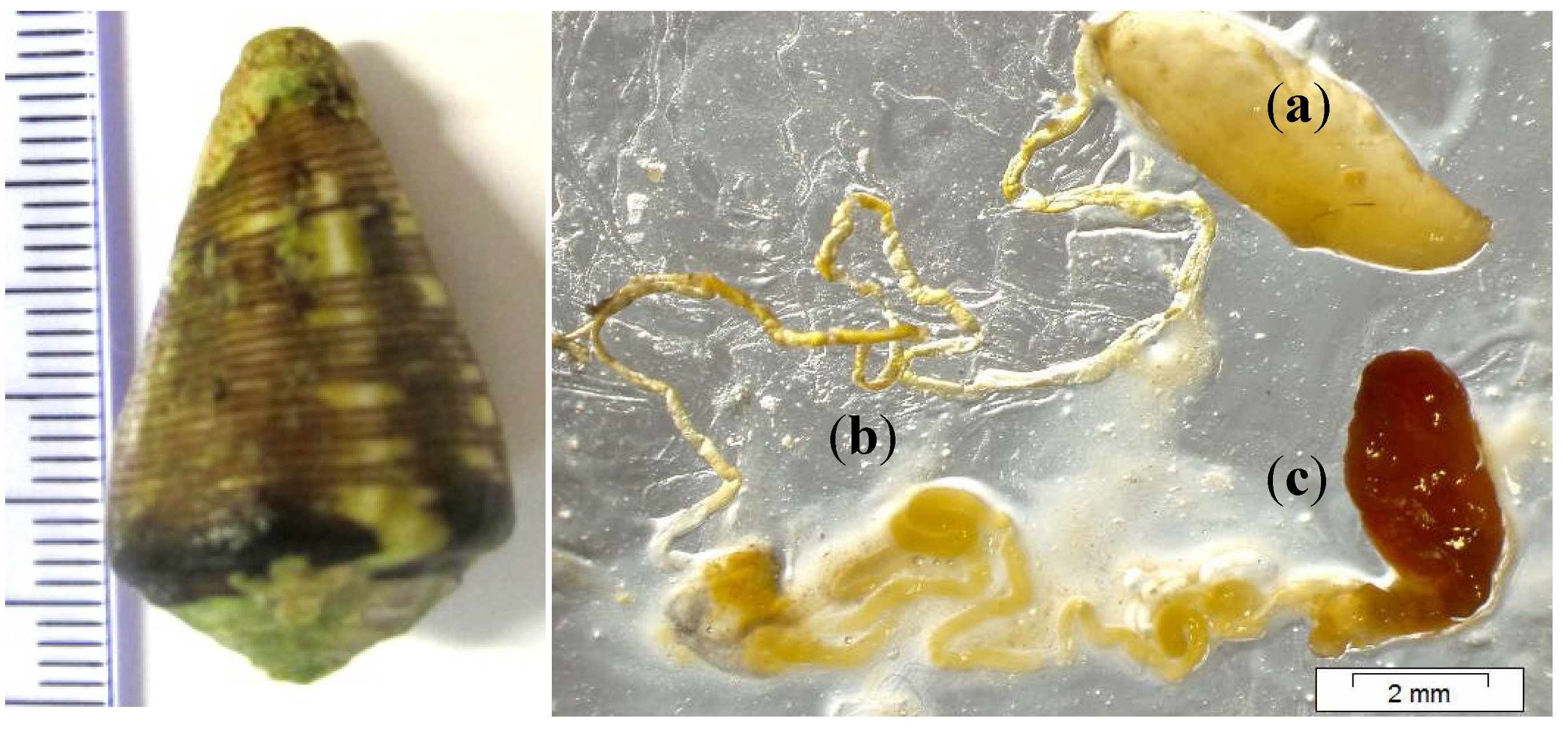
Conopeptides from Cape Verde Conus crotchii

1
CIIMAR/CIMAR—Interdisciplinary Centre of Marine and Environmental Research, Rua dos Bragas 289, Porto 4050-123, Portugal
2
DECM—Department of Engineering and Sea Science, Cape Verde University, Mindelo CP 163, Cape Verde
3
Institute of Molecular Pathology and Immunology of the University of Porto, IPATIMUP, Porto 4200-465, Portugal
4
Faculty of Medicine, University of Porto, Porto 4200-319, Portugal
5
Faculty of Sciences, University of Porto, Porto 4169-007, Portugal
*
Author to whom correspondence should be addressed.
Mar. Drugs 2013, 11(6), 2203-2215; https://doi.org/10.3390/md11062203
Submission received: 1 April 2013 / Revised: 20 May 2013 / Accepted: 27 May 2013 / Published: 19 June 2013
(This article belongs to the Special Issue Marine Peptides and Their Mimetics)

Abstract

:
Marine Cone snails of the genus Conus contain complex peptide toxins in their venom. Living in tropical habitats, they usually use the powerful venom for self-defense and prey capture. Here, we study Conus crotchii venom duct using a peptide mass-matching approach. The C. crotchii was collected on the Cape Verde archipelago in the Boa Vista Island. The venom was analyzed using matrix-assisted laser desorption/ionization time-of-flight mass spectrometry (MALDI-TOF MS). About 488 molecular masses between 700 Da and 3000 Da were searched bymatching with known peptide sequences from UniProtKB protein sequence database. Through this method we were able to identify 12 conopeptides. For validation we considered the error between the experimental molecular mass (monoisotopic) and the calculated mass of less than 0.5 Da. All conopeptides detected belong to the A-, O1-, O2-, O3-, T- and D-superfamilies, which can block Ca2+ channels, inhibit K+ channels and act on nicotinic acetylcholine receptors (nAChRs). Only a few of the detected peptides have a 100% UniProtKB database similarity, suggesting that several of them could be newly discovered marine drugs.

Graphical Abstract

1. Introduction

Cone snails (genus Conus) are venomous predators belonging to the Conidae family. There are ~700 Conus species, all carrying complex arrays of peptide toxins in their venom [1]. Conus species normally live in tropical habitats of shallow water, on sand or near coral reefs and may cause lethal paralysis to their prey. Considered aggressive predators, cone snails are usually classified, depending on their prey, into three groups: vermivorous (worm-hunting), molluscivorous (other gastropods-hunting), and piscivorous cone snails (fish-hunting). However, some Conus can feed on hemichordates, bivalve mollusks and echiuroids, but a few species are considered generalist (e.g., C. californicus). Conus can be also dangerous to humans. The fish-hunting species C. geographus, have caused about three dozen fatalities in human poisoning cases [2,3]. The venom bioactive molecules, known as conopeptides or conotoxins (indiscriminate using of this work), are used to capture prey, as self defense from predators or to prevent competition [4,5]. Some authors have estimated that there are 100–200 distinct peptides per species, but recent work suggested that more than 1000 distinct conopeptides may be found per species [6]. These venoms mainly include linear peptides (usually disulfide-rich) and powerfully folded mini-proteins [7], exhibit various neuropharmacological properties with special incidence on ion channels and receptors [8,9]. Therefore, conopeptides have been considered powerful tools in neuroscience, and, for example, in December 2004, the synthetic version of the peptide ω-conotoxin MVIIA (commercial name Prialt®; Elan Pharmaceuticals, Inc., Dublin, Ireland) from C. magus has been approved by the United States Food and Drug Administration (FDA) to treat chronic pain in humans [10,11,12,13]. Despite this achievement, the overall knowledge of Conus venom proteins and peptides is scarce compared to other animal venom-producers (e.g., snakes, scorpions, spiders and sea anemones), thus providing a huge potential for the discovery of new pharmacological drugs [14]. The majority of Conus venom mini-proteins have a sequence length of 12–35 amino acids, normally with a high incidence of post-translational modifications. There are four classifications: (i) disulfide-rich conopeptides (conotoxins), which have two or more disulfides bridges, and disulfide-poor conopeptides, with one or none disulfide bond (conopressins, contryphans, conantokins, and contulakins); (ii) “gene superfamily” scheme that share a highly conserved sequence; (iii) “cysteine framework” scheme sorts them according to the arrangement of cysteines; and (iv) “pharmacological family” scheme reflects the target specificity of each conopeptide [2,9,10,15].
For the analysis of peptides the matrix-assisted laser desorption/ionization equipped with a time-of-flight (MALDI TOF/TOF) mass analyzer has been one of the most valuable analytical tools. This technique is relatively easy to perform (user friendly), is reliable and at the same time enables high-throughput sample analyses. Through MALDI-TOF, valuable data is generated [16], which contributes to the rapid discovery and characterization of new Conus marine drugs. The masses of peptides produced can be compared and “matched” to known sequences available on databases (e.g., UniProtKB and ConoServer). This methodology has been used to make available the identity of any protein whose full-length sequence is contained therein. Some peptides were identified within 0.5 Da (Daltons) of the predicted value, which was considered to be a sufficient criterion [17].
In the Cape Verde tropical Atlantic waters, there are 52 described Conus species, representing about 10% of the worldwide species diversity and only three are non-endemics (C. ermineus, C. genuanus, and C. tabidus). However, this high Conus diversity does not exist in other Macaronesian islands [18]. The conopeptides distribution by zoogeographic regions indicates a lack of information regarding the Atlantic Ocean. In this regard, 184 experimentally verified sequences have been reported from the Indo Pacific region and 25 from the Eastern Pacific region [19]. Only seven experimentally verified sequences from the Eastern Atlantic and Mediterranean regions, 22 from the Western Atlantic and Caribbean regions [19], and one conopeptide sequence from South African (with 18 endemic Conus species) have yet been reported [5]. In this study, we characterized the conopeptides from the C. crotchii venom duct using mass-matching approach (error ≤0.5 Da). C. crotchii have a heavy shell, with a greenish ground color, normally with fine spiral dark brown lines. It lives in shallow water (5 meters deep), and was observed only on south of Boa Vista island (Santa Mónica beach). The natural habitat is shown in Figure 1. Usually the C. crotchii length is nearly 30 mm. To our knowledge, this is the first description of conopeptides from the venom of a Cape Verde endemic Conus.
Figure 1. The endemic C. crotchii can be found only on Boa Vista island–Santa Mónica beach.
Marinedrugs 11 02203 g001

2. Results and Discussion

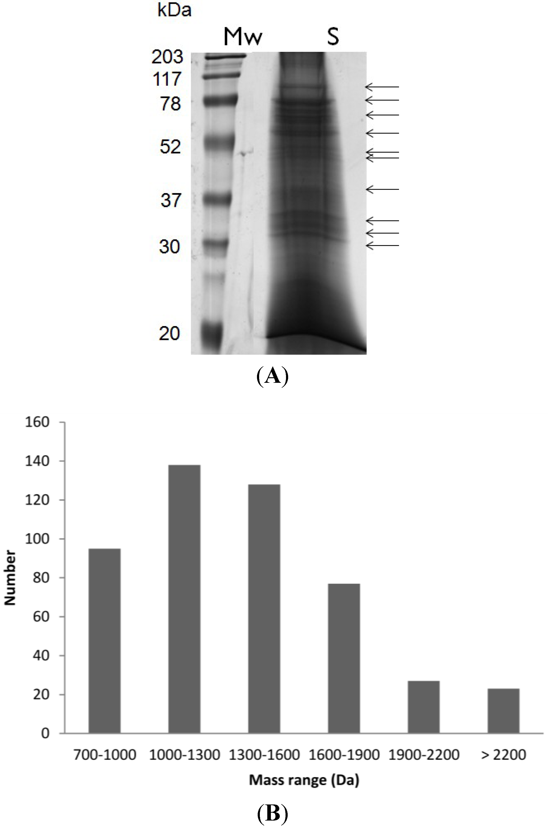
2.1. Peptide Mass Range Distribution

The venom sample was first fractionated by SDS-PAGE (Figure 2A, SDS-PAGE only) and 2DE gel (data not shown). Each protein band was isolated and subjected to reduction, alkylation and trypsin digestion. Peptide samples were subsequently submitted to MALDI-TOF MS analysis, enabling the detection of 488 unique molecular masses, ranging from ~700 Da to ~3000 Da. The same technique was used to characterize C. consors peptides, rendering the detection of similar number of molecular masses (e.g., 550) [20]. The molecular mass distribution of the conopeptides in C. crotchii is shown in Figure 2B. Almost 90% of these masses were between 700 and 1900 Da and only 10% corresponded to large peptides. However, most of the peptides were between 1000 and 1600 Da. The C. crotchii molecular mass range is asymmetrically distributed, as described in the case of other cone snail venoms, namely from C. consors [20], C. textile, C. imperialis and C. marmoreus [6]. The mass range between 1000 and 2000 Da is the best range MALDI-TOF-MS detection for conopeptide with lower hydrophobicity [16].
Figure 2. (A) Proteins separated by sodium dodecyl sulfate polyacrylamide gel electrophoresis (SDS-PAGE) from the venom duct of Conus crotchii. Mw: molecular weight, and S: sample; (B) Molecular mass range distribution of the 488 peptides detected by matrix-assisted laser desorption/ionization time-of-flight mass spectrometry (MALDI-TOF-MS) analysis.
Marinedrugs 11 02203 g002

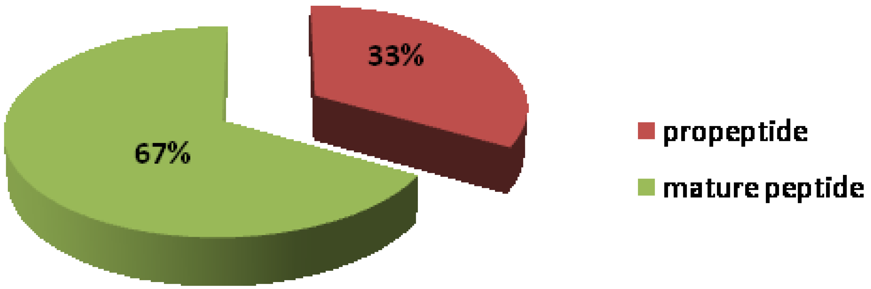
2.2. Peptides Sequence

Conus venom peptides are classified into two groups: the disulfide-poor and the disulfide-rich [21]. The disulfide-rich peptides, also called conotoxins, contain one or more disulphide bridges. In this study, we are able to identify only disulfide-rich peptides by mass-matching (Table 1). These represented only 5% of all “conomass” detected. The 95% of conomasses not identified may thus represent an interesting pool of new conopeptides. Databases from National Center for Biotechnology Information (NCBI), UniProtKB, and Conoserver were used to match peptide masses predicted from sequences with a list of masses experimentally obtained in MASCOT search engine tool. This approach can facilitate the validation and accelerate the discovery of conopeptides [22] despite the high frequency and variability of post-translational modifications (PTM) displayed by conopeptides. Extensive PTM are common in Conus peptides like as hydroxylation of lysine to 5-hydroxylysine, cyclization of N-terminal glutamine to pyroglutamate or amidation of C-terminus [23]. Disulfide bonds are the most common PTM in conopeptides and are found in 220 of the 234 conopeptides isolated at the protein level [19].
Table 1. Peptide mass detected by MALDI-TOF-MS and putative sequence by mass-matching using MASCOT database research.
ConopeptideCalculated Mass (Da)Observed Mass (Da)SequenceCysteine
11916.99
2042.78
1916.90
2042.72
TKTDDDVPLSSLRDNLK
ECCEDGWCCTAAPLTGR
4
2795.44
2232.96
795.48
2233.05
EQHLIR
CCDFVKYVGMNPPADKCR
3
31701.89
2042.73
1701.80
2042.69
LWALMKGPRQCTPK
DAPCDDNNQCCSGLECK
4
42545.052544.99RPECCSDPRCNSTHPELCGGR4
51076.561076.57IRASEGCRK
AVGLIDKMRR
KGDRCGTHLCCPGLR
4
1158.67
1786.82
1158.62
1786.88
61790.81
1693.79
1790.87
1693.80
FQFLNFCCNEK
ILEDIVSTALATCCK
4
71687.88
1970.76
1687.88
1970.66
ASDGGNAAASDLIALTIK
GCCSRPPCALSNPDYCG
4
8798.32
1790.83
798.29
1790.89
CVGVCF G
EQNKTCCGLTNGRPR
4
92042.76
798.34
2042.69
798.32
SGGACNSHDQCCINFCR
KATSTCM
5
101169.51
1190.53
1169.55
1190.55
NFGDTRSCGR
RGKPCPCCR
4
11966.49
2205.99
966.46
2206.08
KCNRFNK
IPNQKCFQHLDDCCSRK
4
12985.45
1352.56
983.46
1352.59
RGHGRSCPG
NGCTCVYHWR
3
The experimental MS spectra data was used in MASCOT database search tool to retrieve peptide sequences [24]. Here the molecular masses (monoisotopic) of the predicted peptides were compared with the estimated experimentally and the mass error was set as less than 0.1 Da. This high-quality conformity between predicted and observed masses lends confidence to the assignments (error ≤0.5 Da) [25]. To estimate the number of Cys residues in the peptide, disulfide bonds were reduced with dithiothreitol (DTT) and the Cys residues alkylated with iodoacetamide (IAA). This procedure is normally used for identifying disulfide-linked peptides and consequently the number of Cys using MALDI-TOF MS [26,27,28,29]. The results suggested the addition of carbamidomethyl group to each sulfur atom and a corresponding increase of 58 Da for each Cys in the peptide. The same chemical modification was suggested for peptide cal12a and cal12b from C. californicus [30].

2.3. BLAST Search for Conotoxins

All reduced and alkylated venom peptides from C. crotchii were analyzed by MS and amino acid sequence data were suggested by a MASCOT database search and blast analysis in UniProtKB. Peptides identified in the venom by MALDI-TOF MS are underlined (Table 2). Signal, propeptide and mature peptide regions are shown in red, blue and black, respectively (Table 2). For MALDI-TOF MS analysis, 33% and 67% propeptide and mature peptide were retrieved, respectively (Figure 3) from the venom duct and venom gland (Figure 4) that can likely be related with. The large percentage (67%) of peptide sequence corresponds to the mature peptide region due to the fact that the sample preparation was done with only the venom duct. Similarly, a total of 12 conotoxins were detected in the C. crotchii venom duct, considering only results with a protein 100% max. identity in UniProtKB (Table 2). A BLAST search on UniProtKB database resulted with an E-value between 1e-12 and 7e-30 and score bits between 151 and 257. The statistics from BLAST alignments was based on the marginally significant criterion E-value of 0.05, with normalized score of ~38 bits [31].
Table 2. Conotoxins identification by BLAST search on UniProtKB database; all 151 results have a 100% Max identity with the protein sequence. Signal, propeptide and mature peptide regions are shown in red, blue and black, respectively. Peptides identified in the venom by MALDI-TOF-MS—MASCOT database are underlined.
Conotoxin (Accession)Protein sequenceE-Value a
TxVA (P81755.2) Marinedrugs 11 02203 i0012e-12
im23.3 (D0PX86.1) Marinedrugs 11 02203 i0027e-21
Ec15a (B0KZ79.1) Marinedrugs 11 02203 i0033e-28
Ai1.2 (P0CB08.1) Marinedrugs 11 02203 i0049e-17
Bu2 (P0CY61.1) Marinedrugs 11 02203 i0057e-30
Ca5.1 (P0C666.1) Marinedrugs 11 02203 i0064e-22
PnMGMR-02 (Q9BP56.1) Marinedrugs 11 02203 i0078e-30
VnMSGL-0123 (Q9BP59.1) Marinedrugs 11 02203 i0082e-15
Eb6.18 (C7T1P1.1) Marinedrugs 11 02203 i0092e-19
Leo-O2 (P0C903.1) Marinedrugs 11 02203 i0101e-12
PVIIA (P56633.2) Marinedrugs 11 02203 i0117e-20
VxXXB (P0C1W6.2) Marinedrugs 11 02203 i0122e-14
a The reported E-values were derived by the BLAST analysis.
Figure 3. Distribution of peptide sequence identified as “Conoprotein”.
Marinedrugs 11 02203 g003
All Cape Verde endemic Conus species are usually regarded as vermivorous, but it is not entirely clear that they are exclusively vermivorous [18]. The toxin’s sequence similarity results indicates that C. crotchii venom presents peptides from vermivorous (50%), but also molluscivorous (33%) and piscivorous (17%) Conus species. Among them, four conotoxins (Eb6.18, Leo-O2, Bu2, PVIIA) could be assigned to the O1-superfamily, three to the A-superfamily (im23.3, Ai1.2, PnMGMR-02), two to the T-superfamily (Ca5.1, TxVA) and one for each O2- (Ec15a), O3- (VnMSGL-0123) and D-superfamily (VxXXB), all of them previously described (Table 3). These results may suggest that the venom from C. crotchii has not only a dietary function but it is used for all kinds of environmental interaction as predators defense. On the other hand, it has already been demonstrated that size and diversity of the conopeptide gene superfamilies on vermivorous species differ significantly [5]. Conus peptides from the same superfamily share the typical arrangement of Cys residues in the mature toxin region, the “Cys pattern”. Each “Cys pattern” generally corresponds to a precise disulfide framework. However, within the same superfamily, thereissome altered loop spacing of amino acids between cysteines. For example, in the superfamily O, conopeptide μO-MrVIA (C. marmoreus) has interval residue number CX6CX9CCX4CX4C (6-9-4-4) and κ-PVIIA (C. purpurascens) has CX6CX6CCX3CX5C (6-6-3-5) [32]. Conopeptides from the superfamily O (cysteine framework “C-C-CC-C-C”) has a O1, O2 and O3 variation, can blockvoltage-gated Ca2+ channels and inhibits voltage-gated K+ channels [19]. However, the A-superfamily conopeptides (cysteine framework “CC-C-C”), one of the most studied superfamiles, together with the superfamilies O and T [19], can act on nicotinic acetylcholine receptors (nAChR) and can also block K+ channels [33].
Table 3. Conotoxins isolated from different Conus species.
NameC. speciesDietSuperfamilyFamilyCys pattern (framework)Reference
Ec15aC. emaciatusvO2unknownC-C-CC-C-C-C-C (XV)[31]
Ca5.1C. caracteristicusvTunknownCC-CC (V)[34]
im23.3C. imperialisvAunknownC-C-C-CC-C (XXIII)[35]
Eb6.18C. ebraeusvO1unknownC-C-CC-C-C (VI/VII)[36]
Leo-O2C. leopardusvO1unknownC-C-CC-C-C (VI/VII)[37]
VxXXBC. vexillumvDαC-CC-C-CC-C-C-C (XX)[38]
TxVAC. textilemTєCC-CC (V)[39]
Ai1.2C. ammairalismAαCC-C-C (I)[31]
PnMGMR-02C. pennaceusmAαCC-C-C (I)[31]
VnMSGL-0123C. ventricosusmO3unknownC-C-CC-C-C (VI/VII)[31]
Bu2C. bullatuspO1unknownC-C-CC-C-C (VI/VII)[40]
PVIIAC. purpurascenspO1κC-C-CC-C-C (VI/VII)[41]
v: vermivorous; m: molluscivorous; p: piscivorous.

3. Experimental Section

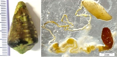
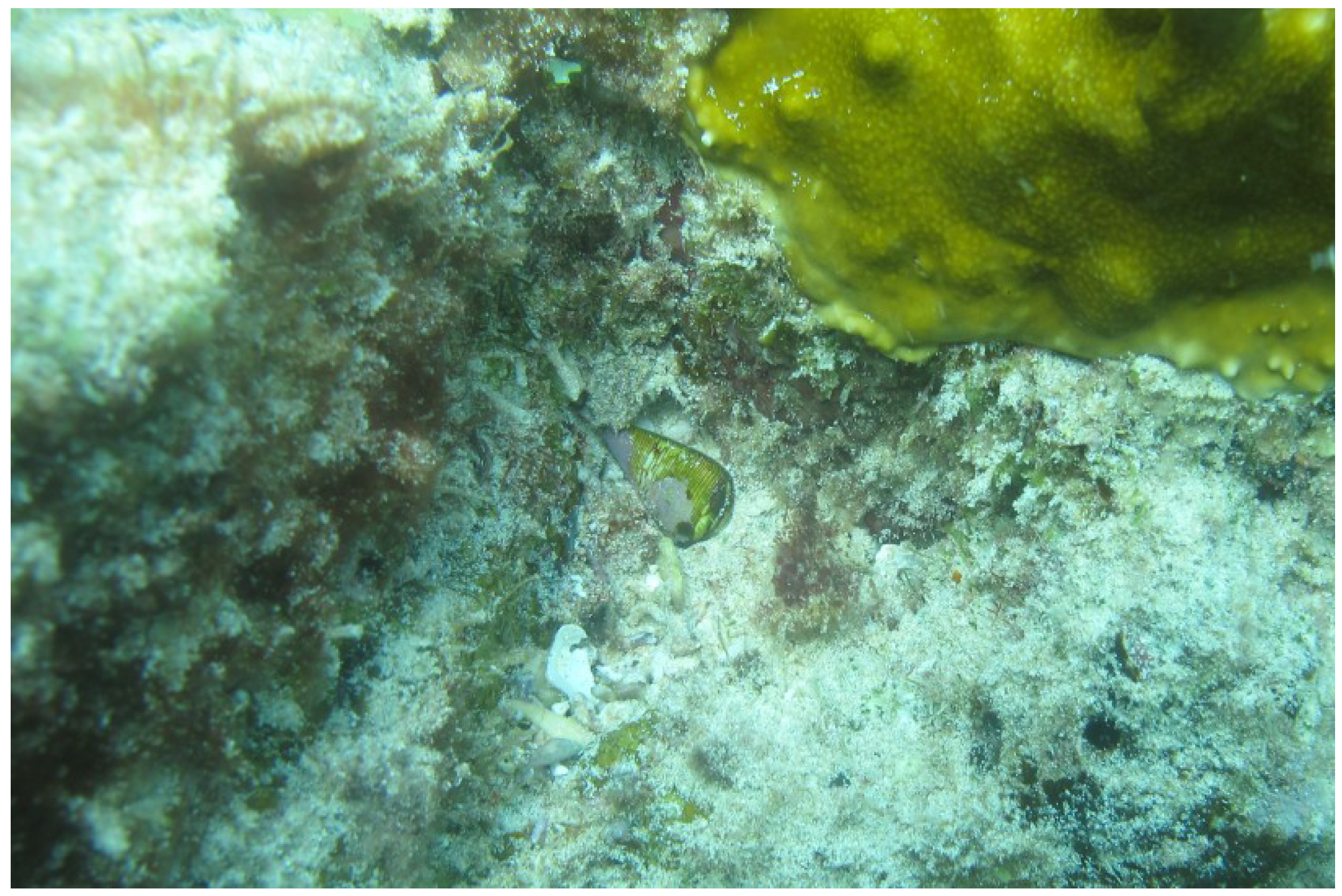
3.1. Cone Snail Specimen and Venom Extraction

The Conus crotchii were obtained from the Boa Vista Island in the Cape Verde archipelago. The venom ducts were dissected on ice and the venom duct removed and diluted in 500 μL of 0.1% formic acidand stored at −80 °C until use. The image was obtained (Figure 4) using magnifying glass Olympus DP72, DF lenses mode (Tokyo, Japan). The venom ducts were mechanically disrupted by ceramic beads (diameter 1.4 mm) using Precellys 24 homogenizer (5400 rpm, 2 × 15 s; Bertin, Montigny-le-Bretonneux, France). Ceramic beads and insoluble materials were removed by centrifugation at 4 °C (16× g for 10 min, twice). After centrifugation, all venom extract were immediately stored at −20 °C prior to analysis.
Figure 4. Shell and venom apparatus of C. crotchii: (a) Venom bulb; (b) Venom duct; (c) Venom gland.
Marinedrugs 11 02203 g004

3.2. Sample Fractionation, SDS-PAGE

Protein samples were mixed with loading buffer with Tris-HCl (0.25 M) pH 6.8, SDS (6%, w/v), glycerol (30%, v/v), β-mercaptoethanol (6.25%, v/v) and proteins separated by sodium dodecyl sulfate polyacrylamide gel electrophoresis (SDS-PAGE) in homogeneous gels (12% acrylamide) according to Laemmli [42]. Gels were stained with colloidal Coomassie Blue [43], gel images acquired using GS-800 calibrated scanner (Bio-Rad, Hercules, CA, USA) and the protein profiles analyzed using the Quantity One software (Bio-Rad, Hercules, CA, USA).

3.3. Protein Reduction, Alkylation and Trypsin Digestion

Proteins were isolated from the SDS-PAGE gels and submitted to in-gel reduction and alkylation to disrupt disulfide bonds [44,45], and thereafter to trypsin digestion. Reduction was achieved by adding 50 μL of DTT (10 mM) prepared in NH4HCO3 (100 mM, pH 8.0) to the protein samples followed by incubation during 45 min at 56 °C. For alkylation the dithiothreitol (DTT) was replaced by 50 μL IAA (55 mM) prepared in NH4HCO3 (100 mM) and the protein samples incubated during 30 min in the dark. For trypsin digestion dried protein gel bands were incubated with 6.7 ng trypsin/μL during 30 min in ice. Thereafter, the excess of trypsin solution was removed and 5–25 μL of NH4HCO3 (50 mM) was added in order to cover the gel pieces. Trypsin digestion proceeded overnight (15 h) at 37 °C. The solution (supernatant) containing the peptides was subsequently collected into an eppendorf tube and stored at −20 °C.

3.4. MALDI-TOF-MS Analysis

The peptide samples were concentrated and cleaned according to the manufacturer’s instructions on a micro C18 ZipTiP column (Millipore, Bedford, MA, USA). The peptides were eluted directly onto the MALDI plate using the matrix α-cyano-4-hydroxycinnamic acid (α-CHCA) at 5 mg/mL prepared in ACN (50%), and formic acid (0.1%). Peptide mass spectrometry analyses were performed by MALDI-TOF/TOF (4700 Proteomics Analyzer, AB SCIEX, Foster City, CA, USA) method described [46,47,48] in reflector positive mode (700–4000 Da). The experimental mass spectra were searched against the UniprotKB protein sequence database with the Mascot (Matrix-Science, London, UK) algorithm, integrated in the GPS Explorer software (AB SCIEX, Foster City, CA, USA). The search parameters were up to two maximum trypsin missed cleavages, mass tolerance of 50 ppm, cysteine carbamidomethylation (fixed modification), methionine oxidation (variable modification) and a charge state of +1.

4. Conclusions

In this work we characterized the peptide profile from C. crotchii using MALDI-TOF and mass-matching. The number of molecular masses studied here resembles the outputs from other studies performed in the genera Conus enabling us to validate our approach. We were able to identify several disulfide-rich conotoxins in C. crotchii venom duct samples that belong to O1-superfamily (Eb6.18, Leo-O2, Bu2, PVIIA), A-superfamily (im23.3, Ai1.2, PnMGMR-02), T-superfamily (Ca5.1, TxVA) and O2- (Ec15a), O3- (VnMSGL-0123) and D-superfamilies (VxXXB). Taking into consideration the mode of action of some of these conotoxins we may consider that the high diversity of conotoxins may not only be related to diet but with all kinds of environmental interaction as predators’ defense. These putative conotoxins may block voltage-gated Ca2+ channels, inhibit voltage-gated K+ channels and act on nAChRs [15,30]. A large number of masses were not assigned in this work. This promises new research potential and the discovery of new bioactive molecules from Cape Verde Conus species.

Acknowledgments

We thank Zinho Évora, Corrine Almeida, Livinho Tavares (Pelé) and Evandro Lopes for providing indispensable field assistance. This work was supported by a PhD grant (SFRH/BD/51477/2011) and was partially funded by the Project MARBIOTECH (reference NORTE-07-0124-FEDER-000047) within the SR & TD Integrated Program MARVALOR—Building research and innovation capacity for improved management and valorization of marine resources, supported by the Programa Operacional Regional do Norte (ON.2—O Novo Norte) and by the European Regional Development Fund, by the project PesT-C/MAR/LA0015/2011 from the Portuguese Foundation for Science and Technology (FCT), INTERREG IV projects ATLANTOX, PHARMATLANTIC, AA was funded by the project PTDC/AAC-AMB/121301/2010 (FCOMP-01-0124-FEDER-019490) from FCT.

Conflicts of Interest

The author declares no conflict of interest.

References

  1. Dobson, R.; Collodoro, M.; Gilles, N.; Turtoi, A.; de Pauw, E.; Quinton, L. Secretion and maturation of conotoxins in the venom ducts of Conus textile. Toxicon 2012, 60, 1370–1379. [Google Scholar] [CrossRef]
  2. Olivera, B.M. Conus venom peptides: Reflections from the biology of clades and species. Annu. Rev. Ecol. Syst. 2002, 33, 25–47. [Google Scholar] [CrossRef]
  3. Terlau, H.; Olivera, B.M. Conus venoms: A rich source of novel ion channel-targeted peptides. Physiol. Rev. 2004, 84, 41–68. [Google Scholar] [CrossRef]
  4. Olivera, B.M. Conus venom peptides, receptor and ion channel targets, and drug design: 50 million years of neuropharmacology. Mol. Biol. Cell 1997, 8, 2101–2109. [Google Scholar]
  5. Kauferstein, S.; Porth, C.; Kendel, Y.; Wunder, C.; Nicke, A.; Kordis, D.; Favreau, P.; Koua, D.; Stöcklin, R.; Mebs, D. Venomic study on cone snails (Conus spp.) from South Africa. Toxicon 2011, 57, 28–34. [Google Scholar] [CrossRef]
  6. Davis, J.; Jones, A.; Lewis, R.J. Remarkable inter- and intra-species complexity of conotoxins revealed by LC/MS. Peptides 2009, 30, 1222–1227. [Google Scholar] [CrossRef]
  7. Favreau, P.; Stöcklin, R. Marine snail venoms: Use and trends in receptor and channel neuropharmacology. Curr. Opin. Pharmacol. 2009, 9, 594–601. [Google Scholar] [CrossRef]
  8. Olivera, B.M.; Cruz, L.J. Conotoxins, in retrospect. Toxicon 2001, 39, 7–14. [Google Scholar] [CrossRef]
  9. Mcintosh, J.M.; Santos, A.D.; Olivera, B.M. Conus peptides targeted to specific nicotinic acetylcholine receptor subtypes. Peptides 1999, 68, 59–88. [Google Scholar]
  10. Essack, M.; Bajic, V.B.; Archer, J.C. Conotoxins that confer therapeutic possibilities. Mar. Drugs 2012, 10, 1244–1265. [Google Scholar] [CrossRef]
  11. Olivera, B.M. Conus peptides: Biodiversity-based discovery and exogenomics. J. Biol. Chem. 2006, 281, 31173–31177. [Google Scholar] [CrossRef]
  12. Luo, S.; Zhangsun, D.; Wu, Y.; Zhu, X.; Xie, L.; Hu, Y.; Zhang, J.; Zhao, X. Identification and molecular diversity of T-superfamily conotoxins from Conus lividus and Conus litteratus. Chem. Biol. Drug Des. 2006, 68, 97–106. [Google Scholar] [CrossRef]
  13. Wen, L.; Yang, S.; Zhou, W.; Zhang, Y.; Huang, P. New conotoxin SO-3 targeting N-type voltage-sensitive calcium channels. Mar. Drugs 1995, 4, 215–227. [Google Scholar] [CrossRef]
  14. Violette, A.; Leonardi, A.; Piquemal, D.; Terrat, Y.; Biass, D. Recruitment of glycosyl hydrolase proteins in a cone snail venomous arsenal: Further insights into biomolecular features of Conus venoms. Mar. Drugs 2012, 10, 258–280. [Google Scholar] [CrossRef]
  15. Olivera, B.M. Conus Snail Venom Peptides. In Handbook of Biologically Active peptides; Kastin, A.J., Ed.; Academic Press: Amsterdam, The Netherlands, 2006; pp. 381–388. [Google Scholar]
  16. Biass, D.; Dutertre, S.; Gerbault, A.; Menou, J.-L.; Offord, R.; Favreau, P.; Stöcklin, R. Comparative proteomic study of the venom of the piscivorous cone snail Conus consors. J. Proteomics 2009, 72, 210–218. [Google Scholar] [CrossRef]
  17. Jakubowski, J.A.; Kelley, W.P.; Sweedler, J.V.; Gilly, W.F.; Schulz, J.R. Intraspecific variation of venom injected by fish-hunting Conus snails. J. Exp. Biol. 2005, 208, 2873–2883. [Google Scholar] [CrossRef]
  18. Cunha, R.L.; Castilho, R.; Rüber, L.; Zardoya, R. Patterns of cladogenesis in the venomous marine gastropod genus Conus from the Cape Verde islands. Syst. Biol. 2005, 54, 634–650. [Google Scholar]
  19. Kaas, Q.; Westermann, J.-C.; Craik, D.J. Conopeptide characterization and classifications: An analysis using ConoServer. Toxicon 2010, 55, 1491–1509. [Google Scholar] [CrossRef]
  20. Biass, D.; Križaj, I.; Leonardi, A.; Dutertre, S. Peptidomics and proteomics of Conus consors cone snail venom. Comp. Gen. Pharmacol. 2009, 72, 37592. [Google Scholar]
  21. Van Der Haegen, A.; Peigneur, S.; Dyubankova, N.; Möller, C.; Marí, F.; Diego-García, E.; Naudé, R.; Lescrinier, E.; Herdewijn, P.; Tytgat, J. Pc16a, the first characterized peptide from Conus pictus venom, shows a novel disulfide connectivity. Peptides 2012, 34, 106–113. [Google Scholar] [CrossRef]
  22. Kaas, Q.; Yu, R.; Jin, A.H.; Dutertre, S.; Craik, D.J. ConoServer: Updated content, knowledge, and discovery tools in the conopeptide database. Nucleic Acids Res. 2012, 40, D325–D330. [Google Scholar] [CrossRef]
  23. Buczek, O.; Bulaj, G.; Olivera, B.M. Conotoxins and the posttranslational modification of secreted gene products. Cell. Mol. Life Sci. 2005, 62, 3067–3079. [Google Scholar] [CrossRef]
  24. Tannu, N.S.; Hemby, S.E. De novo protein sequence analysis of Macaca mulatta. BMC Genomics 2007, 8, 270. [Google Scholar] [CrossRef]
  25. Standing, K. Peptide and protein de novo sequencing by mass spectrometry. Curr. Opin. Struct. Biol. 2003, 13, 595–601. [Google Scholar] [CrossRef]
  26. Sechi, S.; Chait, B.T. Modification of cysteine residues by alkylation. A tool in peptide mapping and protein identification. Peptides 1998, 70, 5150–5158. [Google Scholar]
  27. Jakubowski, J.A.; Sweedler, J.V. Sequencing and mass profiling highly modified conotoxins using global reduction/alkylation followed by mass spectrometry. Anal. Chem. 2004, 76, 6541–6547. [Google Scholar] [CrossRef]
  28. Gray, W.R. Disulfide structures of highly bridged peptides: A new strategy for analysis. Protein Sci. 1993, 2, 1732–1748. [Google Scholar] [CrossRef]
  29. Aguilar, M.B.; López-Vera, E.; Heimer de la Cotera, E.P.; Falcón, A.; Olivera, B.M.; Maillo, M. I-Conotoxins in vermivorous species of the West Atlantic: Peptide sr11a from Conus spurius. Peptides 2007, 28, 18–23. [Google Scholar] [CrossRef]
  30. Gilly, W.F.; Richmond, T.A.; Duda, T.F.; Elliger, C.; Lebaric, Z.; Schulz, J.; Bingham, J.P.; Sweedler, J.V. A diverse family of novel peptide toxins from an unusual cone snail, Conus californicus. J. Exp. Biol. 2011, 214, 147–161. [Google Scholar] [CrossRef]
  31. Altschul, S.F.; Madden, T.L.; Schäffer, A.A.; Zhang, J.; Zhang, Z.; Miller, W.; Lipman, D.J. Gapped BLAST and PSI-BLAST: A new generation of protein database search programs. Nucleic Acids Res. 1997, 25, 3389–3402. [Google Scholar] [CrossRef]
  32. Luo, S.; Zhangsun, D.; Feng, J.; Wu, Y.; Zhu, X.; Hu, Y. Diversity of the O-superfamily conotoxins from Conus miles. J. Peptide Sci. 2007, 13, 44–53. [Google Scholar]
  33. Peng, C.; Ye, M.; Wang, Y.; Shao, X.; Yuan, D.; Liu, J.; Hawrot, E.; Wang, C.; Chi, C. A new subfamily of conotoxins belonging to the A-superfamily. Peptides 2010, 31, 2009–2016. [Google Scholar] [CrossRef]
  34. Peng, C.; Wu, X.; Han, Y.; Yuan, D.; Chi, C.; Wang, C. Identification of six novel T-1 conotoxins from Conus pulicarius by molecular cloning. Peptides 2007, 28, 2116–2124. [Google Scholar] [CrossRef]
  35. Puillandre, N.; Koua, D.; Favreau, P.; Olivera, B.M.; Stöcklin, R. Molecular phylogeny, classification and evolution of conopeptides. J. Mol. Evol. 2012, 74, 297–309. [Google Scholar] [CrossRef]
  36. Duda, T.F.; Chang, D.; Lewis, B.D.; Lee, T. Geographic variation in venom allelic composition and diets of the widespread predatory marine gastropod Conus ebraeus. PLoS One 2009, 4, e6245. [Google Scholar]
  37. Remigio, E.A.; Duda, T.F. Evolution of ecological specialization and venom of a predatory marine gastropod. Mol. Ecol. 2008, 17, 1156–1162. [Google Scholar] [CrossRef]
  38. Loughnan, M.; Nicke, A.; Jones, A.; Schroeder, C.I.; Nevin, S.T.; Adams, D.J.; Alewood, P.F.; Lewis, R.J. Identification of a novel class of nicotinic receptor antagonists: Dimeric conotoxins VxXIIA, VxXIIB, and VxXIIC from Conus vexillum. J. Biol. Chem. 2006, 281, 24745–24755. [Google Scholar] [CrossRef]
  39. Walker, C.S.; Steel, D.; Jacobsen, R.B.; Lirazan, M.B.; Cruz, L.J.; Hooper, D.; Shetty, R.; DelaCruz, R.C.; Nielsen, J.S.; Zhou, L.M.; et al. The T-superfamily of conotoxins. J. Biol. Chem. 1999, 274, 30664–30671. [Google Scholar] [CrossRef]
  40. Hu, H.; Bandyopadhyay, P.K.; Olivera, B.M.; Yandell, M. Characterization of the Conus bullatus genome and its venom-duct transcriptome. BMC Genomics 2011, 12, 60. [Google Scholar]
  41. Shon, K.; Stocker, M.; Terlau, H.; Stu, W.; Jacobsen, R.; Walker, C.; Grilley, M.; Watkins, M.; Hillyard, D.R.; Gray, W.R.; et al. κ-Conotoxin PVIIA is a peptide inhibiting the shaker K+ channel. J. Biol. Chem. 1998, 273, 33–38. [Google Scholar] [CrossRef]
  42. Laemmli, U.K. Cleavage of structural proteins during the assembly of the head of bacteriophage T4. Nature 1970, 227, 680–685. [Google Scholar] [CrossRef]
  43. Neuhoff, V.; Arold, N.; Taube, D.; Ehrhardt, W. Improved staining of proteins in polyacrylamide gels including isoelectric focusing gels with clear background at nanogram sensitivity using Coomassie Brilliant Blue G-250 and R-250. Electrophoresis 1988, 9, 255–262. [Google Scholar] [CrossRef]
  44. Kalume, D.E.; Stenflo, J.; Czerwiec, E.; Hambe, B.; Furie, B.C.; Furie, B.; Roepstorff, P. Structure determination of two conotoxins from Conus textile by a combination of matrix-assisted laser desorption/ionization time-of-flight and electrospray ionization mass spectrometry and biochemical methods. JMS 2000, 35, 145–156. [Google Scholar]
  45. Zugasti-Cruz, A.; Aguilar, M.B.; Falcón, A.; Olivera, B.M.; Heimer de la Cotera, E.P. Two new 4-Cys conotoxins (framework 14) of the vermivorous snail Conus austini from the Gulf of Mexico with activity in the central nervous system of mice. Peptides 2008, 29, 179–185. [Google Scholar] [CrossRef]
  46. Padliya, N.D.; Wood, T.D. Improved peptide mass fingerprinting matches via optimized sample preparation in MALDI mass spectrometry. Anal. Chim. Acta 2008, 627, 162–168. [Google Scholar] [CrossRef]
  47. Romeo, C.; di Francesco, L.; Oliverio, M.; Palazzo, P.; Massilia, G.R.; Ascenzi, P.; Polticelli, F.; Schininà, M.E. Conus ventricosus venom peptides profiling by HPLC-MS: A new insight in the intraspecific variation. J. Sep. Sci. 2008, 31, 488–498. [Google Scholar] [CrossRef]
  48. Puerto, M.; Campos, A.; Prieto, A.; Cameán, A.; de Almeida, A.M.; Coelho, A.V.; Vasconcelos, V. Differential protein expression in two bivalve species; Mytilus galloprovincialis and Corbicula fluminea; exposed to Cylindrospermopsis raciborskii cells. Aquat. Toxicol. 2011, 101, 109–116. [Google Scholar] [CrossRef]

