Article

Synthesis and Evaluation of Some New Aza-B-homocholestane Derivatives as Anticancer Agents

1
Key Laboratory of Beibu Gulf Environment Change and Resources Utilization, College of Chemistry and Life Science, Guangxi Teachers Education University, Nanning 530001, China
2
School of Chemistry and Chemical Engineering, Sun Yat-sen University, Guangzhou 510275, China
3
Clinical Chemistry Program, Department of Chemistry, SI 424, Cleveland State University, Cleveland, OH 44115, USA
*
Author to whom correspondence should be addressed.
Mar. Drugs 2014, 12(4), 1715-1731; https://doi.org/10.3390/md12041715
Submission received: 26 December 2013 / Revised: 24 February 2014 / Accepted: 26 February 2014 / Published: 25 March 2014

Abstract

:
Using analogues of some marine steroidal oximes as precursors, a series of aza-B-homocholestane derivatives possessing different substituted groups at the 3-position of the steroidal nucleus were synthesized. Their biological activity against cancer cell proliferation was determined with multiple cancer cell lines. Aza-B-homocholestane derivatives possessing 3-hydroxyl, 3-hydroximino and 3-thiosemicarbazone groups displayed remarkable cytotoxicity to cancer cells via apoptosis inducing mechanism. Compounds 5, 10, 12, 15 and 18 exhibited better potency to inhibit cancer cell proliferation. In addition, compound 15 was further evaluated with three dimensional (3D) multicellular spheroids assay to determine its potency against spheroid growth. The structure-activity relationship (SAR) generated in the studies is valuable for the design of novel chemotherapeutic agents.

1. Introduction

Natural products derived from marine organisms are important resources of biologically active compounds. Compounds with steroidal skeletons are abundant in marine sponges [1]. However, marine steroids having an oxime group have been less reported in the literatures. (6E)-Hydroximino-24-ethylcholest-4-en-3-one (1) and (6E)-hydroximinocholest-4-en-3-one (2) were two interesting steroidal compounds having 6-oxime group, and were isolated from the sponges Cinachyrella (C. alloclada and C. apion) (Figure 1) [2]. Compound 2 showed cytotoxicity with IC50 of 1.25 μg/mL against P-388, A-549, HT-29 cells, and 2.5 μg/mL toward MEL-28 cells. Based on the results, different types of steroidal oxime derivatives were synthesized and their cytotoxicities were evaluated [3,4,5,6,7,8].
Figure 1. Chemical structures of compounds 1 and 2.
Marinedrugs 12 01715 g001
Aza-homosteroids are a class of steroid compounds that were synthesized and modified in order to increase the biological activity of the lead steroids. The synthesis of some aza-homosteroid compounds with unusual and interesting structures had been reported recently [9,10,11,12,13,14]. These compounds exhibited cytotoxic, antibacterial, antileukemic, and antiandrogenic activities. Studies of aza-homosteroids revealed that the presence of the characteristic group (-NH-CO-) in the aza-homosteroid molecule is important in lowering the acute toxicity and improving anticancer activity of the compounds [15,16].
In our previous work, a series of 3-aza-A-homo and 4-aza-A-homo steroidal lactams were prepared and evaluated against the proliferation of SGC 7901 (human ventriculi carcinoma cell line), HeLa (human cervical carcinoma cell line) and Bel 7404 (human liver carcinoma cell line) [17,18,19]. The results showed that some of these steroidal lactams significantly inhibited the proliferation of the tumor cell lines tested and induced cancer cell apoptosis as well. In order to find more active derivatives as potential antitumor agents, a series of the new aza-B-homecholestane derivatives possessing different aza-position and various substitute groups on the 3-position of steroidal nucleus were designed and synthesized taking analogues of compound 2 as precursors. The antiproliferative activities of these new compounds were evaluated with different cancer cell lines.

2. Results and Discussion

2.1. Chemistry

First, 3-hydroximino-7-aza-B-homocholest-4-en-6-one (7) was synthesized as listed in Scheme 1. The synthesis of steroidal oxime (3) had been reported by our group [7]. Beckman rearrangement of 3 in SOCl2/THF gave 3-acetoxy-7-aza-B-homocholest-4-en-6-one (4). The structure of 4 was confirmed by 1H NMR chemical shifts of 7a-protons at 3.16–3.21 ppm (2H, m). Compound 5 was obtained by deacetylation of 4 in aqueous solution of 13% K2CO3. The oxidation of compound 5 with Jones reagent afforded 7-aza-B-homocholest-4-en-3,6-dione (6). Last, the oxime 7 was produced in a yield of 42% by the reaction of 6 with hydroxylamine hydrochloride in ethanol in the presence of NaOAc. The structure of 7 was confirmed by analysis of the proton and carbon NMR chemical shifts. The downfield chemical shift of 2β-H at 3.453 ppm (dd, J = 15.3 and 4.8 Hz, due to the deshielding influence of the OH in the hydroximino group) and C-3 at 158.6 ppm demonstrated the (E)-configuration and formation of 3-hydroximino in compound 7.
Scheme 1. Synthesis of 3-hydroximino-7-aza-B-homocholest-4-en-6-one.
Marinedrugs 12 01715 g006
Reagents and conditions: (a) SOCl2/THF, 0 °C; (b) 13% K2CO3/CH3OH, stirred, 4 h; (c) Jones reagent/acetone, r. t., 2 h; (d) H2NOH.HCl/Na2Ac.3H2O/EtOH, reflux.
Scheme 2. Synthesis of compounds 915.
Marinedrugs 12 01715 g007
12: X = NOH; 13: X = NOCH3; 14: X = NOCH2Ph; 15: X = NNHCSNH2. Reagents and conditions: (a) SOCl2/THF, 0 °C; (b) 13% K2CO3/CH3OH, reflux, 4 h; (c) Jones reagent/acetone, r. t., 2 h; (d) H2NOH.HCl/Na2Ac.3H2O/EtOH, reflux; (e) CH3ONH2.HCl/95% C2H5OH, 60 °C; (f) PhCH2ONH2/95% C2H5OH, 60 °C; (g) H2NC(S)NHNH2/EtOH, 60 °C.
Similarly, compounds 1215 possessing the 3-substituted-6-aza-B-homocholest-7-one key feature in their structures were synthesized with 8 steps using cholesterol as starting material (Scheme 2). Compound 8 was synthesized based on the known methodology [8]. The structure of 9 was affirmed by proton and carbon NMR spectrums. In the 1H NMR spectrum, the resonances showing of C5-αH at 3.47–3.40 ppm (1H, m), C7a-H at 2.36–2.22 ppm (2H, m) and 7-C at 176.2 ppm demonstrated a formation of NH-CO- bond and a position of 6-NH. The oxidization of 10 by Jones reagent generated compound 11. The reaction of compound 11 with HONH2·HCl, CH3ONH2·HCl, PhCH2ONH2·HCl or thiosemicarbazide afforded the corresponding products 1215. Their structures were confirmed by IR and NMR spectrum, and the mixture of E- and Z-stereoisomer was obtained in preparation of 1315, respectively.
Last, in order to determine if the aza-position has an effect on the compounds’ cytotoxicity, compounds 1721 possessing the structure of 7a-aza-B-homocholest-4(or 5)-ene were prepared (Scheme 3). Beckman rearrangement of 16 provided 3-acetoxy-7a-aza-B-homocholest-5-ene-7-one (17). The downfield chemical shift of C8-H at 3.292 ppm (1H, t, J = 8.4) in 1H NMR spectrum of 17 indicated the position of 7a-aza. In the oxidization of compound 18 by Jones reagent, 5,6-double bond of 18 was transformed into more stable 4,5-double bond of compound 19. Further, compounds 20 and 21 were obtained by the reaction of 19 with HONH2·HCl and thiosemicarbazide respectively. The structures of 20, 21 were confirmed with proton and carbon NMR.
Scheme 3. Synthesis of compounds 1721.
Marinedrugs 12 01715 g008
Reagents and conditions: (a) SOCl2/THF, 0 °C; (b) 13% K2CO3/CH3OH, reflux, 4 h; (c) Jones reagent/acetone, r.t., 2 h; (d) H2NOH.HCl/Na2Ac.3H2O/EtOH, reflux; (e) H2NC(S)NHNH2/EtOH, 60 °C.

2.2. Biological Evaluation

2.2.1. Cytotoxicity Determination

All compounds were evaluated for their antiproliferative activities against SGC 7901 (human gastric carcinoma), HeLa (human cervical carcinoma), Bel 7404 (human liver carcinoma) GNE 2 (nasopharyngeal carcinoma), SPC-A (lung carcinoma) and Tu 686 (laryngocarcinoma) cell lines using the MTT assay. The results were summarized as IC50 values in µM in Table 1.
Table 1. In vitro a antiproliferative activities of aza-B-homocholestane derivatives (IC50 in µM).
CompoundsSGC 7901HeLaBel 7404GNE 2SPC-ATu 686
491.820.814.234.592.585.4
513.226.59.87.27.525.4
6>10031.4>10029.2>10058.6
7>1009.358.4>100>10098.5
825.052.117.434.815.659.8
966.38.315.415.298.558.4
1031.83.211.69.547.642.5
1190.48.78.716.174.56.8
1292.49.131.311.334.828.5
1370.535.869.863.878.736.0
1426.522.937.85.434.023.6
1529.55.59.812.46.628.9
16>100>100>100>10048.5>100
17>100>100>10017.5>10036.2
1811.36.04.820.123.425.3
2025.735.742.243.035.642.5
2124.616.445.239.0ND bND
Cisplatin6.710.123.216.815.217.5
a Data represent the mean values of three independent determinations; b Not determined.
As shown in Table 1, compounds 815 possessing the structure of 3-substituted-6-aza-B-homocholest-7-one had better antiproliferative activity than compounds 47 with the structure of 3-substituted-7-aza-B-homocholest-4-ene-6-one and compounds 17, 18, 20, 21 with the structure of 3-substituted-7a-aza-B-homocholest-4(or 5)-ene-7-one. Compound 5 with 3-hydroxyl group exhibited better antiproliferative activity than the compound 4 with 3-acetoxyl, the 6 with 3-carbonyl and the 7 with 3-hydroximino groups. Comparing to analogues 815, compounds 10, 12, 15 possessing 3-hydroxyl, 3-hydroximino and 3-thiosemicarbazone groups displayed better cytotoxicity. Moreover, compound 18 with 3-hydroxyl group showed the best antiproliferative activity in analogues 17, 18 and 20, 21.
Interestingly, all compounds with 3-hydroxyl, hydroximino and thiosemicarbazone groups exhibited a higher cytotoxicity against HeLa cells. For example, compound 10 with 3-hydroxyl and compound 15 with 3-thiosemicarbazone exhibited IC50 values of 3.2 and 5.5 µM to HeLa cells, respectively. Apparently, all compounds (5, 10, 18) bearing the same 3-hydroxyl and a different aza-position displayed a similar cytotoxicity against these cancer cells. However, compound 15 having same 3-thiosemicarbazone and 6-aza-B-homocholest-7-one key features in its structure showed a better antiproliferative activity to these cancer cells than compound 21 with the structure of 7a-aza-B-homocholest-5-ene-7-one except SGC 7901 cell.
Above results showed that the conversion of a carbonyl group at 3-position to a hydroxyl, hydroximino, or thiosemicarbazone groups would result in a significant increase of the antiproliferative activity, suggesting the importance of these functional groups in the biological function of the compounds. Obviously, the substitution of the 3-hydroxyl group remarkably increased its cytotoxic activity against these cancer cells in comparison with the 3-acetoxyl group.
Overall, compounds 5, 10, 12, 15 and 18 were found to be the most potent compounds as anticancer agents, and they displayed a similar antiproliferative activity, when compared to cisplatin (a positive control).

2.2.2. 3D Multicellular Spheroids Screening

Screening and initial characterization of anticancer drugs typically use monolayer cultures of tumor cells. However, such monolayer cultures do not represent the characteristics of threedimensional (3D) solid tumors. The multicellular tumor spheroid model has an intermediate complexity between in vivo tumors and in vitro monolayer cell cultures. Considering the complexity of the in vivo situation, it is not surprising that many drugs that are effective in a twodimensional cell culture will lose efficacy with the in vivo assays. Limited penetration of the drug into tumor cell masses is one main factor that will lead to poor drug efficacy in animal models. Testing the efficacy of the drug with tumor spheroids may help to predict their in vivo potency [20]. Herein, compound 15 was examined with the 3D spheroid growth assay.
Spheroids were photographed in an inverted phase contrast microscope. A micrometer scale was photographed at the same magnification, and spheroid size was determined and compared.
In Figure 2, the spheroids treated with 15 µM of 15 had a smaller size after 6 days than the untreated control spheroids. The result indicated that compound 15 showed good tumor penetration ability.
Figure 2. Time-lapse images of control untreated 5000-cell H-292 spheroid, and spheroid treated with 20 µM 15. Scale bar is 100 µm.
Marinedrugs 12 01715 g002

2.2.3. Compound 5 Induced Apoptosis of Cancer Cells

To determine the molecular mechanism by which compound 5 inhibited cancer cell proliferation, we further analyzed the cytotoxicity of compound 5 in SGC-7901 cells. As shown in Figure 3, compound 5-induced SGC-7901 cell death could be clearly observed.
Figure 3. Photographs of the unstained cells were taken under Olympus model CKX31 at 100 magnification after treatment of MGC 7901 cells with various doses of compound 5 for 48 h.
Marinedrugs 12 01715 g003
To determine whether the decreased viability of SGC-7901 cells was due to compound 5 induced apoptosis, the cells were treated with compound 5, and Annexin V assay was performed. FITC-conjugated Annexin V is commonly used to determine apoptotic cells at an early stage. As shown in Figure 4, treatment with 5 μg/mL of compound 5 resulted in 8.12% PI/Annexin V double-labeled apoptotic cells (control: 0.57%; cisplatinum: 8.94%) after 24 h incubation (the lower right quadrant and the upper right quadrant which contains early and late apoptotic cells, respectively), suggesting compound 5 is a potent apoptotic inducer in gastric carcinoma cells.
Figure 4. SGC-7901 cells were double-stained with annexin V/PI and analyzed by flow cytometry. Treatment with compound 5 (5 μg/mL) and cisplatinum (10 µg/mL) for 24 h induced apoptosis of SGC-7901 cells.
Marinedrugs 12 01715 g004
The similar result was observed after treating with compound 5 in a dose dependent manner (Figure 5). Treatment with 5 μg/mL, 10 μg/mL, 15 μg/mL of compound 5 for 48 h resulted in 17.98, 30.21, 87.15% PI/Annexin V double-labeled apoptotic cells, proposing compound 5 induced the apoptosis of SGC-7901 cells further.
Figure 5. Dose depended apoptosis induced by compound 5 and cisplatinum (10 µg/mL) for 48 h.
Marinedrugs 12 01715 g005

3. Experimental Section

3.1. Chemistry

The sterols were purchased from the Sinopharm Chemical Reagent Co., Ltd., Shanghai, China. All chemicals and solvents were analytical grade and solvents were purified by general methods before being used. Melting points were determined on an X4 apparatus and were uncorrected. Infrared spectra were measured with a Nicolet FT-360 Spectrophotometer (Thermo Fisher Scientific, New York, NY, USA). The 1H and 13C NMR spectra were recorded in CDCl3 on a Bruker AV-600 spectrometer (Bruker Corporation, Billerica, MA, USA) at working frequencies 600 and 150 MHz, and a Bruker AV-300 spectrometer at working frequencies 300 and 75 MHz, respectively. Chemical shifts are expressed in parts per million (δ) values and coupling constants (J) in Hertz. LREIMS were recorded on a Thermo-DSQ instrument (Thermo Fisher Scientific, New York, NY, USA), while HREIMS were measured on a Agilent 6210 TOFMS instrument (Agilent Technologies, Palo Alto, CA, USA). The cell proliferation assay was undertaken by a MTT method using 96-well plates on MLLTISKAN MK3 analysis spectrometer (Thermo Scientific, Shanghai, China). Annexin V assay was performed using FACS Calibur flow cytometry (Becton Dickinson, Biosciences, Franklin Lakes, NJ, USA).
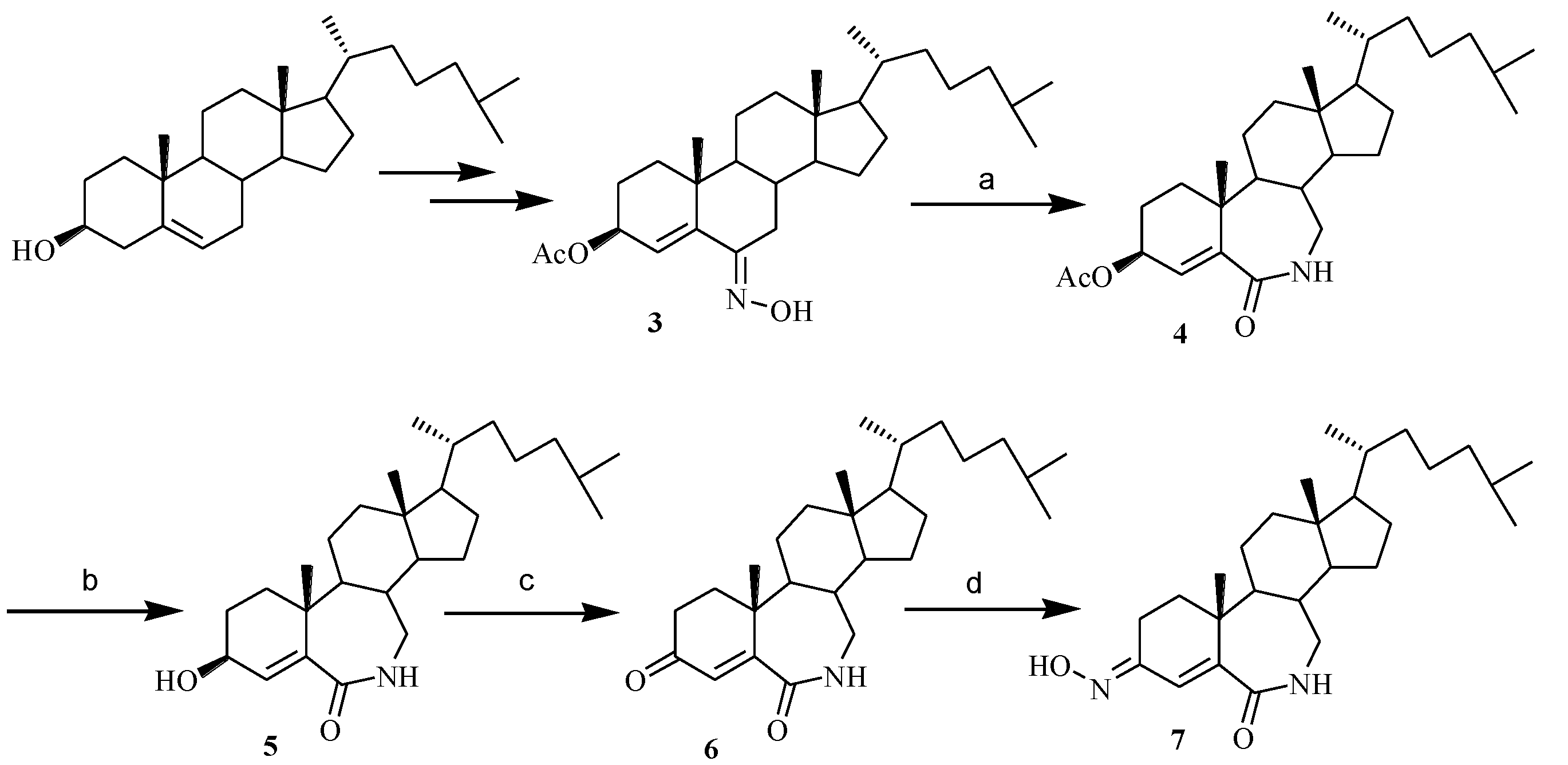
Compounds 3, 16 were prepared according to the procedures in the literature [7].

3.1.1. 3-Acetoxy-7-aza-B-homocholest-4-ene-6-one (4)

The solution of thionyl chloride (1.5 mL) in 5 mL dry THF was added to a solution of the oxime 3 (300 mg) in dry THF (30 mL). The solution was stirred under anhydrous condition for 1 h at 0 °C. The reaction was terminated and water was added to the solution. The solution was neutralized with ammonia and the product was extracted with CH2Cl2 (20 × 3 mL). The combined extract was washed with water, 5% NaHCO3, and saturated brine, dried over anhydrous Na2SO4 and evaporated under reduced pressure to give a crude product which was chromatographed on silica gel (elution: Dichloromethane/methanol = 20/1) to give white solid 216 mg. Yield: 72%, mp 157–159 °C; IR (KBr) ν: 3195, 2938, 1740, 1659, 1470, 1377, 1230, 1136, 1230, 1058 cm−1; 1H NMR (CDCl3, 300MHz) δ: 0.70 (3H, s, 18-CH3), 0.86 (3H, d, J = 6.6, 26- or 27-CH3), 0.88 (3H, d, J = 6.6, 26- or 27-CH3), 0.91 (3H, d, J = 6.6, 21-CH3), 1.21 (3H, s, 19-CH3), 2.14 (3H, s, -OCOCH3), 3.16-3.21 (2H, m, C7a-H), 5.53 (1H, dd, J = 12.3, 4.5, C3-αH), 5.91 (1H, s, C4-H), 6.72 (1H, s, 6-NH); 13C NMR (CDCl3, 75MHz) δ: 169.9 (6-C), 169.8 (COCH3), 156.7 (5-C), 114.7 (4-C), 71.2 (3-C), 56.0 (17-C), 55.5 (14-C), 53.3 (9-C), 45.5 (7a-C), 42.9 (13-C), 42.6 (24-C), 39.6 (12-C), 39.5 (8-C), 36.6 (10-C), 36.1 (22-C), 35.7 (20-C), 34.6 (1-C), 28.1 (16-C), 28.0 (25-C), 24.1 (15-C), 23.8 (2-C), 22.8 (26 or 27-C), 22.5 (26 or 27-C), 21.8 (23-C), 21.4 (11-C), 21.3 (CH3CO), 20.8 (19-C), 18.6 (21-C), 12.0 (18-C); LRESI-MS(m/z): 458.3 [M + H]+ (Supplementary Information: Figure S1).

3.1.2. 3-Hydroxy-7-aza-B-homocholest-4-ene-6-one (5)

20 mL of K2CO3 solution (13%) was added to a solution of compound 4 (1520 mg, 3.32 mmol) in CH3OH (200 mL). The reaction mixture was heated under reflux condition for 1 h. After completion of the reaction as indicated by TLC, the solvent was removed under reduced pressure. 200 mL of CH2Cl2 was added to dissolve solid and the resulting solution was washed with cold water and saturated brine. After drying over anhydrous sodium sulfate, the solvent was removed under reduced pressure, and the resulting crude product was purified by chromatography on silica gel using methanol/dichloromethane (30:1) as elution to give 988 mg of 5 as white solid. Yield: 65%, mp 233–234 °C; IR (KBr) ν: 3366, 2941, 2872, 1654, 1593, 1470, 1380, 1319, 1123, 898 cm−1; 1H NMR (CDCl3, 600MHz) δ: 0.71 (3H, s, 18-CH3), 0.88 (3H, d, J = 6.6, 26- or 27-CH3), 0.89 (3H, d, J = 6.6, 26- or 27-CH3), 0.92 (3H, d, J = 6.6, 21-CH3), 1.15 (3H, s, 19-CH3), 2.16-2.20 (1H, m, C2-H), 2.23 (1H, br s, -OH), 3.16-3.21 (1H, m, C7a-αH), 3.28-3.23 (1H, m, C7a-βH), 4.43 (1H, t, J = 5.4, C3-αH), 6.21 (1H, s, C4-H), 6.38 (1H, s, 7-NH); 13C NMR (CDCl3, 75MHz) δ: 170.7 (6-C), 162.1 (5-C), 114.3 (4-C), 69.6 (3-C), 56.0 (17-C), 55.6 (14-C), 53.1 (9-C), 45.0 (7a-C), 42.7 (13-C), 42.6 (12-C), 39.6 (24-C), 39.5 (8-C), 36.7 (10-C), 36.1 (22-C), 35.7 (20-C), 34.7 (1-C), 28.2 (2-C), 28.1 (16-C), 28.0 (25-C), 24.2 (15-C), 23.8 (23-C), 22.8 (26 or 27-C), 22.6 (26 or 27-C), 21.8 (11-C), 21.7 (19-C), 18.6 (21-C), 12.0 (18-C); HRESI-MS(m/z): 416.3515 [M + H]+ (calcd for C27H46NO2, 416.3529) (Supplementary Information: Figure S2).

3.1.3. 7-Aza-B-homocholest-4-ene-3,6-dione (6)

Jones reagent was added dropwise to the solution of 5 (1 mmol) in 20 mL of acetone in 10 min at room temperature until the reaction solution didn’t fade. The mixture was stirred at room temperature for 1 h, and then neutralized with 10% K2CO3 solution. The suspension was poured over a silica gel column and eluted with ethyl acetate. The solvent was removed under reduced pressure. The residue was purified by chromatography on silica gel using methanol/dichloromethane (30:1) as elution to give 270 mg of 6 as white solid. Yield: 65%, mp 209–210 °C. IR (KBr) ν: 3509, 2933, 2868, 1695, 1658, 1605, 1462, 1376, 1249, 1074 cm−1; 1H NMR (CDCl3, 600MHz) δ: 0.72 (3H, s, 18-CH3), 0.88 (3H, d, J = 6.6, 26-CH3 or 27-CH3), 0.89 (3H, d, J = 6.6, 26 or 27-CH3), 0.93 (3H, d, J = 6.6, 21-CH3), 1.06 (3H, s, 19-CH3), 2.11-2.03 (2H, m, C2-H and C1-H), 2.62 (1H, dd, J = 15, 4.5, C2-H), 3.25-3.21 (1H, m, C7a-αH), 3.35-3.30 (1H, m, C7a-βH), 6.00 (1H, s, C4-H), 6.43 (1H, s, -NH); 13C NMR (75MHz, CDCl3) δ: 205.3 (3-C), 168.7 (6-C), 156.8 (5-C), 121.5 (4-C), 56.4 (17-C), 55.9 (14-C), 51.2 (9-C), 47.1(13-C), 45.9(7a-C), 42.7 (12-C), 40.4 (24-C), 39.5 (8-C), 39.3 (10-C), 36.9(22-C), 36.0 (20-C), 35.7 (1-C), 35.3 (2-C), 28.0 (16-C), 28.0 (25-C), 23.9 (15-C), 23.8 (23-C), 22.8 (26 or 27-C), 22.5 (26-C or 27-C), 21.9 (11-C), 20.5 (19-C), 18.6 (21-C), 12.0 (18-C); HRESI-MS (m/z): 414.3362 [M + H]+ (calcd for C27H44NO2, 414.3372) (Supplementary Information: Figure S3).

3.1.4. 3-Hydroximino-7-aza-B-homocholest-4-ene-6-one (7)

CH3COONa.3H2O (120 mg, 0.33 mmol) and NH2OH.HCl (23 mg, 0.33 mmol) were added to the solution of 120 mg (0.27 mmol) 6 in 20 mL 95% ethanol. After the solution was heated to 60 °C, the mixture was stirred at the temperature for 1 h. Then the reaction was terminated and the majority of solvent was evaporated under reduced pressure. Water was added into the reaction mixture, and the product was extracted with ethyl acetate (20 × 3 mL). The combined extracts were washed with saturated brine, dried, and evaporated under reduced pressure. The residue was subjected to chromatography using petroleum ether/ethyl acetate (5:1) as the eluent to give 50 mg of 7 as white solid. Yield: 42%, mp 285–286 °C. IR (KBr) ν: 3317, 2941, 2859, 2348, 1645, 1600, 1449, 1367, 1257, 963, 930 cm−1; 1H NMR (d6-DMSO, 600MHz) δ: 0.70 (3H, s, 18-CH3), 0.88 (3H, d, J = 6.6, 26- or 27-CH3), 0.89 (3H, d, J = 6.6, 26- or 27-CH3), 0.92 (3H, d, J = 6.6, 21-CH3), 1.08 (3H, s, 19-CH3), 2.07-1.99 (2H, m, ), 3.21-3.16 (1H, m, C7a-αH), 3.38-3.33 (1H, m, C7a-βH), 3.45 (1H, dd, J = 15.3, 4.8, C2-βH), 6.11 (1H, d, J = 1.8, C4-H), 6.33 (1H, s, -NH), 9.50 (H, s, =N-OH); 13C NMR (d6-DMSO, 75MHz) δ: 168.0 (6-C), 158.6 (3-C), 153.3 (5-C), 121.3 (4-C), 56.1 (17-C), 55.9 (14-C), 51.2 (9-C), 44.8 (7a-C), 42.6 (13-C), 36.0 (10-C), 35.5 (22-C), 33.9 (20-C), 30.4 (1-C), 28.2 (2-C), 27.8 (16-C), 27.8 (25-C), 23.6 (15-C), 23.1 (23-C), 22.9 (26 or 27-C), 21.8 (11-C), 20.7 (19-C), 18.9 (21-C), 12.2 (18-C); HRESI-MS (m/z): 429.3463 [M + H]+ (calcd for C27H45N2O2, 429.3481) (Supplementary Information: Figure S4).
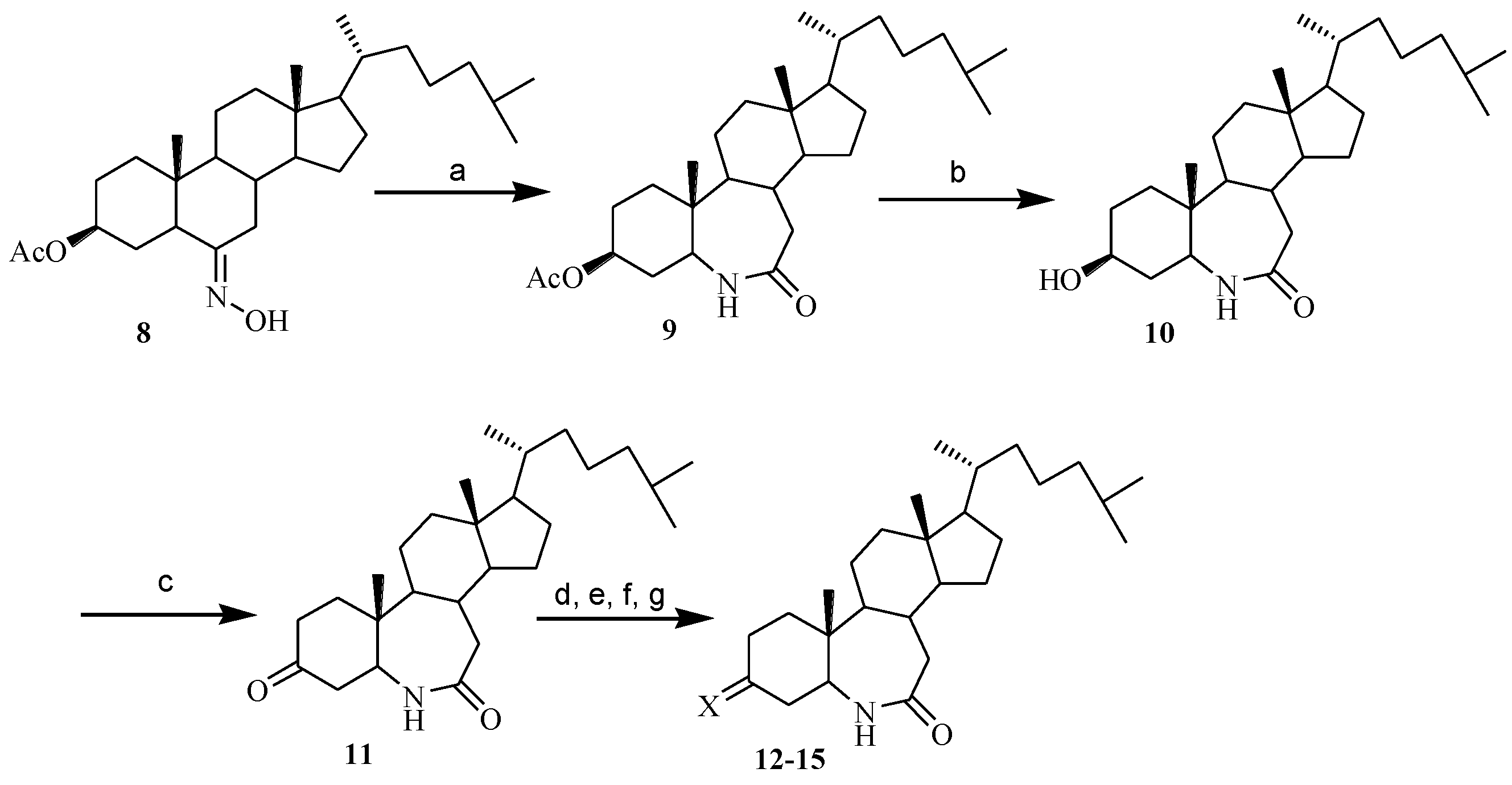
Compounds in Scheme 2 were prepared similarly according to the procedure of Scheme 1.

3.1.5. The Synthesis of 3-Acetoxy-6-aza-B-homocholest-7-one (9)

White solid, yield 93%, mp 222–223 °C; IR(KBr) ν: 3346, 2974, 2945, 2864, 1719, 1662, 1654, 1474, 1364, 1249 cm−1; 1H NMR(CDCl3, 300MHz) δ: 0.68 (3H, s, 18-CH3), 0.86 (3H, s, 19-CH3), 0.87 (6H, d, J = 6.3, 26- and 27-CH3), 0.90 (3H, d, J = 6.3, 21-CH3), 2.05 (3H, s, CH3CO-), 2.36-2.22 (2H, m, C7a-H), 3.47-3.40 (1H, m, C5-H), 4.70-4.60 (1H, m, C3-αH), 5.54 (1H, d, J = 5.1, N-H); 13C NMR (CDCl3, 75MHz) δ: 176.2 (-CONH), 170.5 (COCH3), 70.9 (3-C), 58.7 (14-C), 56.7 (17-C), 56.4 (5-C), 55.6 (9-C), 42.5 (7a-C), 40.2 (13-C), 39.9 (12-C), 39.5 (24-C), 38.7 (20-C), 35.9 (10-C), 35.7 (22-C), 35.3 (1-C), 34.6 (4-C), 34.5 (8-C), 28.0 (16-C), 27.6 (25-C), 27.0 (2-C), 25.6 (23-C), 23.8 (15-C), 23.0 (11-C), 22.8 (27-C), 22.6 (C-26), 21.3 (CH3CO-), 18.6 (21-C), 12.4 (19-C), 11.8 (18-C); HRESI-MS (m/z): 460.3784 [M + H]+ (calcd for C29H50NO3, 460.3791) (Supplementary Information: Figure S5).

3.1.6. The Synthesis of 3-Hydroxy-6-aza-B-homocholest-7-one (10)

White solid, yield: 76%. mp 234–235 °C. IR (KBr) ν: 3321, 2953, 2864, 1650, 1446, 1384, 1053 cm−1; 1H NMR (CDCl3, 600MHz) δ: 0.67 (3H, s, 18-CH3), 0.84 (3H, s, 19-CH3), 0.85 (3H, d, J = 6.6, 26 or 27-CH3), 0.86 (3H, d, J = 6.6, 26 or 27-CH3), 0.88 (3H, d, J = 6.6, 21-CH3), 2.24 (1H, t, J = 13.8, C7a-H), 2.33 (1H, d, J = 13.8, C7a-H), 3.38-3.34 (1H, m, C5-H), 3.61-3.57 (1H, m, C3-H), 5.30 (1H, s, N-H); 13C NMR (CDCl3, 75MHz) δ: 176.3 (7-C), 68.8 (3-C), 58.8 (14-C), 56.9 (17-C), 56.5 (5-C), 55.7 (9-C), 42.5 (7a-C), 40.3 (13-C), 39.9 (12-C), 39.5 (24-C), 38.6 (4-C), 36.0 (10-C), 35.7 (22-C), 35.6 (20-C), 34.6 (1-C), 30.9 (8-C), 28.0 (2-C), 28.0 (25-C), 27.6 (16-C), 25.6 (23-C), 23.8 (15-C), 23.0 (11-C), 22.8 (27-C), 22.6 (26-C), 18.6 (21-C), 12.5 (19-C), 11.8 (18-C); LRESI-MS (m/z): 418.3 [M + H]+(Supplementary Information: Figure S6).

3.1.7. The Synthesis of 6-Aza-B-homocholest-3,7-dione (11)

White solid, yield: 80%. mp 238–240 °C. IR (KBr) ν: 3346, 2966, 2864, 1723, 1658, 1458 cm−1; 1H NMR (CDCl3, 300MHz) δ: 0.72 (3H, s, 18-CH3), 0.88 (6H, d, J = 6.6, 26 and 27-CH3), 0.91 (3H, d, J = 6.6, 21-CH3), 1.07 (3H, s, 19-CH3), 2.52-2.22 (6H, m, C2-, C4- and C7a-H), 3.78-3.66 (1H, m, C5-H), 6.10 (1H, s, N-H); 13C NMR (CDCl3, 75MHz) δ: 207.8 (3-C), 176.4 (7-C), 58.5 (14-C), 58.0 (17-C), 56.5 (9-C), 55.7 (5-C), 43.7 (13-C ), 42.5 (7a-C), 40.2 (4-C), 39.9 (12-C), 39.5 (24-C), 39.0 (10-C), 36.8 (2-C), 36.0 (20-C), 35.9 (22-C), 35.7 (1-C), 34.7 (8-C), 28.0 (16-C), 27.6 (25-C), 25.6 (23-C), 23.8 (15-C), 23.6 (11-C), 22.8 (27-C), 22.5 (26-C), 18.6 (21-C), 12.2 (19-C), 11.8 (18-C); HRESI-MS (m/z): 416.3523 [M + H]+ (calcd for C27H46NO2, 416.3529) (Supplementary Information: Figure S7).

3.1.8. The Synthesis of 3-Hydroximino-6-aza-B-homocholest-7-one (12)

White solid, yield: 85%, mp 248–249 °C. IR (KBr) ν: 3235, 2945, 1650, 1458, 1433, 1380, 1119, 972 cm−1; 1H NMR (CDCl3, 300MHz) δ: 0.70 (3H, s, 18-CH3), 0.84 (3H, s, 19-CH3), 0.85 (3H, d, J = 6.6, 26 or 27-CH3), 0.86 (3H, d, J = 6.6, 26 or 27-CH3), 0.94 (3H, d, J = 6.6, 21-CH3), 3.58-3.44 (1H, m, C5-H), 6.19 (1H, s, N-H); 13C NMR (CDCl3, 75MHz) δ: 176.9 (7-C), 157.5 (3-C), 58.4 (14-C), 57.4 (17-C), 56.5 (5-C), 55.7 (9-C), 42.5 (7a-C), 40.1 (13-C), 39.9 (10-C), 39.5 (12-C), 37.0 (24-C), 36.0 (22-C), 35.7 (20-C), 34.6 (8-C), 29.6 (4-C), 28.0 (16-C), 27.6 (25-C), 27.0 (2-C), 25.6 (1-C), 23.8 (23-C), 23.2 (15-C), 22.8 (26-C), 22.6 (27-C), 22.1 (11-C), 18.6 (21-C), 12.0 (19-C), 11.8 (18-C); HRESI-MS (m/z): 431.3630 [M + H]+ (calcd for C27H47N2O2, 431.3638).

3.1.9. The Synthesis of 3-O-Methyloximino-6-aza-B-homocholest-7-one (13)

Compounds 13 and 14 were prepared similarly according to the procedure of 11, but CH3ONH2·HCl and PhCH2ONH2·HCl were used as reagents instead of NH2OH·HCl.
Compound 13 was a mixture of (E) and (Z)-13 (ratio: E:Z = 0.9:1.1). Yield: 68%, mp 204–205 °C. IR (KBr) ν: 3425, 3222, 1954, 2869, 1673, 1048, 1016 cm1; 1H NMR (CDCl3, 300MHz) δ: 0.66 (3H, s, 18-CH3), 0.83 (3H, d, J = 6.6, 26-CH3 or 27-CH3), 0.84 (3H, d, J = 6.6, 26-CH3 or 27-CH3), 0.86 (3H, d, J = 6.6, 21-CH3), 0.91 (3H, s, 19-CH3), 3.50-3.38 (1H, m, C5-H), 3.01 (Z: 0.55H, br d, J = 14.7, C4-H), 3.31 (E: 0.45H, ddd, J = 14.4, 5.4, 1.5, C2-βH), 3.786 (Z: 3H, s, -OCH3), 3.789 (E: 3H, s, -OCH3), 6.17 (Z: 0.55H, br s, N-H), 6.23 (0.45H, br s, N-H); 13C NMR (CDCl3, 75MHz) δ: 176.6 (7-C), 157.0 (3-C, E-), 156.3 (3-C, Z-), 61.2 (-OCH3), 58.6 (5-C, Z-), 58.4 (14-C), 57.4 (5-C, E-), 56.4 (17-C), 55.6 (9-C), 42.4 (7a-C), 40.2 (13-C), 40.1 (10-C), 39.9 (12-C), 39.4 (24-C), 37.2 (22-C), 36.2 (20-C), 35.9 (4-C, Z-), 35.7(4-C, E-), 34.7 (8-C, Z-), 34.3 (8-C, E-), 28.0 (16-C), 27.7 (2-C, E-), 27.6 (25-C), 27.1 (2-C, Z-), 25.6 (1-C), 23.7 (23-C), 23.1 (15-C), 22.8 (27-C), 22.6 (26-C), 20.8 (11-C), 18.6 (21-C, E-), 12.1 (19-C, Z-), 12.0 (19-C), 11.8 (18-C); HRESI-MS (m/z): 445.3788 [M + H]+ (calcd for C28H49N2O2, 445.3794) (Supplementary Information: Figure S8).

3.1.10. The Synthesis of 3-O-Benzyloximino-6-aza-B-homocholest-7-one (14)

Compound 14 was a mixture of (E) and (Z)-14 (ratio: E:Z = 0.9:1.1). Yield: 63%, mp 195–196 °C. IR(KBr) ν : 3424, 3224, 3088, 3031, 2950, 2868, 1672, 1452, 1366, 1351, 1048, 1015, 734, 697 cm−1; 1H NMR (CDCl3, 300MHz) δ: 0.68 (3H, s, 18-CH3), 0.858 (3H, d, J = 6.6, 26-CH3 or 27-CH3), 0.863 (3H, d, J = 6.6, 26-CH3 or 27-CH3), 0.89 (3H, d, J = 6.6, 21-CH3), 0.93 (3H, s, 19-CH3), 3.13 (0.5H, br d, J = 15.0, C4-H, E-), 3.42-3.36 (0.5H, m, C2-H, Z-), 3.52-3.42 (1H, m, C5-H), 5.05 (2H, s, -OCH2Ph), 5.90 (1H, s, N-H), 7.36-7.30 (5H, m, -C6H5); 13C NMR (CDCl3, 75MHz): 176.4 (7-C), 157.6 (E: 3-C), 156.9 (Z: 3-C), [Z: 137.9, 128.4, 128.4, 128.1, 128.1, 127.8, (-C6H5)], [E: 137.7, 128.4, 128.4, 128.0, 128.0, 127.7, (-C6H5)], 75.5 (-OCH2Ph), 58.5 (Z: 5-C), 58.3 (14-C), 57.4 (E: 5-C), 56.4 (17-C), 55.7 (9-C), 42.5 (7a-C), 42.4 (13-C), 39.9 (10-C), 39.5 (12-C), 37.2 (24-C), 36.2 (22-C), 35.9 (20-C), 35.7 (8-C), 34.7 (Z: 4-C), 34.4 (E: 4-C), 28.0 (16-C), 28.0 (25-C), 27.6 (E: 2-C), 27.1 (Z: 2-C), 25.6 (1-C), 23.8 (22-C), 23.1 (15-C), 22.8 (26-C), 22.6 (27-C), 21.1 (11-C), 18.6 (21-C), 12.1 (E: 19-C), 12.0 (Z: 19-C), 11.8 (18-C); HRESI-MS (m/z): 521.4102 [M + H]+ (calcd for C34H53N2O2, 521.4107) (Supplementary Information: Figure S9).

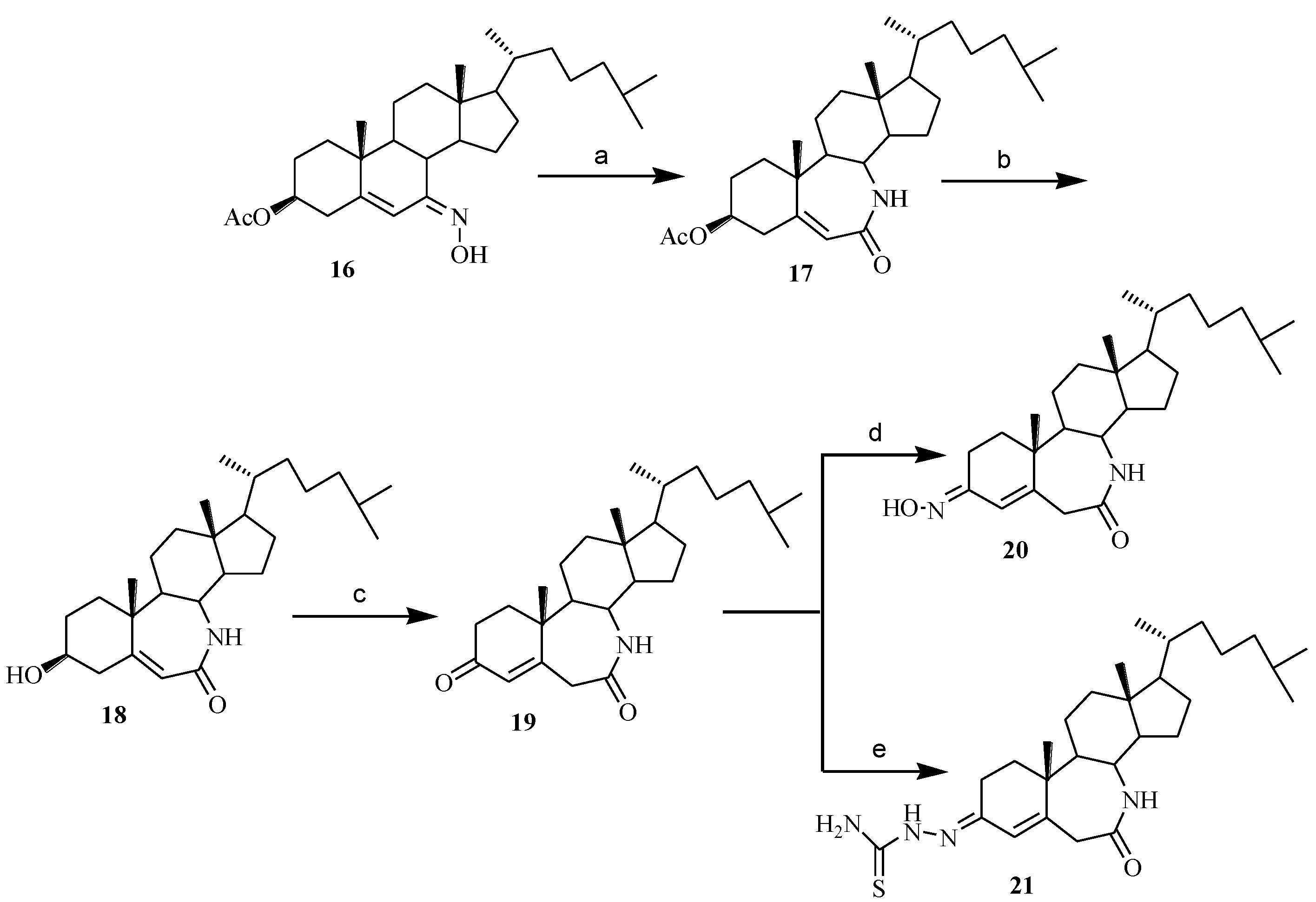
3.1.11. The Synthesis of 6-Aza-7-oxo-B-homocholest-3-thiosemicarbazone (15)

A mixture of compound 11 (100 mg, 0.24 mmol), thiosemicarbazide (24 mg, 0.26 mmol), and a few drops of glacial acetic acid (0.5 mL) in 95% ethanol (20 mL) was stirred at 60–70 °C for 10 h. After completion of the reaction, the majority of solvent was evaporated and some water was added to this solution. The mixture was extracted with CH2Cl2 and the extract was washed with saturated brine, dried with anhydrous sodium sulfate and evaporated under reduce pressure. The resulting residue was chromatographed on a column of silica gel with a mixture of DCM-methanol (20:1) to give 80 mg of compound 15. Yield: 70%, mp 274–276 °C. Compound 15 was a mixture of (E)- and (Z)-15 (ratio: E:Z = 1:1). IR (KBr) ν: 3424, 3237, 2951, 2668, 1658, 1595, 1467, 1386, 1284, 1134, 1078 cm−1; 1H NMR (DMSO, 600 MHz) δ: 0.64 (3H, s, 18-CH3), 0.814 (3H, d, J = 6.6, 26 or 27-CH3), 0.818 (3H, d, J = 6.6, 26 or 27-CH3), 0.84 (1.5H, s, 19-CH3), 0.85 (1.5H, s, 19-CH3), 0.92 (3H, d, J = 6.0, 21-CH3), 2.69 (0.5H, d, J = 12.6, C2-H, E-), 2.81 (1H, br s, C4-H, Z-), 2.97 (0.5H, d, J = 10.8, C4-H, Z-), 3.51 (0.5H, J = 4.8, C5-H, E-), 3.57 (0.5H, J = 4.8, C5-H, Z-), 7.13 (1H, br s, -NH2), 7.29 (0.5H, br s, CON-H-), 7.36 (1H, br s, -NH2), 7.43 (0.5H, br s, CON-H-), 9.57 (0.5H, br s, -NH-, E-), 10.20 (0.5H, br s, -NH-, Z-); 13C NMR (DMSO, 150 MHz) δ: 178.5 (C=S), 177.4 (7-C, Z-), 177.1 (7-C, E-), 156.3 (3-C, Z-), 155.1 (3-C, E-), 59.0 (5-C, Z-), 58.3 (5-C, E-), 58.2 (14-C), 56.5 (17-C), 55.6 (9-C), 42.6 (7a-C), 40.3 (10-C, Z-), 40.2 (10-C, E-), 39.9 (13-C), 39.6 (12-C), 39.4 (24-C), 37.7 (4-C, Z-), 37.2 (4-C, E-), 36.2 (2-C, E-), 36.0 (22-C), 35.8 (20-C), 34.7 (8-C), 30.6 (2-C, Z-), 28.1 (16-C), 27.7 (1-C), 25.8 (25-C), 23.9 (23-C), 23.4 (15-C), 23.3 (11-C), 22.7 (27-C), 22.9 (26-C), 18.7 (21-C), 12.6 (19-C, Z-), 12.4 (19-C, E-), 12.0 (18-C); HRESI-MS (m/z): 489.3620 [M + H]+ (calcd for C28H49N4OS, 489.3627) (Supplementary Information: Figure S10).
Compounds in Scheme 3 were prepared similarly according to the procedure of Scheme 1.

3.1.12. 3-Acetoxy-7a-aza-B-homocholest-5-ene-7-one (17)

Yellow solid, yield: 72%, mp192–194 °C. IR (KBr) ν: 3424, 2942, 2868, 1740, 1659, 1617, 1442 cm−1; 1H NMR (300 MHz, CDCl3) δ: 0.69 (3H, s, 18-CH3), 0.869 (3H, d, J = 6.6, 26 or 27-CH3), 0.874 (3H, s, J = 6.6, 26 or 27-CH3), 0.92 (3H, d, J = 6.6, 21-CH3), 1.27 (3H, s, 19-CH3), 2.05 (3H, s, 2′-CH3), 2.46 (1H, dd, J = 13.2, 3.6, C4-βH), 2.61 (1H, t, J = 11.7, C4-αH), 3.29 (1H, t, J = 8.4, C8-H), 4.76-4.65 (1H, m, C3-αH), 5.66 (1H, br s, -NH-), 5.83 (1H, s, C6-H); 13C NMR (75 MHz, CDCl3) δ: 170.3 (1′-C), 167.4 (7-C), 156.1 (5-C), 122.5 (6-C), 72.5 (3-C), 55.7 (14-C), 55.4 (17-C), 51.7 (8-C), 48.9 (9-C), 43.8 (24-C), 41.8 (12-C), 41.6 (13-C), 39.4 (15-C), 38.6 (10-C), 35.9 (4-C), 35.5 (1-C), 35.4 (22-C), 28.0 (20-C), 27.8 (25-C), 27.0 (2-C), 25.1 (16-C), 23.8 (23-C), 23.2 (11-C), 22.8 (26-C), 22.5 (27-C), 21.3 (2′-C), 19.3 (21-C), 18.6 (19-C), 11.4 (18-C); HRESI-MS (m/z): 458.3623 [M + H]+ (calcd for C29H48NO3, 458.3634) (Supplementary Information: Figure S11).

3.1.13. 3-Hydroxy-7a-aza-B-homocholest-5-ene-7-one (18)

Yellow solid, yield: 74.5%, mp 207–209 °C; IR (KBr) ν: 3383, 2946, 2860, 1663, 1601, 1438 cm−1; 1H NMR (300 MHz, CDCl3) δ: 0.65 (3H, s, 18-CH3), 0.837 (3H, d, J = 6.6, 26 or 27-CH3), 0.842 (3H, s, J = 6.6, 26 or 27-CH3), 0.89 (3H, d, J = 6.6, 21-CH3), 1.24 (3H, s, 19-CH3), 2.40 (1H, dd, J = 13.2, 3.3, C4-βH), 2.53 (1H, t, J = 11.7, 3.6, C4-αH), 3.25 (1H, t, J = 8.1, C8-H), 3.65-3.54 (1H, m, C3-αH), 5.77 (1H, s, C6-H), 6.01 (1H, br s, -NH-); 13C NMR (75 MHz, CDCl3) δ: 168.2 (7-C), 158.6 (5-C), 121.0 (6-C), 70.6 (3-C), 55.6 (14-C), 55.1 (17-C), 51.6 (8-C), 49.1 (9-C), 46.3 (13-C), 44.2 (4-C), 41.7 (24-C), 39.4 (12-C), 38.5 (15-C), 36.0 (1-C), 35.9 (22-C), 35.5 (20-C), 30.5 (10-C), 28.0 (2-C), 27.8 (25-C), 25.1 (16-C), 23.8 (23-C), 23.1 (11-C), 22.8 (26-C), 22.5 (27-C), 19.4 (21-C), 18.6 (19-C), 11.3 (18-C); HRESI-MS (m/z): 416.3515 [M + H]+ (calcd for C27H46NO2, 416.3529) (Supplementary Information: Figure S12).

3.1.14. 7a-Aza-B-homocholest-4-ene-3,7-dione (19)

Faint yellow solid, yield: 43.0%, mp 203–205 °C; IR (KBr) ν: 3367, 2958, 2860, 1675, 1462 cm−1; 1H NMR (300 MHz, CDCl3) δ: 0.72 (3H, s, 18-CH3), 0.845 (3H, d, J = 6.6, 26 or 27-CH3), 0.850 (3H, s, J = 6.6, 26 or 27-CH3), 0.90 (3H, d, J = 6.6, 21-CH3), 1.27 (3H, s, 19-CH3), 2.37-2.28 (2H, m, C2-H), 2.94 (1H, d, J = 12.6, C6-βH), 3.55 (1H, d, J = 12.6, C6-αH), 3.52-3.46 (1H, m, C8-H), 5.82 (1H, br s, -CONH-), 5.93 (1H, s, C4-H); 13C NMR (75 MHz, CDCl3) δ: 198.4 (3-C), 172.5 (7-C), 162.8 (5-C), 126.9 (4-C), 55.8 (17-C), 54.0 (14-C), 52.4 (8-C), 49.2 (9-C), 42.7 (6-C), 41.6 (13-C), 41.3 (24-C), 39.4 (12-C), 38.2 (10-C), 35.9 (15-C), 35.5 (1-C), 34.1 (22-C), 33.4 (20-C), 28.0 (2-C), 27.9 (25-C), 27.7 (16-C), 24.8 (23-C), 23.8 (11-C), 22.8 (26-C), 22.6 (27-C), 21.2 (21-C), 18.6 (19-C), 11.7 (18-C); HRESI-MS (m/z): 414.3362 [M + H]+ (calcd for C28H47N4OS, 414.3372) (Supplementary Information: Figure S13).

3.1.15. 3-Hydroximino-7a-aza-B-homocholest-4-ene-7-one (20)

Faint yellow solid, yield: 77.3%, mp 204–206 °C. IR (KBr) ν: 3268, 2949, 2868, 1658, 1657, 1462, 1366 cm−1; 1H NMR (CDCl3, 300MHz) δ: 0.73 (3H, s, 18-CH3), 0.88 (6H, d, J = 6.6, 26- and 27-CH3), 0.93 (3H, d, J = 6.6, 21-CH3), 1.22 (3H, s, 19-CH3), 2.33-2.22 (2H, m, C2-H), 2.79 (1H, br d, J = 17.4, C2-H), 2.94 (1H, d, J = 13.5, C6-βH), 3.47 (1H, d, J = 13.5, C6-αH), 3.52-3.45 (1H, m, C8-H), 5.77 (1H, br s, -CONH-), 6.12 (1H, s, C4-H), 6.84 (1H, s, -OH); 13C NMR (CDCl3, 75MHz) δ: 174.3 (7-C), 156.1 (3-C), 147.5 (5-C), 121.8 (4-C), 55.9 (14-C), 54.4 (17-C), 52.5 (8-C), 50.0 (13-C), 49.1 (9-C), 42.8 (6-C), 41.0 (24-C), 40.6 (10-C), 39.4 (12-C), 38.5 (15-C), 35.9 (22-C), 35.5 (20-C), 33.0 (1-C), 29.7 (25-C), 28.0 (2-C), 27.7 (16-C), 24.9 (23-C), 23.8 (11-C), 22.8 (26-C), 22.5 (27-C), 21.0 (19-C), 18.6 (21-C), 11.7 (18-C); HRESI-MS (m/z): 429.3463 [M + H]+ (calcd for C27H45N2O2, 429.3481).

3.1.16. 7-Oxo-7a-aza-B-homocholest-4-ene-3-thiosemicarbazone (21)

Faint yellow solid, yield: 31%, mp 230-231 °C; IR(KBr) ν: 3436, 2954, 2856, 1659, 1573, 1409 cm−1; 1 H NMR (CDCl3, 300MHz) δ: 0.75 (3H, s, 18-CH3), 0.88 (3H, d, J = 6.6, 26 or 27-CH3), 0.89 (3H, d, J = 6.6, 26 or 27-CH3), 0.94 (3H, d, J = 6.3, 21-CH3), 1.26 (3H, s, 19-CH3), 2.97 (1H, d, J = 13.2, C6-βH), 3.51 (1H, d, J = 13.2, C6-αH), 3.56-3.48 (1H, m, C8-H), 5.28 (1H, br s, -CONH-), 6.10 (1H, s, C4-H), 6.37 (1H, br s, -NH2), 7.25 (1H, br s, -NH2), 8.64 (1H, s, -NH-CS); 13C NMR (CDCl3, 75MHz) δ: 178.7 (C=S), 173.6 (7-C), 150.5 (5-C), 148.9 (3-C), 124.2 (4-C), 55.9 (14-C), 54.3 (17-C), 52.5 (8-C), 49.2 (9-C), 42.8 (6-C), 41.1 (13-C), 40.5 (24-C), 39.4 (12-C), 38.4 (15-C), 35.9 (22-C), 35.5 (20-C), 32.8 (10-C), 29.7 (1-C), 28.0 (25-C), 24.9 (16-C), 23.8 (23-C), 22.8 (26-C), 22.5 (27-C), 22.4 (2-C), 21.0 (11-C), 20.0 (21-C), 18.6 (19-C), 11.7 (18-C); HRESI-MS (m/z): 487.3491 [M + H]+ (calcd for C28H47N4OS, 487.3471) (Supplementary Information: Figure S14).

3.2. Biology

3.2.1. Assay for Cell Viability

The cell proliferation assay was undertaken by a MTT method using 96-well plates on a MLLTISKAN MK3 analysis spectrometer. GNE 2 (nasopharyngeal carcinoma), SPC-A (lung carcinoma) and Tu 686 (laryngic carcinoma) cell lines were obtained by Guangxi Medical University (Guangxi, China); MGC 7901 (human gastric carcinoma) and HeLa (human cervical carcinoma) cell lines were obtained by Guangxi Traditional Chinese Medical University. Cells were grown in RPMI-1640 supplemented with 10% cosmic calf serum (Hyclone) and antibiotics in a humidified atmosphere of 5% CO2 at 37 °C. The viability of these cells was determined using the colorimetric CellTiter 96 aqueous Cell Proliferation Assay (MTT) according to the instruction provided by the manufacturer (Promega, Madison, WI, USA). Briefly, cells (1–3 × 10⁴ cells per well) were seeded in 96 wells plates. One day after seeding, the cells were treated with or without different concentration of each compound and reincubated for 72 h. After the cells were washed with sterile phosphate buffer saline (PBS), 190 µL of RPMI-1640 and 10 µL of the tetrazolium dye (MTT) (5 mg/mL) solution were added to each well, and the cells were incubated for an additional 4 h. The medium was discarded; 200 µL of DMSO was added to dissolve the purple formazan crystals formed. The absorbance (A) at 492 nm was measured using a MLLTISKAN MK3 analysis spectrometer. The IC50 value was calculated in µmol/L in Table 1 as the concentration of drug yielding 50% cell survival. The effect of compound 5 on the morphology of treated human carcinoma cells was investigated by the light microscope and then photographed by TE2000-U Nikon (Tokyo, Honshu, Japan) inverted microscope.

3.2.2. 3D Multicellular Spheroids

H292 (lung adenocarcinoma) cancer cells were obtained from ATCC, Manassas, VA. C. Monolayer cultures were incubated in RPMI-1640 supplemented with 10% fetal bovine serum, 100 units/mL penicillin. The cell cultures were kept at 37 °C in a humidified 5% CO2 and 95% air incubator. Uniform single-spheroid H292 lung carcinoma cells were cultured as follows. The 96-well flat-bottom plates were coated with 70 μL of a 1.5% agarose (weight/volume) solution in distilled water (freshly autoclaved). During the coating process, the agarose solution was maintained at ≥60 °C followed by cooling and setting at room temperature for 40 min. Then the cells were plated at a density of 5000 cells/well in 80 μL of RPMI-1640 (10% FBS), and allowed to form spheroid in 48 h. The spheroids were then treated with 20 μL of a 25% solution of growth factor–reduced Matrigel in cell culture medium, resulting in a final volume of 100 μL with 5% Matrigel. Spheroids were cultured for one more day to reach an average diameter of 100 μm under standard tissue culture conditions (37 °C, 5% CO2). For drug treatment, 100 μL fresh medium with various concentration of drugs were added at day 3 (final matrigel concentration became 2.5% as well). Spheroid morphological images in 96-well microplate were carried out manually on an inverted microscope equipped with camera. Spheroid diameters and volumes were determined from their images. The treatment was quart replicated, and the spheroid images were taken every other day. The suppression of the spheroid growth was normalized with control treatment (0.1% DMSO) [21,22].

3.2.3. Annexin V Staining Assay

Apoptosis was detected with an annexin V-FITC kit purchased from BD Pharmingen (San Diego, CA, USA) according to the manufacturer’s instructions. SGC-7901 cells were seeded in 35 mm culture dishes and allowed to attach overnight. The cells were treated with different concentration of compound 5 for 24 h respectively, collected, and washed twice with PBS. To detect early and late apoptosis, both adherent and floating cells were harvested together and resuspended in annexin V binding buffer at a concentration of 106 cells/mL. Subsequently, 5 μL of FITC-conjugated annexin V and 5 μL of propidium iodide were added to 100 μL of the cell suspension (105 cells). The cells were incubated for 15 min at room temperature in the dark. Finally, 400 μL of annexin V binding buffer was added to each tube, and cells were analyzed by a two color cytometry using FACS Calibur (Becton Dickinson, Biosciences, Franklin Lakes, NJ, USA).

4. Conclusions

We have prepared a series of aza-B-homocholestane derivatives having different substituted groups at 3-postion of the steroidal nucleus, taking analogues of marine steroidal oximes as precursors. The antiproliferative activity of the synthesized compounds against SGC-7901, HeLa, Bel-7404, GNE 2, SPC-A and Tu 686 cell lines was investigated. The results showed that aza-B-homocholestane derivatives possessing 3-hydroxyl, 3-hydroximino and 3-thiosemicarbazone groups showed remarkable cytotoxic activity. In the synthesized compounds, compounds 5, 10, 12, 15 and 18 were found to be the most potent compounds as anticancer agents, and they displayed a similar antiproliferative activity as cisplatin did. The result of 3D multicellular spheroids screening of 15 showed also distinct antiproliferative activity, and Annexin V staining assay indicated that compound 5 was able to effectively induce tumor cells apoptosis. Compounds 5 and 15 are now submitted to further acute toxicity and antitumor activity studies in animal models, and the relative possible results will be reported in due course. Our findings provide new evidence showing the relationship between the chemical structure and biological activities, and may be useful for the design of novel chemotherapeutic drugs.

Conflicts of Interest

The authors declare no conflict of interest.

Acknowledgments

The authors acknowledge the financial support of the Natural Science Foundation of Guangxi Province (No: 2010GXNSFD013019) , Natural Science Fund of Education Department of Guangxi province (No. 201202ZD059) and The Training Project for Excellent Middle-aged/Young Teachers in Guangxi Higher Education Institutions.

Author Contributions

J. G. Cui was responsible for the natural science foundation of guangxi province (No: 2010GXNSFD013019), and conceived the design of synthetic route and was responsible for the guidance of all synthetic experiments. Y. H. Huang, C. J. Chen and Q. F. Lin synthetized Aza-B-homocholestane derivatives. H. C. Song was responsible for the guidance of partial synthetic experiments. Y. H. Huang screened cell lines in vitro. Y. H. Huang and C. F. Gan performed the apoptotic experiment. B. Su, A. M. Zhou and Y. H. Huang screened the 3D Multicellular Spheroids.

References

  1. Blunt, J.W.; Copp, B.R.; Munro, M.H.G.; Northcote, P.T.; Prinsep, M.R. Marine natural products. Nat. Prod. Rep. 2011, 28, 196–268. [Google Scholar] [CrossRef]
  2. Rodríguez, J.; Nunez, L.; Pexinho, S.; Jiménez, C. Isolation and synthesis of the first natural 6-hydroximino-4-en-3-one steroids from the sponges Cinachyrella spp. Tetrahedron Lett. 1997, 38, 1833–1836. [Google Scholar] [CrossRef]
  3. Deive, N.; Rodríguez, J.; Jiménez, C. Synthesis of cytotoxic 6E-hydroximino-4-ene steroids: Structure/activity studies. J. Med. Chem. 2001, 44, 2612–2618. [Google Scholar] [CrossRef]
  4. Rega, M.; Jiménez, C.; Rodríguez, J. 6E-Hydroximinosteroid homodimerization by cross-metathesis processes. Steroids 2007, 72, 729–735. [Google Scholar] [CrossRef]
  5. Bazin, M.A.; Loiseau, P.M.; Bories, C.; Letourneux, Y.; Rault, S.; Kihel, L.E. Synthesis of oxysterols and nitrogenous sterols with antileishmanial and trypanocidal activities. Eur. J. Med. Chem. 2006, 41, 1109–1116. [Google Scholar] [CrossRef]
  6. Poza, J.; Rega, M.; Paz, V.; Alonso, B.; Rodríguez, J.; Salvador, N.; Antonio, F.; Carlos, J. Synthesis and evaluation of new 6-hydroximinosteroid analogs as cytotoxic agents. Bioorg. Med. Chem. 2007, 15, 4722–4740. [Google Scholar] [CrossRef]
  7. Cui, J.G.; Fan, L.; Huang, L.L.; Liu, H.L.; Zhou, A.M. Synthesis and evaluation of some steroidal oximes as cytotoxic agents: Structure/activity studies (I). Steroids 2009, 74, 62–72. [Google Scholar] [CrossRef]
  8. Cui, J.G.; Fan, L.; Huang, Y.M.; Xin, Y.; Zhou, A.M. Synthesis and evaluation of some steroidal oximes as cytotoxic agents: Structure/Activity Studies (II). Steroids 2009, 74, 989–995. [Google Scholar] [CrossRef]
  9. Ibrahim-Ouali, M.; Rocheblave, L. Recent advances in azasteroids chemistry. Steroids 2008, 73, 375–407. [Google Scholar] [CrossRef]
  10. Krstić, N.M.; Bjelaković, M.S.; Žižak, Ž.; Pavlović, M.D.; Juranić, Z.D.; Pavlović, V.D. Synthesis of some steroidal oximes, lactams, thiolactams and their antitumor activities. Steroids 2007, 72, 406–414. [Google Scholar] [CrossRef]
  11. Anna, I.K.; Manolis, A.; Evagelia, S.A.; Athanasios, P.; George, N.P.; Sotiris, S.N. Rational design, synthesis, and in vivo evaluation of the antileukemic activity of six new alkylating steroidal esters. Bioorg. Med. Chem. 2008, 16, 5207–5215. [Google Scholar] [CrossRef]
  12. Vatmurge, N.S.; Hazra, B.G.; Pore, V.S. Synthesis and antimicrobial activity of β-lactam–bile acid conjugates linked via triazole. Bioorg. Med. Chem. Lett. 2008, 18, 2043–2047. [Google Scholar] [CrossRef]
  13. Mohamed, E.F.; Gamal, A.E.; Emad, F.E.; Hanaa, M.R.; Mohamed, A.T. Novel modified steroid derivatives of androstanolone as chemotherapeutic anti-cancer agents. Eur. J. Med. Chem. 2009, 44, 3936–3946. [Google Scholar] [CrossRef]
  14. Dhingraa, N.; Bhardwaj, T.R.; Mehta, N.; Mukhopadhyay, T.; Kumar, A.; Kumar, M. Synthesis, antiproliferative, acute toxicity and assessment of antiandrogenic activities of some newly synthesized steroidal lactams. Eur. J. Med. Chem. 2010, 45, 2229–2236. [Google Scholar] [CrossRef]
  15. Catsoulacos, P.; Politis, D.; Wampler, G.L. Antitumor activity of homo aza-steroidal ester of [p-bis(2-chloroethyl)amino]phenyl acetic acid and [p-bis(2-chloroethyl)amino]phenyl butyric acid. Cancer Chemother. Pharmacol. 1983, 10, 129–132. [Google Scholar] [CrossRef]
  16. Catsoulacos, P.; Catsoulacos, D. Antitumor activity of homo-aza-steroidal esters of p-N,N-bis(2-chloroetheryl)amino phenoxy acetic acid. Anticancer Res. 1993, 13, 1203–1208. [Google Scholar]
  17. Huang, Y.M.; Chen, S.J.; Cui, J.G.; Gan, C.F.; Liu, Z.P.; Wei, Y.L.; Song, H.C. Synthesis and cytotoxicity of A-homo-lactam derivatives of cholic acid and 7-deoxycholic acid. Steroids 2011, 76, 690–694. [Google Scholar] [CrossRef]
  18. Huang, Y.M.; Cui, J.G.; Chen, S.J.; Gan, C.F.; Zhou, A.M. Synthesis and antiproliferative activity of some steroidal lactams. Steroids 2011, 76, 1346–1350. [Google Scholar] [CrossRef]
  19. Huang, Y.M.; Cui, J.G.; Zeng, Q.X.; Zeng, C.; Chen, Q.; Zhou, A.M. 6-hydroximino-4-aza-A-homo-cholest-3-one and related analogue as a potent inducer of apoptosis in cancer cells. Steroids 2012, 77, 829–834. [Google Scholar] [CrossRef]
  20. Tung, Y.C.; Hsiao, A.Y.; Allen, S.G.; Torisawa, Y.S.; Ho, M.; Takayama, S. High-throughput 3D spheroid culture and drug testing using a 384 hanging drop array. Analyst 2011, 136, 473–478. [Google Scholar] [CrossRef]
  21. Yuhas, J.M.; Li, A.P.; Martinez, A.O.; Landman, A.J. A simplified method for production and growth of multicellular tumor spheroids. Cancer Res. 1977, 37, 3639–3643. [Google Scholar]
  22. Lama, R.; Zhang, L.; Naim, J.M.; Williams, J.; Zhou, A.; Su, B. Development, validation and pilot screening of an in vitro multi-cellular three-dimensional cancer spheroid assay for anti-cancer drug testing. Bioorg. Med. Chem. 2013, 21, 922–931. [Google Scholar] [CrossRef]

