Article

Nocapyrones: α- and γ-Pyrones from a Marine-Derived Nocardiopsis sp.

1
Biotechnology Research Center and Department of Biotechnology, Toyama Prefectural University, 5180 Kurokawa, Imizu, Toyama 939-0398, Japan
2
Shokei Gakuin University, 4-10-1 Yurigaoka, Natori, Miyagi 981-1295, Japan
3
School of Nutrition and Dietetics, Kanagawa University of Human Services, 1-10-1 Heisei-cho, Yokosuka, Kanagawa 238-8566, Japan
4
Okayama University of Science, 1-1 Ridai-cho, Okayama, Okayama 700-0005, Japan
5
Tokyo University of Marine Science and Technology, 4-5-7 Konan, Minato-ku, Tokyo 108-8477, Japan
6
Center for Advanced Marine Core Research, Kochi University, B200 Monobe, Nankoku, Kochi 783-8502, Japan
*
Author to whom correspondence should be addressed.
Mar. Drugs 2014, 12(7), 4110-4125; https://doi.org/10.3390/md12074110
Submission received: 5 May 2014 / Revised: 4 June 2014 / Accepted: 20 June 2014 / Published: 8 July 2014
(This article belongs to the Special Issue Advances and New Perspectives in Marine Biotechnology)

Abstract

:
One new α-pyrone (nocapyrone R (1)), and three known γ-pyrones (nocapyrones B, H and L (24)) were isolated from the culture extract of a Nocardiopsis strain collected from marine sediment. Structures of these compounds were determined on the basis of spectroscopic data including NMR and MS. γ-Pyrones 24 were found to induce adiponectin production in murine ST-13 preadipocyte cells but the α-pyrone 1 had no activity. The absolute configuration of the anteiso-methyl branching in 4 was determined by HPLC comparison of a degraded product of 4 with standard samples as a 2:3 enantiomeric mixture of (R)- and (S)-isomers.

Graphical Abstract

1. Introduction

Natural products have been playing an important role for the development of novel therapeutics owing to their enormous and unpredictable structural diversity [1]. Specifically, actinomycetes are continuously providing structurally diverse secondary metabolites possessing pharmaceutically useful bioactivities. More than 70% of microbial antibiotics have been discovered from actinomycetes [2]. In recent years, in addition to the terrestrial species, actinomycetes from marine environments are attracting a substantial attention as a new resource of novel drug candidates [3]. The differences between the marine and terrestrial environments are likely reflected in the genetic divergences. Marine-derived actinomycetes are recognized to actually produce a range of chemically distinctive secondary metabolites that terrestrial actinomycetes are not able to produce [4]. Consequently, the number of novel metabolites from marine actinomycetes has been increasing in recent years.
The increasing incidents of type 2 insulin-resistant diabetes, as a result of growing obesity rates, are causing a serious social and economic problem [5]. Adipose tissue secretes various types of biologically active adipokines, such as free fatty acids, tumor necrosis factor-α, adiposin, resistin, and leptin, to regulate energy homeostasis [6]. Adiponectin is one of such adipokines secreted exclusively by mature adipocytes [7] and this proteineous substance functions to regulate the glucose and lipid metabolism [8]. Recent research revealed that the mRNA expression of adiponectin is reduced in obese diabetic murine model [9] and type 2 diabetic patients [10,11,12], and also plasma level of adiponcetin is significantly lower in obese diabetic mice and humans [13,14,15]. Therefore, the replenishment of adiponectin by transcriptional induction in adipocytes is believed to provide a new effective therapeutic approach to insulin resistance, type 2 diabetes, and related diseases. Thiazolidinediones represented by rosiglitazone (Avandia) and pioglitazone (Actos) are widely-used orally available drugs for type 2 diabetes. These synthetic compounds activate transcription by PPARγ (peroxisome proliferator activated receptor γ) primarily in adipose tissues and induce the elevation of adiponectin plasma level. Meanwhile, several research groups have demonstrated that serious side effects are caused by rosiglitazone; the use of this drug is associated with a 43% increase in myocardial infraction and 64% increase in the risk of death from cardiovascular causes [16]. Thus, the development of side effect-free new agents for type 2 diabetes is desperately desired. In our screening program for discovering new lead scaffolds for adiponectin inducers from natural products [17,18,19], a marine-derived Nocardiopsis strain was found to produce a new α-pyrone (1) along with three known γ-pyrones, nocapyrones B, H, and L (24, Figure 1). Interestingly, nocapyrone L (4) was isolated as an enantiomeric mixture, which is noticeable in marine natural products. Herein, we describe isolation, structure determination, and biological activities of these compounds.
Figure 1. Structures of nocapyrones R (1), B (2), H (3), and L (4).
Marinedrugs 12 04110 g001

2. Results and Discussion

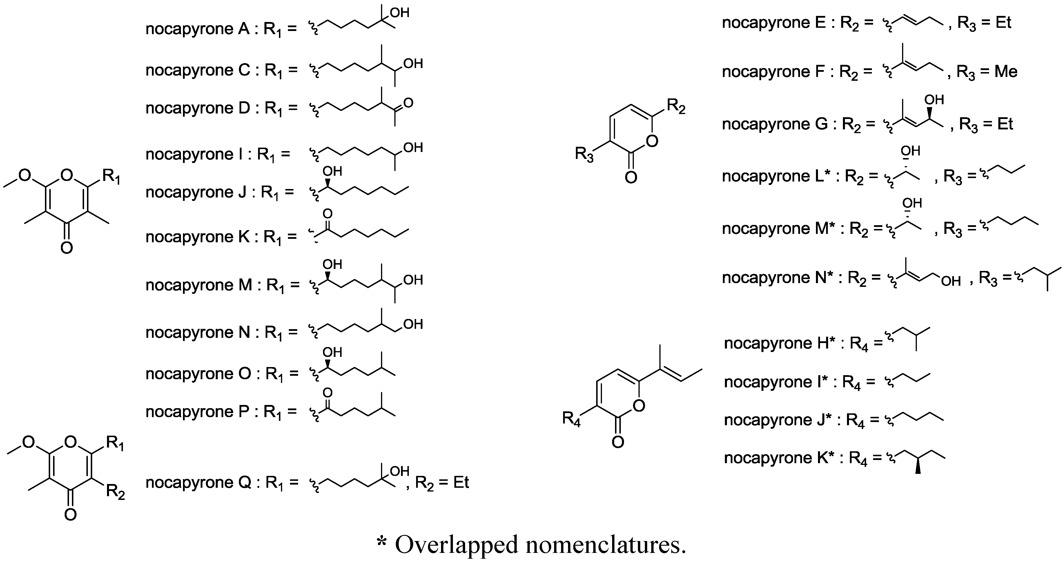
2.1. Structure Analysis and Characterization

2.1.1. Isolation

The producing strain TP-A0876 was isolated from a sediment sample collected at −775 m in Ishikari gulf, Hokkaido, Japan. On the basis of the result of 16S rRNA gene sequence similarity, the isolate was identified as Nocardiopsis sp. The strain TP-A0876 was cultured in A-3M medium at 30 °C for 6 days and the whole culture broth was extracted with 1-butanol. The crude extract was subjected to silica gel and ODS column chromatographies, followed by HPLC purification, to yield nocapyrone R (1) along with nocapyrones B, H, and L (24), three known compounds previously isolated from marine Nocardiopsis strains by other groups [20,21].

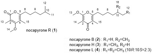
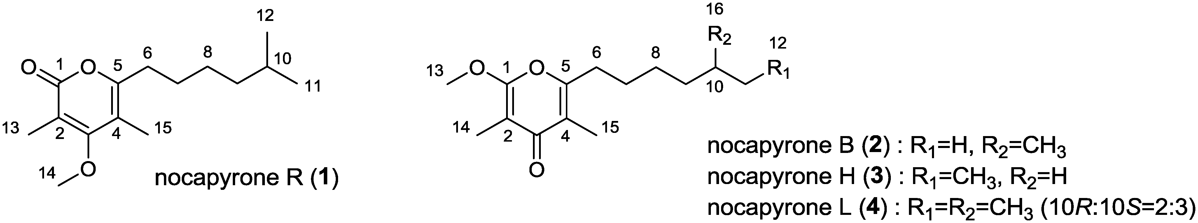
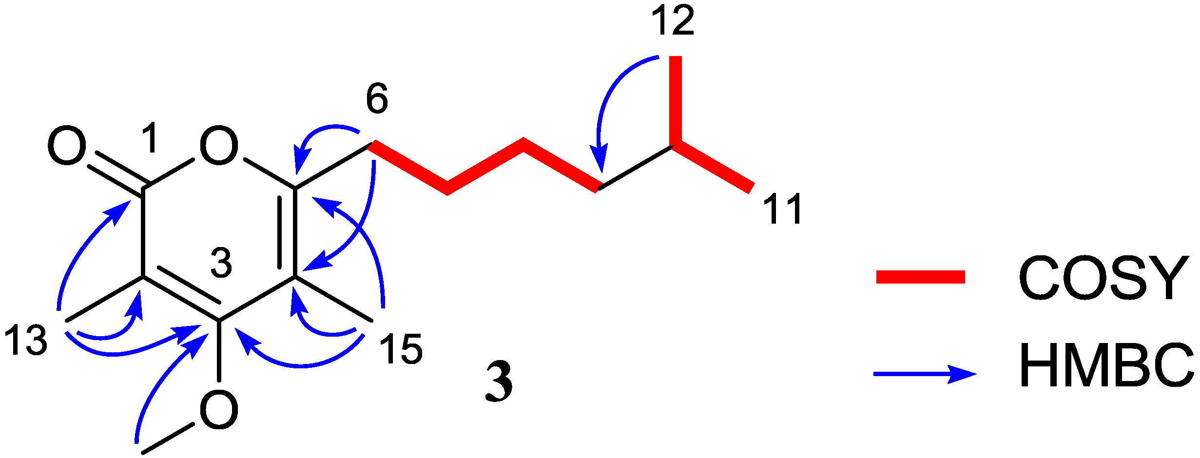
2.1.2. Nocapyrone R (1)

Nocapyrone R (1) was isolated as colorless amorphous. The molecular formula of 1 was determined as C15H24O3 by high resolution ESITOFMS (Electrospray Ionization Time Of Flight Mass Spectrometry) analysis that showed a pseudomolecular ion at m/z 275.1619 [M + Na]+ (calcd for C15H24O3Na, 275.1623), which was corroborated by the 1H and 13C NMR data. The IR spectrum indicated the presence of alkene (2944 cm−1) and carbonyl (1660 cm−1) groups. 1H and 13C NMR data in combination with the HSQC (Heteronuclear Single Quantum Coherence) analysis revealed the presence of 15 carbons assignable to one carbonyl carbon, four sp2 quaternary carbons, four sp3 methylenes, one sp3 methine, and five methyl groups (one is oxygenated) (Table 1, Supplementary Figures S1, S2 and S4). The NMR spectra of nocapyrone R (1) showed the resonances for a pyrone unit as well as two methyl (C-13 and C-15) and one methoxy (C-14) substituents (Table 1, Supplementary Figures S1 and S2): however, the UV spectrum showing absorption maximum at λmax 290 nm was different from that for γ-pyrone (24: UV λmax 252 nm). Furthermore, the ester carbonyl absorption of IR spectrum and the ester carbonyl signal of 13C NMR suggested the presence of an ester/lactone functionality. Protons of two olefinic methyl groups (H3-13 and H3-15) had HMBC correlations with C-3, to which was correlated the methoxy protons (H3-14), establishing the α-pyrone substructure with a methoxy group at C-3 flanked by two olefinic methyl groups (Supplementary Figure S5). The 1H-1H COSY spectrum provided two fragments, H2-6/H2-7/H2-8/H2-9 and H3-11/H-10/H3-12. These two fragments were assembled into an alkyl chain bearing an isopropyl terminus by HMBC correlations from H3-12 to C-9, C-10, and C-11. This was consistent with the presence of two doublet methyl proton signals for H3-11 and H3-12 (Supplementary Figures S3 and S5). The alkyl side chain was attached to C-5 by HMBC correlations from H2-6 to C-4 and C-5, completing the structure of 1 (Figure 2).
Table 1. 1H and 13C NMR data for nocapyrone R (1) in CDCl3.
PositionδC aδH mult (J in Hz) bCOSY bHMBC b,c
1166.3, qC
2109.3, qC
3168.4, qC
4109.0, qC
5159.3, qC
631.1, CH22.49, t (7.8)74, 5, 7
727.7, CH21.61, m6, 85
827.1, CH21.31, m7, 910
938.7, CH21.18, m87, 11, 12
1027.9, CH1.52, m 11, 12
1122.6, CH30.86, d (6.5)10, 129, 10, 12
1222.6, CH30.86, d (6.5)10, 119, 10, 11
1310.2, CH32.03, s 1, 2, 3
1460.2, CH33.80, s 3
1510.1, CH31.92, s 3, 4, 5
a recorded at 100 MHz; b recorded at 500 MHz; c HMBC correlations are from proton(s) stated to the indicated carbon.
Figure 2. 2D-NMR correlations for nocapyrone R (1).
Marinedrugs 12 04110 g002

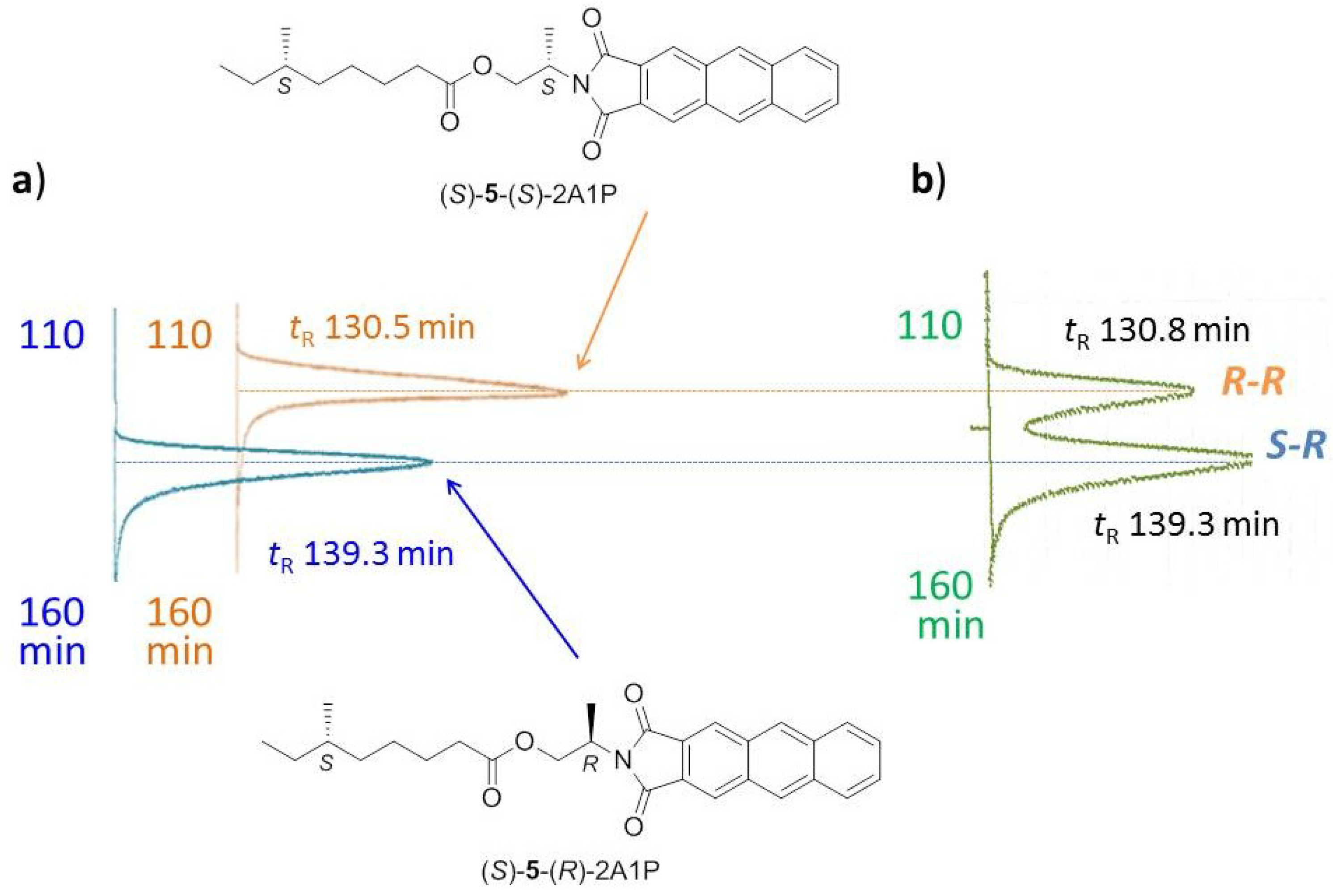
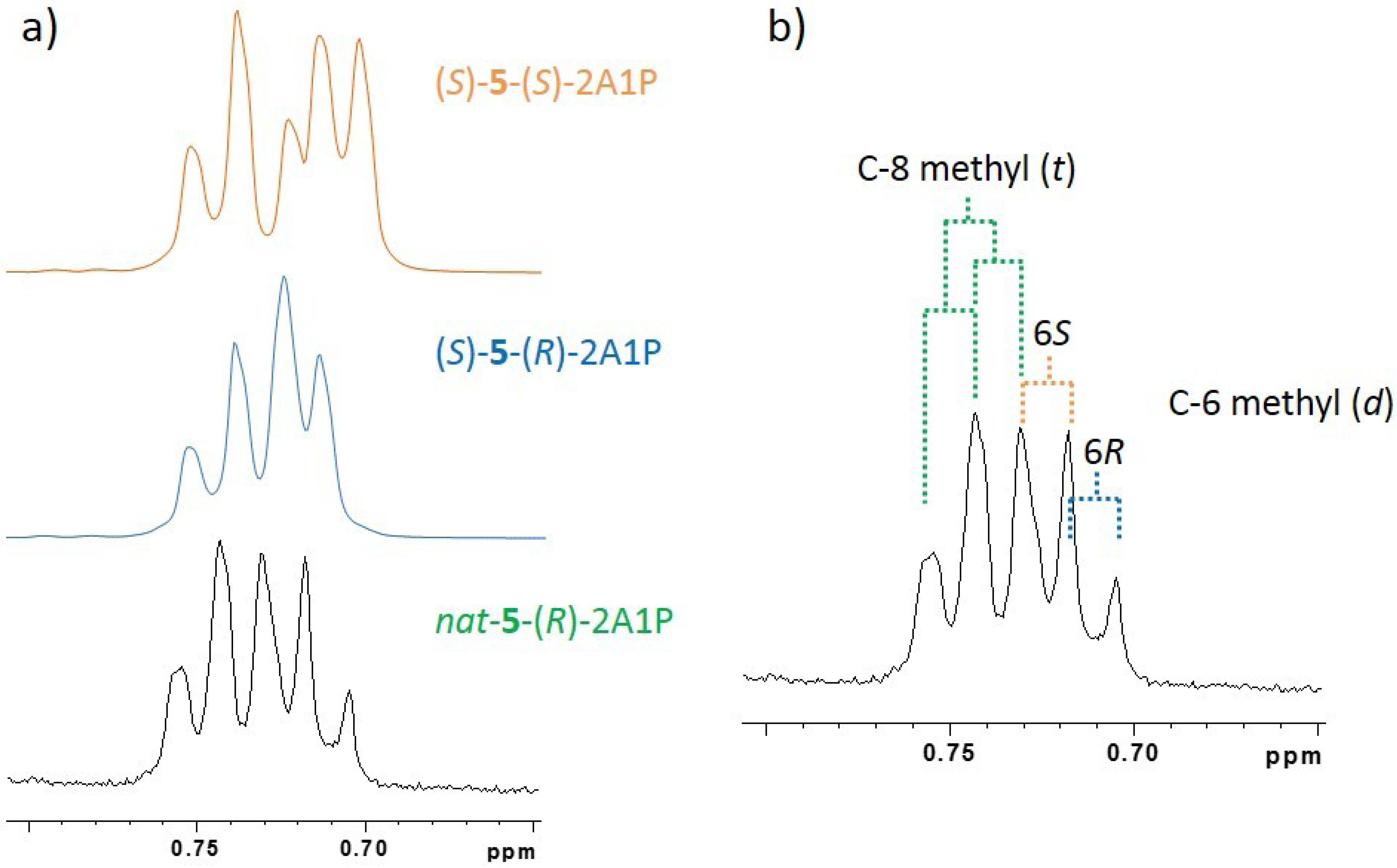
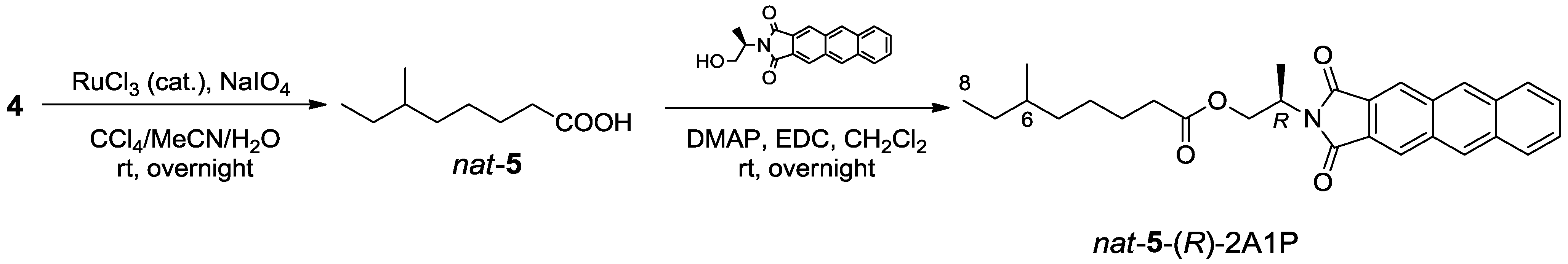
2.1.3. Absolute Configuration of Nocapyrone L (4)

Nocapyrone L (4) has been reported as a secondary metabolite of Nocadiopsis sp. isolated from a mollusk as a symbiotic bacterium [21]. Due to the remoteness of the branched methyl chiral center (C-10) from the modifiable functional groups, the absolute configuration of 4 had not been assigned. From the structural analogy of 4 to germicidin A, we hypothesized that 4 could be biosynthesized through condensation of an amino acid-derived starter with malonate extenders [22]. Consequently, the anteiso-subunit known to be originated from l-isoleucine is assumed to have S configuration. Interestingly, the specific rotation of 4 we isolated showed [α]D25 = −2.2 (c = 0.1, CHCl3), while that of reported one is [α]D25 = +9.0 (c = 0.1, CHCl3) [21]. In order to clarify the absolute configuration of 4, Ohrui-Akasaka method was applied (Scheme 1) [23]. The pyrone unit of 4 was oxidatively degraded by the treatment with a catalytic amount of ruthenium (III) chloride and sodium periodate in a biphasic solvent system (CCl4–MeCN–H2O) to give 6-methyloctanoic acid 5 [24]. This anteiso-fatty acid was then esterified with a chiral anthracene reagent, (R)-2-(anthracene-2,3-dicarboximido)propanol [(R)-2A1P], to yield nat-5-(R)-2A1P (Supplementary Figure S6). HPLC analysis of this derivative was performed on Develosil ODS-HG-3 column (4.6 mm × 150 mm, column temp. −58.5 °C) with the eluent of MeOH/MeCN/THF (4:3:1) at a flow rate of 0.2 mL/min. (S)-6-Methyloctanoic acid [(S)-5], which was prepared from commercially available (S)-6-methyloctanol, was labeled with (R)-2A1P or (S)-2A1P to give reference samples, (S)-5-(R)-2A1P and (S)-5-(S)-2A1P (Supplementary Figures S7 and S8). Retention times of reference samples were 130.5 min for (S)-5-(S)-2A1P (corresponding to (R)-5-(R)-2A1P diastereomer) and 139.3 min for (S)-5-(R)-2A1P (Figure 3a). Surprisingly, nat-5-(R)-2A1P derived from 4 showed two peaks for (R)-5-(R)-2A1P and (S)-5-(R)-2A1P in a ratio of 2:3 (Figure 3b). Consequently, nocapyrone L (4) was confirmed as an enantiomeric mixture of (R)- and (S)-isomers in a ratio of 2:3. This was supported by the 1H NMR signal for C-6 methyl protons of nat-5-(R)-2A1P, which showed overlapped signals for those of (S)-5-(S)-2A1P and (S)-5-(R)-2A1P (Figure 4).
Scheme 1. Degradation of 4 to 5 and labeling with (R)-2A1P.
Marinedrugs 12 04110 g010
Figure 3. Determination of absolute configuration of anteiso-methyl group by HPLC. (a) HPLC chromatogram of standard (S)-5-(S)-2A1P (right graph) and (S)-5-(R)-2A1P (left graph); (b) HPLC chromatogram of nat-5-(R)-2A1P.
Marinedrugs 12 04110 g003
Figure 4. Expanded 1H NMR spectra of nat-5-(R)-2A1P, standard (S)-5-(S)-2A1P and (S)-5-(R)-2A1P. (a) comparison of nat-5-(R)-2A1P (bottom) with standard (S)-5-(S)-2A1P (top) and (S)-5-(R)-2A1P (middle); (b) analysis of 1H NMR signal of nat-5-(R)-2A1P.
Marinedrugs 12 04110 g004

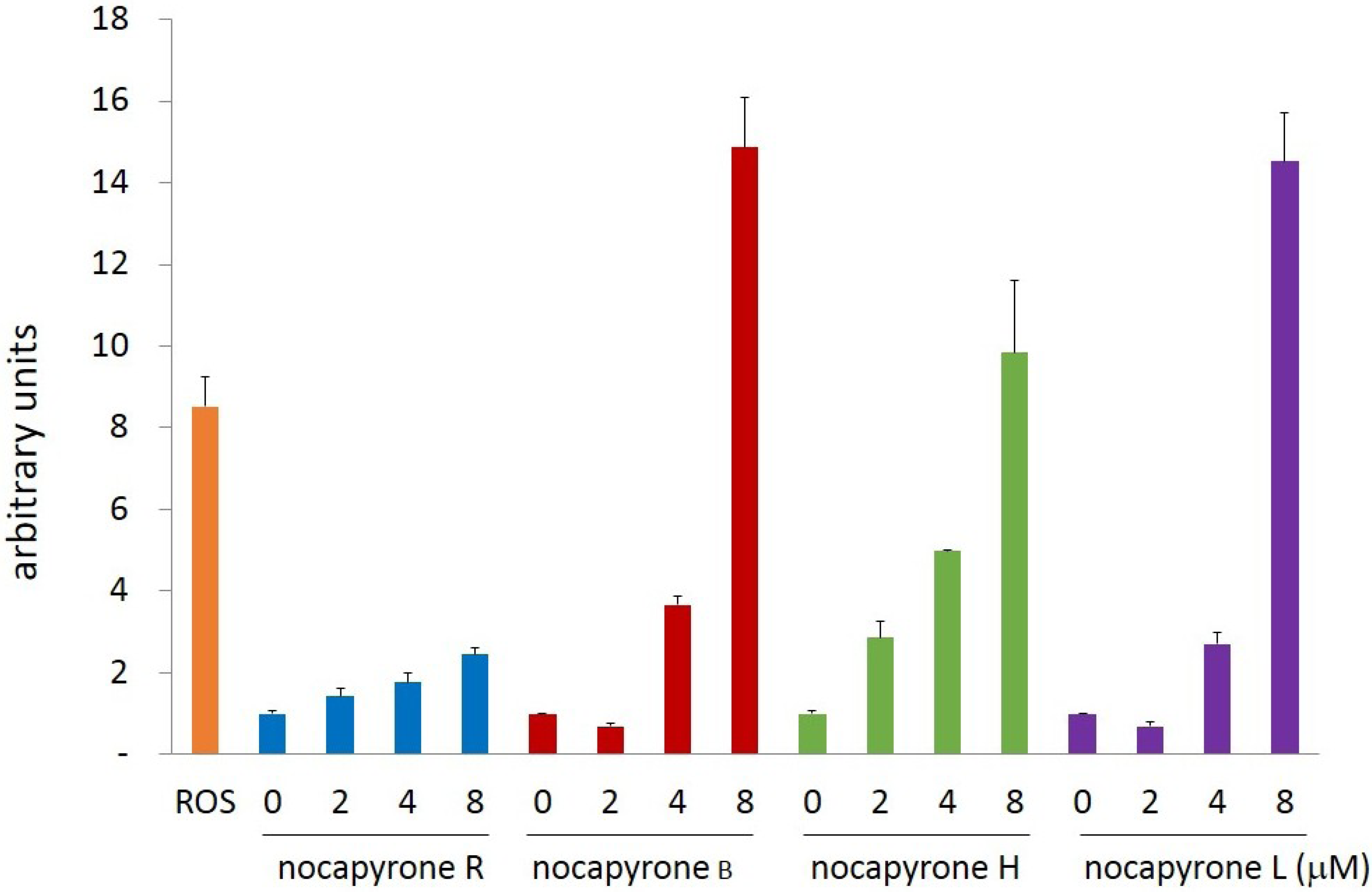
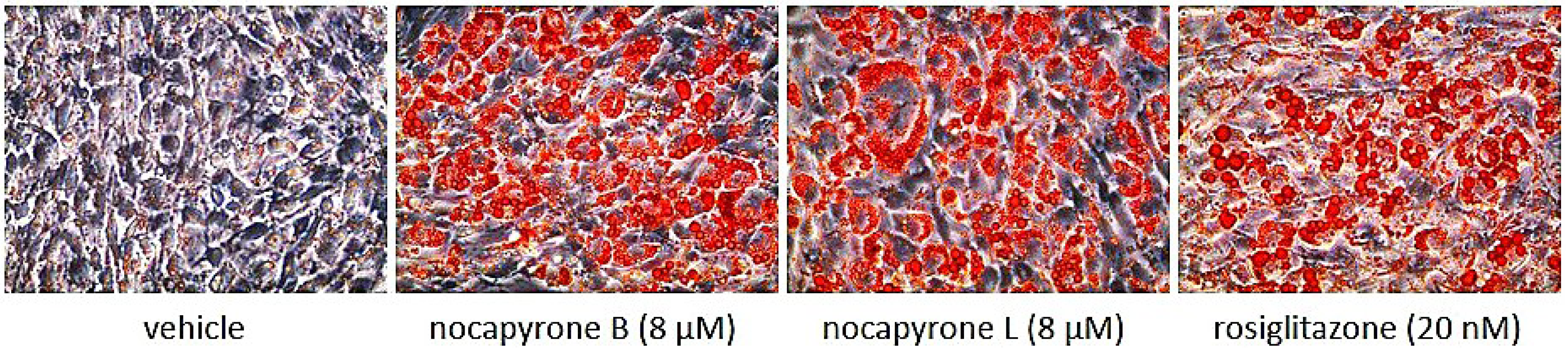
2.2. Biological Activities

Activity of compounds 14 to promote adiponectin-production in adipocyte cells was assessed by measuring the mRNA expression level of adiponectin-coding gene [25]. After the treatment of murine ST-13 preadipocyte cells with the compounds for 11 days, the total mRNA was subjected to quantitative real-time PCR analysis. All the γ-pyrones (24) enhanced the expression of adiponectin mRNA in a concentration-dependent manner while the α-pyrone (1) showed no such effect (Figure 5). Especially, 2 and 4 more strongly induced adiponectin production at 8 μM and at the same concentration the accumulation of lipid droplets were observed (Figure 6, oil red O staining) that indicates the differentiation to mature adipocyte cells. Compound 2 was further examined for the transcriptional activation of PPARγ that is the main target of the antidiabetic drug, thiazolidinediones. In the luciferase reporter assay, troglitazone activated the transcription through PPARγ but 2 did not (Figure 7), confirming that 2 is not a ligand for PPARγ and its mode of action is different from thiazolidinediones. These effects on adipocytes or adiponectin-inducing activity have not been described for nocapyrones and other related pyrone compounds.
Figure 5. Effect of nocapyrones on expression of adiponectin mRNA (ROS = rosiglitazone, 20 nM).
Marinedrugs 12 04110 g005
Figure 6. Adipocyte differentiation induced by nocapyrones B and L.
Marinedrugs 12 04110 g006
Figure 7. Effect of nocapyrone B on activation of PPARγ in luciferase ligand assay system.
Marinedrugs 12 04110 g007

3. Experimental Section

3.1. General Experimental Procedures

Optical rotation was measured using a JASCO DIP-3000 polarimeter (JASCO Corporation, Hachioji, Japan). UV spectra were recorded on a Hitachi U-3210 spectrophotometer (Hitachi High-Technologies Corporation, Tokyo, Japan). IR spectra were measured on a Perkin-Elmer Spectrum 100 (Perkin-Elmer Japan Co. Ltd., Yokohama, Japan). NMR spectra were obtained on a Bruker AVANCE 400 (Bruker BioSpin K.K., Yokohama, Japan) or a Bruker AVANCE 500 spectrometer (Bruker BioSpin K.K., Yokohama, Japan). HR-ESI-TOFMS were recorded on a Bruker microTOF focus (Bruker Daltonics K.K., Yokohama, Japan). Cosmosil 75C18-PREP (Nakalai Tesque, Inc., Kyoto, Japan, 75 μm) was used for ODS column chromatography.

3.2. Microorganism

Strain TP-A0876 was isolated from a sediment sample collected at a depth of 775 m off the island of Hokkaido (N 42°56′80″, E 140°06′32″), Japan in 2006 (August), using a piston corer. The strain was identified as a member of the genus Nocardiopsis on the basis of 100% 16S rRNA gene sequence (1455 nucleotides; GenBank accession number AB488799) similarity to Nocardiopsis sp. 123 (accession number AY036002).

3.3. Fermentation

Strain TP-A0876 cultured on a Bn-2 agar plate soluble starch 0.5%, glucose 0.5%, meat extract (Kyokuto Pharmaceutical Industrial Co., Ltd., Tokyo, Japan) 0.1%, yeast extract (Difco Laboratories, Surrey, United Kingdom) 0.1%, NZ-case (Wako Pure Chemical Industries, Ltd., Osaka, Japan) 0.2%, NaCl 0.2%, CaCO3 0.1% and agar 1.5%] was inoculated into 500-mL K-1 flasks each containing 100 mL of the V-22 seed medium consisting of soluble starch 1%, glucose 0.5%, NZ-case 0.3%, yeast extract 0.2%, Tryptone (Difco Laboratories) 0.5%, K2HPO4 0.1%, MgSO4·7H2O 0.05%, and CaCO3 0.3% (pH 7.0). The flasks were placed on a rotary shaker (200 rpm) at 30 °C for 4 days. The seed culture (3 mL) was transferred into 500-mL K-1 flasks each containing 100 mL of the A-3M production medium consisting of glucose 0.5%, soluble starch 2.0%, glycerol 2.0%, yeast extract 0.3%, Pharmamedia (Trader’s Protein) 1.5%, and Diaion HP-20 (Mitsubishi Chemical Holdings Corporation, Tokyo, Japan) 1%. The pH of the medium was adjusted to 7.0 before sterilization. The inoculated flasks were placed on a rotary shaker (200 rpm) at 30 °C for 7 days.

3.4. Extraction and Isolation

At the end of the fermentation period, 100 mL of 1-butanol was added to each flask, and they were allowed to shake for one hour. The mixture was centrifuged at 5000 rpm for 10 min and the organic layer was separated from the aqueous layer containing the mycelium. Evaporation of the solvent gave 7.8 g of extract from 2 L of culture. The crude extract (7.8 g) was subjected to silica gel column chromatography with a step gradient of CHCl3/MeOH (1:0, 20:1, 10:1, 4:1, 2:1, 1:1, and 0:1 v/v). Fractions 2 (20:1) and 3 (10:1) were combined and concentrated in vacuo to give brown oil (0.97 g) which was further fractionated by reverse-phase ODS column chromatography with a gradient of MeCN/distilled water (2:8, 3:7, 4:6, 5:5, 6:4, 7:3, and 8:2 v/v). Fractions 4 (5:5) and 5 (6:4) were combined and evaporated and the remaining aqueous solution was extracted with EtOAc. The organic layer was washed with brine, dried over anhydrous Na2SO4, filtered, and evaporated to dryness. The residual brown solid (0.28 g) was then subjected to HPLC purification using an ODS column (Cosmosil ARII, 10 mm × 250 mm) with 55% MeCN in distilled water at 3.0 mL/min to give nocapyrone B (2, tR 28.1 min, 10 mg), nocapyrone H (3, tR 31.3 min, 2.3 mg) and a mixture of nocapyrones R and L (1 and 4, tR 41.2 min, 3.8 mg). 1 and 4 were separated by using a cholesterol-bonded reverse-phase column (Cosmosil Cholester, 10 mm × 250 mm) with 55% MeCN in distilled water at 3.0 mL/min to give 4 (tR 29.9 min, 2.1 mg) and 1 (tR 35.0 min, 0.8 mg).
Nocapyrone R (1): colorless amorphous solid; UV (MeOH) λmax (log ε) 290 (4.04) nm; IR (ATR) νmax 2944, 1660 cm−1; 1H and 13C-NMR data, see Table 1; HRESITOFMS m/z 275.1619 [M + Na]+ (calcd for C15H24O3Na, 275.1623).

3.5. Determination of the Absolute Configuration of the Anteiso-Methyl Group in 4 by Ohrui-Akasaka Method

Oxidative Degradation of 4 to Yield 5: A solution of nocapyrone L (4, 4 mg, 15 μmol) in a mixture of MeCN (320 μL) and H2O (240 μL) was stirred with NaIO4 (19.2 mg, 89 μmol) until the salt was dissolved. To this solution were added CCl4 (320 μL) and 1 mg/mL solution of RuCl3 hydrate in 0.1 M sodium phosphate buffer (240 μL, pH 7.6) and the biphasic mixture was vigorously stirred at room temperature for 17 h. The reaction mixture was passed through Celite and the filter cake was washed with MeCN. After evaporation of the organic solvent from the filtrate, the aqueous solution was acidified with 2 M HCl and extracted with EtOAc. The EtOAc layer was concentrated and the remaining material was dissolved in MeOH (1 mL) and THF (1 mL). To this solution was added 1 M NaOH (1 mL) and the mixture was stirred at room temperature for 12 h. The reaction mixture was then acidified with 2 M HCl and extracted with EtOAc. The EtOAc layer was washed with water and brine, dried over anhydrous Na2SO4, and concentrated in vacuo to give 6-methyloctanoic acid (5, 1.2 mg) which was used for the next reaction without further purification:1H NMR (CDCl3, 500 MHz) δ 0.84 (3H, d, J = 6.0 Hz), 0.85 (3H, t, J = 7.6 Hz), 1.12 (2H, m), 1.28–1.35 (5H, m), 1.62 (2H, m), 2.36 (1H, t, J = 7.5 Hz).
Preparation of nat-5-(R)-2A1P: 6-Methyloctanoic acid (5) obtained by degradation of nocapyrone L (4) was reacted with (R)-2-(anthracene-2,3-dicarboximido)propanol [(R)-2A1P] (2 mg, 6.6 μmol) in dry CH2Cl2 (1 mL) in the presence of EDAC (2 mg, 10 μmol) and DMAP (trace amount) at room temperature for 17 h. The reaction mixture was diluted with ice-water and extracted with EtOAc. The organic layer was concentrated under reduced pressure and the residue was chromatographed over a silica gel column (n-hexane–EtOAc = 1:0~1:1) to give (R)-2A1P ester derivative of naturally occurring 6-methyloctanoic acid [nat-5-(R)-2A1P, 0.8 mg].
Preparation of standard (S)-6-methyloctanoic acid: To a solution of (S)-6-methyl-1-octanol (10 mg, 69 μmol, Wako Pure Chemical Industries, Ltd., Osaka, Japan) in acetone (1 mL) was added Jones reagent (0.4 mL, 80 μmol) dropwise. After stirring for 3 h at room temperature, the reaction mixture was extracted with EtOAc and the organic layer was concentrated in vacuo to give (S)-6-methyloctanoic acid (7.0 mg): 1H NMR (CDCl3, 500 MHz) δ 0.85 (3H, t, J = 5.5 Hz), 0.87 (3H, d, J = 7.0 Hz), 1.25–1.35 (5H, m), 1.33 (2H, m), 1.62 (2H, m), 2.36 (3H, t, J = 7.5 Hz).
Preparation of standard (S)-5-(R)-2A1P and (S)-5-(S)-2A1P: In the same manner as described for the preparation of nat-5-(R)-2A1P, (S)-5-(R)-2A1P and (S)-5-(S)-2A1P were prepared by the reaction of (S)-6-methyloctanoic acid with (R)- and (S)-2A1P, respectively.
(S)-5-(R)-2A1P: 1H NMR (CDCl3, 500 MHz) δ 0.72 (3H, d, J = 5.2 Hz), 0.74 (3H, t, J = 6.7 Hz), 0.97 (2H, m), 1.11–1.25 (5H, m), 1.49 (2H, m), 1.57 (2H, d, J = 7.1 Hz), 2.22 (2H, t, J = 7.7 Hz), 4.61 (1H, t, J = 6.1 Hz), 4.43 and 4.73 (2H, m), 7.62 (1H, dd, J = 6.5, 2.9 Hz), 8.07 (1H, dd, J = 6.3, 2.8 Hz), 8.48 (1H, s), 8.60 (1H, s).
(S)-5-(S)-2A1P: 1H NMR (CDCl3, 500 MHz) δ 0.71 (3H, d, J = 5.9 Hz), 0.74 (3H, t, J = 6.8 Hz), 0.96 (2H, m), 1.13–1.25 (5H, m), 1.48 (2H, m), 1.57 (2H, d, J = 7.1 Hz), 2.23 (2H, t, J = 7.7 Hz), 4.62 (1H, t, J = 6.1 Hz), 4.44 and 4.74 (2H, m), 7.62 (1H, dd, J = 7.0, 3.1 Hz), 8.07 (1H, dd, J = 6.3, 2.8 Hz), 8.48 (1H, s), 8.60 (1H, s).
HPLC analysis: nat-5-(R)-2A1P derived from nocapyrone L (4) and synthetic (S)-5-(R)-2A1P and (S)-5-(S)-2A1P were analyzed by HPLC under the following conditions. Column: Develosil ODS-HG-3 (4.6 mm × 150 mm, Nomura Chemical); mobile phase: MeOH/MeCN/THF=4:3:1; column temperature: −58.5 °C; flow rate: 0.2 mL/min. The column was cooled by using Cryocool CC-100 (Neslab Instruments Inc., Portsmouth, NH, USA). HPLC peaks were detected by monitoring fluorescence intensity at 460 nm with the excitation at 362 nm by using an FP-920 fluorescence detector (JASCO Corporation, Hachioji, Japan). Retention times were 130.5 min for (S)-5-(S)-2A1P and 139.3 min for (S)-5-(R)-2A1P. Natural product-derived nat-5-(R)-2A1P gave peaks at 130.8 min and 139.3 min in a ratio of 2:3.

3.6. Biological Activity Study

Adipocyte differentiation assay was carried out according to the procedure previously described [25].

3.6.1. Real-Time Quantitative PCR Analysis

The total RNA (1 μg) was reverse-transcribed to cDNA using a Super Script TM II RT (Invitrogen, Tokyo, Japan). To quantitatively estimate the mRNA levels of several genes, PCR amplification was performed using a Light Cycler system (Roche Diagnostic Co., Tokyo, Japan). Real-time PCR was carried out in a total volume of 20 μL containing 500 nM each of gene-specific primers, cDNA, and SYBRP remix Ex Tag (Takara, Kyoto, Japan). Expression was normalized to glyceraldehyde-3-phosphate dehydrogenase (GAPDH). Thermal cycling conditions for the PCR were 95 °C for 5 min, followed by 45 cycles of 95 °C for 5 s, 60 °C for 15 s, and 72 °C for 10 s, then a melting curve analysis from 65 °C to 95 °C, every 0.1 °C. The primer sequences used were: adiponectin, 5′-GAAGCCGCTTATATGTATCG-3′ (forward) and 5′-GCCGTCATAATGATTCTGT-3′ (reverse); GAPDH, 5′-CCAGAACATCATCCCTGC-3′ (forward) and 5′-CCACGACGGACACATT-3′ (reverse); fatty acid-binding protein (aP2), 5′-GAAATCACCGCAGACG-3′ (forward) and 5′-ACATTCCACCACCAGC-3′ (reverse); PPARγ2, 5′-CTGTTGACCCAGAGCA-3′ (forward) and 5′-GCGAGTGGTCTTCCAT-3′ (reverse).

3.6.2. Oil Red O Staining

ST-13 cells treated with nocapyrones B and L for 11 days were washed three times with phosphate-buffered saline (PBS), fixed with 10% formalin neutral buffer solution (Wako Pure Chemical, Osaka, Japan) at room temperature for 10 min, and then washed with distilled water to remove formalin solution. Furthermore, the cells were rinsed with 60% 2-propanol for 5 min, stained with 0.24% oil red O at room temperature for 20 min, and then were photographed under a phase contrast microscope (×100 magnification) equipped with a CCD camera (Leica Microsystems Japan, Tokyo, Japan).

3.6.3. Luciferase Reporter Assay

An expression plasmid containing the ligand binding domain of human PPARγ fused to the GAL4 DNA binding domain (pPPARγ-GAL4), and the luciferase reporter plasmid, 17m2G TATA Luc (p17m2G), were kindly donated by Dr. S. Kato (University of Tokyo, Tokyo, Japan). We transiently transfected COS-1 cells (6 × 10⁵ cells) with pPPARγ-GAL4 (0.25 μg) and p17m2G (1 μg) using Effectene Transfection Reagent (QIAGEN, Tokyo, Japan). Transfections were performed in triplicate in 24-well plates according to the manufacture’s instructions. After 16 h, the transfected cells received the indicated concentrations of nocapyrones B (or troglitazone), and were cultured for additional 24 h at 37 °C in a 5% CO2 incubator. Then cells were harvested and luciferase activities were measured by a Steady-Glo® Luciferase Assay System (Promega, Madison, WI, USA) according to manufacturer’s instructions.

4. Conclusions

In this study, a marine-derived actinomycete Nocardiopsis sp. TP-A0876 was isolated from a sediment sample and its secondary metabolites were investigated. Purification from the 1-butanol extract of the culture broth resulted in isolation of one α-pyrone and three γ-pyrones. Based on the spectroscopic analysis, the α-pyrone was confirmed as a new compound, whereas other three γ-pyrones were identified as nocapyrones B (2), H (3), and L (4). Biological testing in a set of assays proved that γ-pyrone compounds (24) have ability to induce preadipocyte differentiation and adiponectin production in murine ST13 preadipocyte cells. Since the α-pyrone compound 1 showed no activity, the γ-pyrone structure is proposed to play a key role in this activity.
A large number of pyrone–containing marine natural products are known [26,27,28,29,30,31,32]. Specifically, a series of nocapyrones (A–Q) have been continuously reported from marine-derived Nocardiopsis strains with a wide range of bioactivity (Figure 8). Nocapyrones B (2) and H (3) modulate nerve cell depolarization, activate or inhibit Ca2+ flux into a panel of human cells overexpressing various transient receptor potential (TRP) channels depending upon the agent and channel subtype, together with cytotoxicity against human breast adenocarcinoma, but have no antibacterial activity [21]. Nocapyrones E–G are described to have modest antibacterial activity against Bacillus subtilis [30]. Nocapyrone H* inhibits NO production in LPS-stimulated BV-2 microglial cells and neuro-protective effect [31], and nocapyrones H*, I*, and M* were shown to suppress gene expression in quorum sensing [32]. However, there is no report on the activity of these pyrones for preadipocyte differentiation or adiponectin production. Although the effectiveness is lower than thiazolidinediones, different mode of action of the γ-pyrones may provide an opportunity of drug development from this new therapeutic scaffold for insulin resistance type 2 diabetes. The concise synthetic strategies [33,34,35,36] and interesting bioactivities of structurally related compounds have been reported [37,38]. On the base of these studies, further synthetic and biological studies of nocapyrone families could be inspiring.
The absolute configuration of anteiso-methyl group is known and believed to be S in most cases because it is derived from l-isoleucine. However, the anteiso-methyl branching in 4 was confirmed as a mixture of (R)- and (S)-configurations in a ratio of 2:3. Along with the advances in synthetic and analytical techniques, several enantiomeric mixtures have been discovered in natural products (Figure 9) [39,40,41]. Many of them are marine-derived: trichodenone A, an antitumor metabolite of Trichoderma harzianum isolated from marine sponge (Halichondria okadai), is an (R)- and (S)-enantiomeric mixture [39]; pericosines B and C, metabolites of Periconia byssoide isolated from sea hare Aplysia kurodai, bearing four chiral centers are also enantiomeric mixtures [40]. Recently, the enantiomeric mixture regarding to the anteiso-methyl asymmetry was found in the insect pheromone. 4,8-Dimethyldecanal, the male aggregation pheromone of the red flour beetle (Tribolium castaneum), consists of four diastereomers, (4R, 8R)-, (4R, 8S)-, (4S, 8R)-, and (4S, 8S)-forms in a ratio of 4:4:1:1 [41]. To the best of our knowledge, nocapyrone L (4) is the first microbial metabolite proved to comprise an enantiomeric pair of R- and S-configurations at the anteiso-methyl substituent. The sec-methyl group at C-10 of nocapyrones C [20] and M [21] are reported to be an R/S-mixture but these compounds have a hydroxyl group on the neighboring carbon [21]. Biosynthetic origin of their anteiso-methyl portion should be clarified. Further investigation, focused on the biosynthetic precursors and gene analysis with our nocapyrone H (4), are under way to identify the enzymatic reactions responsible for the formation of enantiomeric mixture [42].
Figure 8. Nocapyrones isolated from marine-derived Nocardiopsis.
Marinedrugs 12 04110 g008
* Overlapped nomenclatures.
Figure 9. Natural products existing as mixtures of enantiomers.
Marinedrugs 12 04110 g009

Supplementary Files

  • Supplementary File 1:

    Supplementary Information (PDF, 477 KB)

  • Author Contributions

    Conceived and designed the experiments: Yasuhiro Igarashi. Extraction, isolation and characterization: Youngju Kim (nocapyrones R and H) and Hiromu Ogura (nocapyrones B and L). Determination of absolute configuration of nocapyrone L: Hiromu Ogura and Kazuaki Akasaka. Evaluation of biological activities: Tsutomu Oikawa and Nobuyasu Matsuura. Isolation of microorganism: Chiaki Imada and Hisato Yasuda. Wrote the manuscript: Youngju Kim and Yasuhiro Igarashi. Read and approved the final manuscript: Youngju Kim and Yasuhiro Igarashi.

    Conflicts of Interest

    The authors declare no conflict of interest.

    References

    1. Harvey, A.L. Natural products in drug discovery. Drug Discov. Today 2008, 13, 894–901. [Google Scholar] [CrossRef]
    2. Bérdy, J. Bioactive microbial metabolites. J. Antibiot. 2005, 58, 1–26. [Google Scholar] [CrossRef]
    3. Lam, K.S. Discovery of novel metabolites from marine actinomycetes. Curr. Opin. Microbiol. 2006, 9, 245–251. [Google Scholar] [CrossRef]
    4. Haefner, B. Drugs from the deep: Marine natural products as drug candidates. Drug Discov. Today 2003, 8, 536–544. [Google Scholar] [CrossRef]
    5. Candib, L.M. Obesity and diabetes in vulnerable populations: Reflection on proximal and distal causes. Ann. Fam. Med. 2007, 5, 547–556. [Google Scholar] [CrossRef]
    6. Trayhurn, P.; Wood, I.S. Signalling role of adipose tissue: Adipokines and inflammation in obesity. Biochem. Soc. Trans. 2005, 33, 1078–1081. [Google Scholar] [CrossRef]
    7. Simons, P.J.; van den Pangaart, P.S.; van Roomen, C.P.; Aerts, J.M.; Boon, L. Cytokine-mediated modulation of leptin and adiponectin secretion during in vitro adipogenesis: Evidence that tumor necrosis factor-alpha- and interleukin-1beta-treated human preadipocytes are potent leptin producers. Cytokine 2005, 32, 94–103. [Google Scholar] [CrossRef]
    8. Tsao, T.S.; Lodish, H.F.; Fruebis, J. ACRP30, a new hormone controlling fat and glucose metabolism. Eur. J. Pharmacol. 2002, 440, 213–221. [Google Scholar] [CrossRef]
    9. Hu, E.; Liang, P.; Spiegelman, B.M. AdipoQ is a novel adipose-specific gene dysregulated in obesity. J. Biol. Chem. 1996, 271, 10697–10703. [Google Scholar]
    10. Hotta, K.; Funahashi, T.; Arita, Y.; Takahashi, M.; Matsuda, M.; Okamoto, Y.; Iwahashi, H.; Kuriyama, H.; Ouchi, N.; Maeda, K.; et al. Plasma concentrations of a novel, adipose-specific protein, adiponectin, in type 2 diabetic patients. Arterioscler. Thromb. Vasc. Biol. 2000, 20, 1595–1599. [Google Scholar] [CrossRef]
    11. Lihn, A.S.; Østergard, T.; Nyholm, B.; Pedersen, S.B.; Richelsen, B.; Schmitz, O. Adiponectin expression in adipose tissue is reduced in first-degree relatives of type 2 diabetic patients. Am. J. Physiol. Endocrinol. Metab. 2003, 284, 443–448. [Google Scholar]
    12. Tiikkainen, M.; Häkkinen, A.M.; Korsheninnikova, E.; Nyman, T.; Mäkimattila, S.; Yki-Järvinen, H. Effects of rosiglitazone and metformin on liver fat content, hepatic insulin resistance, insulin clearance, and gene expression in adipose tissue in patients with type 2 diabetes. Diabetes 2004, 53, 2169–2176. [Google Scholar] [CrossRef]
    13. Yang, W.S.; Jeng, C.Y.; Wu, T.J.; Tanaka, S.; Funahashi, T.; Matsuzawa, Y.; Wang, J.P.; Chen, C.L.; Tai, T.Y.; Chuang, L.M. Synthetic peroxisome proliferator-activated receptor-gamma agonist, rosiglitazone, increases plasma levels of adiponectin in type 2 diabetic patients. Diabetes Care 2002, 25, 376–380. [Google Scholar] [CrossRef]
    14. Yamauchi, T.; Kamon, J.; Waki, H.; Terauchi, Y.; Kubota, N.; Hara, K.; Mori, Y.; Ide, T.; Murakami, K.; Tsuboyama-Kasaoka, N.; et al. The fat-derived hormone adiponectin reverses insulin resistance associated with both lipoatrophy and obesity. Nat. Med. 2001, 7, 941–946. [Google Scholar] [CrossRef]
    15. Kadowaki, T.; Yamauchi, T. Adiponectin and adiponectin receptors. Endocr. Rev. 2005, 26, 439–451. [Google Scholar] [CrossRef]
    16. Nissen, S.E.; Wolski, K. Effect of rosiglitazone on the risk of myocardial infarction and death from cardiovascular causes. N. Engl. J. Med. 2007, 356, 2457–2471. [Google Scholar] [CrossRef]
    17. Igarashi, Y.; Tanaka, Y.; Ikeda, M.; Oikawa, T.; Kitani, S.; Nihira, T.; Mongkol, P.; Janhom, M.; Panbangred, W. Prajinamide, a new modified peptide from a soil-derived Streptomyces. J. Antibiot. 2012, 65, 157–159. [Google Scholar] [CrossRef]
    18. Igarashi, Y.; Yu, L.; Ikeda, M.; Oikawa, T.; Kitani, S.; Nihira, T.; Bayanmunkh, B.; Panbangred, W. Jomthonic acid, a modified amino acid from a soil-derived Streptomyces. J. Nat. Prod. 2012, 75, 986–990. [Google Scholar] [CrossRef]
    19. Indananda, C.; Igarashi, Y.; Ikeda, M.; Oikawa, T.; Thamchaipenet, A. Linfuranone A, a new polyketide from plant-derived Microbispora sp. GMKU 363. J. Antibiot. 2013, 66, 675–677. [Google Scholar] [CrossRef]
    20. Schneemann, I.; Ohlendorf, B.; Zinecker, H.; Nagel, K.; Wiese, J.; Imhoff, J.F. Nocapyrones A–D, γ-pyrones from a Nocardiopsis strain isolated from the marine sponge Halichondria panicea. J. Nat. Prod. 2010, 73, 1444–1447. [Google Scholar] [CrossRef] [Green Version]
    21. Lin, Z.; Torres, J.P.; Ammon, M.A.; Marett, L.; Teichert, R.W.; Reilly, C.A.; Kwan, J.C.; Hughen, R.W.; Flores, M.; Tianero, M.D.; et al. A bacterial source for mollusk pyrone polyketides. Chem. Biol. 2013, 24, 73–81. [Google Scholar]
    22. Gregory, L.C. Mining microbial genomes for new natural products and biosynthetic pathways. Microbiology 2008, 154, 1555–1569. [Google Scholar] [CrossRef]
    23. Akasaka, K.; Imizumi, K.; Ohrui, H. Enantiomeric separation of branched fatty acids having chiral centers remote from the carboxyl group by labelling with chiral fluorescent derivatization reagents. Enantiomer 1998, 3, 169–174. [Google Scholar]
    24. Nunez, M.T.; Martin, V.S. Efficient oxidation of phenyl groups to carboxylic acids with ruthenium tetraoxide. A simple synthesis of (R)-γ-caprolactone, the pheromone of Trogoderma granarium. J. Org. Chem. 1990, 55, 1928–1932. [Google Scholar] [CrossRef]
    25. Ikeda, M.; Kurotobi, Y.; Namikawa, A.; Kuranuki, S.; Matsuura, N.; Sato, M.; Igarashi, Y.; Nakamura, T.; Oikawa, T. Norlichexanthone isolated from fungus P16 promotes the secretion and expression of adiponectin in cultured ST-13 adipocytes. Med. Chem. 2011, 7, 250–256. [Google Scholar] [CrossRef]
    26. Singh, M.P.; Kong, F.; Janso, J.E.; Arias, D.A.; Suarez, P.A.; Bernan, V.S.; Petersen, P.J.; Weiss, W.J.; Carter, G.; Greenstein, M. Novel α-pyrones produced by a marine Pseudomonas sp. F92S91: Taxonomy and biological activities. J. Antibiot. 2003, 56, 1033–1044. [Google Scholar] [CrossRef]
    27. Trisuwan, K.; Rukachaisirikul, V.; Sukpondma, Y.; Preedanon, S.; Phongpaichit, S.; Rungjindamai, N.; Sakayaroj, J. Epoxydons and a pyrone from the marine-derived fungus Nigrospora sp. PSU-F5. J. Nat. Prod. 2008, 71, 1323–1326. [Google Scholar] [CrossRef]
    28. Yu, K.; Ren, B.; Wei, J.; Chen, C.; Sun, J.; Song, F.; Dai, H.; Zhang, L. Verrucisidinol and Verrucosidinol Acetate, Two pyrone-type polyketides isolated from a marine derived fungus, Penicillium aurantiogriseum. Mar. Drugs 2010, 8, 2744–2754. [Google Scholar]
    29. Carbone, M.; Ciavatta, M.L.; Wang, J.R.; Cirillo, I.; Mathieu, V.; Kiss, R.; Mollo, E.; Guo, Y.W.; Gavagnin, M. Extending the record of polypropionates from marine pulmonate mollusks. J. Nat. Prod. 2013, 76, 2065–2073. [Google Scholar]
    30. Fu, P.; Liu, P.; Qu, H.; Wang, Y.; Chen, D.; Wang, H.; Li, J.; Zhu, W. A-pyrones and diketopiperazine derivatives from the marine-derived actinomycete Nocardiopsis dassonvillei HR10–5. J. Nat. Prod. 2011, 74, 2219–2223. [Google Scholar] [CrossRef]
    31. Kim, M.C.; Kwon, O.W.; Park, J.S.; Kim, S.Y.; Kwon, H.C. Nocapyrones H–J, 3,6-disubstituted α-pyrones from the marine actinomycete Nocardiopsis sp. KMF-001. Chem. Pharm. Bull. 2013, 61, 511–515. [Google Scholar] [CrossRef]
    32. Fu, P.; Liu, P.; Gong, Q.; Wang, Y.; Wanga, P.; Zhu, W. α-Pyrones from the marine-derived actinomycete Nocardiopsis dassonvillei subsp. dassonvillei XG-8–1. RSC Adv. 2013, 3, 20726–20731. [Google Scholar]
    33. Shimamura, H.; Sunazuka, T.; Hizoura, T.; Hirose, T.; Shiomi, K.; Omura, S. Total synthesis and biological evaluation of verticipyrone and analogues. Org. Lett. 2007, 9, 65–67. [Google Scholar] [CrossRef]
    34. Lipshutz, B.H.; Amorelli, B. Carboalumination/Ni-catalyzed couplings. A short synthesis of verticipyrone. Tetrahedron Lett. 2009, 50, 2144–2146. [Google Scholar] [CrossRef]
    35. De Paolis, M.; Rosso, H.; Henrot, M.; Prandi, C.; d’Herouville, F.; Maddaluno, J. A concise route to α′-methoxy-γ-pyrones and verticipyrone based upon the desymmetrization of α,α′-dimethoxy-γ-pyrone. Chem. Eur. J. 2010, 16, 11229–11232. [Google Scholar]
    36. Rosso, H.; de Paolis, M.; Collin, V.C.; Dey, S.; Hecht, S.M.; Prandi, C.; Richard, V.; Maddaluno, J. One-pot regio- and stereoselective synthesis of α′-methoxy-γ-pyrones: Biological evaluation as mitochondrial respiratory complex inhibitors. J. Org. Chem. 2011, 76, 9429–9437. [Google Scholar] [CrossRef]
    37. Sharma, P.; Powell, K.J.; Burnley, J.; Awaad, A.S.; Moses, J.E. Total Synthesis of polypropionate-derived γ-pyrone natural products. Synthesis 2011, 2011, 2865–2892. [Google Scholar]
    38. Wilk, W.; Waldmann, H.; Kaiser, M. Gamma-pyrone natural products—A privileged compound class provided by nature. Bioorg. Med. Chem. 2009, 17, 2304–2309. [Google Scholar] [CrossRef]
    39. Usami, Y.; Ikura, T.; Amagata, T.; Numata, A. First total synthesesand configurational assignments of cytotoxic trichodenones A–C. Tetrahedron Asymmetry 2000, 11, 3711–3725. [Google Scholar] [CrossRef]
    40. Usami, Y.; Okada, Y.; Yamada, T. Natural pericosines B and C as enantiomeric mixtures: direct evidence by chiral HPLC analysis. Chirality 2001, 23, E7–E11. [Google Scholar] [CrossRef]
    41. Akasaka, K.; Tamogami, S.; Beeman, R.W.; Mori, K. Pheromone synthesis. Part 245: Synthesis and chromatographic analysis of the four stereoisomers of 4,8-dimethyldecanal, the male aggregation pheromone of the red flour beetle, Tribolium castaneum. Tetrahedron 2011, 67, 201–209. [Google Scholar] [CrossRef]
    42. Komaki, H.; Ichikawa, N.; Hosoyama, A.; Fujita, N.; Igarashi, Y. Draft genome sequence of marine-derived actinomycete Nocardiopsis sp. TP-A0876, a producer of polyketide pyrones. Genome Announc. 2014, in press. [Google Scholar]

    Share and Cite

    MDPI and ACS Style

    Kim, Y.; Ogura, H.; Akasaka, K.; Oikawa, T.; Matsuura, N.; Imada, C.; Yasuda, H.; Igarashi, Y. Nocapyrones: α- and γ-Pyrones from a Marine-Derived Nocardiopsis sp. Mar. Drugs 2014, 12, 4110-4125. https://doi.org/10.3390/md12074110

    AMA Style

    Kim Y, Ogura H, Akasaka K, Oikawa T, Matsuura N, Imada C, Yasuda H, Igarashi Y. Nocapyrones: α- and γ-Pyrones from a Marine-Derived Nocardiopsis sp. Marine Drugs. 2014; 12(7):4110-4125. https://doi.org/10.3390/md12074110

    Chicago/Turabian Style

    Kim, Youngju, Hiromu Ogura, Kazuaki Akasaka, Tsutomu Oikawa, Nobuyasu Matsuura, Chiaki Imada, Hisato Yasuda, and Yasuhiro Igarashi. 2014. "Nocapyrones: α- and γ-Pyrones from a Marine-Derived Nocardiopsis sp." Marine Drugs 12, no. 7: 4110-4125. https://doi.org/10.3390/md12074110

    Article Metrics

    Back to TopTop