Article

Pseudaboydins A and B: Novel Isobenzofuranone Derivatives from Marine Fungus Pseudallescheria boydii Associated with Starfish Acanthaster planci

1
School of Pharmaceutical Sciences, Sun Yat-sen University, Guangzhou 510006, China
2
Guangdong Technology Research Center for Advanced Chinese Medicine, Guangzhou 510006, China
3
Institute of Chinese Medical Sciences, Guangdong Pharmaceutical University, Guangzhou 510006, China
4
School of Chemistry and Chemical Engineering, Sun Yat-sen University, Guangzhou 510275, China
*
Authors to whom correspondence should be addressed.
Mar. Drugs 2014, 12(7), 4188-4199; https://doi.org/10.3390/md12074188
Submission received: 4 June 2014 / Revised: 3 July 2014 / Accepted: 3 July 2014 / Published: 14 July 2014

Abstract

:
Two novel isobenzofuranone derivatives, pseudaboydins A (1) and B (2), along with five known compounds, including (R)-2-(2-hydroxypropan-2-yl)-2,3-dihydro-5-hydroxybenzofuran (3), (R)-2-(2-hydroxypropan-2-yl)-2,3-dihydro-5-methoxybenzofuran (4), 3,3′-dihydroxy-5,5′-dimethyldiphenyl ether (5), 3-(3-methoxy-5-methylphenoxy)-5-methylphenol (6) and (−)-regiolone (7), were isolated from the culture broth of the marine fungus, Pseudallescheria boydii, associated with the starfish, Acanthaster planci. Their structures were elucidated primarily based on NMR and MS data. The absolute configurations of 14 were determined by CD spectroscopy and single-crystal X-ray diffraction studies. The cytotoxic and antibacterial activities of 14 were evaluated. Pseudaboydin A (1) showed moderate cytotoxic activity against human nasopharyngeal carcinoma cell line HONE1, human nasopharyngeal carcinoma cell line SUNE1 and human glandular lung cancer cell line GLC82 with IC50 values of 37.1, 46.5 and 87.2 μM, respectively.

1. Introduction

In the course of our investigation on the secondary metabolites of marine fungi associated with marine organisms in coral reefs, a variety of novel and/or bioactive metabolites were isolated [1,2,3,4,5,6,7]. The starfish, Acanthaster planci, is found easily in the coral reef of the South China Sea and is notorious for preying on corals and is devastating to the coral reef. The arms and upper surface of Acanthaster planci are covered by long and sharp spines. If the spines sting a person, it will cause tissue swelling for a week or more. Recently, a fungus was isolated from the inner tissue of Acanthaster planci, and it was identified as Pseudallescheria boydii based on internal transcribed spacer (ITS) sequence similarity. The marine origin of this fungal species is unprecedented. The terrigenous Pseudallescheria boydii is mainly distributed in soil and polluted waters. Pseudallescheria boydii is deemed to be a human pathogenic fungus and can cause a variety of infections, especially in the immunocompromised and immunocompetent patients. However, up to now, the literature reports on the secondary metabolites of this fungus are still limited [8,9,10,11,12,13]. The literature survey prompted us to investigate the metabolites of the fungus, Pseudallescheria boydii. This fungal strain was cultured in glucose-peptone-yeast extract (GPY) medium. Two new isobenzofuranone derivatives, pseudaboydins A (1) and B (2), along with five known compounds, (R)-2-(2-hydroxypropan-2-yl)-2,3-dihydro-5-hydroxybenzofuran (3), (R)-2-(2-hydroxypropan-2-yl)-2,3-dihydro-5-methoxybenzofuran (4), 3,3′-dihydroxy-5,5′-dimethyldiphenyl ether (5), 3-(3-methoxy-5-methylphenoxy)-5-methylphenol (6) and (−)-regiolone (7), were isolated from the EtOAc extracts of fungal culture broth (Figure 1). Herein, we describe the isolation, structure determination and biological activity evaluation of these compounds.
Figure 1. Chemical structures of Compounds 17.
Marinedrugs 12 04188 g001

2. Results and Discussion
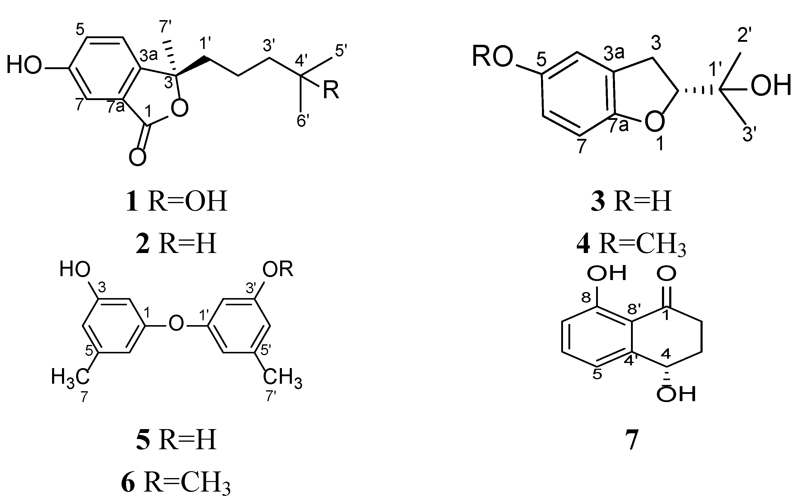
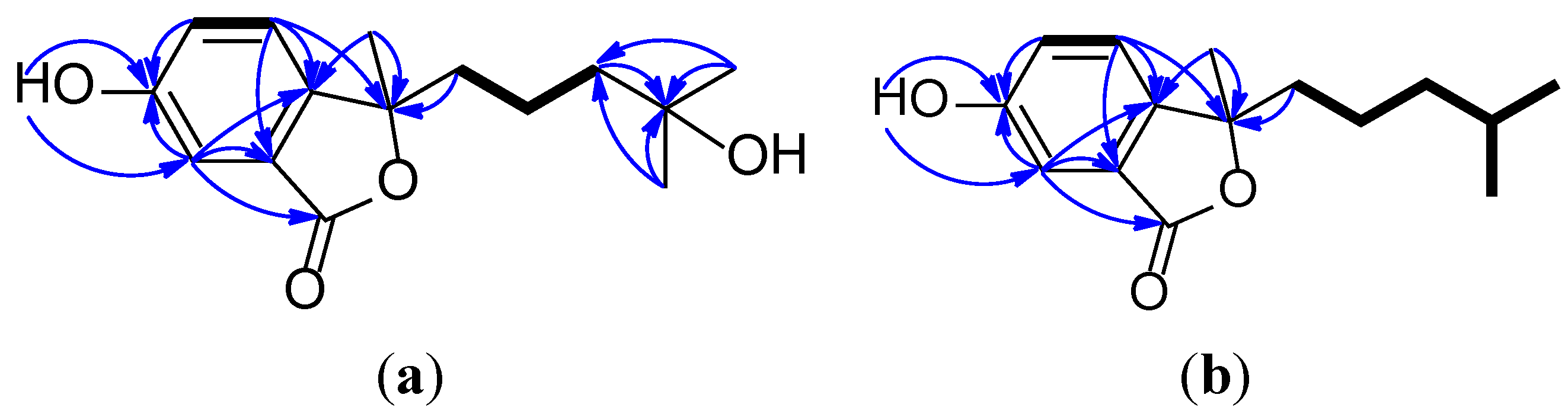
Pseudaboydin A (1) was obtained as a white solid. The molecular formula was determined to be C15H20O4 by HREIMS at m/z 264.1355 [M]+ (calcd. 264.1362) (Supplementary Figure S2), requiring six degrees of unsaturation. The IR spectrum indicated the presence of hydroxyl groups (3217 cm−1), a carbonyl group (1680 cm−1) and a benzene ring (3073, 1574 and 1507 cm−1). The UV maximum absorption at 217, 243 and 298 nm displayed the carbonyl group conjugated with the benzene ring. The 13C-NMR and DEPT spectra displayed three methyls, three methylenes, three methines and six quaternary carbons (Table 1, and Supplementary Figures S4 and S5). The 1H-1H COSY cross-peaks of H-4/H-5, H2-1′/H2-2′ and H2-2′/H2-3′ led to the identification of two isolated proton spin-systems of –CH–CH– and –CH2–CH2–CH2– (Figure 2a, and Supplementary Figure S8). By interpretation of the three aromatic protons at δH 7.13 (d, J = 8.0 Hz, H-4), 7.56 (d, J = 8.0 Hz, H-5) and 7.55 (s, H-7) (Supplementary Figure S3), a 1, 2, 4-trisubstituted benzene ring in the molecule was suggested, which was confirmed by HMBC correlations of H-4/C-3a, H-5/C-6, H-7/C-6 and H-7/C-7a (Supplementary Figure S7). The phenolic hydroxy group at δH 9.33 (brs) showed HMBC correlations with C-5, C-6 and C-7, indicated that it connected to C-6. The HMBC correlations of H-4/C-3 and H-7/C-1 established an isobenzofuranone skeleton. The methylene (H2-3′) and two singlet methyl groups at δH 1.28 and 0.95 (Supplementary Figure S6) showed HMBC correlations with the oxygenated quaternary carbon C-4′ and deduced the only side chain –CH2–CH2–CH2–C(OH)(CH3)2 in the molecule. This side chain and another methyl group were connected to C-3 based on the HMBC correlations of H2-1′/C-3 and H3-7′/C-3.
Table 1. 1H and 13C-NMR data of Compounds 1 and 2 obtained at 400 and 100 MHz, resp., in CDCl3, δ in ppm.
Position12
δCTypeδH, Multiple, ( J in Hz)δCTypeδH, Multiple, ( J in Hz)
1171.8C 171.6C
2
377.7C 79.1C
3a137.1C 135.5C
4124.7CH7.13, d (8.0)126.5CH7.08, d (8.0)
5121.5CH7.56, d (8.0)121.3CH7.54, d (8.0)
6157.1C 156.1C
7119.0CH7.55, s119.3CH7.57, s
7a129.7C 129.8C
1′33.9CH22.45, brd (13.6)
1.65, m
42.9CH21.92, dt (14.4, 8.0)
1.80, dt (14.4, 8.0)
2′16.7CH21.71, m21.7CH21.29, m
3′36.8CH21.52, m39.1CH21.16, m
4′75.4C 27.8CH1.49, nine (6.4)
5′32.0CH31.28, s22.6CH30.82, d (6.4)
6′24.8CH30.95, s22.4CH30.82, d (6.4)
7′31.4CH31.50, s29.0CH31.68, s
6-OH 9.33, brs 9.34, brs
4′-OH 1.80, brs
Figure 2. 1H-1H COSY (bold line) and main HMBC (arrow) correlations of 1 and 2 (a and b).
Marinedrugs 12 04188 g002
The circular dichroism (CD) spectrum of 1 (Figure 3a) exhibited a positive Cotton effect at 303 nm (n–π* transition) and a negative Cotton effect at 210.2 nm (π–π* transition). The α, β-unsaturated-γ-lactone core has a planar conformation; Uchida and Kuriyama researched the π–π* CD of α, β-unsaturated-γ-lactones and found the correlation of the Cotton effect with the configuration of the more polarizable bond at the γ-carbon atom [14]. Gawronski et al. reported the simple CD method for the determination of the absolute configuration of 5-alkyl and 5-alkoxy-substituted 2(5H)-furanones [15]. The chiral center of 1 exists at C-3. The side chain –CH2–CH2–CH2–C(OH)(CH3)2 makes a greater contribution to negative π–π* and positive n–π* Cotton effects than the methyl group. According to Uchida and Gawronski’s empirical rules, the γ-carbon atom (C-3) was assigned to be R configuration. Therefore, Compound 1 was determined as (R)-3-methyl-3-(4-hydroxy-4-methylpentyl)-6-hydroxy isobenzofuran-1(3H)-one.
Figure 3. Circular dichroism (CD) spectra of Compounds 14 (ad) in MeCN solution.
Marinedrugs 12 04188 g003
Pseudaboydin B (2) was isolated as a white solid. The molecular formula of 2 was established as C15H20O3 on the basis of HREIMS (m/z 248.1408 [M]+, calcd. 248.1412) (Supplementary Figure S10) and NMR data (Table 1, and Supplementary Figures S11–S16). The general features of its UV, IR and NMR spectra data were very similar to those of 1. The 13C-NMR and DEPT spectra displayed three methyls, three methylenes, four methines and five quaternary carbons. A quick identification was made that oxygenated quaternary carbon C-4′ was replaced by a methine. Two methyl doublets at δH 0.82 (d, J = 6.4 Hz, H3-5′ and H3-6′) connected to the methine at δH 1.49 (nine, J = 6.4 Hz, H-4′). The 1H-1H COSY cross-peaks of H2-1′/H2-2′, H2-2′/H2-3′, H2-3′/H-4′, H-4′/H3-5′ and H-4′/H3-6′ also confirmed the presence of the aliphatic side chain 4-methylpentyl (Figure 2b). By careful analysis of HMQC, 1H-1H COSY and HMBC spectra data, Compound 2 was assigned as 3-methyl-3-(4-methylpentyl)-6-hydroxyisobenzofuran-1(3H)-one. The stereochemistry of Compound 2 was also established by CD spectrum (Figure 3b). 2 showed a very similar CD spectrum with that of 1. Thus, the absolute configuration of 2 was also definitely assigned as 3R.
Compounds 3 and 4 were identified as 2-(2-hydroxypropan-2-yl)-2,3-dihydro-5-hydroxybenzofuran (3) and 2-(2-hydroxypropan-2-yl)-2,3-dihydro-5-methoxybenzofuran (4). Compound 3 was previously isolated from the fungus, Acremonium murorum [16]; 4 was obtained by synthesis [17]. The NMR data of 3 recorded in DMSO-d6 (Table 2, and Supplementary Figures S17 and S18) were slightly different from the reference data recorded in CD3OD, and the 13C-NMR data (Supplementary Figures S19 and S20) of 4 were not reported previously. Furthermore, the absolute configurations of their chiral centers were not yet reported. Fortunately, we obtained a single crystal of 3 from the MeOH solution. In the crystal, the molecules are arranged in a helical manner and connected by strong O-H...O hydrogen bonds. The helicates are further joined together by O-H...O hydrogen bonds to form a discrete molecular layer with hydrophobic substituents pointing outwards. With this kind of layer-type arrangement, the crystal structure is stabilized by the weak van der Waals’ interactions. The absolute configuration of 3 was determined as 2R by analysis of the single-crystal X-ray data (Figure 4). The Flack absolute structure parameter is 0.2 (4) [18]. In the CD spectrum, 3 showed a positive Cotton effect at 233 nm and a negative Cotton effect at 211.6 nm (Figure 3c). Finally, the CD spectrum of 4 (Figure 3d) was almost identical with that of 3. Thus, the absolute configuration of 4 was also determined as 2R.
Table 2. 1H and 13C-NMR data of Compounds 3 and 4 obtained at 400 and 100 MHz, resp., δ in ppm.
Position3 a4 b
δCTypeδH, Multiple, (J in Hz)δCTypeδH, Multiple, (J in Hz)
1
288.6CH4.42, dd (9.6, 8.4)89.4CH4.58, dd (9.2, 8.8)
330.5CH23.06, dd (16.0, 8.4);
2.98, dd (16.0, 9.6)
31.2CH23.17, dd (16.0, 8.8);
3.10, dd (16.0, 9.2)
3a128.1C 128.1C
4111.9CH6.59, d (2.4)111.2CH6.75, d (1.6)
5150.9C 154.1C
6113.3CH6.42, dd (8.4, 2.4)112.8CH6.64, dd (8.8, 1.6)
7108.3CH6.50, d (8.4)108.9CH6.68, d (8.8)
7a152.5C 153.7C
1′70.1C 71.8C
2′24.7CH31.09, s23.9CH31.20, s
3′26.2CH31.11, s26.2CH31.33, s
5-OH 8.69, s
1′-OH 4.50, s 1.97, brs
5-OCH3 56.0CH33.75, s
a Measured in DMSO-d6 and DMSO-d6 was used as an internal standard (δC 39.51, δH 2.50); b measured in CDCl3 and CDCl3 was used as an internal standard (δC 77.0, δH 7.26).
Figure 4. Crystal structure of 3. Thermal ellipsoids are plotted at a 30% probability level.
Marinedrugs 12 04188 g004
Compounds 57 were determined as 3,3′-dihydroxy-5,5′-dimethyldiphenyl ether [19], 3-(3-methoxy-5-methylphenoxy)-5-methyl phenol [20] and (−)-regiolone [21] respectively, by comparing their spectroscopic data (Supplementary Figures S21–S28) with the literature values.
Compounds 14 were evaluated for in vitro cytotoxicity against human nasopharyngeal carcinoma cell line HONE1, human nasopharyngeal carcinoma cell line SUNE1 and human glandular lung cancer cell line GLC82. As a result, Compound 1 showed moderate cytotoxic activity against HONE1, SUNE1 and GLC82 with IC50 values of 37.1, 46.5 and 87.2 μM, respectively. Compounds 24 displayed no cytotoxicity against the tested cancer cell lines (IC50 > 100 μM). Additionally, Compounds 14 were also evaluated for their antibacterial activity against MRSAST239, standard Staphylococcus aureus (ATCC29213) and Escherichia coli (ATCC295225), using a broth dilution method (Mueller–Hinton broth). Vancomycin and ampicillin sodium were used as positive control. However, Compounds 14 did not exhibit significant activity at a concentration lower than 256 μg/mL.

3. Experimental Section

3.1. General Experimental Procedures

Preparative HPLC was performed using a Shimadzu LC-20AT HPLC pump (Shimadzu Corporation, Nakagyo-ku, Kyoto, Japan) equipped with an SPD-20A dual λ absorbance detector (Shimadzu Corporation, Nakagyo-ku, Kyoto, Japan) and a Shim-pack PRC-ODS HPLC column (250 mm × 20 mm, Shimadzu Corporation, Nakagyo-ku, Kyoto, Japan). Optical rotations were measured using a Schmidt and Haensch Polartronic HNQW5 optical rotation spectrometer (SCHMIDT + HAENSCH GmbH & Co., Berlin, Germany). CD spectra were measured on a JASCO J-810 circular dichroism spectrometer (JASCO International Co. Ltd., Hachioji, Tokyo, Japan). IR spectra were recorded on a PerkinElmer Frontier FT-IR spectrophotometer (PerkinElmer Inc., Waltham, MA, USA). UV spectra were recorded on a Shimadzu UV-Vis-NIR spectrophotometer (Shimadzu Corporation, Nakagyo-ku, Kyoto, Japan). 1D and 2D NMR spectra were recorded on a Bruker Avance II 400 spectrometer (Bruker BioSpin AG, Industriestrasse 26, Fällanden, Switzerland). The chemical shifts are relative to the residual solvent signals (CDCl3: δH 7.26 and δC 77.0; CD3OD: δH 3.31 and δC 49.15; DMSO-d6: δH 2.50 and δC 39.51). Mass spectra were obtained on a Thermo DSQ EI low-resolution mass spectrometer (Thermo Fisher Scientific, Waltham, MA, USA) and a Thermo MAT95XP EI high-resolution mass spectrometer (Thermo Fisher Scientific, Waltham, MA, USA). X-ray diffraction data were acquired on a Bruker SMART APEX CCD X-ray single-crystal diffractometer (Bruker AXS GmbH, Karlsruhe, Germany).

3.2. Fungal Strain and Culture Method

Marine fungus Pseudallescheria boydii was isolated from the inner tissue of sea star Acanthaster planci collected from Hainan Sanya National Coral Reef Reserve, Hainan, China. A voucher specimen was deposited in the School of Pharmaceutical Sciences, Sun Yat-sen University, Guangzhou, China.
The fermentation medium was glucose 10 g/L, peptone 5 g/L, yeast extract 2 g/L, sea water 1 L and pH 7.5 (GPY medium). Fungal mycelia were cut and transferred aseptically to 500-mL Erlenmeyer flasks, each containing 200 mL sterilized GPY liquid medium. The flasks were incubated at 28 °C on a rotary shaker (120 rpm) for 20 days.

3.3. Extraction and Isolation

Eighty liters of liquid culture were filtered through cheesecloth. The culture broth was extracted three times with EtOAc (80 L) and then was concentrated by low-temperature rotary evaporation to give an extract (30.9 g). The extract was subjected to column chromatography (diameter: 8 cm, length: 70 cm) over silica gel (200 g) eluting with petroleum ether (2 L)-EtOAc (2 L) (100:0–0:100) followed by EtOAc (2 L)-MeOH (2 L) (100:0–0:100) to yield 15 fractions (code: Fr. 1–Fr. 15). Fr. 8 (1.08 g) was further separated by RP-HPLC (MeCN-H2O, 4:6) to afford Compounds 1 (18 mg) and 2 (26 mg). Fr. 12 (3.22 g) was further separated by RP-HPLC (MeOH-H2O, 4:6) to afford Compounds 3 (1608 mg) and 4 (50 mg). Fr. 5 (1.58 g) was purified by RP-HPLC (MeOH-H2O, 6:4) to give Compounds 5 (65 mg), 6 (18 mg) and 7 (12 mg).
Pseudaboydin A ((R)-3-methyl-3-(4-hydroxy-4-methylpentyl)-6-hydroxyisobenzofuran-1(3H)-one, (1): white solid. Marinedrugs 12 04188 i001
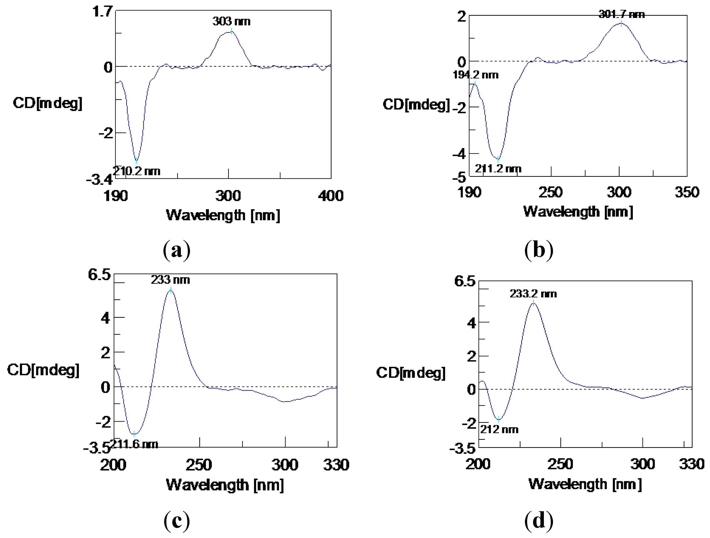
: +200° (c = 0.025, MeOH). CD (MeCN) λmax (Δε): 210.2 (−5.41), 303 (+2.01) nm. IR (KBr): 3217, 3073, 2978, 1680, 1574, 1507, 1419, 1389, 1354, 1287, 1230, 1161, 1065, 957, 891, 763 cm−1. UV (MeOH) λmax (ε): 217 nm (8152), 243 nm (7142), 298 nm (2247). 1H and 13C-NMR: Table 1. LREIMS: m/z 264, 246, 231, 203, 189, 181, 165, 147, 133, 119, 105, 91, 69. HREIMS: m/z 264.1355 [M]+ (calcd. for C15H20O4+: 264.1362).
Pseudaboydin B ((R)-3-methyl-3-(4-methylpentyl)-6-hydroxyisobenzofuran-1(3H)-one, (2): white solid. Marinedrugs 12 04188 i001: +53.6° (c = 0.078, MeOH). CD (MeCN) λmax (Δε): 211.2 (−7.22), 301.7 (+2.78) nm. IR (KBr): 3377, 3073, 2952, 1687, 1575, 1510, 1467, 1380, 1294, 1223, 1127, 1052, 947, 890, 766 cm−1. UV (MeOH) λmax (ε): 219 nm (11,833), 244 nm (12,258), 299 nm (4202). 1H and 13C-NMR: Table 1. LREIMS: m/z 248, 233, 203, 192, 178, 165, 147, 133, 119, 105, 91, 77. HREIMS: m/z 248.1408 [M]+ (calcd. for C15H20O3+: 248.1412).
(R)-2-(2-hydroxypropan-2-yl)-2,3-dihydro-5-hydroxybenzofuran (3): colorless solid. Marinedrugs 12 04188 i001: +81.3° (c = 0.49, MeOH). CD (MeCN) λmax (Δε): 211.6 (−3.21), 233 (+6.53) nm. 1H and 13C-NMR: Table 2. LREIMS: m/z 194, 176, 161, 136, 123, 107, 77, 59.
(R)-2-(2-hydroxypropan-2-yl)-2,3-dihydro-5-methoxybenzofuran (4): colorless oil. Marinedrugs 12 04188 i001: +42.5° (c = 0.41, MeOH). CD (MeCN) λmax (Δε): 212 (−2.32), 233.2 (+6.48) nm. 1H and 13C-NMR: Table 2. LREIMS: m/z 208, 190, 175, 150, 135, 121, 107, 91, 77.
3,3′-Dihydroxy-5,5′-dimethyldiphenyl ether (5): colorless oil. 13C-NMR (CDCl3, 100 MHz) δC: 158.5 (C-1, 1′), 157.6 (C-3, 3′), 140.1 (C-5, 5′), 111.2 (C-6, 6′), 110.1 (C-4, 4′), 103.0 (C-2, 2′), 21.1 (C-7, 7′). 1H NMR (CDCl3, 400 MHz) δH: 9.44 (brs, 3-OH, 3′-OH), 6.35 (brs, H-6, 6′), 6.25 (brs, H-4, 4′), 6.17 (brs, H-2, 2′), 2.18 (s, H-7, 7′).
3-(3-Methoxy-5-methylphenoxy)-5-methylphenol (6): colorless oil. 13C-NMR (CDCl3, 100 MHz) δC: 160.6 (C), 158.3 (C), 157.8 (C), 156.5 (C), 140.9 (C), 140.6 (C), 112.2 (CH), 111.9 (CH), 110.9 (CH), 110.0 (CH), 103.2 (CH), 102.3 (CH), 55.3 (CH3), 21.6 (CH3), 21.4 (CH3). 1H NMR (CDCl3, 400 MHz) δH: 6.49 (s, 1H), 6.43 (s, 1H), 6.41 (s, 1H), 6.39 (s, 2H), 6.30 (s, 1H), 4.91 (brs, OH), 3.76 (s, OCH3), 2.29 (s, CH3), 2.26 (s, CH3). 13C-NMR (CD3OD, 100 MHz) δC: 162.4 (C), 159.8 (C), 159.72 (C), 159.70 (C), 141.8 (2 × C), 112.9 (CH), 112.2 (CH), 111.9 (CH), 110.7 (CH), 104.4 (CH), 103.2 (CH), 55.9 (CH3), 21.8 (CH3), 21.7 (CH3). 1H NMR (CD3OD, 400 MHz) δH: 6.46 (s, 1H), 6.35 (s, 2H), 6.30 (s, 1H), 6.25 (s, 1H), 6.18 (s, 1H), 3.70 (s, OCH3), 2.24 (s, CH3), 2.20 (s, CH3). LREIMS: m/z 244, 229, 213, 201, 183, 173, 169, 158, 145, 122, 115.
(−)-Regiolone (7): colorless oil. Marinedrugs 12 04188 i001: −9.6° (c = 0.12, MeOH). 13C-NMR (CDCl3, 100 MHz) δC: 204.3 (C-1), 162.7 (C-8), 145.9 (C-4′), 137.0 (C-6), 117.8 (C-7), 117.4 (C-5), 115.2 (C-8′), 67.7 (C-4), 34.6 (C-2), 31.2 (C-3). 1H NMR (CDCl3, 400 MHz) δH: 12.41 (brs, 8-OH), 7.49 (dd, J = 8.4, 7.2 Hz, H-6), 7.02 (d, J = 7.2 Hz, H-5), 6.92 (d, J = 8.4 Hz, H-7), 4.91 (dd, J = 8.0, 4.0 Hz, H-4), 3.00 (ddd, J = 17.6, 8.4, 4.8 Hz, H-2a), 2.64 (ddd, J = 17.6, 8.0, 4.8 Hz, H-2b), 2.34 (dddd, J = 15.2, 8.4, 4.8, 4.0 Hz, H-3a), 2.19 (dddd, J = 15.2, 8.0, 8.0, 4.8 Hz, H-2b), 1.97 (s, 4-OH). LREIMS: m/z 178, 160, 150, 132, 121, 104, 93, 77, 65, 51.

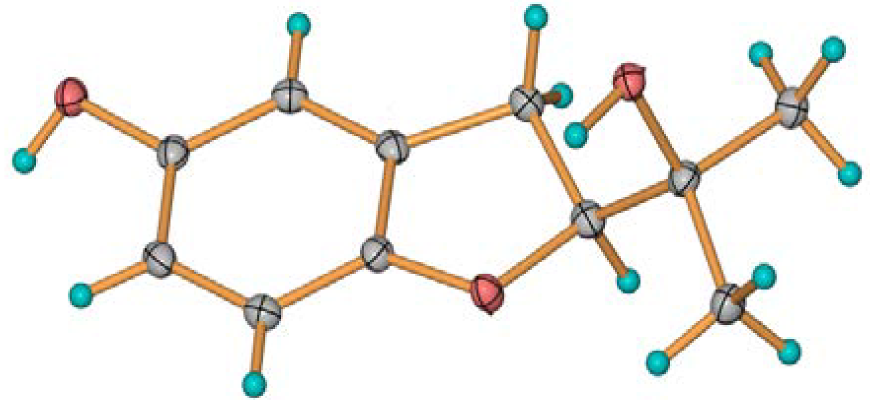
3.4. Crystal Structure Determination of 3

A crystal of 3 was obtained from MeOH solution. C11H14O3, M = 194.22, colorless block, orthorhombic system, space group P2(1)2(1)2(1), a = 6.2067(3), b = 9.1962(3), c = 16.9308(7) Å. V = 966.37(7) Å3. Z = 4, d = 1.335 g/cm3, crystal size 0.40 × 0.39 × 0.41 mm3. The absolute flack parameter is 0.2 (4). X-ray diffraction data were collected on a Bruker SMART APEX CCD diffractometer with Cu Kα radiation (λ = 1.54184 Å) at a temperature of 293 K. The data were processed using CrysAlis. The structures were solved by a direct method. H-atoms were added in ideal positions and refined as riding models. The structures were refined using full-matrix least-squares based on F2 with the program, SHELXL [22,23].
The Cambridge Crystallographic Data Centre (CCDC) 1004603 contains the supplementary crystallographic data of Compound 3 [24].

3.5. Cytotoxicity Assay

The in vitro cytotoxicities of 14 were assayed using a standard protocol [1,2,3]. Hirsutanol A, a potent anticancer agent isolated from marine fungal metabolites, was used as a positive control, and it showed cytotoxic activities against the tested cancer cell lines, HONE1, SUNE1 and GLC82, with IC50 values of 17.4, 3.5 and 10.1 μM, respectively [4].

3.6. Antibacterial Activity Assay

Vancomycin and ampicillin sodium were used as a positive control. The MIC values were determined using a broth dilution method (Mueller–Hinton broth) based on the National Committee on Clinical Laboratory Standards (NCCLS). The starting concentrations of the tested compounds were 256 µg/mL (from 256 to 0.25). The solution of compound in DMSO (10 µL) was added to 90 µL of bacterial culture (1 × 10⁶ CFU/mL) in the first well of flat-bottomed 96-well tissue culture plates. The solution was then double diluted. The bacterial culture solution containing the appropriate compound (50 µL) was discarded from the last well in order to ensure 100-µL volume of bacterial culture in every well. A set of tubes containing only inoculated broth was kept as a control. The plate was incubated at 37 °C overnight in an electro-heating standing-temperature cultivator before the measurement of the absorbance value. The optical density values at 600 nm were measured using a multifunction microplate reader (PowerWaveTM XS2, BioTek® Instruments Inc., Winooski, VT, USA).

4. Conclusions

Isobenzofuranones, fused-ring aromatic γ-lactones, showed various biological activities and have attracted much attention from synthetic chemists [25]. However, naturally occurring isobenzofuranones are few and mainly found in plants and endophytic fungi [26,27,28,29,30]. Moreover, the absolute configurations at the C-3 position of many substituted isobenzofuranone still remain unknown. Pseudaboydins A (1) and B (2) are unprecedented isobenzofuranone derivatives with a methylpentyl side chain. The absolute configurations of 1 and 2 were unambiguously determined as 3R by CD spectra analysis. The stereochemistry of Compound 3 was first elucidated as 2R by single-crystal X-ray diffraction analysis. Compound 4 was also assigned as having 2R absolute configuration for the CD, similar to 3.

Supplementary Files

  • Supplementary File 1:

    Supplementary Information (PDF, 2391 KB)

  • Acknowledgments

    This project is financially supported by the National Natural Science Foundation of China (Nos. 30973633 and J1103305), the Guangdong Provincial Science and Technology Research Program (No. 2012A031100005), the Guangdong Natural Science Foundation (No. S2012010010653), the Special Financial Fund of Innovative Development of Marine Economic Demonstration Project (GD2012-D01-001), the Guangzhou Science and Technology Research Program (Nos. 2011Y1-00036 and 2014J4100059) and the Instrumental Analysis and Research Center of Sun Yat-Sen University Funding (No. 2013-06).

    Author Contributions

    Conceived and designed the experiments: Wen-Jian Lan, Hou-Jin Li. Performed the experiments: Wei Liu, Wan-Ling Liang, Zeng Xu, Xiu Le, Jun Xu, Chi-Keung Lam, De-Po Yang, Hou-Jin Li, Lai-You Wang. Wrote the paper: Wen-Jian Lan, Wei Liu, Hou-Jin Li.

    Conflicts of Interest

    The authors declare no conflict of interest.

    References

    1. Li, H.J.; Jiang, W.H.; Liang, W.L.; Huang, J.X.; Mo, Y.F.; Ding, Y.Q.; Lam, C.K.; Qian, X.J.; Zhu, X.F.; Lan, W.J. Induced marine fungus Chondrostereum sp. as a means of producing new sesquiterpenoids Chondrosterins I and J by using glycerol as the carbon source. Mar. Drugs 2014, 12, 167–175. [Google Scholar] [CrossRef]
    2. Li, H.J.; Chen, T.; Xie, Y.L.; Chen, W.D.; Zhu, X.F.; Lan, W.J. Isolation and structural elucidation of Chondrosterins F-H from the marine fungus Chondrostereum sp. Mar. Drugs 2013, 11, 551–558. [Google Scholar] [CrossRef]
    3. Li, H.J.; Xie, Y.L.; Xie, Z.L.; Chen, Y.; Lam, C.K.; Lan, W.J. Chondrosterins A–E, triquinane-type sesquiterpenoids from soft coral-associated fungus Chondrostereum sp. Mar. Drugs 2012, 10, 627–638. [Google Scholar] [CrossRef]
    4. Li, H.J.; Lan, W.J.; Lam, C.K.; Yang, F.; Zhu, X.F. Hirsutane sesquiterpenoids from the marine-derived fungus Chondrostereum sp. Chem. Biodivers. 2011, 8, 317–324. [Google Scholar] [CrossRef]
    5. Lan, W.J.; Zhao, Y.; Xie, Z.L.; Liang, L.Z.; Shao, W.Y.; Zhu, L.P.; Yang, D.P.; Zhu, X.F.; Li, H.J. Novel sorbicillin analogues from the marine fungus Trichoderma sp. associated with the seastar Acanthaster planci. Nat. Prod. Commun. 2012, 7, 1337–1340. [Google Scholar]
    6. Zhao, Y.; Li, S.Q.; Li, H.J.; Lan, W.J. Lanostane triterpenoids from the fungus Ceriporia lacerate. Chem. Nat. Compd. 2013, 49, 653–656. [Google Scholar] [CrossRef]
    7. Xie, Z.L.; Li, H.J.; Wang, L.Y.; Liang, W.L.; Liu, W.; Lan, W.J. Trichodermaerin, a new lactone from the marine fungus Trichoderma erinaceum associated with the sea star Acanthaster planci. Nat. Prod. Commun. 2013, 8, 67–68. [Google Scholar]
    8. Chang, Y.C.; Deng, T.S.; Pang, K.L.; Hsiao, C.J.; Chen, Y.Y.; Tang, S.J.; Lee, T.H. Polyketides from the littoral plant associated fungus Pseudallescheria boydii. J. Nat. Prod. 2013, 76, 1796–1800. [Google Scholar]
    9. Su, H.J.; Lin, M.J.; Tsou, Y.J.; Ko, W.H. Pseudallin, a new antibiotic produced by the human pathogenic fungus Pseudallescheria boydii, with ecological significance. Bot. Stud. 2012, 53, 239–242. [Google Scholar]
    10. Ko, W.H.; Tsou, Y.J.; Ju, Y.M.; Hsieh, H.M.; Ann, P.J. Production of a fungistatic substance by Pseudallescheria boydii isolated from soil amended with vegetable tissues and its significance. Mycopathologia 2010, 169, 125–131. [Google Scholar] [CrossRef]
    11. Pavlaskova, K.; Nedved, J.; Kuzma, M.; Zabka, M.; Sulc, M.; Sklenar, J.; Novak, P.; Benada, O.; Kofronova, O.; Hajduch, M.; et al. Characterization of Pseudacyclins A–E, a suite of cyclic peptides produced by Pseudallescheria boydii. J. Nat. Prod. 2010, 73, 1027–1032. [Google Scholar]
    12. Nirma, C.; Eparvier, V.; Stien, D. Antifungal agents from Pseudallescheria boydii SNB-CN73 isolated from a Nasutitermes sp. Termite. J. Nat. Prod. 2013, 76, 988–991. [Google Scholar]
    13. Pinto, M.R.; Rodrigues, M.L.; Travassos, L.R.; Haido, R.M.T.; Wait, R.; Barreto-Bergter, E. Characterization of glucosylceramides in Pseudallescheria boydii and their involvement in fungal differentiation. Glycobiology 2002, 12, 251–260. [Google Scholar]
    14. Uchida, I.; Kuriyama, K. The π–π* circular dichroism of α, β-unsaturated γ-lactones. Tetrahedron Lett. 1974, 15, 3761–3764. [Google Scholar] [CrossRef]
    15. Gawronski, J.K.; van Oeveren, A.; van der Deen, H.; Leung, C.W.; Feringa, B.L. Simple circular dichroic method for the determination of absolute configuration of 5-substituted 2(5H)-furanones. J. Org. Chem. 1996, 61, 1513–1515. [Google Scholar] [CrossRef]
    16. Tanaka, M.; Nara, F.; Yamasato, Y.; Ono, Y.; Ogita, T. F-11334s, new inhibitors of membrane-bound neutral sphingomyelinase. J. Antibiot. 1999, 52, 827–830. [Google Scholar]
    17. Maria, D.B.; Giovanni, V.; Paola, V.F.; Giovanni, F. Fungal metabolites. 28. The chemistry of Lactarius fuliginosus and Lactarius picinus. Tetrahedron 1992, 48, 7331–7344. [Google Scholar] [CrossRef]
    18. Flack, H.D.; Bernardinelli, G. The use of X-ray crystallography to determine absolute configuration. Chirality 2008, 20, 681–690. [Google Scholar] [CrossRef]
    19. Takenaka, Y.; Tanahashi, T.; Nagakura, N.; Hamada, N. Phenyl ethers from cultured lichen mycobionts of Graphis scripta var. serpentina and G. rikuzensis. Chem. Pharm. Bull. 2003, 51, 794–797. [Google Scholar]
    20. Wang, J.F.; Lu, Z.Y.; Liu, P.P.; Wang, Y.; Li, J.; Hong, K.; Zhu, W.M. Cytotoxic polyphenols from the fungus Penicillium expansum 091006 endogenous with the mangrove plant Excoecaria agallocha. Planta Med. 2012, 78, 1861–1866. [Google Scholar]
    21. Talapatra, S.K.; Karmacharya, B.; de, S.C.; Talapatra, B. (−)-Regiolone, an α-tetralone from Juglans regia: Structure, stereochemistry and conformation. Phytochemistry 1988, 27, 3929–3932. [Google Scholar]
    22. Sheldrick, G.M. SADABS: Program for Empirical Absorption Correction of Area Detector Data; University of Göttingen: Göttingen, Germany, 1996. [Google Scholar]
    23. Sheldrick, G.M. SHELXTL 5.10 for Windows NT: Structure Determination Software Programs; Bruker Analytical X-ray Systems, Inc.: Madison, WI, USA, 1997. [Google Scholar]
    24. CCDC CIF Depository Request Form. Available online: http://www.ccdc.cam.ac.uk/cgi-bin/catreq.cgi (accessed on 26 May 2014).
    25. Logrado, L.P.; Santos, C.O.; Romeiro, L.A.S.; Costa, A.M.; Ferreira, J.R.O.; Cavalcanti, B.C.; Manoel de Moraes, O.; Costa-Lotufo, L.V.; Pessoa, C.; dos Santos, M.L. Synthesis and cytotoxicity screening of substituted isobenzofuranones designed from anacardic acids. Eur. J. Med. Chem. 2010, 45, 3480–3489. [Google Scholar]
    26. Huang, X.Z.; Zhu, Y.; Guan, X.L.; Tian, K.; Guo, J.M.; Wang, H.B.; Fu, G.M. A novel antioxidant isobenzofuranone derivative from fungus Cephalosporium sp. AL031. Molecules 2012, 17, 4219–4224. [Google Scholar]
    27. Tayone, W.C.; Honma, M.; Kanamaru, S.; Noguchi, S.; Tanaka, K.; Nehira, T.; Hashimoto, M. Stereochemical investigations of isochromenones and isobenzofuranones isolated from Leptosphaeria sp. KTC727. J. Nat. Prod. 2011, 74, 425–429. [Google Scholar] [CrossRef]
    28. Ding, G.; Liu, S.C.; Guo, L.D.; Zhou, Y.G.; Che, Y.S. Antifungal metabolites from the plant endophytic fungus Pestalotiopsis foedans. J. Nat. Prod. 2008, 71, 615–618. [Google Scholar]
    29. Strobel, G.; Ford, E.; Worapong, J.; Harper, J.K.; Arif, A.M.; Grant, D.M.; Fung, P.C.W.; Chau, R.M.W. Isopestacin, an isobenzofuranone from Pestalotiopsis microspora, possessing antifungal and antioxidant activities. Phytochemistry 2002, 60, 179–183. [Google Scholar]
    30. Youla, S.T.; Kelvin, K.O.; Alan, K.W. Phytotoxic metabolites of Phomopsis convolvulus, a host-specific pathogen of field bindweed. Can. J. Chem. 1992, 70, 2276–2284. [Google Scholar]

    Share and Cite

    MDPI and ACS Style

    Lan, W.-J.; Liu, W.; Liang, W.-L.; Xu, Z.; Le, X.; Xu, J.; Lam, C.-K.; Yang, D.-P.; Li, H.-J.; Wang, L.-Y. Pseudaboydins A and B: Novel Isobenzofuranone Derivatives from Marine Fungus Pseudallescheria boydii Associated with Starfish Acanthaster planci. Mar. Drugs 2014, 12, 4188-4199. https://doi.org/10.3390/md12074188

    AMA Style

    Lan W-J, Liu W, Liang W-L, Xu Z, Le X, Xu J, Lam C-K, Yang D-P, Li H-J, Wang L-Y. Pseudaboydins A and B: Novel Isobenzofuranone Derivatives from Marine Fungus Pseudallescheria boydii Associated with Starfish Acanthaster planci. Marine Drugs. 2014; 12(7):4188-4199. https://doi.org/10.3390/md12074188

    Chicago/Turabian Style

    Lan, Wen-Jian, Wei Liu, Wan-Ling Liang, Zeng Xu, Xiu Le, Jun Xu, Chi-Keung Lam, De-Po Yang, Hou-Jin Li, and Lai-You Wang. 2014. "Pseudaboydins A and B: Novel Isobenzofuranone Derivatives from Marine Fungus Pseudallescheria boydii Associated with Starfish Acanthaster planci" Marine Drugs 12, no. 7: 4188-4199. https://doi.org/10.3390/md12074188

    Article Metrics

    Back to TopTop