Article

Bioactive Isopimarane Diterpenes from the Fungus, Epicoccum sp. HS-1, Associated with Apostichopus japonicus

1
Key Laboratory for Applied Microbiology of Shandong Province, Biotechnology Center of Shandong Academy of Sciences, Jinan 250014, China
2
School of Chemistry and Chemical Engineering, Sun Yat-Sen University, Guangzhou 510275, China
3
Shandong Provincial Key Laboratory of Test Technology for Material Chemical Safety, Jinan 250014, China
*
Author to whom correspondence should be addressed.
Mar. Drugs 2015, 13(3), 1124-1132; https://doi.org/10.3390/md13031124
Submission received: 24 November 2014 / Revised: 8 February 2015 / Accepted: 13 February 2015 / Published: 2 March 2015
(This article belongs to the Special Issue Bioactive Compounds from Marine Microbes)

Abstract

:
One new isopimarane diterpene (1), together with two known compounds, 11-deoxydiaporthein A (2) and iso-pimara-8(14),15-diene (3) were isolated from the culture of Epicoccum sp., which was associated with Apostichopus japonicus. Their structures were determined by the analysis of 1D and 2D NMR, as well as mass spectroscopic data. The absolute configuration of Compound 1 was deduced by a single-crystal X-ray diffraction experiment using CuKα radiation. In the bioactivity assay, both Compounds 1 and 2 exhibited α-glucosidase inhibitory activity with IC50 values of 4.6 ± 0.1 and 11.9 ± 0.4 μM, respectively. This was the first report on isopimarane diterpenes with α-glucosidase inhibitory activity.

1. Introduction

The incidence and prevalence of type 2 diabetes, representing over 90% of all case of diabetes, are increasing rapidly as time passes. The International Diabetes Federation predicted that the number of people with diabetes would rise from 266 million in 2011 to 552 million by 2030 [1]. Alpha-glucosidase is a key enzyme for breaking down carbohydrates for absorption [2]. Agents inhibiting α-glucosidase are effective therapeutic agents for diabetes [3]. The development of new drugs for use against α-glucosidase is therefore urgently needed.
Marine fungi have attracted more attention for their good bioactivity against α-glucosidase [4,5,6]. In the course of our continuing search for biologically-active substances from fungi derived from mangrove and sea cucumber [7,8,9], we had screened extracts from a number of fungi, and those with interesting biological activities were routinely subjected to chemical exploration. The ethyl acetate extract from the fungus, Epicoccum sp. HS-1, associated with Apostichopus japonicus, showed α-glucosidase inhibition activity. One new isopimarane diterpene (1), together with two known compounds, 11-deoxydiaporthein A (2) and isopimara-8(14),15-diene (3), were isolated from the extract. To date, biomedical and pharmaceutical studies of isopimarane diterpene have shown interesting activities, such as cytotoxicity [10,11], acetylcholinesterase inhibitory activity [12] and anti-HIV-1 bioactivity [13]. To our knowledge, there have been no previous reports of the α-glucosidase inhibition activity of these. The details of the isolation, structural elucidation and the results of the α-glucosidase inhibition study of the isolated compounds are reported herein.

2. Results and Discussion

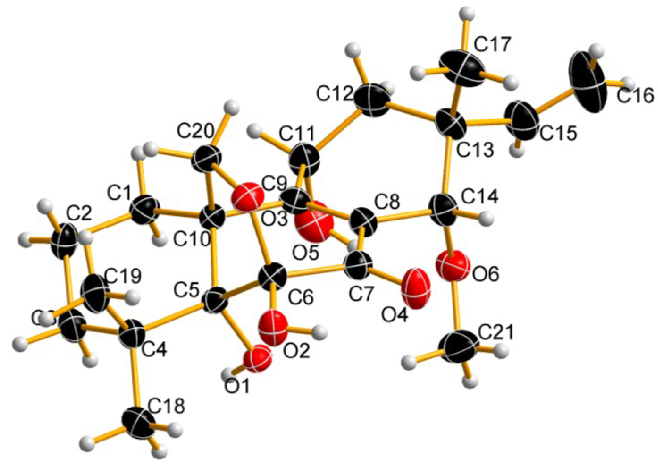
2.1. Chemical Structure Elucidation

Compound 1 was isolated as colorless crystals. The molecular formula was established as C21H30O6 on the basis of HR-ESI-TOF/MS data for m/z 401.1938 [M + Na]+ with seven degrees of unsaturation. Twenty seven protons were bound to carbons, and three exchangeable hydrogens were present. Detailed analysis of the 1H, 13C, HMQC, as well as DEPT spectra of Compound 1 revealed the presence of one methoxy group at δH 3.23/57.6 (21-CH3O), two oxygenated methylene groups at δH 4.43 and 3.22/69.8 (H-20a, H-20b), two oxygenated methine groups at δH 4.42/ 62.8 (H-11) and δH 4.15/80.0 (H-14), three methyl groups at δH 0.81/25.5 (CH3-17), δH 1.18/28.1 (CH3-19) and δH 1.51/23.8 (CH3-18), sp3 methylene groups at δH 2.15 and 1.67/23.3 (H-1a, H-1b), δH 1.82/17.8 (H-2), δH 1.60 and 1.28/37.1 (H-3a, H-3b), as well as δH 2.30 and 1.78/40.8 (H-12a, H-12b), an sp2 methylene group at δH 5.07 and 5.11/112.3 (H-16a, H-16b) and an sp2 methine group at δH 6.20/145.3 (H-15). In addition, five sp3 and two sp2 non-hydrogenated carbon signals appeared at δC 36.8, 41.2, 52.9, 81.2, 105.0, 133.2, 165.7, as well as a carbonyl group at 193.4. On the basis of these data, Compound 1 was hypothesized to have a diterpene skeleton.
Compound 1 was also presumed to have four rings in its structure based on the unsaturation degree, since it contained two double bonds and a carbonyl group. The COSY spectrum of Compound 1 revealed the presence of three spin systems, including CH2(1)CH2(2)CH2(3), CH(11)CH2(12) and CH(15)CH2(16). The extension of the spin systems and attachments of functional groups were confirmed by HMBC correlations. The HMBC correlations of CH3-18 and CH3-19 to C-4 and C-5 indicated that CH3-18 and CH3-19 were attached to C-4 at δC 36.8, CH3-17 to C-13, C-14 and C-15, H-15 to C-13 and C-16, indicating that both CH3-17 and CH (15) = CH2 (16) were attached to C-13, and 21-OCH3 to C-14, suggesting that OCH3 was linked to C-14. H-20a with C-6 in HMBC allowed the linkage of oxygenated an sp3 methylene carbon C-20 at δC 69.8 to the hemi-acetyl carbon C-6 at δC 105.0 through oxygen to form a dihydrofuran ring. Ultimately, the chemical structure of Compound 1 was identified as shown in Figure 1. Detailed 1H and 13C NMR data are presented in Table 1. More detailed spectra are available at the Supplementary Figures S1–S7.
Figure 1. Structures of isolated Compounds 13.
Marinedrugs 13 01124 g001
Table 1. 1H (MeOD, 600 MHz) and 13C (MeOD, 150 MHz) NMR data of Compound 1 a.
PositionδC, mult.δHHMBC (H→C)
123.3, CH2a, 2.15 (dt, 4.8, 13.8); b, 1.67 (m)C-2, 10
217.8, CH21.82 (2H, dt, 6.0,13.0)C-1, 3
337.1, CH2a, 1.60 (dt, 3.0, 13.8); b, 1.28 (m)C-2, 5
436.8, C
581.2, C
6105.0, C
7193.4, C
8133.2, C
9165.7, C
1052.9, C
1162.8, CH4.42 (dd, 2.0, 8.4)C-8, 9, 12
1240.8, CH2a, 2.30 (dd, 2.0, 14.4); b, 1.78 (dd, 9.0, 14.4)C-9, 11, 13, 15
1341.2, C
1480.0, CH4.15 (s)C-7, 9, 21-OCH3
15145.3, CH6.20 (dd, 11.4, 18.6)C-16
16112.3, CH2a, 5.07 (d, 12.0); b, 5.11 (d, 18.0)C-14, 15
1725.5, CH30.81 (s)C-13, 14, 15
1823.8, CH31.51 (s)C-4, 5, 19
1928.1, CH31.18 (s)C-4, 5, 18
2069.8, CH2a, 4.43 (d, 9.0); b, 3.22 (d, 9.0)C-5, 6, 9
2157.6, CH33.23 (s)C-14
a δ in ppm, J in Hz.
The relative configuration of Compound 1 was established on the basis of NOESY correlations of H-20a (δ 4.43, d, J = 9.0 Hz)/CH3-18 (δ 1.51, s)/H-11 (δ 4.42, dd, J = 2.0, 8.4 Hz) and CH3-17 (δ 0.81, s)/H-14 (δ 4.15, s). The coupling constants derived from coupling between H-11 (δ 4.42, dd, J = 2.0, 8.4 Hz), H-12a (δ 2.30, t, J = 2.0, 14.4 Hz) and H-12b (δ 1.78, t, J = 9.0, 14.4 Hz) confirmed the equatorial position of H-11.
The absolute configuration of 1 was further confirmed by a single-crystal X-ray diffraction experiment using CuKα radiation (Figure 2) with Cambridge Crystallographic Data Centre (CCDC) No. 1000803, which was defined as 5R, 6S, 10S, 11R, 13R and 14S.
The structure of Compound 2 was identified as 11-deoxydiaporthein A by comparison of its NMR, MS and CD data with those available in the literature [14]. Compound 2 showed a positive Cotton effect at 217 nm, which was similar to that of 11-deoxydiaporthein A isolated from marine fungus, Cryptosphaeria eunomia var. eunomia [14], and showed only mild activity against Mycobacterium tuberculosis with a MIC value of 200 μg/mL [15]. Compound 3 was identified as ent-pimara-8(14),15-diene by comparison of its NMR and MS data [16], which was named as isopimara-8(14),15-diene in this paper. Isopimara-8(14),15-diene was obtained from engineered Aspergillus nidulans and showed significantly higher DPPH radical scavenging activity than beta-carotene [16].
Figure 2. Perspective oak ridge thermal ellipsoid plot (ORTEP) drawing of Compound 1.
Marinedrugs 13 01124 g002

2.2. Biological Activity

All compounds were tested for their in vitro inhibitory activities against α-glucosidase according to a previously described method with modifications [17]. The results are listed in Table 2. Compounds 1 and 2 exhibited more effective inhibitory activities, with IC50 values of 4.6 ± 0.1 and 11.9 ± 0.4 μM, respectively, than resveratrol, which was used as the positive control, while Compound 3 did not exhibit inhibitory activities (IC50 > 200 μM) in this test (Table 2).
Table 2. Inhibitory effects of the isolates against α-glucosidase.
CompoundsIC50 (μM)
14.6 ± 0.1
211.9 ± 0.4
3>200.0
Resveratrol a31.2 ± 4.4
a Positive control.

3. Experimental Section

3.1. General Experimental Procedures

Optical rotations were measured on an Anton Paar MCP 300 polarimeter (Ashland, VA, USA) at 30 °C. UV data were recorded on a Shimadzu UV-240 spectrophotometer. CD spectra were recorded on a JASCO J-815 spectropolarimeter using a Nicolet Magna-IR 750 spectrophotometer. 1H and 13C NMR spectra were recorded on a Bruker AVANCE 600 (600 and 150 MHz). Chemical shifts were reported in δ (ppm), using tetramethylsilane (TMS) as the internal standard, and coupling constants (J) were in Hertz (Hz). HR-ESI-TOF/MS was performed on an Agilent 6530 high-resolution mass spectrometer. Single-crystal data were measured on an Agilent Gemini Ultra diffractometer (CuKα radiation). Column chromatography (CC) was performed on silica gel (200–300 mesh, Qingdao Marine Chemical Factory, Qingdao, China) and a Sephadex LH-20 (Amersham Pharmacia Biotech AB, Stockholm, Sweden). Precoated silica gel plates (G60, F-254, Yan Tai Zi Fu Chemical Group Co., Yantai, China) were used for thin-layer chromatography. Semipreparative HPLC was performed on a Waters HPLC system consisting of a 2695 pump, a 2998 PDA (potato dextrose agar), an autosampler and a YMC C18 column (250 × 10 mm, 5 μm); flow rate: 2.0 mL/min. Compounds 13 were isolated as the secondary metabolites from cultures of the Apostichopus japonicus-derived fungus strain, Epicoccum sp. HS-1. Resveratrol was purchased from Adamas-beta Ltd. Co. (Shanghai, China).

3.2. Fungal Material

The HS-1 strain [8] was cultured on slants of PDA at 25 °C for 7 days. Agar plugs were cut into small pieces (1-cm diameter each) under aseptic conditions, and 10 pieces were used to inoculate into five Erlenmeyer flasks (500 mL), each containing 100 mL of media (glucose 10 g/L, peptone 2 g/L, yeast extract 1 g/L and crude sea salt 3 g/L). Five flasks of inoculated media were incubated at 25 °C on a rotary shaker (170 r.p.m.) for seven days to prepare the seed culture. The mycelium was aseptically transferred to 100 Erlenmeyer flasks (500 mL) containing a total of 20 L of liquid medium consisting of glucose (10 g/L), peptone (2 g/L), yeast extract (1 g/L) and crude sea salt (3 g/L), then incubated at room temperature for 28 days.

3.3. Extraction and Isolation

The fermentation medium was filtered through cheesecloth. The filtrate was concentrated below 50 °C to a final volume of 5 L and extracted three times with ethyl acetate (1:1, v/v), and the extracts were pooled and concentrated to dryness with a rotary evaporator at 45 °C. The resulting crude extract (5.2 g) was subjected to vacuum liquid chromatography (VLC) with silica gel (60, <0.663 mm, Merck, Darmstadt, Germany). The column system was sequentially eluted by 500 mL of each of the following solvents: petroleum, petroleum/ethyl acetate (9:1, 4:1, 7:3, 1:1, 1:4, v/v); ethyl acetate, ethyl acetate/MeOH (4:1, 7:3, 1:1, 1:4, 0:5 v/v). At last, 10 fractions were obtained, all of which were monitored by thin liquid chromatography (TLC) in 254 and 310 nm. Fraction 2 was further purified by a Sephadex LH-20 using CH2Cl2/MeOH (1:1, v/v) to obtain Compound 3 (14.0 mg). Fraction 5 (30.6 mg) was separated on a Sephadex LH-20 using CH2Cl2/MeOH (1:1, v/v) to give Compound 2 (7.1 mg). Fraction 4 (21.0 mg) was purified by a Sephadex LH-20 using CH2Cl2/MeOH (1:1, v/v) to give ten subfractions (Fraction 4-1–4-10), and Fraction 4-5 and Fraction 4-6 were combined and then subjected to high performance liquid chromatography (HPLC) over octadecylsilane (ODS) (YMC, 250 × 10 mm) (30% MeOH-H2O, v/v, 5 min; 30%–100% MeOH-H2O, v/v, 45 min; 100% MeOH, 55 min ) to give Compound 1 (retention time (tR) 38.4 min; 4.4 mg).
Isopimarane diterpene (1): colorless crystals. UV (MeOH): λmax (log ε): 191 (0.62) and 248 (0.31) nm. CD (MeOH) λmax (θ in mdeg) 191.2 (38.1), 193.9 (−4.85), 250.0 (−3.68), 266.7 (−5.03); [α]30D −242. (c 0.024 MeOH); HR-ESI-TOF/MS: m/z 401.1938 [M + Na]+ (calcd. for 401.1940). For the 1H and 13C NMR data, see Table 1.
X-ray crystallographic analysis of 1: Colorless crystals of 1 were obtained from MeOH by slow evaporation. X-ray diffraction data were measured on an Agilent Gemini Ultra diffractometer (CuKα radiation, λ = 1.54178 Å) at 293 K. The structure was solved by direct methods (SHELXS-97) and refined using full-matrix least squares difference Fourier techniques. All non-hydrogen atoms were refined anisotropically, and all hydrogen atoms were placed in idealized positions and refined as riding atoms with the relative isotropic parameters. Monoclinic, C21H30O6, space group P21, a = 10.9167 (4) Å, b = 7.5903 (2) Å, c = 12.2976 (4) Å, α = γ = 90.00, β = 109.063 (4), V = 803.78 (5) Å3, Z = 2, Dcalcd = 1.305 g/cm3, μ = 0.775 mm−1, F(000) = 408, Flack = −0.23 (17). Crystal size: 0.41 × 0.38 × 0.15 mm3. Independent reflections: 2947 [Rint = 0.0207]. The final indices were R1 = 0.0319, wR2 = 0.0801 (I > 2σ (I)).

3.4. Assays for Inhibitory Activity

All of the assays were carried out using 0.01 M KH2PO4/K2HPO4 buffer, pH 7.0, using a Shimadzu 2450 spectrophotometer. Enzyme solutions were prepared to give 2.0 units/mL in 2-mL aliquots. The assay medium contained phosphate buffer, pH 7.0 (950 µL), 10 µL of enzyme, 20 µL DMSO or inhibitor (dissolved in DMSO) and 20 µL of 0.01 M substrate (p-nitrophenyl (PNP) glycosides (3 mg/mL). The substrate was added to the assay medium containing enzyme and buffer with the inhibitor after 20 min of incubation time, and then, the increase in absorbance was measured at 400 nm for 1-min intervals at 37 °C. The percentage of inhibitory activity was calculated according to the equation: η (%) = ((B − S)/B) × 100% (B, the assay medium with DMSO; S, the assay medium with inhibitor). Values of IC50 were calculated according to the inhibitory activity curve. All measurements were done in triplicate from two independent experiments. The reported IC50 was the average value of two independent experiments.

4. Conclusions

Epicoccum sp. associated with Apostichopus japonicus was cultivated in liquid media, and a chemical investigation of cultures of this strain led to the isolation of one new isopimarane diterpene (1), together with two known compounds, 11-deoxydiaporthein A (2) and isopimara-8(14),15-diene (3). The absolute configuration of Compound 1 was deduced as 5R, 6S, 10S, 11R, 13R and 14S by a single-crystal X-ray diffraction experiment with CuKα radiation. All isolated compounds were evaluated for their α-glucosidase inhibitory activity, and both Compounds 1 and 2 exhibited α-glucosidase inhibitory activity with IC50 values of 4.6 ± 0.1 and 11.9 ± 0.4 μM, respectively. To our best knowledge, this was the first report on isopimarane diterpenes with α-glucosidase inhibitory activity. These results suggest that fungi associated with sea cucumber could be a good resource of anti-α-glucosidase agent.

Supplementary Files

Supplementary File 1

Acknowledgments

We gratefully acknowledge the financial support of the National Natural Science Foundation of China (No. 81202452), the Prize Fund for Excellent Middle Aged and Young Scientists of Shandong Province (BS2009HZ004, BS2013HZ016), the NSFC-NRF Scientific Cooperation Program (No. 81411140251) and the Funds for Excellent Youth of Shandong Academy of Sciences.

Author Contributions

Conceived and designed the experiments: Xuekui Xia, Zhigang She. Performed the experiments including fermentation, extraction, isolation and bioactivity test: Xuekui Xia, Jun Qi, Airong Jia, Yayue Liu, Changheng Liu, Cuiling Gao. Analyzed the NMR and X-ray diffraction data: Xuekui Xia, Yayue Liu, Yonggang Zhang. Wrote the paper: Xuekui Xia. Read and approved the final manuscript: Cuiling Gao, Zhigang She, Xuekui Xia.

Conflicts of Interest

The authors declare no conflicts of interest.

References

  1. Lyssenko, V.; Laakso, M. Genetic screening for the risk of Type 2 diabetes. Diabetes Care 2013, 36, 5120–5126. [Google Scholar] [CrossRef]
  2. Van de Laar, F.A.; Lucassen, P.L.; Akkermans, R.P.; van de Lisdonk, E.H.; de Grauw, W.J. Alpha-glucosidase inhibitors for people with impaired glucose tolerance or impaired fasting blood glucose. Cochrane Database Syst. Rev. 2006, 18, CD005061. [Google Scholar]
  3. Ahamad, J.; Naquvi, K.J.; Mir, S.R.; Ali, M.; Shuaib, M. Review on role of natural α-glucosidase inhibitors for management of diabetes mellitus. Int. J. Biomed. Res. 2011, 2, 374–380. [Google Scholar]
  4. Blunt, J.W.; Copp, B.R.; Keyzers, R.A.; Munro, M.H.; Prinsep, M.R. Marine natural products. Nat. Prod. Rep. 2014, 31, 160–258. [Google Scholar] [CrossRef] [PubMed]
  5. Blunt, J.W.; Copp, B.R.; Keyzers, R.A.; Munro, M.H.; Prinsep, M.R. Marine natural products. Nat. Prod. Rep. 2013, 30, 237–323. [Google Scholar] [CrossRef] [PubMed]
  6. Bhattacharjee, R.; Mitra, A.; Dey, B.; Pal, A. Exploration of Anti-diabetic Potentials Amongst Marine Species. Indo Global J. Pharmaceut. Sci. 2014, 4, 65–73. [Google Scholar]
  7. Xiao, Z.E.; Huang, H.R.; Shao, C.L.; Xia, X.K.; Ma, L.; Huang, X.S.; Lu, Y.J.; Lin, Y.C.; Long, Y.H.; She, Z.G. Asperterpenols A and B, new sesterterpenoids isolated from a mangrove endophytic fungus Aspergillus sp. 085242. Org. Lett. 2013, 15, 2522–2525. [Google Scholar] [CrossRef] [PubMed]
  8. Xia, X.K.; Zhang, J.Y.; Zhang, Y.G.; Wei, F.; Liu, X.; Jia, A.R.; Liu, C.H.; Li, W.; She, Z.G.; Lin, Y.C. Pimarane diterpenes from the fungus Epicoccum sp. HS-1 associated with Apostichopus japonicus. Bioorg. Med. Chem. Lett. 2012, 22, 3017–3019. [Google Scholar] [CrossRef] [PubMed]
  9. Liu, Y.Y.; Xia, G.X.; Li, H.X.; Ma, L.; Ding, B.; Lu, Y.J.; He, L.; Xia, X.K.; She, Z.G. Vermistatin derivatives with α-Glucosidase inhibitory activity from the mangrove endophytic fungus Penicillium sp. Planta Med. 2014, 80, 912–917. [Google Scholar] [CrossRef] [PubMed]
  10. Wang, X.N.; Bashyal, B.P.; Wijeratne, E.M.K.; U’Ren, J.M.; Liu, M.X.; Gunatilaka, M.K.; Arnold, A.E.; Gunatilaka, A.A.L. Smardaesidins A–G, isopimarane and 20-nor-isopimarane diterpenoids from Smardaea sp., a fungal endophyte of the moss Ceratodon purpureus. J. Nat. Prod. 2011, 74, 2052–2061. [Google Scholar]
  11. Maslovskaya, L.A.; Savchenko, A.I.; Gordon, V.A.; Reddell, P.W.; Pierce, C.J.; Parsons, P.G.; Williams, C.M. EBC-316, 325–327, and 345: New pimarane diterpenes from Croton insularis found in the Australian rainforest. Aust. J. Chem. 2015. doi: org/10.1071/CH14550. [Google Scholar]
  12. Yilmaz, A.; Caglar, P.; Dirmenci, T.; Goren, N.; Topcu, G. A novel isopimarane diterpenoid with acetylcholinesterase inhibitory activity from Nepeta sorgerae, an endemic species to the Nemrut Mountain. Nat. Prod. Commun. 2012, 7, 693–696. [Google Scholar] [PubMed]
  13. Huang, S.Z.; Ma, Q.Y.; Fang, W.W.; Xu, F.Q.; Peng, H.; Dai, H.F.; Zhou, J.; Zhao, Y.X. Three new isopimarane diterpenoids from Excoecaria acerifolia. J. Asian Nat. Prod. Res. 2013, 15, 750–755. [Google Scholar] [CrossRef] [PubMed]
  14. Yoshida, S.; Kito, K.; Ooi, T.; Kanoh, K.; Shizuri, Y.; Kusumi, T. Four pimarane diterpenes frommarine fungus: Chloroform incorporated in crystal lattice for absolute configuration analysis by X-ray. Chem. Lett. 2007, 36, 1386–1387. [Google Scholar] [CrossRef]
  15. Dettrakul, S.; Kittakoop, P.; Isaka, M.; Nophichai, S.; Suyarnsestakorn, C.; Tanticharoen, M.; Thebtaranonth, Y. Antimycobacterial pimarane diterpenes from the fungus Diaporthe sp. Bioorg. Med. Chem. Lett. 2003, 13, 1253–1255. [Google Scholar] [CrossRef] [PubMed]
  16. Bromanm, K.; Viljanen, K.; Moreira, V.M.; Yli-Kauhaluoma, J.; Ruohonen, L.; Nakari-Setala, T. Isolation and purification of ent-pimara-8(14), 15-diene from engineered Aspergillus nidulans by accelerated solvent extraction combined with HPLC. Anal. Methods 2014, 6, 1227–1234. [Google Scholar] [CrossRef]
  17. Du, Z.Y.; Liu, R.R.; Shao, W.Y.; Mao, X.P.; Ma, L.; Gu, L.Q.; Huang, Z.S.; Chan, A.S.C. α-Glucosidase inhibition of natural curcuminoids and curcumin analogs. Eur. J. Med. Chem. 2006, 41, 213–218. [Google Scholar] [CrossRef] [PubMed]

