
A New Cyclic Hexapeptide and a New Isocoumarin Derivative from the Marine Sponge-Associated Fungus Aspergillus similanensis KUFA 0013

 , , and
Abstract
:1. Introduction

2. Results and Discussion

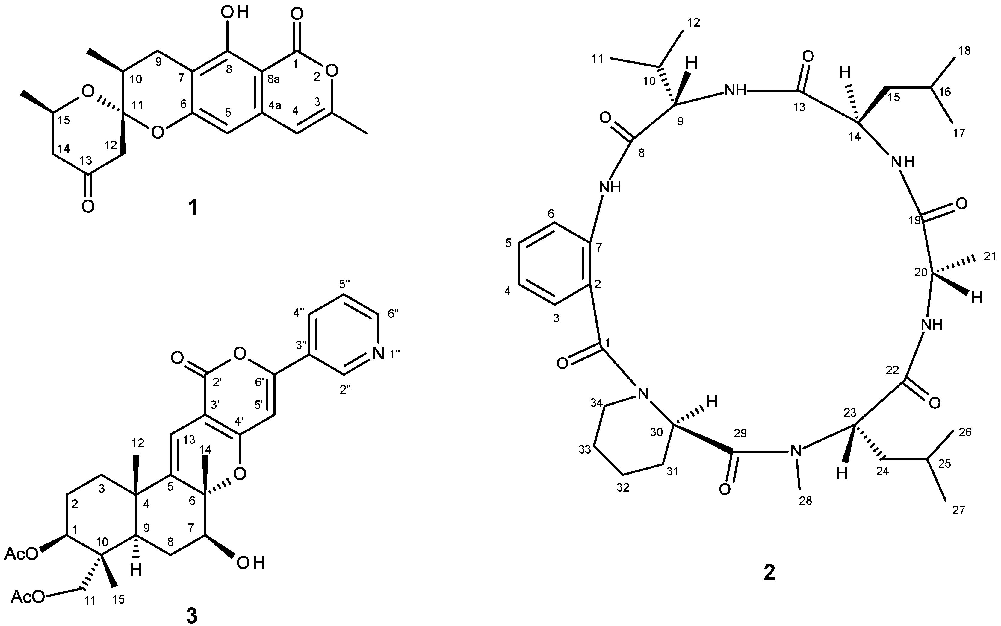
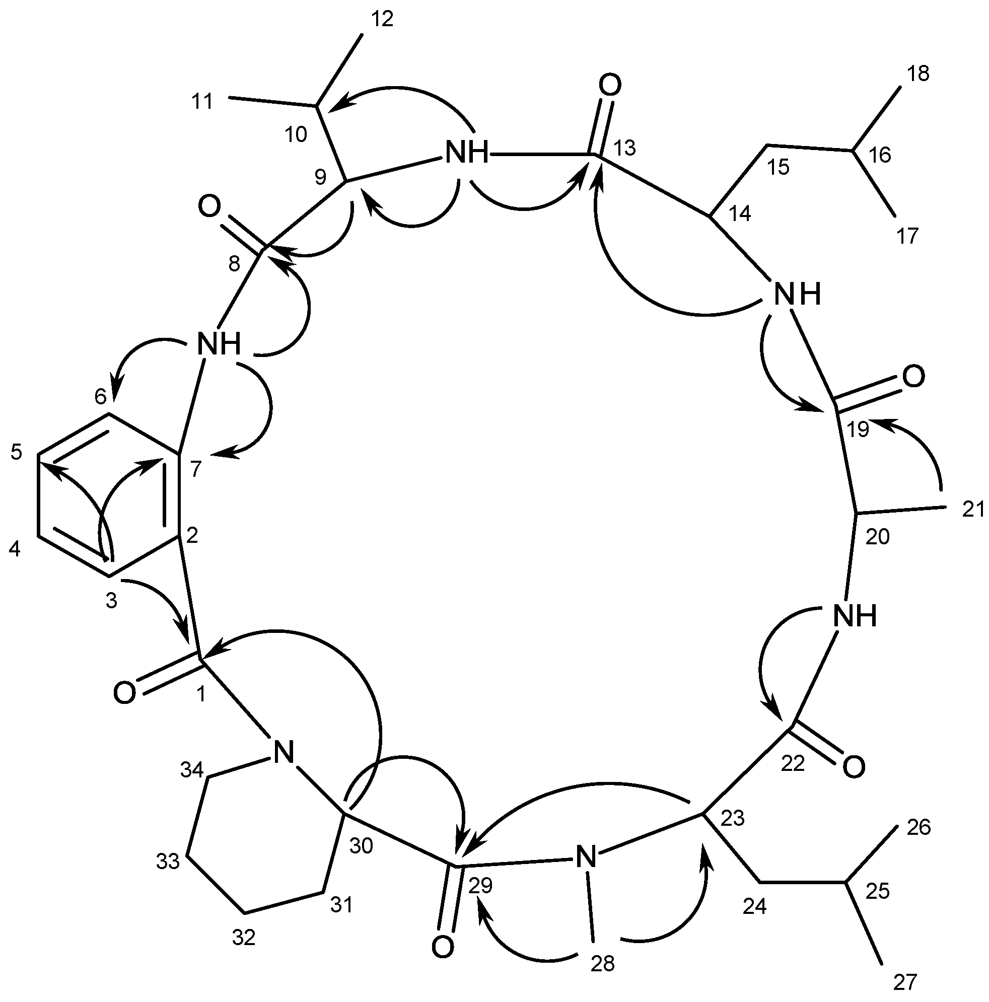
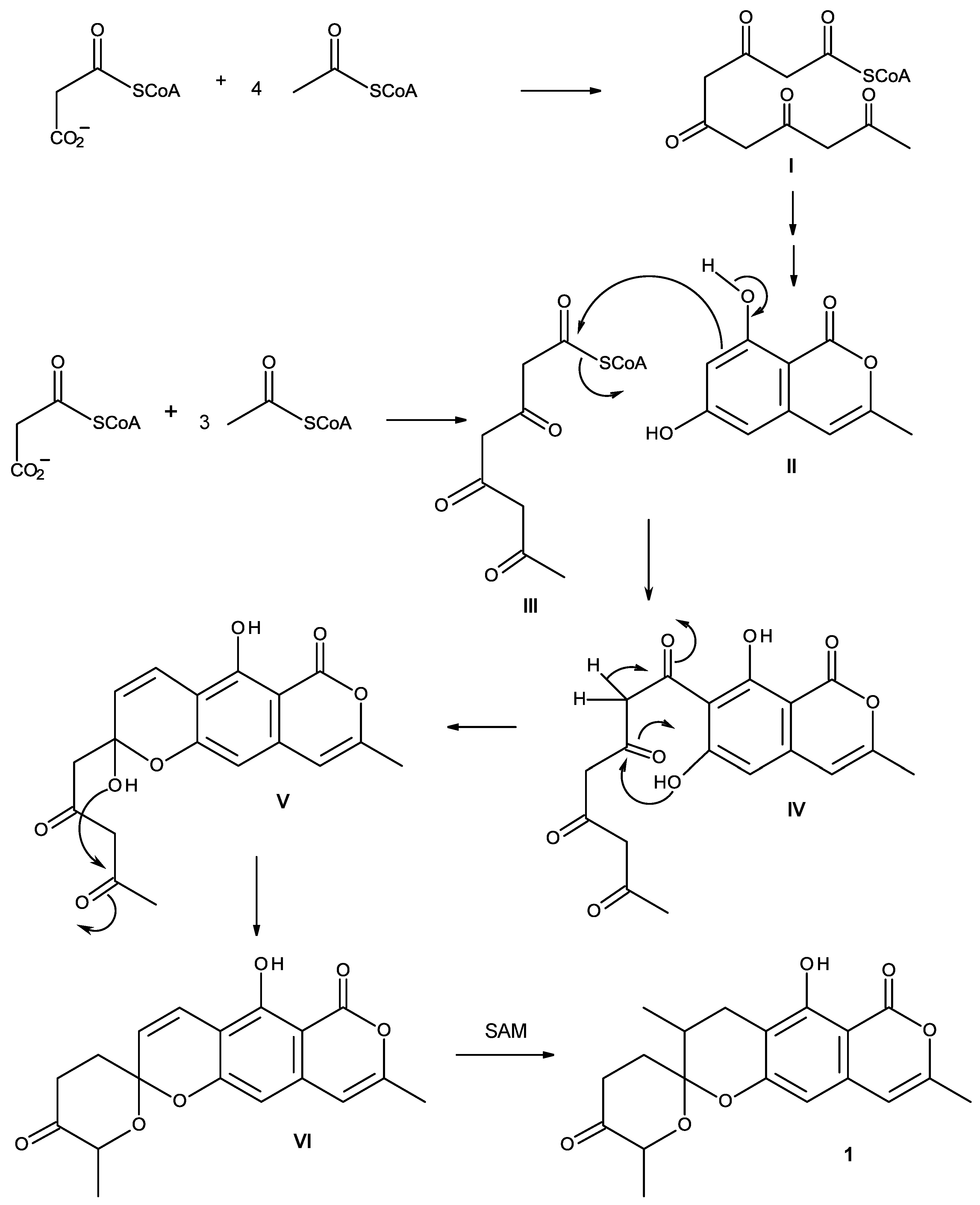
| Position | δC, Type | δH, (J in Hz) | COSY | HMBC | NOESY |
|---|---|---|---|---|---|
| 1 | 166.8, CO | - | |||
| 3 | 153.4, C | - | |||
| 4 | 104.4, CH | 6.13, d (0.9) | CH3-3 | C-3, 5, 8a | CH3-3 |
| 4a | 136.7, C | - | |||
| 5 | 103.0, CH | 6.26, s | C-4, 6, 7, 8a | ||
| 6 | 158.3, C | - | |||
| 7 | 110.0, C | - | |||
| 8 | 160.0, C | - | |||
| 8a | 99.6, C | - | |||
| 9α | 23.6, CH2 | 2.81, dd (16.8, 5.6) | H-9β, H-10 | C-6, 7, 10, 11 | H-9β, H-10, CH3-10 |
| 9β | 2.57, brd (16.8) | H-9α, H-10 | C-7, 10, 11 | H-9α, H-10, CH3-10 | |
| 10 | 33.9, CH | 1.98, m | H-9α, H-9β, CH3-10 | H-9α,H-9β, CH3-10 | |
| 11 | 102.9, C | - | |||
| 12α | 47.2, CH2 | 2.55, dd (14.0, 1.9) | H-12β | C-10, 11, 13 | H-12β |
| 12β | 2.81, brd (14.0) | H-12α | C-10, 11, 13 | H-12α | |
| 13 | 205.3, CO | - | |||
| 14α | 48.3, CH2 | 2.28, ddd (14.7, 11.3, 0.7) | H-14β, H-15 | C-13, 15 | H-14β, CH3-15 |
| 14β | 2.48, ddd (14.7, 2.9, 1.9) | H-14α, H-15 | C-13 | H-14β, CH3-15 | |
| 15 | 67.2, CH | 4.15, m | H-14α, H-14β, CH3-15 | H-14β, CH3-15 | |
| CH3-3 | 19.4, CH3 | 2.24, s | H-4 | C-3, 4 | H-4 |
| CH3-10 | 15.9, CH3 | 1.23, d (6.2) | H-10 | C-9, 10, 11 | H-9β, H-10, H-12α, 2β |
| CH3-15 | 21.6, CH3 | 1.21, d (6.2) | H-15 | C-14, 15 | H-14α, H-14β, H-15 |
| OH-8 | - | 11.35, s | C-7, 8, 8a |


| Position | δC, Type | δH, (J in Hz) | COSY | HMBC | NOESY | |
|---|---|---|---|---|---|---|
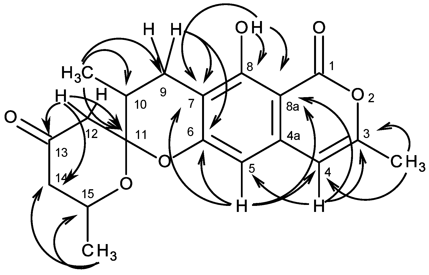
| Anthranilic acid | 1 | 170.2, CO | - | |||
| 2 | 122.7, C | - | ||||
| 3 | 127.1, CH | 7.20, dd (7.7, 1.5) | H-4 | C-1, 5, 7 | H-34 | |
| 4 | 123.4, CH | 7.13, ddd (7.9, 7.9, 1.0) | H-3, 5 | C-2, 6 | ||
| 5 | 131.7, CH | 7.47, ddd (7.9, 7.9, 1.6) | H-4, 6 | C-3, 7 | ||
| 6 | 123.9, CH | 8.29, d (8.3) | H-5 | C-2, 4 | H-12 | |
| 7 | 137.0, C | - | ||||
| NH | - | 9.41, brs | C-6, 7, 8 | NH (Val), H-9, 12 | ||
| 8 | 170.7, CO | - | ||||
| 9 | 59.3, CH | 4.32, dd (7.4, 3.3) | H-10, NH | C-8, 10, 11, 12 | H-10, 11 | |
| 10 | 29.9, CH | 2.68, m | H-9, 11, 12 | H-9, 11, 12 | ||
| 11 | 19.8, CH3 | 1.06, d (6.9) | H-10 | C-9, 10, 12 | ||
| 12 | 16.2, CH3 | 0.94, d (7.0) | H-10 | C-9, 10, 11 | ||
| NH | - | 7.43, d (7.5) | H-9 | C-9, 10, 13 | H-9, 11, 12, 14 | |
| 13 | 174.2, CO | - | ||||
| 14 | 50.9, CH | 4.57, m | H-15, NH | H-15, 18 | ||
| 15 | 36.2, CH2 | 2.02, m; 1.77, m | H-14, 16 | |||
| 16 | 24.4, CH | 1.77, m | H-15, 17, 18 | |||
| 17 | 23.3, CH3 | 0.97, d (6.5) | H-16 | C-15, 16, 18 | ||
| 18 | 21.7, CH3 | 0.88, d (6.4) | H-16 | C-15, 16, 17 | ||
| NH | - | 8.02, d (7.9) | H-14 | C-13, 19 | NH (Ala), H-14, 15, 17 | |
| 19 | 174.3, CO | - | ||||
| 20 | 47.9, CH | 4.82, dd (9.7, 7.3) | H-21, NH | C-19, 21 | H-21 | |
| 21 | 18.4, CH3 | 1.29, d (7.3) | H-20 | C-19, 20 | ||
| NH | - | 7.64, d (7.9) | H-20 | C-22 | C-21, 23, 28 | |
| N-Me Leu | 22 | 169.3, CO | - | |||
| 23 | 65.1, CH | 3.49, dd (9.0, 4.7) | H-24 | C-22, 24, 28, 29 | ||
| 24 | 37.8, CH2 | 1.95, m; 2.20, m | H-23, 25 | |||
| 25 | 25.5, CH | 1.65, m | H-24, 26, 27 | |||
| 26 | 23.2, CH3 | 0.97, d (6.5) | H-25 | C-24, 25, 27 | ||
| 27 | 22.1, CH3 | 0.99, d (6.5) | H-25 | C-24, 25, 26 | ||
| 28 | 37.9, CH3 | 3.20, s | C-23, 29 | 23, 30, 32, 34α | ||
| 29 | 168.9, CO | - | ||||
| 30 | 61.4, CH | 3.71, dd (11.3, 2.5) | H-31 | C-1, 29 | H-34α | |
| 31 | 28.1, CH2 | 2.05, m | H-30, 32 | |||
| 32 | 24.5, CH2 | 2.07, m | H-31, 33 | |||
| 33 | 27.4, CH2 | 1.56, m | H-32, 34 | |||
| 34α | 52.5, CH2 | 3.16, dd (13.2 2.3) | H-33 | H-34β | ||
| 34β | 4.14, dd (14.4, 2.4) | H-34α |

| Position | δC, Type | δH, (J in Hz) | COSY | HMBC | NOESY |
|---|---|---|---|---|---|
| 1 | 72.8, CH | 4.64, t (8.5) | H-2 | ||
| 2 | 22.9, CH2 | 1.79, m | H-1, 3 | ||
| 3 | 35.2, CH2 | 1.98, m | H-2 | ||
| 4 | 38.4, C | - | |||
| 5 | 146.1, C | - | |||
| 6 | 85.9, C | - | |||
| 7 | 75.7, CH | 3.85, dd (10.6, 4.2) | H-8 | ||
| 8 | 27.3, CH2 | 1.70, m | H-7 | ||
| 9 | 40.7, CH | 1.48, m | H-8 | ||
| 10 | 40.3, C | - | |||
| 11 | 64.4, CH2 | 3.75, s | C-1, 9 | H3-15 | |
| 12 | 23.8, CH3 | 1.19, s | C-3, 4, 5 | H3-14, 15 | |
| 13 | 109.4, CH | 6.16, s | C-4, 6, 2″, 4″ | ||
| 14 | 20.1, CH3 | 1.45, s | C-5, 6, 7 | H3-12 | |
| 15 | 12.7, CH3 | 0.84, s | C-1, 9, 10, 11 | H2-11, H3-12 | |
| 2′ | 161.6, C | - | |||
| 3′ | 100.3, C | - | |||
| 4′ | 160.2, C | - | |||
| 5′ | 98.7, CH | 7.11, s | C-3′, 4′, 6′, 3″ | ||
| 6′ | 156.8, C | - | |||
| 2″ | 146.5, CH | 9.0, d (1.7) | H-4″ | C-3″, 6″ | H-5′ |
| 3″ | 126.9, C | - | |||
| 4″ | 132.8, CH | 8.25, dt (8.7, 2.2) | H-2″, 5″ | H-5′, 5″ | |
| 5″ | 123.9, CH | 7.54, dd (7.9, 4.8) | H-4″, 6″ | C-3″ | H-4″, 6″ |
| 6″ | 151.3, CH | 8.68, dd (4.8, 1.5) | H-2″, 5″ | C-2″, 4″ | H-5″ |
| OAc-1 | 170.1, CO | - | |||
| 20.5, CH3 | 2.00, s | CO (Ac) | |||
| OAc-11 | 169.8, CO | - | |||
| 20.8, CH3 | 2.00, s | CO (Ac) |
3. Experimental Section
3.1. General Procedure
3.2. Extraction and Isolation
3.2.1. Similanpyrone C (1)
3.2.2. Similanamide (2)
3.2.3. Pyripyropene T (3)
3.3. Amino Acids Analysis of Acidic Hydrolysate of Compound 2
3.3.1. Acid Hydrolysis
3.3.2. Chiral HPLC Analysis
4. Conclusions
Supplementary Files
Supplementary File 1Acknowledgments
Author Contributions
Conflicts of Interest
References
- Bugni, T.S.; Ireland, C.M. Marine-derived fungi: A chemically and biologically diverse group of microorganisms. Nat. Prod. Rep. 2004, 21, 143–163. [Google Scholar] [CrossRef] [PubMed]
- Saleem, M.; Ali, M.S.; Hussain, S.; Jabbar, A.; Ashraf, M.; Lee, Y.S. Marine natural products of fungal origin. Nat. Prod. Rep. 2007, 24, 1142–1152. [Google Scholar] [CrossRef] [PubMed]
- Rateb, M.E.; Ebel, R. Secondary metabolites of fungi from marine habitats. Nat. Prod. Rep. 2011, 28, 290–344. [Google Scholar] [CrossRef] [PubMed]
- Liu, X.-H.; Miao, F.-P.; Liang, X.-R.; Ji, N.-Y. Ergosteroid derivatives from an algicolous strain of Aspergillus ustus. Nat. Prod. Res. 2014, 28, 1182–1186. [Google Scholar] [CrossRef] [PubMed]
- Yang, G; Sandjo, L.; Yun, K.; Leutou, A.S.; Kim, G.-D.; Choi, H.D.; Kang, S.J.; Hong, J.; Son, B.W. Flavusides A and B, antibacterial cerebrosides from the marine-derived fungus Aspergillus flavus. Chem. Pharm. Bull. 2011, 59, 1174–1177. [Google Scholar]
- Sun, L.-L.; Shao, C.-L.; Chen, J.–F.; Guo, Z.-Y.; Fu, X.-M.; Chen, M.; Chen, Y.-Y.; Li, R.; de Voogd, N.J.; She, Z.-G.; Lin, Y.-C.; Wang, C.-Y. New bisabolane sesquiterpenoids from a marine-derived fungus Aspergillus sp. isolated from the sponge Xestospongia testudinaria. Bioorg. Med. Chem. Lett. 2012, 22, 1326–1329. [Google Scholar] [CrossRef] [PubMed]
- Kitano, M.; Yamada, T.; Amagata, T.; Minoura, K.; Tanaka, R.; Numatra, A. Novel pyridino-α-pyrone sesquiterpene type pileotin prduced by a sea urchin-derived Aspergillus sp. Tetrahedron Lett. 2012, 53, 4192–4194. [Google Scholar] [CrossRef]
- Liu, H.-B.; Edrada-Ebel, R.; Wang, Y.; Schulz, B.; Draeger, S.; Müller, W.E.G.; Wray, V.; Lin, W.-H.; Proksch, P. Ophiobolin sesterterpenoids and pyrrolidine alkaloids from the sponge-derived fungus Aspergillus ustus. Helv. Chim. Acta 2011, 94, 623–631. [Google Scholar] [CrossRef]
- Zhang, D.; Fukuzawa, S.; Satake, M.; Li, X.; Kuranaga, T.; Niitsu, A.; Yoshizawa, K.; Tachibana, T. Ophiobolin O and 6-epi-ophiobolin O, two new cytotoxic sesterterpenes from the marine-derived fungus Aspergillus sp. Nat. Prod. Commun. 2012, 7, 1411–1414. [Google Scholar] [PubMed]
- Sun, H.-F.; Li, X.-M.; Cui, C.-M.; Gao, S.-S.; Li, C.-S.; Huang, C.-G.; Wang, B.-G. Asperolides A–C, tetranorlabdane diterpenoids from the marine alga-derived endophytic fungus Aspergillus wentii EN-48. J. Nat. Prod. 2012, 75, 148–152. [Google Scholar] [CrossRef] [PubMed]
- Zhou, Y.; Debbab, A.; Wray, V.; Lin, W.; Schulz, B.; Trepos, R.; Pile, C.; Hellio, C.; Proksch, P.; Aly, A.H. Marine bacterial inhibitors from the sponge-derived fungus Aspergillus sp. Tetrahedron Lett. 2014, 55, 2789–2792. [Google Scholar] [CrossRef] [Green Version]
- Zhang, Y.; Li, X.-M.; Wang, B.-G. Anthraquinone derivatives produced by marine-derived fungus Aspergillus versicolor EN-7. Biosci. Biotechnol. Biochem. 2012, 76, 1774–1776. [Google Scholar] [CrossRef] [PubMed]
- Huang, H.; Wang, F.; Luo, M.; Chen, Y.; Song, Y.; Zhang, W.; Zhang, S.; Ju, J. Halogenated anthraquinones from the marine-derived fungus Aspergillus sp. SCSIO F063. J. Nat. Prod. 2012, 75, 1346–1352. [Google Scholar] [CrossRef]
- Chen, M.; Fu, X.-M.; Kong, C.-J.; Wang, C.-Y. Nucleoside derivatives from the marine-derived fungus Aspergillus versicolor. Nat. Prod. Res. 2014, 28, 895–900. [Google Scholar] [CrossRef] [PubMed]
- Cai, S.; Kong, X.; Wang, W.; Zhou, H.; Zhu, T.; Li, D.; Gu, Q. Aspergilazine A, a diketopiperazine dímer with a rare N-1 to C-6 linkage, from a marine-derived fungus Aspergillus taichungensis. Tetrahedron Lett. 2012, 53, 2615–2617. [Google Scholar] [CrossRef]
- He, F.; Han, Z.; Peng, J.; Qian, P.-Y.; Qi, S.-H. Antifouling indole alkaloids from two marine-derived fungi. Nat. Prod. Commun. 2013, 8, 329–332. [Google Scholar] [PubMed]
- Ji, I.-Y.; Liu, Z.-H.; Miao, F.-P.; Qiao, M.-F. Aspeverin, a new alkaloid from algicolous strain of Aspergillus versicolor. Org. Lett. 2013, 15, 2327–2329. [Google Scholar] [CrossRef] [PubMed]
- Tsukamoto, S.; Umaoka, H.; Yoshikawa, K.; Ikeda, T.; Hirota, H. Notoamide O, a structurally unprecedented prenylated índole alkaloid, and notamides P-R from a marine-derived fungus, Aspergillus sp. J. Nat. Prod. 2010, 73, 1438–1440. [Google Scholar] [CrossRef] [PubMed]
- Wang, Y.; Lin, C.-L.; Bai, J.; Zhang, L.-M.; Wu, X.; Zhang, L.; Pei, Y.-H.; Jing, Y.-K.; Hua, H.-M. 2,5-Diketopiperazines from the marine-derived fungus Aspergillus fumigatus YK-7. Chem. Biodivers. 2012, 9, 385–393. [Google Scholar] [CrossRef] [PubMed]
- Song, F.; Liu, X.; Guo, H.; Ren, B.; Chen, C.; Piggott, A.M.; Yu, K.; Gao, H.; Wang, Q.; Liu, M.; et al. Brevianamides with antitubercular potential from a marine-derived isolate of Aspergillus versicolor. Org. Lett. 2012, 14, 4770–4773. [Google Scholar] [CrossRef] [PubMed]
- Chen, M.; Shao, C.-L.; Fu, X.-M.; Xu, R.-F.; Zheng, J.-J.; Zhao, D.-L.; he, Z.-G.; Wang, C.-Y. Bioactive indole alkaloids and phenyl ether derivatives from a marine-derived Aspergillus sp. fungus. J. Nat. Prod. 2013, 76, 547–553. [Google Scholar] [CrossRef] [PubMed]
- Peng, J.; Zhang, X.-Y.; Tu, Z.-C.; Xu, X.-Y.; Qui, S.-H. Alkaloids from the deep-sea-derived fungus Aspergillus westerdijkiae DFFSCS013. J. Nat. Prod. 2013, 76, 983–987. [Google Scholar] [CrossRef] [PubMed]
- He, F.; Bao, J.; Zhang, X.-Y.; Tu, Z.-C.; Shi, Y.-M.; Qi, S.-H. Asperterrestide A, a cytotoxic tetrapeptide from the marine-derived fungus Aspergillus terreus SCSGAF0162. J. Nat. Prod. 2013, 76, 1182–1186. [Google Scholar] [CrossRef] [PubMed]
- Zhou, L.-N.; Gao, H.-Q.; Cai, S.-X.; Zhu, T.-J.; Gu, Q.-Q.; Li, D.-H. Two new cyclic pentapeptides from the marine-derived fungus Aspergillus versicolor. Hel. Chim. Acta 2011, 96, 1065–1070. [Google Scholar] [CrossRef]
- Zhuang, Y.; Teng, X.; Wang, Y.; Liu, P.; Wang, H.; Li, J.; Li, G.; Zhu, W. Cyclopeptides and polyketides from coral-associated fungus, Aspergillus versicolor LCJ-5-4. Tetrahedron 2011, 67, 7085–7089. [Google Scholar] [CrossRef]
- Liu, S.; Shen, Y. A new cyclic peptide from the marine fungal strain Aspergillus sp. AF119. Chem. Nat. Compd (Engl. Transl.) 2011, 47, 786–788. [Google Scholar] [CrossRef]
- Bao, J.; Zhang, X.-Y.; Xu, X.-Y.; He, F.; Nong, X.-H.; Qi, S.-H. New cyclic tripeptides and asteltoxins from gorgonian-derived fungus Aspergillus sp. SCSGAF0076. Tetrahedron 2013, 69, 2113–2117. [Google Scholar] [CrossRef]
- Ebada, S.S.; Fischer, T.; Hamacher, A.; Du, F.-Y.; Roth, Y.O.; Kassack, M.U.; Wang, B.-G.; Roth, E.H. Psychrophilin E, a new cyclopeptide, from co-fermentation of two marine alga-derived fungi of the genus Aspergillus. Nat. Prod. Res. 2014, 28, 776–781. [Google Scholar] [CrossRef] [PubMed]
- Prompanya, C.; Dethoup, T.; Bessa, L.J.; Pinto, M.M.M.; Gales, L.; Costa, P.M.; Silva, A.M.S.; Kijjoa, A. New isocoumarin derivatives and meroterpenoids from the marine sponge-associated fungus Aspergillus similanensis sp. nov. KUFA 0013. Mar. Drugs 2014, 12, 5160–5173. [Google Scholar] [CrossRef] [PubMed]
- Kai, K.; Yoshikawa, H.; Kuo, Y.-H.; Akiyama, K.; Hayashi, H. Determination of absolute structure of cyclic peptides, PF1171A and PF1171C, from unidentified ascomycete OK-128. Biosci. Biotechnol. Biochem. 2010, 74, 1309–1311. [Google Scholar] [CrossRef] [PubMed]
- Masuda, Y.; Tanaka, R.; Kai, K.; Ganesan, A.; Doi, T. Total synthesis and biological evaluation of PF1171A, C, F and G, cyclic hxapeptides with insecticidal activity. J. Org. Chem. 2014, 79, 7844–7853. [Google Scholar] [CrossRef] [PubMed]
- Huang, H.; She, Z.; Lin, Y.; Vrijmoed, L.L.P.; Lin, W. Cyclic peptide from an endophytic fungus obtained from a mangrove leaf (Kandelia candel). J. Nat. Prod. 2007, 70, 1696–1699. [Google Scholar] [CrossRef] [PubMed]
- Berthod, A.; Liu, Y.; Bagwill, C.; Armstrong, D.W. Facile liquid chromatographic enantioresolution of native amino acids and peptides using a teicoplanin chiral stationary phase. J. Chromatogr. A 1996, 731, 123–127. [Google Scholar] [CrossRef] [PubMed]
- Péter, A.; Török, G.; Armstrong, D.W. High-performance liquid chromatographic separation of enantiomers of unusual amino acids on a teicoplanin chiral stationary phase. J. Chromatogr. A 1998, 793, 283–296. [Google Scholar] [CrossRef] [PubMed]
- Kučerová, G.; Vozka, J.; Kalílová, K.; Geryx, R.; Plecita, D.; Pajponova, T.; Tesařová, E. Enantioselective separation of unusual amino acids by high performance liquid chromatography. Sep. Purif. Technol. 2013, 119, 123–128. [Google Scholar] [CrossRef]
- Wu, Z.-C.; Li, S.; Nam, S.-J.; Liu, Z.; Zhang, C. Nocardiamides A and B, two cyclohexapeptides from the marine-derived Actinomycete nocardiopsis sp. CNX037. J. Nat. Prod. 2013, 76, 694–701. [Google Scholar] [CrossRef] [PubMed]
- Cai, G.; Napolitano, J. G.; McAlpine, J. B.; Wang, Y.; Jaki, B.U.; Suh, J.-V.; Yang, S.H.; Lee, I.-A.; Franzblau, S.G.; Pauli, G.F.; Cho, S. Hytramycins V and I, Anti-mycobacterium tuberculosis hexapeptides from a Streptomyces hygroscopicus strain. J. Nat. Prod. 2013, 76, 2009–2018. [Google Scholar] [CrossRef] [PubMed]
- Song, Y.; Li, Q.; Liu, X.; Chen, Y.; Zhang, Y.; Sun, A.; Zhang, W.; Zhang, J.; Ju, J. Cyclic hexapeptides from the deep South China Sea-derived Streptomyces scopuliridis SCSIO ZJ46 active against pathogenic Gram-positive bacteria. J. Nat. Prod. 2014, 77, 1937–1941. [Google Scholar] [CrossRef]
- Kijjoa, A.; Wattanadilok, R.; Campos, N.; Maria São José Nascimento, M.S.J.; Pinto, M.; Herz, W. Anticancer activity evaluation of kuanoniamines A and C isolated from the marine sponge Oceanapia sagittaria, collected from the Gulf of Thailand. Mar. Drugs 2007, 5, 6–22. [Google Scholar] [CrossRef] [PubMed]
- Gomes, N.M.; Bessa, L.J.; Buttachon, S.; Costa, P.M.; Buaruang, J.; Tida Dethoup, T.; Silva, A.M.S.; Kijjoa, A. Antibacterial and antibiofilm activities of tryptoquivalines and meroditerpenes isolated from the marine-derived fungi Neosartorya paulistensis, N. laciniosa, N. tsunodae, and the soil fungi N. fischeri and N. siamensis. Mar. Drugs 2014, 12, 822–839. [Google Scholar] [CrossRef] [PubMed]
© 2015 by the authors; licensee MDPI, Basel, Switzerland. This article is an open access article distributed under the terms and conditions of the Creative Commons Attribution license (http://creativecommons.org/licenses/by/4.0/).
Share and Cite
Prompanya, C.; Fernandes, C.; Cravo, S.; Pinto, M.M.M.; Dethoup, T.; Silva, A.M.S.; Kijjoa, A. A New Cyclic Hexapeptide and a New Isocoumarin Derivative from the Marine Sponge-Associated Fungus Aspergillus similanensis KUFA 0013. Mar. Drugs 2015, 13, 1432-1450. https://doi.org/10.3390/md13031432
Prompanya C, Fernandes C, Cravo S, Pinto MMM, Dethoup T, Silva AMS, Kijjoa A. A New Cyclic Hexapeptide and a New Isocoumarin Derivative from the Marine Sponge-Associated Fungus Aspergillus similanensis KUFA 0013. Marine Drugs. 2015; 13(3):1432-1450. https://doi.org/10.3390/md13031432
Chicago/Turabian StylePrompanya, Chadaporn, Carla Fernandes, Sara Cravo, Madalena M. M. Pinto, Tida Dethoup, Artur M. S. Silva, and Anake Kijjoa. 2015. "A New Cyclic Hexapeptide and a New Isocoumarin Derivative from the Marine Sponge-Associated Fungus Aspergillus similanensis KUFA 0013" Marine Drugs 13, no. 3: 1432-1450. https://doi.org/10.3390/md13031432