Article

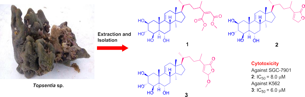
Topsensterols A–C, Cytotoxic Polyhydroxylated Sterol Derivatives from a Marine Sponge Topsentia sp.

1
Key Laboratory of Marine Drugs, The Ministry of Education of China, School of Medicine and Pharmacy, Ocean University of China, Qingdao 266003, China
2
Marine Science & Technology Institute, College of Environmental Science & Engineering, Yangzhou University, 196#, Huayang West Street, Yangzhou 225127, China
*
Author to whom correspondence should be addressed.
These authors contributed equally to this work.
Mar. Drugs 2016, 14(8), 146; https://doi.org/10.3390/md14080146
Submission received: 14 June 2016 / Revised: 16 July 2016 / Accepted: 26 July 2016 / Published: 1 August 2016
(This article belongs to the Collection Bioactive Compounds from Marine Invertebrates)

Abstract

:
Three new polyhydroxylated sterol derivatives topsensterols A–C (13) have been isolated from a marine sponge Topsentia sp. collected from the South China Sea. Their structures were elucidated by detailed analysis of the spectroscopic data, especially the NOESY spectra. Topsensterols A–C (l3) possess novel 2β,3α,4β,6α-tetrahydroxy-14α-methyl Δ9(11) steroidal nuclei with unusual side chains. Compound 2 exhibited cytotoxicity against human gastric carcinoma cell line SGC-7901 with an IC50 value of 8.0 μM. Compound 3 displayed cytotoxicity against human erythroleukemia cell line K562 with an IC50 value of 6.0 μM.

Graphical Abstract

1. Introduction

Most marine invertebrates are soft bodied and have a sedentary life style, resulting in the formation of distinctive chemical protection strategy [1,2]. The chemical means of defense necessitate the production of toxic compounds that can deter predators and paralyze their prey [2]. Undoubtedly, marine invertebrates represent a rich source of structurally novel and mechanistically unique secondary metabolites for the discovery of new drug leads [3]. Especially, sponges (Porifera) including the genus of Topsentia have been recognized as outstanding producers of sterols possessing novel side chains with unique alkylation and dealkylation patterns and displaying a variety of biological activities [4,5,6,7].
In our continuing efforts to discover new bioactive substances from marine invertebrates, a chemical investigation of a sponge of Topsentia sp. collected from the South China Sea was carried out. As a result, three new polyhydroxylated steroids, topsensterols A–C (13) (Figure 1), were isolated from its n-butanol extract. Compounds 2 and 3 showed significant cytotoxicity against human gastric carcinoma cell line SGC-7901 and human erythroleukemia cell line K562, respectively. Herein, we report the isolation, structural elucidation, plausible biogenetic pathway and biological evaluation of these steroids.

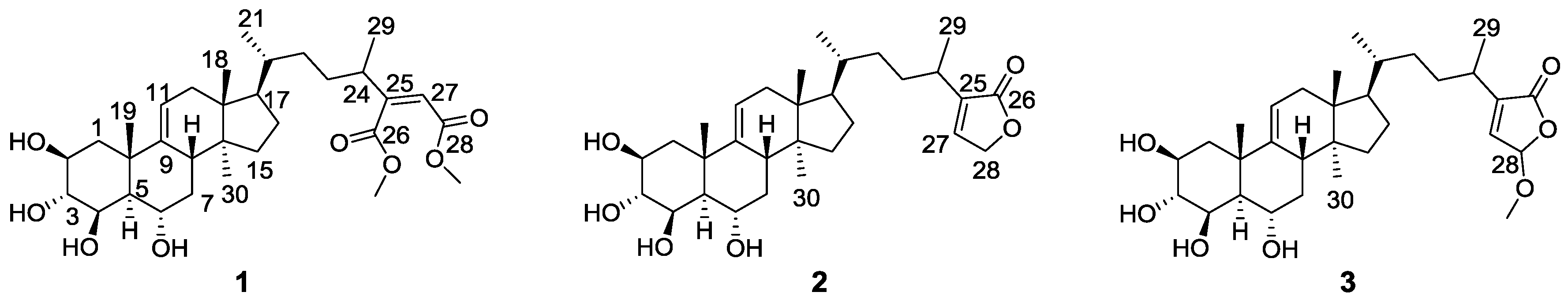
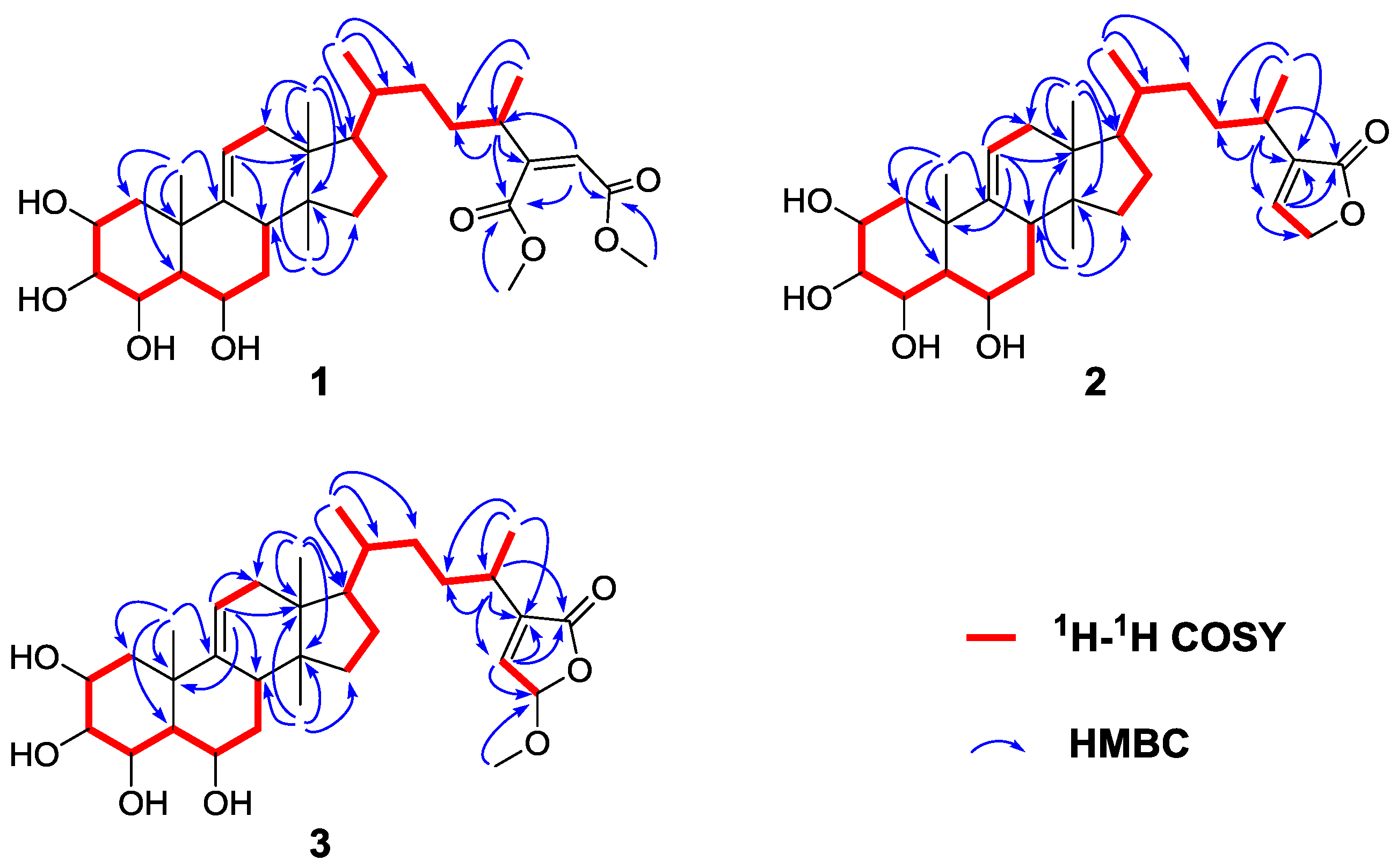
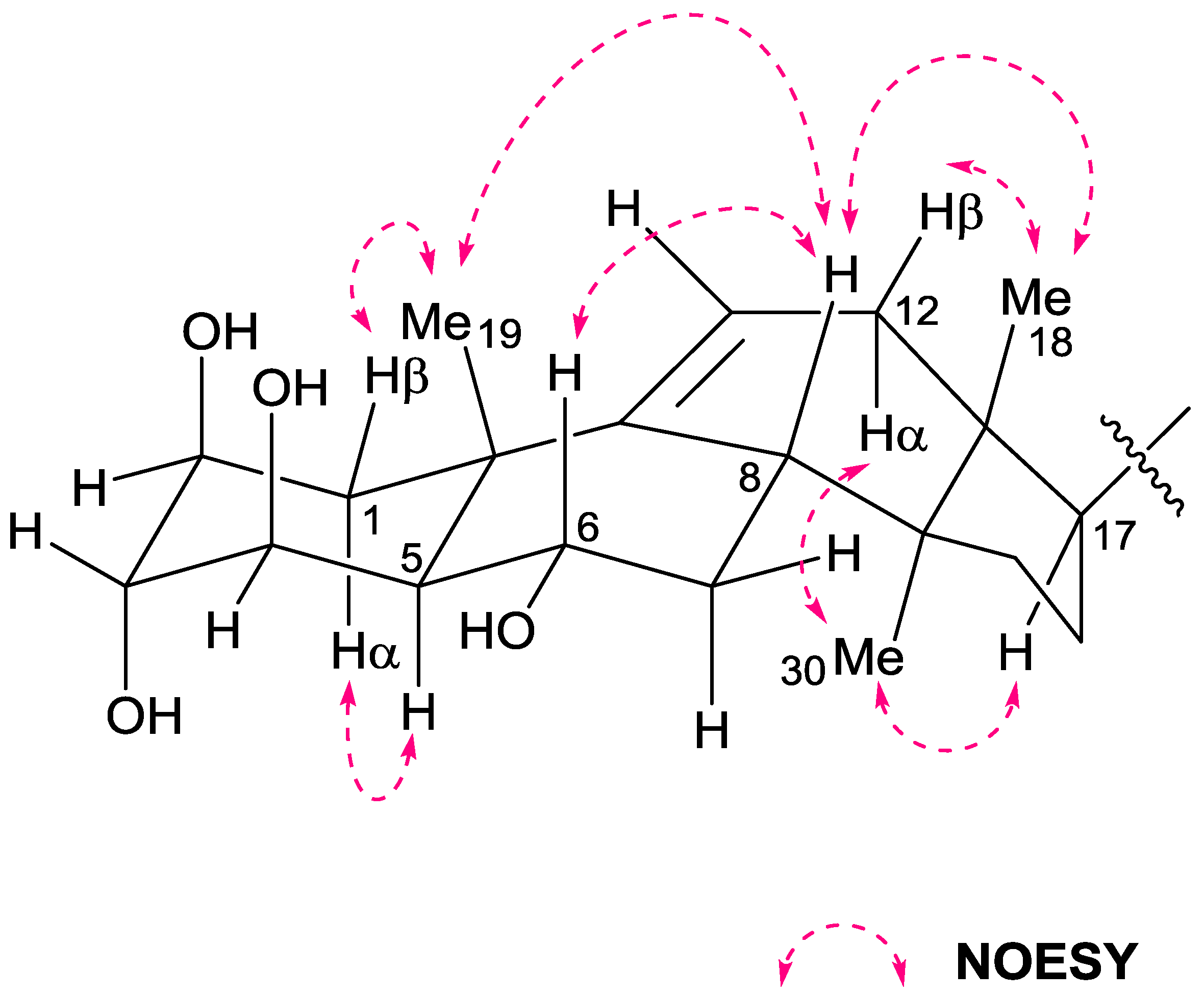
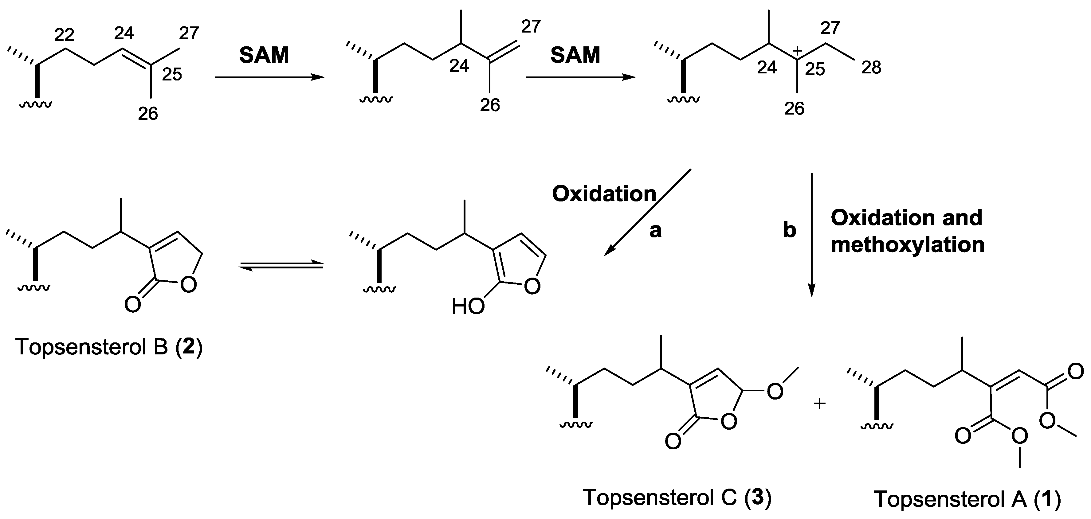
2. Results and Discussion

The frozen sample of sponge Topsentia sp. (5.1 kg, wet weight) was exhaustively extracted with 95% EtOH (8 L × 5 times) at room temperature, and the EtOH solution was evaporated under reduced pressure to give the crude extract. This extract was further partitioned between H2O and EtOAc. After evaporation of the organic solvents, the remaining aqueous suspension was extracted with n-butanol. Then the organic layer was concentrated to offer the n-butanol extract (27.1 g). This extract was subjected to a silica gel column chromatograph (CC) and the fractions were purified by octadecylsilyl silica gel CC and semi-preparative HPLC to obtain compounds 13.
Topsensterol A (1) was obtained as a white amorphous powder. Its molecular formula was determined as C32H50O8 on the basis of HR-ESI-MS (Supplementary Materials, Figure S9), indicating 8 degrees of unsaturation. The 1H NMR spectrum (Table 1, Supplementary Materials, Figure S1) showed five methyl singlets including two methoxy signals (δH 3.70, 3.66, 1.34, 0.82, and 0.70) and two methyl doublets (δH 1.12 and 0.91) together with four oxygenated methine signals (δH 4.25, 4.11, 3.98, and 3.91). In addition, two olefinic signals (δH 5.86 (s), 5.32 (d, J = 5.2 Hz)) were observed. The 13C NMR spectrum (Table 2, Supplementary Materials, Figure S2) exhibited two ester carbonyls, two trisubstituted double bonds, seven methyls, seven methylenes, nine methines including four oxygenated methines, and three quaternary sp3 carbons. Therefore, a steroid nature was suggested for this molecule. The HMBC correlations (Figure 2, Supplementary Materials, Figure S5) observed from Me-30 to C-8, C-14, and C-15 demonstrated that a methyl group anchored at C-14 of the steroid nucleus. Interpretation of the COSY correlations (Figure 2, Supplementary Materials, Figure S4) from H-1 to H-8 indicated a contiguous sequence including four oxygenated methines, assigning four hydroxy groups were at C-2, C-3, C-4, and C-6, respectively. The COSY crosspeaks of H-11/H-12 and HMBC correlations from H-11 to C-8, C-10, and C-13 inferred the Δ9(11)-unsaturated structure in steroid nucleus. The above spectroscopic data indicated that 1 belongs to 14α-methyl Δ9(11)-unsaturated steroids previously isolated from marine sponge, Topsentia sp. [8]. In addition, HMBC correlations from 26-OMe to C-26 and from 28-OMe to C-28 showed 26-OMe and 28-OMe were connected to C-26 and C-28, respectively. HMBC correlations from H-27 to C-24, C-25, C-26, and C-28, and from H-24 to C-23, C-25, and C-26 revealed the double bond was between C-25 and C-27. Consequently, a 2-substituted-dimethyl maleate unit was suggested in the side chain. Therefore the planar structure of 1 was established.
The stereochemistry of the steroid nucleus was established on the basis of coupling constants, COSY and NOESY data. Small coupling constants between H-2 and both H2-1 suggested that H-2 adopted an equatorial orientation. A large coupling constant between H-5 and H-6 (J = 10.8 Hz) and a small coupling constant between H-5 and H-4 (J = 2.4 Hz), implied that both H-5 and H-6 were axial, and H-4 was equatorial. In the COSY spectrum, a conspicuous W-type long-range crosspeaks between H-2 and H-4 further confirmed both H-2 and H-4 were equatorial. While W-type long-range crosspeaks between H-lβ and H-3 indicated that H-3 occupied an equatorial orientation. Therefore, the hydroxyl groups 2-OH, 3-OH, 4-OH, and 6-OH occupied the axial, axial, axial, and equatorial orientation, respectively. In the NOESY spectrum (Figure 3, Supplementary Materials, Figures S6 and S7), the absence of a crosspeak between Me-19 and H-5 suggested a trans-fused A/B ring junction. The NOESY crosspeaks of Me-19/H-8, Me-18/H-8, Me-18/H-12β, and H-6/H-8 indicated these protons were β-orientation, while crosspeaks of Me-30/H-17, and Me-30/H-12α revealed these protons were α-orientation. Furthermore, NOESY crosspeaks observed between Me-29 and H-27 suggested the double bond occupied Z-configuration, confirming the presence of a 2-substituted-dimethyl maleate unit in 1.
Careful inspection of the NMR spectra of 13 (Table 1 and Table 2) showed that the common and highly conserved signals of the steroidal nuclei for 13 are strikingly similar, including the signals of three angular methyl groups, a C-9(11)-double bond, four oxygenated methines, and four hydroxyl groups. Combined with their NOESY data and comparison with the configurations of the known similar metabolites such as topsentiasterol sulfates A–F, chlorotopsentiasterol sulfate D, and iodotopsentiasterol sulfate D also isolated from the marine sponge Topsentia sp. [7,8], compounds 13 were suggested to possess the same 2β,3α,4β,6α-tetrahydroxy-14α-methyl Δ9(11) sterol framework. The differences between these compounds were restricted to the respective aliphatic side chains.
Topsensterol B (2) was isolated as a white amorphous powder with a molecular formula of C30H46O6 determined on the basis of HR-ESI-MS (Supplementary Materials, Figure S16). The COSY spectrum (Supplementary Materials, Figure S13) revealed the connectivities of H3-21/H-20/H2-22/H2-23/H-24/H3-29 and the terminal γ-lactone in the side chain was deduced by interpretation of HMBC data (Supplementary Materials, Figure S14) together with NMR signals (Table 1 and Table 2, Supplementary Materials, Figures S10 and S11) for an oxymethylene (δH 4.87, δC 71.7), a trisubstituted double bond (δH 7.32, δC 139.6, 146.1) and an ester carbonyl (δC 176.3). These NMR signals also corresponded well with those reported for 2-alkyl butenolide [8].
Topsensterol C (3), with molecular formula of C31H48O7, was also isolated as a white amorphous powder. The 1H and 13C NMR signals (Table 1 and Table 2, Supplementary Materials, Figures S17 and S18) of the side chain were very similar to those of 2. The only significant difference was that a methoxy (δH 3.58, δC 57.0) and an oxygenated methine (δH 5.87, δC 104.0) in 3 replaced the oxymethylene (δH 4.87, δC 71.7) in 2. Interpretation of HMBC data (Supplementary Materials, Figure S21) of 3 indicated that a 4-methoxy-2-alkyl butenolide moiety connected at the terminal of the side chain for 3.
Polyhydroxylated sterols with various novel side chains are common secondary metabolites from marine sponges. In present study, topsensterol A (1) possesses a unique side chain terminated with a 2-substituted-dimethyl maleate unit. To the best of our knowledge, it is the first report of polyhydroxylated sterol with a 2-substituted-dimethyl maleate side chain. The plausible biosynthesis mechanism to form the side chains of 13 was proposed in Scheme 1. According to the literature [6,7], the side chains present in 13 are likely formed via methylations by S-adenosylmethionine (SAM) to the side chain present in parkeol, along with oxidation and methoxylation. Scheme 1 illustrates that: (i) the addition of a methyl from SAM to C-24 and a loss of a proton to generate the 25,27-double bond; (ii) SAM adds a methyl to C-27 to form the C-25 cation; (iii) the C-25 cation followed by oxidation and tautomerization to form topsensterol B (2) (pathway a); and (iv) the C-25 cation followed by oxidation and methoxylation to form topsensterol C (3) and topsensterol A (1) (pathway b).
Marine sponges of the genus Topsentia were reported to produce various structurally unique steroids including polyhydroxylated sterols [9] and polysulfated steroids [10,11,12]. In the present study, all of the isolated polar steroids were polyhydroxylated sterols, as the desulfated derivatives of similar sterol sulfates [8]. It seems that our isolated compounds maybe a group of desulfated artifacts. However, the desulfation of polysulfated sterols should be not so easy to occur during the ordinary extraction and separation processes. To date many polysulfated sterols have been reported to be obtained from marine sponges via ordinary separation processes, indicating that polysulfated sterols are stable enough. More importantly, on the basis of biogenetic considerations, the 2β,3α,4β,6α-tetrahydroxy-14α-methyl Δ9(11) steroidal nuclei pattern could be biosynthesized directly from parkeol in marine sponges [6,13,14]. Given the above, compounds 13 were more likely to be produced by ecological conditions, but the probability of desulfated artifacts could not be ruled out.
Compounds 13 were assessed for their cytotoxic activity against human gastric carcinoma SGC-7901 and human erythroleukemia K562 cell lines by MTT method [15]. Compound 2 exhibited cytotoxicity against SGC-7901 and K562 cell lines with IC50 values of 8.0 and 20 μM, respectively. Compound 3 exhibited cytotoxicity against SGC-7901 and K562 cell lines with IC50 values of 28 and 6.0 μM, respectively. However, compound 1 showed no cytotoxicity against the two cell lines. The above results revealed that the terminal butenolide moiety in the side chain may play a key role in the cytotoxicity.
Compounds 13 were also tested for their antimicrobial activity against human pathogenic bacteria including Staphylococcus aureus (ATCC 51650), Methicillin-resistant Staphylococcus aureus (ATCC 9551), and Candida albicans (ATCC 10231) using the method developed by Fromtling et al. [16]. However, no compound was found to be active against these bacteria.

3. Experimental Section

3.1. General Experimental Procedures

Optical rotation values were measured on a Jasco-P-1020 digital polarimeter (Jasco Corp., Tokyo, Japan). NMR spectra were recorded on a Bruker AV-400 NMR spectrometer (Bruker Corp., Fallanden, Switzerland), and chemical shifts were recorded as δ values (400 MHz for 1H and 100 MHz for 13C). ESIMS spectra were obtained from a Micromass Q-TOF spectrometer (Waters Corp., Milford, MA, USA). Semipreparative HPLC was performed on a Waters 1525 system using a semipreparative C18 column (5 μm, 10 × 250 mm, Kromasil, Sweden) coupled with a Waters 2996 photodiode array detector (Waters Corp., Milford, MA, USA). Silica gel (200–300 mesh, Qingdao Haiyang Chemical Factory, Qingdao, China), and octadecylsilyl (ODS) silica gel (45–60 mm; Merck KGaA, Darmstadt, Germany) were used for column chromatography. Thin-layer chromatography (TLC) was performed on precoated silica gel 60 GF254 plates (Yantai Zifu Chemical Group Co., Yantai, China).

3.2. Animal Material

The marine sponge Topsentia sp. was collected from Xuwen, Guangdong Province, China, in April 2006, and was identified by Nicole J. de Voogd, National Museum of Natural History, The Netherlands. A voucher specimen (GD-XW-20060007) was deposited at the Key Laboratory of Marine Drugs, Ministry of Education, Ocean University of China, Qingdao, China.

3.3. Extraction and Isolation

The n-butanol extract (27.1 g) was subjected to a silica gel column chromatograph (CC) and eluted with a gradient of CHCl3/MeOH (50:1–0:1, v:v) to collect 3 fractions (Fr.A–Fr.C). Fr.A displayed cytotoxicity and the 1H NMR features of diverse sterol analogues. This fraction was fractionated upon a octadecylsilyl silica gel CC and eluted with a gradient of H2O/MeOH (50:1–0:1, v:v) to obtain three subfractions (Fr.A1–Fr.A3). Fr.A1 was subjected to semi-preparative HPLC eluted with MeOH/H2O (80/20, v/v) to obtain compounds 2 (4.2 mg) and 3 (6.4 mg). Fr.A2 was subjected to semi-preparative HPLC eluted with MeOH/H2O (85/15, v/v) to yield compound 1 (2.8 mg).
Topsensterol A (1): white amorphous powder; [α] D 20 +11 (c 0.1, MeOH); for 1H and 13C NMR data, see Table 1 and Table 2; HRESIMS m/z 563.3600 [M + H]+ (calcd. for C32H51O8+, 563.3584).
Topsensterol B (2): white amorphous powder; [α] D 20 +14 (c 0.1, MeOH); for 1H and 13C NMR data, see Table 1 and Table 2; HRESIMS m/z 520.3621 [M + NH4]+ (calcd. for C30H50NO6+, 520.3633).
Topsensterol C (3): white amorphous powder; [α] D 20 +17 (c 0.1, MeOH); for 1H and 13C NMR data, see Table 1 and Table 2; HRESIMS m/z 550.3732 [M + NH4]+ (calcd. for C31H52NO7+, 550.3738).

4. Conclusions

In summary, three new polyhydroxylated steroid derivatives topsensterols A–C (l3) possessing unusual side chains were successfully isolated from the marine sponge Topsentia sp. The plausible biosynthesis mechanism to form the uncommon side chains of 13 was proposed. Compounds 2 and 3 exhibited significant cytotoxicities, demonstrating that the terminal butenolide moiety in the side chain may act an important part in the cytotoxicity.

Supplementary Materials

The following are available online at www.mdpi.com/1660-3397/14/8/146/s1, HRESIMS, 1D and 2D NMR data of compounds l3.

Acknowledgments

We thank Chang-Lun Shao, School of Medicine and Pharmacy, Ocean University of China, for collection of the sponge samples. This work was supported by the National Natural Science Foundation of China (No. 41130858; 41322037; U1406402-1), and the Taishan Scholars Program, China.

Author Contributions

Min Chen and Xu-Dong Wu contributed to extraction, isolation, NMR analysis, structure elucidation and manuscript preparation; Qing Zhao contributed to bioactivities test; and Chang-Yun Wang was the project leader, organized and guided the experiments and wrote the manuscript.

Conflicts of Interest

The authors declare no conflict of interest.

References

  1. Jiang, C.S.; Muller, W.E.; Schroder, H.C.; Guo, Y.W. Disulfide- and multisulfide-containing metabolites from marine organisms. Chem. Rev. 2012, 112, 2179–2207. [Google Scholar] [CrossRef] [PubMed]
  2. Haefner, B. Drugs from the deep: Marine natural products as drug candidates. Drug Discov. Today 2003, 8, 536–544. [Google Scholar] [CrossRef]
  3. Gomes, N.G.; Dasari, R.; Chandra, S.; Kiss, R.; Kornienko, A. Marine invertebrate metabolites with anticancer activities: Solutions to the “supply problem”. Mar. Drugs 2016, 14. [Google Scholar] [CrossRef] [PubMed]
  4. Aiello, A.; Fattorusso, E.; Menna, M. Steroids from sponges: Recent reports. Steroids 1999, 64, 687–714. [Google Scholar] [CrossRef]
  5. Dai, J.; Sorribas, A.; Yoshida, W.Y.; Kelly, M.; Williams, P.G. Topsentinols, 24-isopropyl steroids from the marine sponge Topsentia sp. J. Nat. Prod. 2010, 73, 1597–1600. [Google Scholar] [CrossRef] [PubMed]
  6. Whitson, E.L.; Bugni, T.S.; Chockalingam, P.S.; Concepcion, G.P.; Harper, M.K.; He, M.; Hooper, J.N.A.; Mangalindan, G.C.; Ritacco, F.; Ireland, C.M. Spheciosterol sulfates, PKCζ inhibitors from a Philippine sponge Spheciospongia sp. J. Nat. Prod. 2008, 71, 1213–1217. [Google Scholar] [CrossRef] [PubMed]
  7. Guzii, A.G.; Makarieva, T.N.; Denisenko, V.A.; Dmitrenok, P.S.; Burtseva, Y.V.; Krasokhin, V.B.; Stonik, V.A. Topsentiasterol sulfates with novel iodinated and chlorinated side chains from the marine sponge Topsentia sp. Tetrahedron Lett. 2008, 49, 7191–7193. [Google Scholar] [CrossRef]
  8. Fusetani, N.; Takahashi, M.; Matsunaga, S. Topsentiasterol sulfates, antimicrobial sterol sulfates possessing novel side chains, from a marine sponge, Topsentia sp. Tetrahedron 1994, 50, 7765–7770. [Google Scholar] [CrossRef]
  9. Luo, X.; Li, F.; Shinde, P.B.; Hong, J.; Lee, C.O.; Im, K.S.; Jung, J.H. 26,27-Cyclosterols and other polyoxygenated sterols from a marine sponge Topsentia sp. J. Nat. Prod. 2006, 69, 1760–1768. [Google Scholar] [CrossRef] [PubMed]
  10. McKee, T.C.; Cardellinaii, J.H.; Tischler, M.; Snader, K.M.; Boyd, M.R. Ibisterol sulfate, a novel HIV-inhibitory sulfated sterol from the deep water sponge Topsentia sp. Tetrahedron Lett. 1993, 34, 389–392. [Google Scholar] [CrossRef]
  11. Sun, H.H.; Gross, S.S.; Gunasekera, M.; Koehn, F.E. Weinbersterol disulfates A and B, antiviral steroid sulfates from the sponge Petrosia weinbergi. Tetrahedron 1991, 47, 1185–1190. [Google Scholar] [CrossRef]
  12. Fusetani, N.; Matsunaga, S.; Konosu, S. Bioactive marine metabolites II. Halistanol sulfate, an antimicrobial novel steroid sulfate from the marine sponge halichondria cf. moorei bergquist. Tetrahedron Lett. 1981, 22, 1985–1988. [Google Scholar] [CrossRef]
  13. Sarma, N.S.; Krishna, M.S.R.; Rao, S.R. Sterol ring system oxidation pattern in marine sponges. Mar. Drugs 2005, 3, 84–111. [Google Scholar] [CrossRef]
  14. Massey, I.J.; Djerassi, C. Mass spectrometry in structural and stereochemical problems. 252. Structural and stereochemical applications of mass spectrometry in the marine sterol field. Synthesis and electron impact induced mass spectral fragmentation of Δ24- and Δ24(28)-3β-hydroxy-Δ5-sterols. J. Org. Chem. 1979, 44, 2448–2456. [Google Scholar]
  15. Mosmann, T. Rapid colorimetric assay for cellular growth and survival: Application to proliferation and cytotoxicity assays. J. Immunol. Methods 1983, 65, 55–63. [Google Scholar] [CrossRef]
  16. Fromtling, R.A.; Galgiani, J.N.; Pfaller, M.A.; Espinel-Ingroff, A.; Bartizal, K.F.; Bartlett, M.S.; Body, B.A.; Frey, C.; Hall, G.; Roberts, G.D. Multicenter evaluation of a broth macrodilution antifungal susceptibility test for yeasts. Antimicrob. Agents Chemother. 1993, 37, 39–45. [Google Scholar] [CrossRef] [PubMed]
Figure 1. Chemical structures of compounds 13 (relative configuration).
Marinedrugs 14 00146 g001
Figure 2. 1H-1H COSY and key HMBC correlations of compounds 13.
Marinedrugs 14 00146 g002
Figure 3. Key NOESY correlations for the sterol nucleus of compound 1.
Marinedrugs 14 00146 g003
Scheme 1. Plausible biosynthesis mechanism to form the side chains of 13.
Marinedrugs 14 00146 sch001
Table 1. 1H NMR spectroscopic data (400 MHz, CD3OD) for 13.
H123
1.96 (dd, 13.0, 4.8)2.00 (m)1.98 (m)
2.40 (m)2.46 (br d, 13.0)2.46 (br d, 14.0)
24.25 (br s)4.28 (br s)4.28 (br s)
33.91 (t, 2.4)3.98 (t, 2.8)3.97 (t, 3.2)
43.98 (br d, 2.4)4.04 (br d, 2.8)4.03 (br d, 3.2)
51.41 (dd, 10.8, 2.0)1.46 (dd, 10.8, 2.0)1.45 (dd, 10.8, 2.0)
64.11 (td, 10.8, 4.4)4.16 (td, 10.8, 4.4)4.15 (td, 10.8, 4.4)
1.51 (m)1.53 (m)1.53 (m)
1.91 (m)1.91 (m)1.91 (m)
81.95 (m)1.95 (m)1.95 (m)
115.32 (d, 5.2)5.36 (d, 5.2)5.36 (br d, 5.2)
12α2.12 (d, 17.5)2.15 (br d, 17.5)2.15 (br d, 17.5)
12β1.93 (m)1.93 (m)1.93 (m)
15a1.38 (m)1.38 (m)1.38 (m)
15b1.44 (m)1.44 (m)1.44 (m)
16a1.37 (m)1.37 (m)1.37 (m)
16b1.92 (m)1.92 (m)1.92 (m)
171.66 (q, 9.4)1.69 (q, 9.4)1.68 (q, 9.4)
180.70 (s)0.73 (s)0.73 (s)
191.34 (s)1.38 (s)1.38 (s)
201.42 (m)1.42 (m)1.42 (m)
210.91 (d, 6.4)0.95 (d, 6.4)0.95 (d, 6.4)
22a1.06 (m)1.11 (m)1.11 (m)
22b1.43 (m)1.48 (m)1.48 (m)
23a1.42 (m)1.47 (m)1.47 (m)
23b1.54 (m)1.55 (m)1.55 (m)
242.42 (m)2.52 (q, 6.4)2.53 (q, 6.4)
275.86 (s)7.32 (br s)6.93 (br s)
28 4.87 (br s)5.87 (br s)
291.12 (d, 6.8)1.21 (d, 6.8)1.20 (d, 6.8)
300.82 (s)0.85 (s)0.85 (s)
26-OCH33.70 (s)
28-OCH33.66 (s) 3.58 (s)
Table 2. 13C NMR spectroscopic data (100 MHz, CD3OD) for 13.
C123
139.9, CH239.7, CH239.8, CH2
271.5, CH71.2, CH71.3, CH
372.5, CH72.0, CH72.2, CH
472.8, CH72.4, CH72.6, CH
548.1, CH48.8, CH49.0, CH
666.7, CH66.4, CH66.5, CH
737.9, CH237.5, CH237.6, CH2
841.4, CH41.0, CH41.1, CH
9147.9, C147.4, C147.5, C
1039.8, C39.5, C39.5, C
11117.0, CH116.9, CH116.9, CH
1238.4, CH238.1, CH238.1, CH2
1345.7, C45.4, C45.4, C
1448.1, C47.8, C47.9, C
1534.9, CH234.7, CH234.7, CH2
1629.0, CH228.8, CH228.8, CH2
1752.3, CH51.9, CH51.9, CH
1815.1, CH315.1, CH315.1, CH3
1927.1, CH327.0, CH327.0, CH3
2037.5, CH37.0, CH37.1, CH
2118.9, CH318.9, CH318.9, CH3
2234.7, CH234.4, CH234.5, CH2
2332.7, CH232.5, CH232.4, CH2
2440.5, CH31.8, CH31.9, CH
25157.6, C139.6, C144.2, C
26170.7, C176.3, C172.8, C
27119.4, CH146.1, CH143.1, CH
28167.1, C71.7, CH2104.0, CH
2919.3, CH318.8, CH318.8, CH3
3019.0, CH318.9, CH318.9, CH3
26-OCH352.3, CH3
28-OCH352.7, CH3 57.0, CH3

