Article

Dichotocejpins A–C: New Diketopiperazines from a Deep-Sea-Derived Fungus Dichotomomyces cejpii FS110

1
State Key Laboratory of Applied Microbiology Southern China, Guangdong Provincial Key Laboratory of Microbial Culture Collection and Application, Guangdong Open Laboratory of Applied Microbiology, Guangdong Institute of Microbiology, Guangzhou 510070, China
2
Guangdong Provincial Key Laboratory of Bioengineering Medicine, National Engineering Research Center of Genetic Medicine, College of Life Science and Technology, Jinan University, Guangzhou 510632, China
*
Author to whom correspondence should be addressed.
Mar. Drugs 2016, 14(9), 164; https://doi.org/10.3390/md14090164
Submission received: 3 August 2016 / Revised: 2 September 2016 / Accepted: 5 September 2016 / Published: 9 September 2016

Abstract

:
Three new diketopiperazines, dichotocejpins A–C (13), together with eight known analogues (411), were isolated from the culture of the deep-sea sediment derived fungus Dichotomomyces cejpii FS110. Their structures, including absolute configurations, were elucidated by a combination of HRESIMS, NMR, X-ray crystallography, and ECD calculations. Compounds 46, 1011 showed significant cytotoxic activities against MCF-7, NCI-H460, HepG-2, and SF-268 tumor cell lines. Compound 1 exhibited excellent inhibitory activity against α-glucosidase with an IC50 of 138 μM.

1. Introduction

Marine microorganisms are a rich source of structurally unique and bioactive metabolites. Even considering the trend of recent years that many marine natural products research efforts are directed towards microorganisms, there has been a sharp upward swing in the number of new metabolites reported from marine microorganisms [1,2]. Deep-sea organisms survive under extreme conditions such as an absence of light, low levels of oxygen, and intensely high pressures. These factors may result in the production of structurally unique secondary metabolites. Although thousands of natural products with diverse structural types have been reported from marine flora and fauna, the secondary metabolites derived from the deep-sea fungi at depths of over 1000 m below the surface are limited [3,4]. In our ongoing effort to search for structurally diverse and biologically significant metabolites from deep-sea fungi [5,6,7,8], we found that a culture broth of Dichotomomyces cejpii FS110, which was isolated from deep-sea sediment, showed strong growth inhibitory effects against four human tumor cell lines [9]. Bioassay-guided separation of the EtOAc extract of culture broth yielded three new diketopiperazines (13), along with eight known analogues. All of the isolated compounds (111) were evaluated for their cytotoxic activities against the SF-268, MCF-7, NCI-H460, and HepG-2 tumor cell lines and inhibitory activity against α-glucosidase. Herein, the isolation, structural elucidation, cytotoxic activities, and α-glucosidase inhibitory activities of all these isolates are described.

2. Results and Discussion

The fermentation broth of the deep-sea-derived fungus D. cejpii FS110 was extracted with EtOAc and then concentrated under reduced pressure to give an extract. The EtOAc extract was subjected to various column chromatography to afford compounds 111 (Figure 1). Three new structures were identified by combination of spectroscopic analysis, single crystal X-ray diffraction, and ECD calculation, while eight known analogues were identified as 6-deoxy-5a,6-didehydrogliotoxin (4) [10], gliotoxin (5) [11,12], acetylgliotoxin (6) [13], bis-N-norgliovictin (7) [14], 3-benzyl-6-(hydroxymethyl)-3-(methylthio)piperazine-2,5-dione (8) [15], bisdethiobis(methylthio)gliotoxin (9) [16], 6-acetylbis(methylthio)gliotoxin (10) [17], and 1,2,3,4-tetrahydro-2-methyl-3-methylene-1,4-dioxopyrazino[1,2-a]indole (11) [18] by the comparison of their spectroscopic data (Figures S26–S47) with those in the literature.

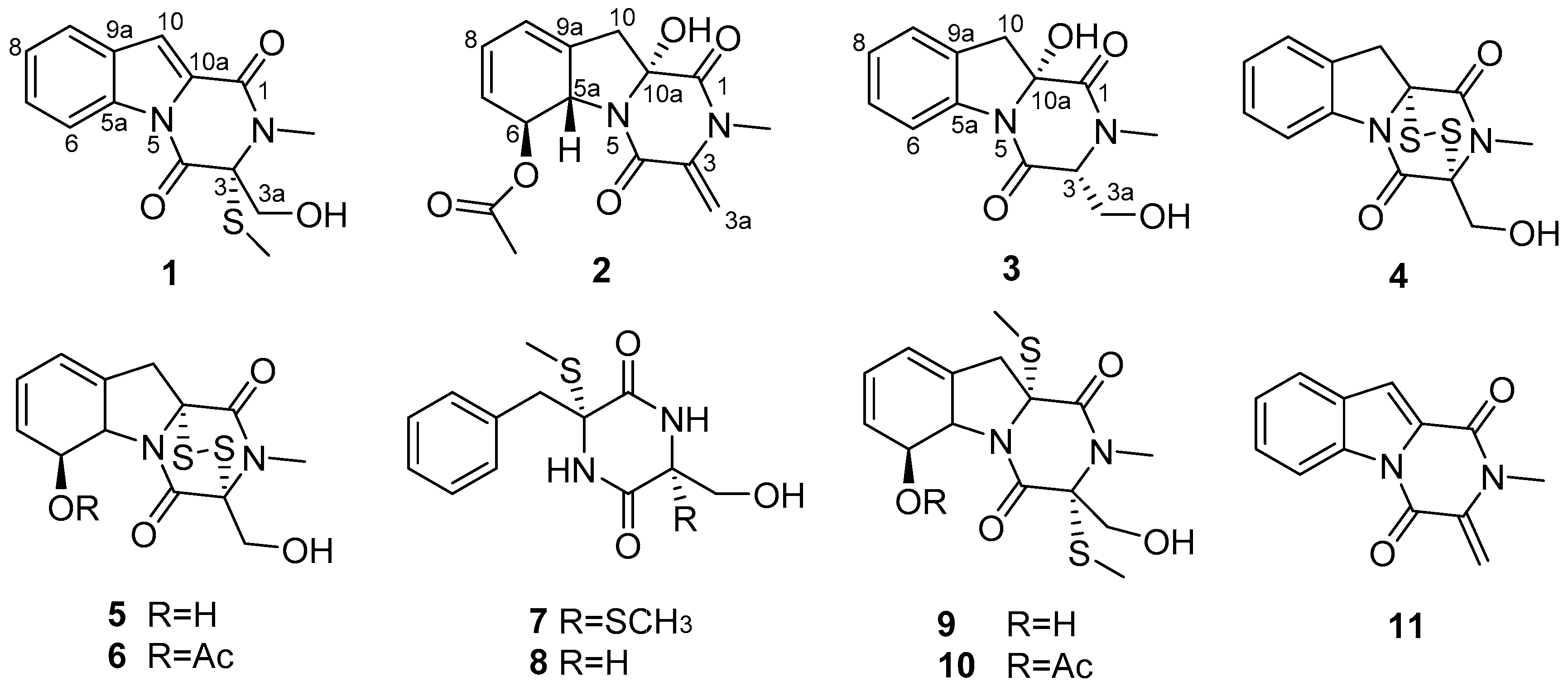
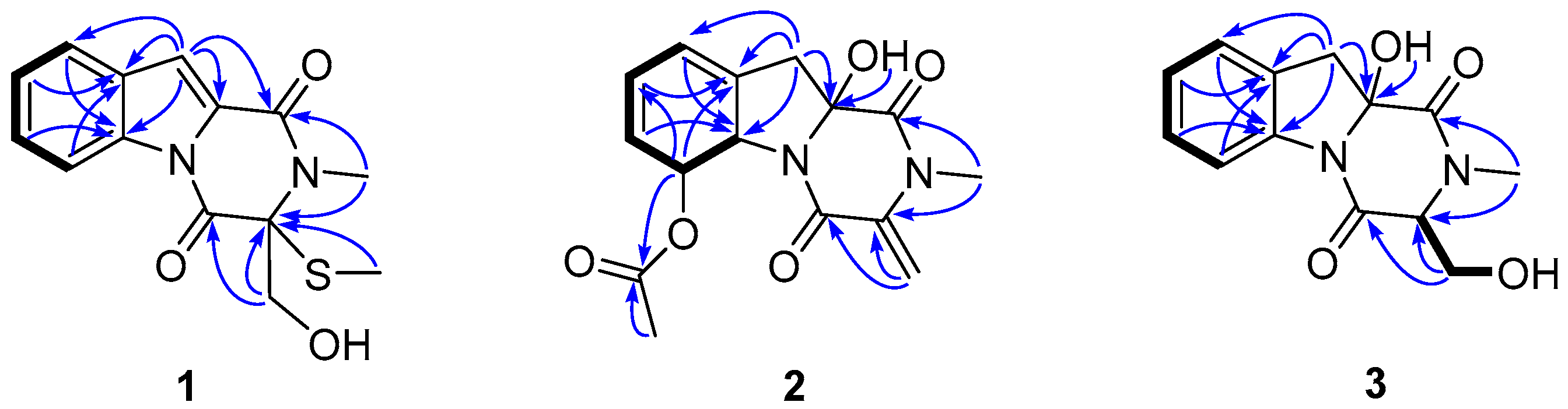
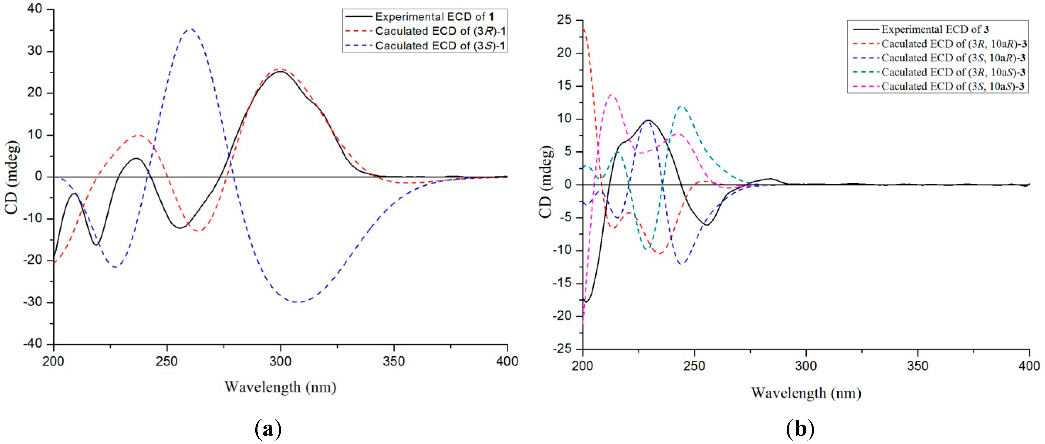
2.1. Identification of New Compounds

Compound 1, a pale yellow solid, had the molecular formula of C14H14N2O3S, as determined by HRESIMS (m/z 291.0798 [M + H]+, C14H15N2O3S, calcd. for 291.0803, Figure S6), corresponding to nine degrees of unsaturation. The 1H NMR spectrum (Figure S1) revealed the presence of two singlet methyls [δH 1.90 (3H, s) and 3.32 (3H, s)], one O-substituted sp3 methylene [δH 4.08 (1H, dd, J = 12.0, 7.8 Hz) and δH 4.46 (1H, dd, J = 12.0, 4.8 Hz)], an aromatic proton [δH 7.30 (1H, d, J = 1.0 Hz)], and a 1,2-disubstituted benzene ring [δH 7.26 (1H, ddd, J = 8.0, 7.2, 1.0 Hz), δH 7.42 (1H, dt, J = 8.0, 1.0 Hz), δH 7.47 (1H, ddd, J = 8.3, 7.2, 1.0 Hz) and δH 8.44 (1H, dt, J = 8.3, 1.0 Hz)]. The 13C NMR, in combination with DEPT experiments (Figure S2), resolved 14 carbon resonances attributed to two amide carbonyls (δC 158.0, 163.5), three sp2 quaternary carbons, five sp2 methines, one sp3 quaternary carbon, one sp3 methylene, and two methyls (Table 1). The 1D NMR data of 1 showed resonances characteristic of a diketopiperazine framework similar to that of neosartin B [19], with the exception of a thiomethyl at δC 12.2 replacing a methoxyl of the latter, and this assignment was supported by the HMBC correlations (Figure 2) from the thiomethyl (δH 1.90) to the severely upfiled-shifted C-3 (δC 76.7 in 1 and 93.6 in neosartin B). Detailed 2D analyses (HSQC, 1H–1H COSY, and HMBC, Figures S3–S5) supported the planar structure of 1 as depicted (Figure 2). HMBC correlations from the N-methyl (δH 3.32) to an amide carbonyl (δC 158.0, C-1) and a sp3 quaternary carbon (δC 76.7, C-3) and from the O-substituted sp3 methylene (δH 4.46 and 4.08, H2-3a) to another amide carbonyl (δC 163.5, C-4), and the sp3 quaternary carbon revealed the diketopiperazine framework. COSY correlations of H-6/H-7/H-8/H-9 and HMBC correlations from H-10 (δH 7.30) to C-9 (δC 122.8), C-9a (δC 134.9), C-5a (δC 129.1), C-10a (δC 127.4), and C-1 (δC 158.0) and from H-6 (δH 8.44) to C-9a (δC 134.9) suggested the remaining fragments. The absolute structure of 1 was deduced by comparison of the experimental and simulated electronic circular dichroism (ECD) spectra generated by time-dependent density functional theory (TDDFT) calculations (B3LYP/6-31G (d)) with Gaussian 09 [20]. The experimental ECD spectrum of 1 showed an ECD curve with Cotton effects around 300 (+), 256 (−) and 237 (+) nm, respectively (Figure 3a), which was in good accordance with the calculated ECD spectrum for (3R)-1, indicating that 1 had a 3R-configuration. Compound 1 was given the trivial name dichotocejpin A.
Compound 2, a colorless crystal, had the molecular formula of C15H16N2O5, as determined by HRESIMS (m/z 327.0953 [M + Na]+, C15H16N2O5Na; calcd. for 327.0957, Figure S15), corresponding to nine degrees of unsaturation. The 1H NMR spectrum (Figure S9) revealed the presence of two singlet methyls [δH 2.02 (3H, s) and 3.11 (3H, s)], one sp3 methylene [δH 2.75 (1H, d, J = 15.3 Hz) and 2.88 (1H, d, J = 15.3 Hz)], two O- or N-substituted sp3 methines [δH 4.98 (1H, d, J = 14.0 Hz) and 5.75 (1H, d, J = 14.0 Hz)], one terminal double bond methylene [δH 5.02 (1H, d, J = 1.1 Hz) and 5.48 (1H, d, J = 1.1 Hz)], three sp2 methines [δH 5.54 (1H, d, J = 9.6 Hz), 5.94 (1H, m) and 5.99 (1H, m)], and a deshielded hydroxyl group [δH 7.14 (1H, s)]. The 13C NMR, in combination with DEPT experiments (Figure S10), resolved 15 carbon resonances attributed to three ester or peptide carbonyl carbons, two sp2 quaternary carbons, three sp2 methines, one sp2 methylene, one sp3 methylene, and two methyls. The above-mentioned information was quite similar to the diketopiperazine bis(dethio)-10a-methylthio-3a-deoxy-3,3a-didehydrogliotoxin [10], except for the presence of a acetoxyl group and the absence of a single methyl in 2, which was further confirmed by 1H–1H COSY correlations of C-6/C-7/C-8/C-9 (Figure 2) and HMBC correlations from H-6 (δH 5.75) to the acetyl (δC 169.9) (Figures S11–S14).
As molecules possessing quaternary chiral center widely exist in nature, it has proven challenging via conventional spectroscopic methods to assign which antipodal series compound 2 belongs to. We thus resorted to a single-crystal X-ray diffraction experiment (Figure 4) using Cu Kα radiation (λ = 1.54184 Å). The structure including the absolute configuration of 2 was confirmed, and C-5a, C-6, and C-10a were assigned to S, S, R (absolute structure parameter: −0.12). Thus, the structure of 2 was established and given the trivial name dichotocejpin B.
Compound 3, a white solid, had the molecular formula of C13H14N2O4, as determined by HRESIMS (m/z 261.0888 [M − H], C13H13N2O4, calcd. for 261.0875, Figure S23), corresponding to eight degrees of unsaturation. The 1D NMR data of 3 (Figures S18 and S19) showed that it possessed the same diketopiperazine skeleton as 1, and was quite similar to the known compound 6-deoxy-5a,6-didehydrogliotoxin [10]. The main differences between them were the presence of a triplet hydrogen and the downfield-shifted C-3 (δC 66.1 in 3 and δC 74.0 in 6-deoxy-5a,6-didehydrogliotoxin) and C-10a in 3C 88.0 in 2 and δC 74.0 in 6-deoxy-5a,6-didehydrogliotoxin). The molecular weight of 3 was 46 amu, less than that of 6-deoxy-5a,6-didehydrogliotoxin, indicating the absence of the disulphide bond between C-3 and C-10a. HMBC correlations from OH-10a to C-10, C-10a, and C-1 revealed 3 was a 10a-OH derivative of 6-deoxy-5a,6-didehydrogliotoxin. This was supported by detailed 2D NMR spectra (Figure 2, Figures S20–S22) analyses.
The absolute structure of 3 was also deduced by comparison of the experimental and calculated ECD spectra generated by TDDFT calculations. As illustrated in Figure 3b, the experimentally acquired CD spectrum for 3 agreed well with the ECD curve computed for (3S, 10aR)-3. Thus, the most likely absolute configuration of 3 was established as 3S, 10aR. Compound 3 was given the trivial name dichotocejpin C.

2.2. Cytotoxicity Assay

The in vitro cytotoxicities of compounds 111 were evaluated against the SF-268, MCF-7, NCI-H460, and HepG-2 tumor cell lines by the Sulforhodamine B (SRB) method. Compounds 46, which have a disulfide bond, exhibited the most potent inhibitory activities against the four tumor cell lines with IC50 values in the range of 0.08–1.52 μM (Table 2). Compounds 10 and 11 also showed significant inhibitory activities, whereas compounds 1, 2, 3, and 79 were inactive or weak against these tumor cell lines.

2.3. α-Glucosidase Inhibitory Activity Assay

All compounds were evaluated in vitro for α-glucosidase inhibitory activities. Compound 1 showed excellent inhibitory activity against α-glucosidase (IC50 = 138 μM), being stronger than the positive control acarbose (an oral antidiabetic agent, IC50 = 463 μM).

3. Materials and Methods

3.1. General Experimental Procedures

Optical rotations were measured using an Anton Paar MCP-500 (Anton Paar, Graz, Austria). Circular dichroism (CD) measurements were carried out under N2 gas on a Jasco 820 spectropolarimeter (Jasco Corporation, Kyoto, Japan). IR spectra were recorded on a Shimadzu IR Affinity-1 spectrophotometer (Shimadzu Corporation, Kyoto, Japan). UV spectra were measured on a Shimadzu UV-2600 spectrophotometer (Shimadzu Corporation, Kyoto, Japan). NMR spectra were recorded on a Bruker Avance-500 spectrometer (Bruker Corporation, Fremont, CA, USA) and referenced to the signals of tetramethylsilane as an internal standard. HRESIMS was measured on a Thermo MAT95XP high-resolution mass spectrometer (Thermo Fisher Scientific, Bremen, Germany) and ESIMS was measured on an Agilent Technologies 1290-6430A Triple Quad LC/MS (Agilent Technologies Inc., Santa Clara, CA, USA). A Shimadzu LC-20 AT (Shimadzu Corporation, Kyoto, Japan) equipped with an SPD-M20A PDA detector (Shimadzu Corporation, Kyoto, Japan) was used for HPLC, a YMC-pack ODS-A column (250 × 10 mm, 5 μm, 12 nm) was used for semi-preparative HPLC separation, and a YMC-pack SIL (250 × 20 mm, 5 μm, 12 nm, YMC CO., Ltd., Kyoto, Japan) and a YMC-pack ODS-A column (250 × 20 mm, 5 μm, 12 nm, YMC CO., Ltd., Kyoto, Japan) were used for preparative HPLC separation. Column chromatography: commercial silica gel (SiO2; 200–300 mesh; Qingdao Marine Chemical Plant, Qingdao, China) and Sephadex LH-20 gel (Amersham Biosciences, Uppsala, Sweden). All solvents were of analytical grade (Guangzhou Chemical Regents Company, Ltd., Guangzhou, China).

3.2. Fungal Material and Identification

The fungal strain FS110 was isolated from a deep-sea sedimental sample in the South China Sea (19°0.368′ N, 117°58.233′ E; depth 3941 m). The isolate was identified as Dichotomomyces cejpii FS110 based on ITS rDNA sequence analysis [9]. The sequence of the ITS region of the strain FS110 has been submitted to GenBank (Accession No. KF706672). By using BLAST (nucleotide sequence comparison program) to search the GenBank database, FS110 has 99.8% similarity with Dichotomomyces cejpii NRRL 26980 (Accession No. EF669956). The strain was preserved at the Guangdong Provincial Key Laboratory of Microbial Culture Collection and Application, Guangdong Institute of Microbiology. Working stocks were prepared on potato dextrose agar (PDA) slants and stored at 4 °C.

3.3. Fermentation, Extraction, and Isolation

A well-grown slant culture of D. cejpii FS110 was used for preparation of the seed culture. Three pieces (0.5 × 0.5 cm2) of mycelial agar plugs of this strain were inoculated into 250 mL of PD medium (potato 200 g/L, glucose 20 g/L, KH2PO4 3 g/L, MgSO4∙7H2O 1.5 g/L, vitamin B1 10 mg/L, sea salt 15 g/L) in a 500 mL Erlenmeyer flask, and incubated for 2 days in a rotary shaker (200 r/m) at 28 °C. The seed cultures (10%) were then aseptically transferred into 500 mL of PD medium in 1000 mL Erlenmeyer flasks and shaken (120 r/m) at 28 °C for 7 days. A total of 100 L of fermentation broth was filtered to give the broth and mycelia. The broth was partitioned sequentially with EtOAc (4 × 25 L) to yield a dark brown oily residue (40.0 g), which was subjected to column chromatography on silica gel eluted with petroleum ether/EtOAc in linear gradient (30:1→1:1) and followed by CHCl3/MeOH in linear gradient (10:1→0:1) to give 26 fractions (F1–F26). F2 was washed with MeOH to get 4 (4.8 mg). F6 was separated on a preparative reversed-phase (RP) HPLC system equipped with a YMC column (MeOH/H2O, 50:50, 10 mL/min) to yield 1 (1.4 mg, tR = 17 min) and 6 (1.0 g, tR = 25 min). F10 was separated by column chromatography on Sephadex LH-20 (CHCl3/MeOH, 1:1, v/v) to yield 5 (2.0 mg). F11 was subjected to a Sephadex LH-20 column eluting with CHCl3/MeOH (1:1), then further purified by column chromatography on silica gel eluted with petroleum ether/EtOAc (5:1) to yield 7 (4.8 mg). F12 was separated by column chromatography on Sephadex LH-20 (CHCl3/MeOH, 1:1, v/v), then further separated by semi-preparative HPLC on a C-18 column (YMC*GEL ODS-A, 120A S-5 μm, 250 × 10 mm, MeOH/H2O, 40:60, 2 mL/min) to yield 2 (5.0 mg, tR = 16.0 min), 9 (75.0 mg, tR = 25.0 min), and 10 (75.0 mg, tR = 31.0 min). F13 was separated by column chromatography on Sephadex LH-20 (CHCl3/MeOH, 1:1, v/v) and then further purified by semi-preparative HPLC on a C-18 column (YMC*GEL ODS-A, 120A S-5 μm, 250 × 10 mm, MeOH/H2O, 40:60, 2 mL/min) to yield 11 (3.3 mg, tR = 18 min). F17 was separated by preparative HPLC on a C-18 column (YMC*GEL ODS-A, 120A S-5 μm, 250 × 20 mm, MeOH/H2O, 50:50, 10 mL/min) to give 5 fractions (F17.1–F17.5). F17.2 was further purification by semi-preparative HPLC on a C-18 column (YMC*GEL ODS-A, 120A S-5 μm, 250 × 10 mm, MeCN/H2O, 30:70, 2 mL/min) to give 8 (50.2 mg, tR = 34 min). F17.3 was further purification by semi-preparative HPLC on a C-18 column (YMC*GEL ODS-A, 120A S-5 μm, 250 × 10 mm, MeCN/H2O, 20:80, 2 mL/min) to give 3 (9.0 mg, tR = 19 min).
Dichotocejpin A (1): pale yellow solid; [α] D 25 +126.2 (c 0.1, MeOH); CD (MeOH, c 0.25 mg/mL) 209, 219, 237, 256 and 300 nm (∆ε −1.19, −4.93, +1.35, −3.71 and +7.63); UV (MeOH) λmax (log ε) 207 (4.37), 244 (4.34), 296 (4.15) nm (Figure S8); IR νmax 3372, 2955, 2922, 2853, 1709, 1636, 1587, 1429, 1391, 1358, 1259, 1026 cm−1 (Figure S7); 1H and 13C NMR data, see Table 1; HRESIMS m/z 291.0798 ([M + H]+, calcd for 291.0803).
Dichotocejpin B (2): colorless crystal; [α] D 25 +93.5 (c 0.1, MeOH); CD (MeOH, c 0.25 mg/mL) 217, 233 and 268 nm (∆ε −1.91, +1.00 and −4.68); UV (MeOH) λmax (log ε) 205 (4.55), 232 (4.59) nm (Figure S17); IR νmax 3273, 2958, 2924, 2855, 1732, 1682, 1612, 1367, 1258, 1026 cm−1 (Figure S16); 1H and 13C NMR data, see Table 1; HRESIMS m/z 327.0953 ([M + Na]+, calcd for 327.0957).
Dichotocejpin C (3): white solid; [α] D 25 +44.4 (c 0.1, MeOH); CD (MeOH, c 0.25 mg/mL) 230, 256 and 284 nm (∆ε +2.98, −1.85 and +0.29); UV (MeOH) λmax (log ε) 206 (4.26), 246 (3.88), 277 (3.48), 284 (3.46) nm (Figure S25); IR νmax 3294, 2941, 1672, 1643, 1603, 1487, 1431, 1400, 1078, 1056 cm−1 (Figure S24); 1H and 13C NMR data, see Table 1; HRESIMS m/z 261.0888 ([M − H], calcd for 261.0875).

3.4. Quantum-Chemical ECD Calculation

The quantum-chemical ECD calculation methods were used to establish the absolute configurations of compounds 1 and 3. The compounds were charged using the Gasteiger–Hückel method and the preliminary random conformational search was performed with the SYBYL 8.0 software package using the MMFF94 molecular mechanics force field. The geometry optimizations were then performed by using DFT at the B3LYP/6-31G (d) level as implemented in the Gaussian 09 program package. The stable conformers obtained were subsequently submitted to ECD calculations by TDDFT calculations (B3LYP/6-31G (d)) with Gaussian 09.

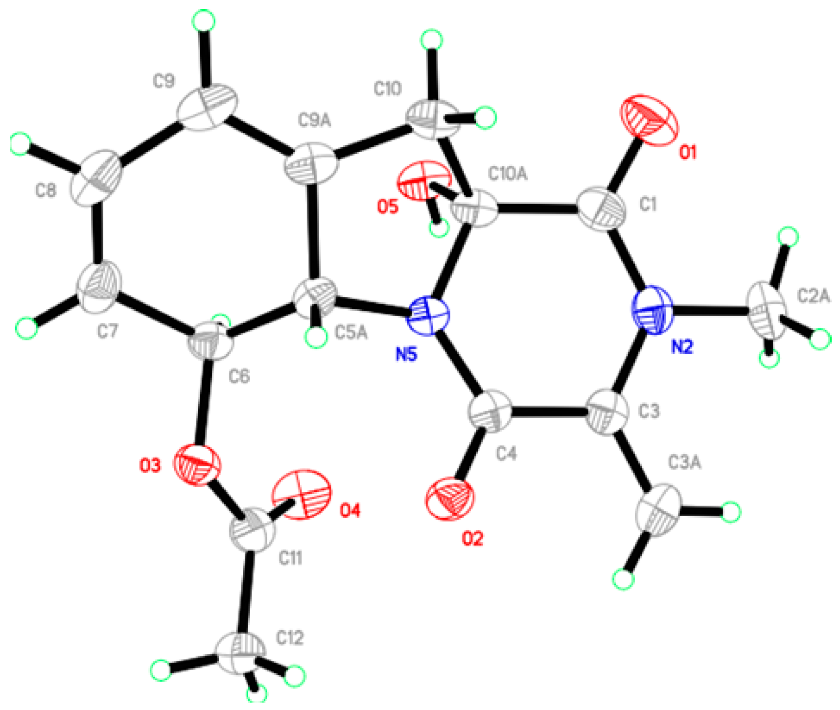
3.5. Crystallographic Data for 2

X-ray crystal data for dichotocejpin B (2). C15H16N2O5, M = 304.30 g/mol, orthorhombic, crystal size 0.400 × 0.320 × 0.230 mm3, space group P212121, a = 8.58255(15) Å, b = 10.5549(2) Å, c = 16.0203 (3) Å, α = β = γ = 90°, V = 1451.24(5) Å3, Z = 4, Dcalcd = 1.393 mg/m3, F(000) = 640. μ = 0.889 mm−1, Cu Kα radiation, λ = 1.54184 Å, T = 150 (2) K, 5.018° ≤ 2θ ≤ 66.890°, 9943 reflections collected, 2531 unique (Rint = 0.0291). Final GooF = 1.123, R1 = 0.0310, wR2 = 0.0748 based on 2531 reflections with I > 2σ(I) (refinement on F2), 424 parameters, 42 restraint. Lp and absorption corrections applied, m = 0.091 mm_1. Flack parameter = −0.12 (9). Crystallographic data for 1 have been deposited with the Cambridge Crystallographic Data Centre as supplementary publication No. CCDC 1491672. Copies of the data can be obtained, free of charge, on application to the CCDC, 12 Union Road, Cambridge CB2 1EZ, UK (fax, +44-(0)-1223-336033; e-mail, [email protected]).

3.6. Cytotoxicity Assay

Cytotoxicities of compounds (111) were assayed against four tumor cell lines SF-268, MCF-7, NCI-H460, and HepG-2. Assays were performed by the SRB method [21].

3.7. α-Glucosidase Inhibitory Activity Assay

An assay of α-glucosidase inhibitory activity was performed as previously described [22].

4. Conclusions

In this study, eleven diketopiperazines, including three new ones, were isolated from the deep-sea-derived fungus Dichotomomyces cejpii. All the chemical structures, including absolute configurations, were established. Compounds 46, 1011 exhibited significant cytotoxic activities against SF-268, MCF-7, NCI-H460, and HepG-2 tumor cell lines. By comparing the structures of these compounds, compounds 46, with a disulfide bridge, showed stronger cytotoxicities, which is accordance with the investigations reported in the literature [3,10,19]. Compound 1 is the first case of thio-diketopiperazines with excellent inhibitory activity against α-glucosidase, which might be useful for further developing α-glucosidase inhibitor and antidiabetic agents.

Supplementary Materials

The HRESIMS, IR, UV, and 1D and 2D-NMR spectra of compounds 13 (Figures S1–S25), ESIMS and 1D-NMR data of 411 (Figures S26–S47), can be accessed at: https://www.mdpi.com/1660-3397/14/9/164/s1.

Acknowledgments

This work was supported financially by the National Natural Science Foundation of China (31272087); the Science and Technology Program of Guangzhou, China (201607020018); the National High Technology Research and Development Program of China (863 Program, 2012AA092104); the Guangdong Provincial Project for Science and Technology (2014A020217012, 2015A030302061, 2016A020222022); and the Guangdong Provincial Innovative Development of Marine Economy Regional Demonstration Projects (GD2012-D01-002).

Author Contributions

Z.F. fractionated the extract, isolated the compounds, and elucidated structures; Z.-H.S. elucidated structures and wrote the paper; Z.L. performed the calculated ECD spectra experiment; Y.-C.C. and H.-H.L. performed the bioassays; H.-X.L. analyzed the data; W.-M.Z. designed and coordinated the study and reviewed the manuscript.

Conflicts of Interest

The authors declare no conflict of interest.

References

  1. Blunt, J.W.; Copp, B.R.; Keyzers, R.A.; Munro, M.H.G.; Prinsep, M.R. Marine natural products. Nat. Prod. Rep. 2016, 36, 382–431. [Google Scholar] [CrossRef] [PubMed] [Green Version]
  2. Blunt, J.W.; Copp, B.R.; Munro, M.H.G.; Northcote, P.T.; Prinsep, M.R. Marine natural products. Nat. Prod. Rep. 2011, 28, 196–268. [Google Scholar] [CrossRef] [PubMed]
  3. Horikoshi, K. Barophiles: Deep-sea microorganisms adapted to an extreme environment. Curr. Opin. Microbiol. 1998, 1, 291–295. [Google Scholar] [CrossRef]
  4. Niu, S.W.; Liu, D.; Hu, X.X.; Proksch, P.; Shao, Z.Z.; Lin, W. Spiromastixones A–O, antibacterial chlorodepsidones from a deep sea-derived Spiromastix sp. fungus. J. Nat. Prod. 2014, 77, 1021–1030. [Google Scholar] [CrossRef] [PubMed]
  5. Sun, L.; Li, D.L.; Tao, M.H.; Dan, F.J.; Zhang, W.M. Two new sesquiterpenes from the marine fungus Eutypella scoparia FS26 from the South China Sea. Helv. Chim. Acta 2012, 1, 157–162. [Google Scholar] [CrossRef]
  6. Sun, L.; Li, D.L.; Tao, M.H.; Chen, Y.C.; Zhang, Q.B.; Dan, F.J.; Zhang, W.M. Two new polyketides from the marine fungus Eutypella scoparia FS26. Nat. Prod. Res. 2012, 27, 1298–1304. [Google Scholar] [CrossRef] [PubMed]
  7. Sun, L.; Li, D.L.; Tao, M.H.; Chen, Y.C.; Dan, F.J.; Zhang, W.M. Scopararanes C–G: New oxygenated pimarane diterpenes from the marine sediment-derived fungus Eutypella scoparia FS26. Mar. Drugs 2012, 10, 539–550. [Google Scholar] [CrossRef] [PubMed]
  8. Gao, X.W.; Liu, H.X.; Sun, Z.H.; Chen, Y.C.; Tan, Y.Z.; Zhang, W.M. Secondary metabolites from the deep-sea derived fungus Acaromyces ingoldii FS121. Molecules 2016, 21, 371. [Google Scholar] [CrossRef]
  9. Yang, X.L.; Chen, Y.C.; Li, H.H.; Zhang, W.M. Molecular identification of 23 marine fungal strains and their activities against plant pathogenic fungi and cytotoxic activities. Biotechnol. Bull. 2014, 8, 132–137. [Google Scholar]
  10. Sun, Y.; Takada, K.; Takemoto, Y.; Yoshida, M.; Nogi, Y.; Okada, S.; Matsunaga, S. Gliotoxin analogues from a marine-derived fungus, Penicillium sp., and their cytotoxic and histone methyltransferase inhibitory activities. J. Nat. Prod. 2012, 75, 111–114. [Google Scholar] [CrossRef] [PubMed]
  11. Mourad, K. Gliotoxin: Uncommon 1H couplings and revised 1H and 13C-NMR assignments. J. Nat. Prod. 1990, 53, 717–719. [Google Scholar]
  12. Katharine, R.W.; Joseline, R.; Ang, K.H.; Karen, T.; Jennifer, E.C.; James, M.; Phillip, C. Assessing the trypanocidal potential of natural and semi-synthetic diketopiperazines from two deep water marine-derived fungi. Bioorg. Med. Chem. 2010, 18, 2566–2574. [Google Scholar]
  13. Didier, V.D.; Junji, I.; Kazuro, S.; Hong, Y.; Hideo, T.; Satoshi, O. Inhibition of farnesyl-protein transferase by gliotoxin and acetylgliotoxin. J. Antibiot. 1992, 45, 1802–1805. [Google Scholar]
  14. Kirby, G.W.; Rao, G.V.; Robins, D.J. New co-metabolites of gliotoxin in Gliocladium virens. J. Chem. Soc. Perkin Trans. 1 1988, 301–304. [Google Scholar] [CrossRef]
  15. Zhao, W.Y.; Zhu, T.J.; Han, X.X.; Fan, G.T.; Liu, H.B.; Zhu, W.M.; Gu, Q.Q. A new gliotoxin analogue from a marine-derived fungus Aspergillus fumigatus Fres. Nat. Prod. Res. 2009, 23, 203–207. [Google Scholar] [CrossRef] [PubMed]
  16. Kirby, G.W.; Robins, D.J.; Sefton, M.A.; Talekar, R.R. Biosynthesis of bisdethiobis (methylthio) gliotoxin, a new metabolite of Gliocladium deliquescens. J. Chem. Soc. Perkin Trans. 1 1980, 119–121. [Google Scholar] [CrossRef]
  17. Afiyatullov, S.S.; Kalinovskii, A.I.; Pivkin, M.V.; Dmitrenok, P.S.; Kuznetsova, T.A. Alkaloids from the marine isolate of the fungus Aspergillus fumigatus. Chem. Nat. Compd. 2005, 41, 236–238. [Google Scholar] [CrossRef]
  18. Ajay, K.B.; Das, K.G.; Funke, P.T.; Irene, K.; Shukla, O.P.; Khanchandani, K.S.; Suhadolnik, R.J. Biosynthetic studies on gliotoxin using stable isotopes and mass spectral methods. J. Am. Chem. Soc. 1968, 90, 1038–1041. [Google Scholar]
  19. Liang, W.L.; Le, X.; Li, H.J.; Yang, X.L.; Chen, J.X.; Xu, J.; Liu, H.L.; Wang, L.Y.; Wang, K.T.; Hu, K.C.; et al. Exploring the chemodiversity and biological activities of the secondary metabolites from the marine fungus Neosartorya pseudofischeri. Mar. Drugs 2014, 12, 5657–5676. [Google Scholar] [CrossRef] [PubMed]
  20. Frisch, M.J.; Trucks, G.W.; Schlegel, H.B.; Scuseria, G.E.; Robb, M.A.; Cheeseman, J.R.; Scalmani, G.; Barone, V.; Mennucci, B.; Petersson, G.A.; et al. Gaussian 09W; Revision A.02; Gaussian Inc.: Wallingford, CT, USA, 2009. [Google Scholar]
  21. Skehan, P.; Storeng, R.; Scudiero, D.; Monks, A.; McMahon, J.; Vistica, D.; Warren, J.T.; Bokesch, H.; Kenney, S.; Boyd, M.R. New colorimetric cytotoxicity assay for anticancer-drug screening. J. Natl. Cancer Inst. 1990, 82, 1107–1112. [Google Scholar] [CrossRef] [PubMed]
  22. Liu, Y.; Yang, Q.; Xia, G.; Huang, H.; Li, H.; Ma, L.; Lu, Y.; He, L.; Xia, X.; She, Z. Polyketides with α-glucosidase inhibitory activity from a mangrove endophytic fungus, Penicillium sp. HN29-3B1. J. Nat. Prod. 2015, 78, 1816–1822. [Google Scholar] [CrossRef] [PubMed]
Figure 1. Structures of compounds 111 isolated from Dichotomomyces cejpii FS110.
Marinedrugs 14 00164 g001
Figure 2. Key COSY (▬) and HMBC (→) correlations for compounds 13.
Marinedrugs 14 00164 g002
Figure 3. (a) Experimental ECD spectra of dichotocejpin A (1) in MeOH and calculated ECD spectra of (3R)-1 and (3S)-1 (b) Experimental ECD spectra of dichotocejpin C (3) in MeOH and calculated ECD spectra of (3R,10aR)-3, (3S,10aR)-3, (3R,10aS)-3, and (3S,10aS)-3. The calculated ECD spectra were computed at the B3LYP/6-31G (d) level.
Marinedrugs 14 00164 g003
Figure 4. ORTEP diagram of compound 2.
Marinedrugs 14 00164 g004
Table 1. 1H (500 MHz) and 13C (125 MHz) NMR data for compounds 13 (δ in ppm, J in Hz).
Position1 a2 b3 b
δH, mult. (J in Hz)δC, TypeδH, mult. (J in Hz)δC, TypeδH, mult. (J in Hz)δC, Type
1 158.0, C 164.2, C 165.2, C
3 76.7, C 138.4, C4.29, t (3.6)66.1, CH
3a4.46, dd (12.0, 7.8)
4.08, dd (12.0, 4.8)
64.2, CH25.48, d (1.1)
5.02, d (1.1)
102.1, CH23.96, m60.5, CH2
4 163.5, C 157.0, C 164.6, C
5a 129.1, C4.98, d (14.0)63.0, CH 128.8, C
68.44, dt (8.3, 1.0)116.9, CH5.75, d (14.0)75.3, CH7.97, d (7.7)115.5, CH
77.47, ddd (8.3, 7.2, 1.0)128.3, CH5.54, d (9.6)127.4, CH7.26, t (7.7)127.3, CH
87.26, ddd (8.0, 7.2, 1.0)125.9, CH5.99, m125.4, CH7.13, td (7.7, 1.1)124.7, CH
97.42, dt (8.0, 1.0)122.8, CH5.94, m118.5, CH7.35, d (7.7)125.2, CH
9a 134.9, C 135.9, C 140.0, C
107.30, d (1.0)115.1, CH2.88, d (15.3)
2.75, d (15.3)
41.7, CH23.53, d (17.2)
3.14, d (17.2)
39.7, CH2
10a 127.4, C 88.7, C 88.0, C
N-CH33.32, s28.2, CH33.11, s29.6, CH32.98, s32.2, CH3
S-CH31.90, s12.2, CH3
OH-3a3.39, t (7.1) 6.53, brs
OH-10a 7.14, s 6.88, brs
OAc 2.02, s169.9, C
21.3, CH3
a Recorded in CDCl3; b Recorded in DMSO-d6.
Table 2. IC50 values of compounds 111 against four tumor cell lines and α-glucosidase.
CompoundsIC50 (μM) a
SF-268MCF-7NCI-H460HepG-2α-glucosidase
135.7 ± 2.129.5 ± 2.3>10028.9 ± 3.0138 ± 6.7
2>100>100>100>100>500
3>100>100>100>100>500
41.35 ± 0.050.68 ± 0.021.27 ± 0.041.52 ± 0.03>500
50.24 ± 0.100.08 ± 0.00.24 ± 0.010.21 ± 0.01>500
60.25 ± 0.030.22 ± 0.040.32 ± 0.020.49 ± 0.07>500
7>100>100>100>100>500
8>100>100>100>100>500
9>100>100>100>100>500
1034.0 ± 3.63.1 ± 0.105.4 ± 0.607.0 ± 0.17>500
113.3 ± 0.284.67 ± 1.412.3 ± 0.242.29 ± 0.30>500
positive control2.37 ± 0.35 b3.09 ± 0.27 b2.43 ± 0.15 b1.39 ± 0.18 b463 ± 35 c
a x ¯ ± s , n = 3 ; b Positive control was cisplatin; c Positive control was acarbose.

