Article

Molecular Characterization of Voltage-Gated Sodium Channels and Their Relations with Paralytic Shellfish Toxin Bioaccumulation in the Pacific Oyster Crassostrea gigas

1
Laboratoire des Sciences de l’Environnement Marin (LEMAR), Institut Universitaire Européen de la Mer, Université de Bretagne Occidentale, UMR 6539 CNRS/UBO/IRD/Ifremer, 29280 Plouzané, France
2
Laboratory of Immunology and Pathology of Invertebrates, Department of Molecular Biology, Exact and Natural Sciences Center, Federal University of Paraíba—Campus I, 58051-900 João Pessoa, PB, Brazil
3
UMR 5805 EPOC, CNRS—Équipe Écotoxicologie Aquatique, Université de Bordeaux, Station Marine d’Arcachon, 33120 Arcachon, France
4
Laboratoire Phycotoxines, IFREMER, BP 21105, 44311 Nantes, France
5
Ifremer, UMR 6539 LEMAR CNRS/UBO/IRD/Ifremer, 29280 Plouzané, France
*
Authors to whom correspondence should be addressed.
Mar. Drugs 2017, 15(1), 21; https://doi.org/10.3390/md15010021
Submission received: 16 November 2016 / Revised: 4 January 2017 / Accepted: 6 January 2017 / Published: 19 January 2017
(This article belongs to the Special Issue Marine Neurotoxins)

Abstract

:
Paralytic shellfish toxins (PST) bind to voltage-gated sodium channels (Nav) and block conduction of action potential in excitable cells. This study aimed to (i) characterize Nav sequences in Crassostrea gigas and (ii) investigate a putative relation between Nav and PST-bioaccumulation in oysters. The phylogenetic analysis highlighted two types of Nav in C. gigas: a Nav1 (CgNav1) and a Nav2 (CgNav2) with sequence properties of sodium-selective and sodium/calcium-selective channels, respectively. Three alternative splice transcripts of CgNav1 named A, B and C, were characterized. The expression of CgNav1, analyzed by in situ hybridization, is specific to nervous cells and to structures corresponding to neuromuscular junctions. Real-time PCR analyses showed a strong expression of CgNav1A in the striated muscle while CgNav1B is mainly expressed in visceral ganglia. CgNav1C expression is ubiquitous. The PST binding site (domain II) of CgNav1 variants possess an amino acid Q that could potentially confer a partial saxitoxin (STX)-resistance to the channel. The CgNav1 genotype or alternative splicing would not be the key point determining PST bioaccumulation level in oysters.

1. Introduction

Phycotoxins are natural compounds produced by phytoplanktonic species that can be responsible for many human illnesses and poisoning linked to contaminated seafood consumption. With favorable environmental conditions, toxic microalgae can proliferate and aggregate to form harmful algal blooms (HAB). These natural phenomena have increased in recent years, both in frequency and in a worldwide geographical distribution [1]. The HAB are a major health risk [2], can cause economic losses associated with fishery or aquaculture closure and sale prohibition, and can have ecological consequences on marine ecosystems [3,4]. Paralytic shellfish poisoning (PSP) is one of the highest threats to human health among poisoning by phycotoxins. The toxins involved in this syndrome are the paralytic shellfish toxins (PST), produced by dinoflagellates, mainly of the genus Alexandrium.
Suspension-feeders such as bivalve molluscs that consume phytoplankton can bioaccumulate large amounts of toxins during these blooms. Consumption of PST-contaminated shellfish represents one of the main vectors of PSP illnesses in humans. In Europe, shellfish toxin content is subjected to health regulations defined by the European Union (CE Regulation No. 854/2004), which prohibits the harvesting and sale of shellfish with more than 80 μg of STX equivalent to 100 g of shellfish meat. PST are composed of many toxic derivatives of saxitoxin (STX), the most potent toxin. STX and its derivatives bind to the voltage-gated sodium channels (Nav) and block conduction of action potential. The Nav channel plays a crucial role in membrane excitability in nerve cells, which makes it the target of many neurotoxins produced by animals or plants, such as STX and tetrodotoxin (TTX).
Nav channels are large and complex transmembrane proteins responsible for electrical excitability of cells [5]. Increased sodium permeability induces membrane depolarization, producing action potentials and electrical conduction [6]. Nav channels are composed of a main α subunit, responsible for the selectivity of the channel and an accessory subunit that can complete functions of the Nav channel. Expression of the α subunit alone may be sufficient to produce sodium currents in the heterologous system. Two types of genes encoding the α subunit Nav channel exist in invertebrates (coding for Nav1 and Nav2); whereas, vertebrates possess at least nine α subunit Nav genes, all coding for the Nav1 channel family [7]. Accessory subunits are called β subunit in mammals, and have homologous in other vertebrates [8,9]. Accessory subunits have been characterized in insects with a tipE subunit in Drosophila, and tipE-homologous in other insects [10,11]. The β subunits modulate gating and membrane expression of Nav channels [12]. The α subunit consists of 4 homologous domains (I–IV) (Figure 1). Each domain has 6 transmembrane segments (S1–S6) connected by intra- and extra-cellular loops [13,14,15]. The S4 segments are charged positively and are responsible for voltage sensitivity [6,14]. The loop between S5 and S6 segments forms the ion-selectivity filter and is named pore-loop or P segment [16]. The selectivity filter contains a specific pattern of amino acids selective for sodium ions only: D400 (of rat Nav1.4) in domain I, E755 in domain II, K1237 in domain III, and A1529 in domain IV [17]. STX, like TTX, is known to bind at the P segment, or site 1, of Nav [6,14].
In the softshell clam, Mya arenaria, on the eastern coast of North America, two populations were studied by Bricelj et al. [18]: one of them, regularly exposed to bloom of Alexandrium spp., was proved to be resistant to PST effects upon neuromuscular impairment, clams accumulating high levels of PST without dying. The other population, never exposed to Alexandrium blooms, was referred to as sensitive because of the death of experimentally-exposed individuals [18]. A polymorphism of a single amino acid at the STX binding site of the α subunit Nav sequence was shown to be associated with decreased affinity for STX up to 1000-fold, explaining the lower nerve sensitivity and the higher PST accumulation in resistant populations of softshell clams exposed to Alexandrium spp. compared to the sensitive population [4]. Alternatively, post-transcriptional regulations also can generate molecular diversity in Nav α subunit and have been associated with contrasting phenotypes of Nav sensitivity to neurotoxins. In insects, alternative splicing leads to the formation of two distinct variants of para channels (the Nav1 channels of insects) with different sensitivity to a pyrethroid insecticide, one being 100-fold less sensitive to insecticide than the second, as demonstrated in the German cockroach Blattella germanica [19]. In the Pacific oyster, Crassostrea gigas, high inter-individual variability in PST bioaccumulation was measured in oysters exposed to a toxic strain of A. minutum [20]. This variability could originate from physiological plasticity of oysters, for example from a variation of feeding behaviour between oysters as proposed by Haberkorn et al. [20], and/or different sensitivities of the voltage-gated sodium channel.
The present study investigated the implication of the voltage-gated sodium channel and its potential isoforms in the variability of PST accumulation in oysters. First, the analysis of the two sequences of Nav α subunit available in the C. gigas genome database (annotated Nav5 and Nav9) [21], allowed determination of the phylogenetic status of Nav genes in different invertebrate species. Nav9 and Nav5 were proposed to be renamed CgNav1 and CgNav2, respectively, in accordance with phylogenetic positioning. Then, the study focused only on CgNav1, assessing the expression of CgNav1 α subunit by in situ hybridization and real-time PCR in all tissues of oysters. The existence of CgNav1 isoforms, either resulting from genetic polymorphism or from alternative mRNA splicing, was also investigated. Three alternative splice variants were characterized. Finally, we examined the link between PST accumulation and the expression of splice variants in oysters experimentally exposed to A. minutum.

2. Results

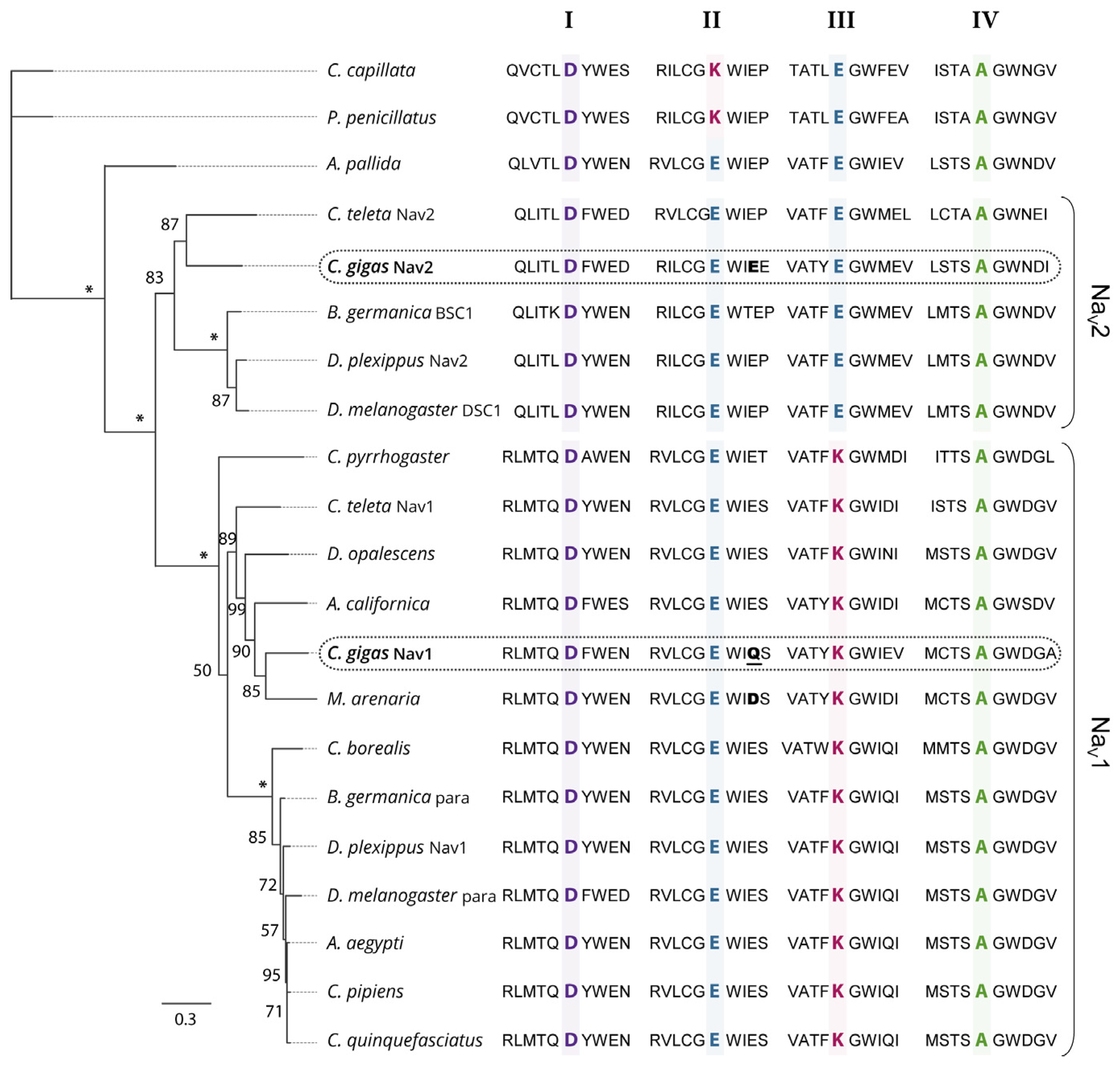
2.1. Phylogenetic Analysis of Nav Channels in Crassostrea gigas

Phylogenetic relationships between the protein sequences of Nav α subunit characterized in C. gigas (EKC22630 and EKC21550) and the Nav previously reported in other invertebrate species were studied (Figure 2). Analyses revealed that sequences annotated Nav9 (EKC22630) and Nav5 (EKC21550) channels in C. gigas did not cluster together but branched at the root defining the two clades of Nav channels (Nav1 and Nav2): the Nav9 sequence appeared grouped with Nav1 channels, but Nav5 grouped with Nav2. Accordingly, we renamed Nav9 as CgNav1 and Nav5 as CgNav2. The Nav1 cluster includes para type channel and Nav2 includes BSC1/DSC1 type channel found in insects. The Pacific oyster Nav1 channel showed a very close phylogenetic relationship with the Nav channel of the clam M. arenaria and with para-like channels characterized in other mollusc species: in the gastropod Aplysia californica and in the cephalopod Doryteuthis opalescens. Contrastingly, C. gigas Nav2 channel presented a close relationship with the Nav2 channel of Capitella teleta, BSC1 and DSC1 channels of the insects Blatella germanica and Drosophila melanogaster, respectively. To our knowledge, CgNav2 is the first member of Nav2 cluster characterized in bivalves. Analyses of the selectivity filters revealed a DEKA motif for the Nav1 of C. gigas as observed in all Nav1 channels. This sequence gives the channel selectivity to sodium ions only. For the Nav2 of C. gigas, a DEEA motif was revealed as observed in the Nav2 channels of anthozoans and bilaterians. These results suggest that the Nav2 channel is likely to be selective to sodium and calcium ions. Considering these results, only the CgNav1 channel was considered for further characterization steps in the study of relationships between paralytic shellfish toxins accumulation and sodium channel characteristics, as the sodium channel is known to be the first target of PST.

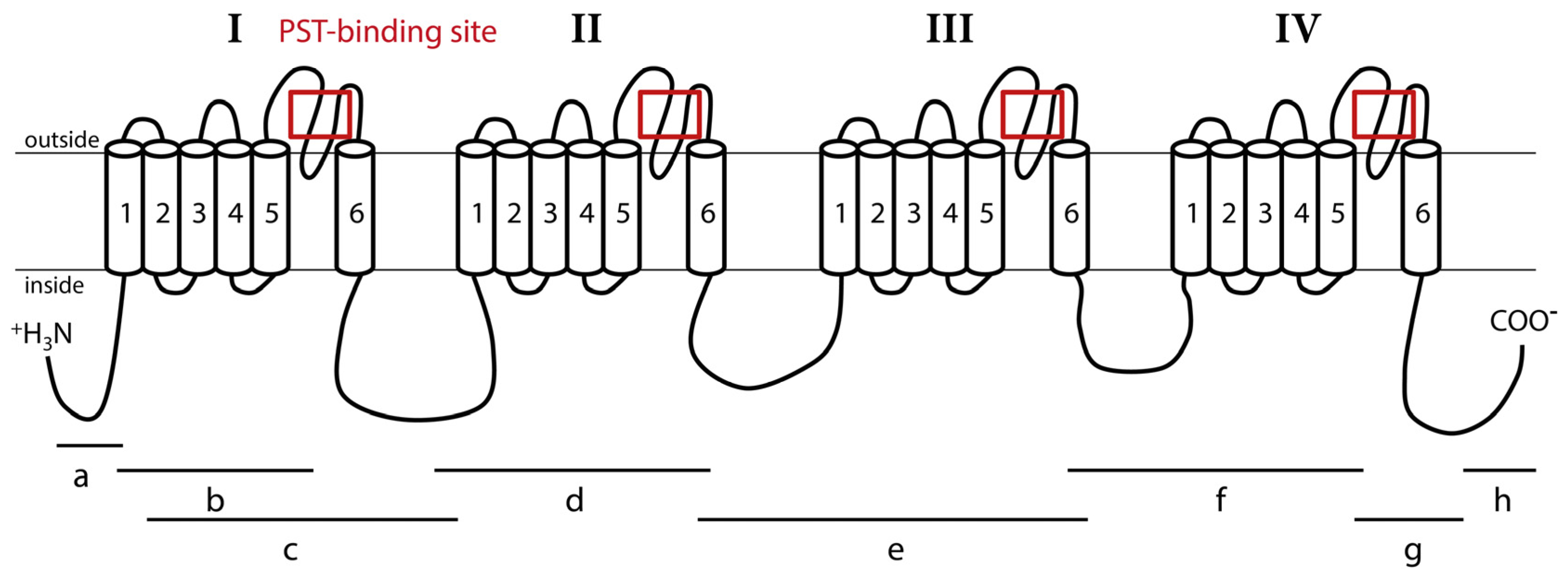
2.2. Structure of CgNav1 α Subunit

The CgNav1 α subunit was amplified and sequenced step by step using 8 overlapping cDNA fragments covering the entire open reading frame (ORF) (Figure 1). The 8 overlapping fragments correspond to: 575 bp from 5′UTR to segment 1 of domain I (IS1), 444 bp from IS1 to IS5-IS6, 1813 bp from IS1 to IS6-IIS1, 904 bp from IS6-IIS1 to IIS6-IIIS1, 1337 bp from IIS6-IIIS1 to IIIS6-IVS1, 736 bp from IIIS6 to IVS5-IVS6, 724 bp from IVS5-IVS6 to stop codon and 533 bp from stop codon to 3′UTR. The consensus sequence created using Geneious software has been compared to the NaV9 α subunit genomic reference sequence (CGI_10001852). This allowed the identification of 25 exons (including an alternate exon, see Section 2.4) and 24 introns (Figure 3). The exons were numbered from 1 to 25. The size of exons ranges from 27 bp to 1235 bp and the size of introns ranges from 89 bp to 2389 bp.

2.3. CgNav1 DNA Polymorphism

The polymorphism was investigated in the DNA sequences of the region surrounding the PST binding sites of the 4 domains (I, II, III, and IV) of CgNav1 α subunit. The size of amplicons was 428 bp, 136 bp, 328 bp and 301 bp for domains I, II, III and IV, respectively. In the 644 sequences analysed (4 regions sequenced per gene, analysed on 161 oysters sampled from all 4 populations) only 3 non-synonymous polymorphisms were identified. These are located outside of the 10 amino acids constituting the PST binding sites. Thus, the protein sequence of the PST binding site appeared perfectly conserved in all the individuals analysed. The number of SNP (Single Nucleotid Polymorphism), calculated per population and per domain of the CgNav1 α subunit, varied from 0 to 5 in exonic regions and 2 to 7 in intronic zones (Table 1). The level of SNP was 1/61 bp in coding regions and 1/40 bp in non-coding regions. Global genetic diversity calculated as the mean ∏t of the 4 domains was similar in all populations (∏t mean = 0.016).

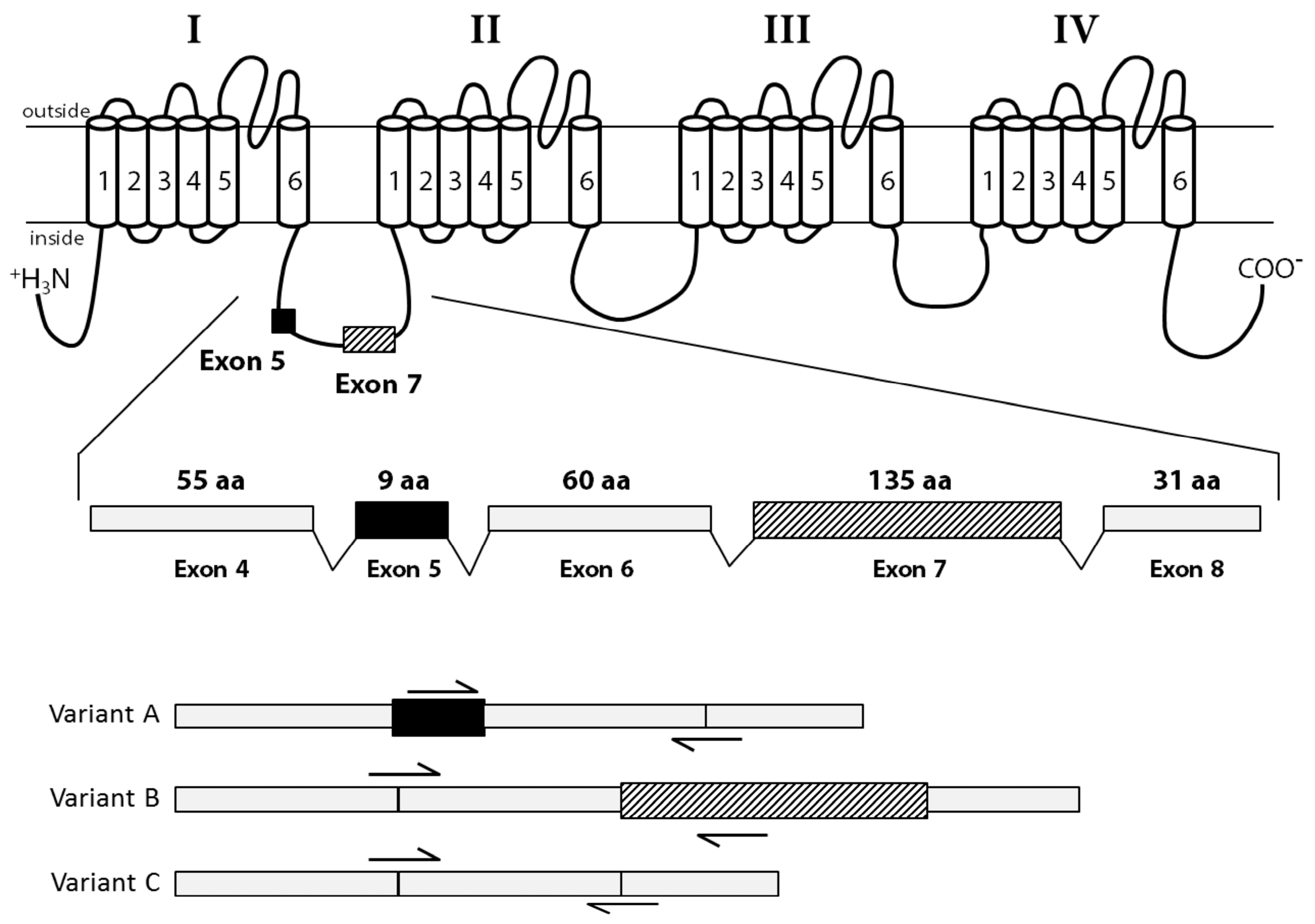
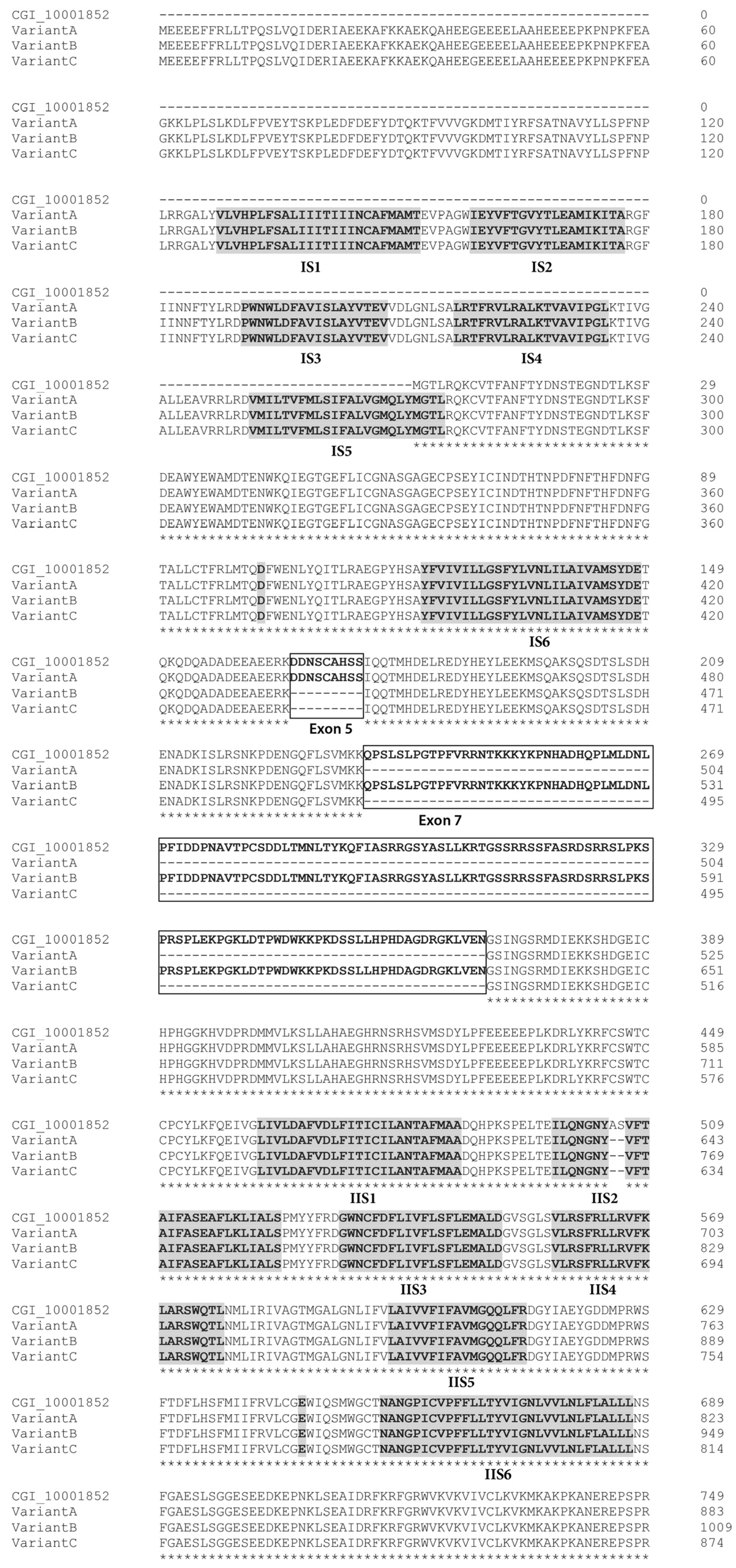
2.4. Identification of CgNav1 Splice Variants

The cDNA fragments “a” to “h” (Figure 1) were individually amplified in 9 tissues of 5 oysters, to reveal potential splice variants. Splice variants were discriminated on the basis of size using electrophoresis. The fragment “c” (from 361 base pair (bp) to 2234 bp length) was the unique amplicon presenting size variations between samples. Sequencing analyses and alignment of CgNav1 cDNA to CGI_10001852 revealed the existence of three different splice variants named A, B and C. The alignment of variant sequences on the CGI_10001852 reference sequence allowed determination of which exons were spliced (Figure 3). Sequences were deposited in GenBank with accession numbers KY020155, KY020156 and KY020157 for variant A, B and C, respectively.
Variant A lacks exon 7 (135 amino acids), which encodes a region of the intracellular inter-domain between domain I and II (ID I-II) rich in proline and serine residues. Variant B lacks exon 5 (9 amino acids), which also encodes a part of the ID I-II. Finally, variant C lacks both 5 and 7 exons. Exons 5 and 7 seem to be mutually exclusive exons, as no complete sequence with both exons has been found. Variants A and C also have retained a part of the intron 14 (33 bp), that could correspond to an alternate exon, which is predicted to encode a part of the intracellular ID II-III. We named this alternate exon “15”. All the characteristics of transcript variants are summarized in Table 2 and Figure 4.
Analyses of the predicted protein structure of splice variants showed that exon 5 has three serine residues, which are polar amino acids and could be phosphorylated. Exon 7 has many important residues. There are 15 proline residues at the beginning and the end of the exon and 17 serine residues in the middle of the exon, all corresponding to many phosphorylation sites. The alternate exon 15, which is present in variants A and C, had only one proline residue. These results were confirmed by the prediction of the phosphorylated sites in both exons. Exon 5 has two protein kinase C (PKC) predicted sites; whereas, exon 7 had 12 protein kinase A (PKA) predicted sites.

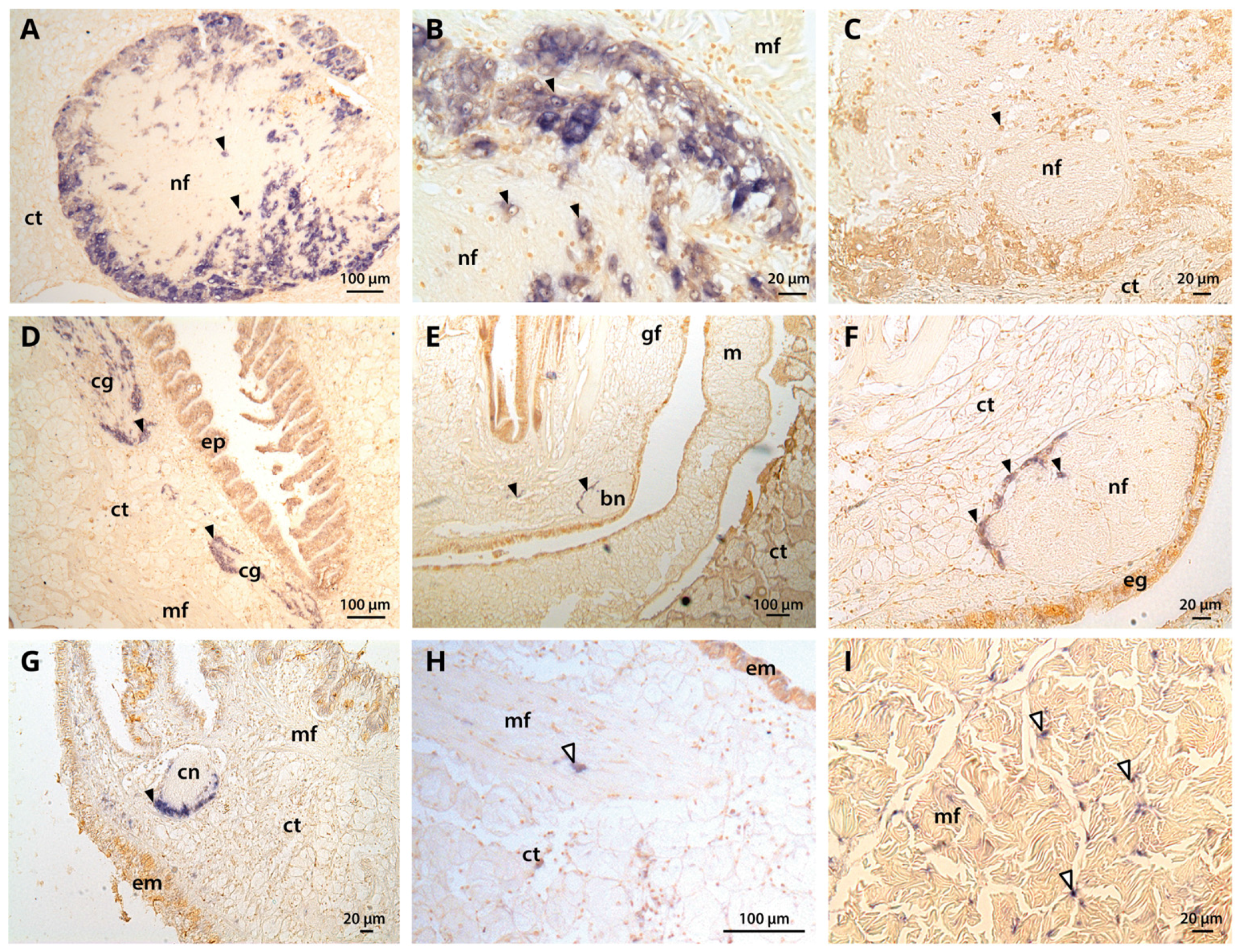
2.5. Tissue-Level CgNav1 α Subunit Expression Patterns

Expression of CgNav1 channel was investigated by real-time PCR and by in situ hybridization (Figure 5) using primers amplifying a sequence common to all the variants. Real-time PCR analyses showed that CgNav1 gene appeared predominantly expressed in visceral ganglia (relative quantification, Qr = 14). CgNav1 is also expressed in striated muscle (Qr = 3.2). The expression of CgNav1 in the gills (Qr = 2.3) is twice as intense as in the mantle (Qr = 1) and four times higher than in the labial palps (Qr = 0.5). The CgNav1 gene is expressed less in the smooth muscle (Qr = 0.3) and almost absent in gonad (Qr = 0.2), heart, and digestive gland (Qr = 0.1 for both).
In situ hybridization showed that CgNav1 is selectively expressed in the nerve cells of the visceral ganglia, located on the peryphery of the ganglia (Figure 5A–C). CgNav1 mRNA were also detected in the nerve cells of cerebral ganglia at the base of the labial palps but not in the adjacent connective tissue (Figure 5D). As in visceral ganglia, nerve cells are located on the periphery of the cerebral ganglia but seem less abundant. Clear staining was detected in the nerve cells of the branchial nerve near the gill axis (Figure 5E,F), and in the nerve cells of the circumpallial nerve which runs along the mantle edge Figure 5G). Staining spots of 5–6 μm-size were observed abundantly among the muscle fibres of the striated muscle and sporadically in mantle (Figure 5H); in both tissues, the staining was not localized in a delimitated cell structure (Figure 5I). According to the description of the adductor muscle of the eastern oyster by Morrison [22], this staining could be localized in the nerve ending that often occurr close to the sarcolemma, corresponding to neuromuscular junctions. No signal was observed in the smooth muscle, neither inside the muscle fibres themselves nor in the nerve fibres. In all tissues, the observed staining of CgNav1 mRNA corresponds to the nerve cell bodies or the neuromuscular junctions but not to the axons of the neurons.

2.6. Expression Patterns of CgNav1 α Subunit Splice Variants

The expression of the splice variants A, B and C of CgNav1 α subunit was examined by real-time PCR using variant-specific primers (Table 3 and Figure 6). Variant A is most expressed in striated muscle (Qr = 10.4), then in decreasing order, in mantle (Qr = 4.8), smooth muscle (Qr = 0.6), gills (Qr = 0.1), and no expression was detected in visceral ganglia, labial palps, digestive gland, or heart. Variant B is mainly expressed in visceral ganglia (Qr = 24.1) and weakly expressed, in decreasing order, in mantle (Qr = 2.4) (inter-organ comparisons, n = 5, p < 0.001), gills (Qr = 2.1), labial palps (Qr = 1.4) (n = 5, p = 0.016), digestive gland (Qr = 0.4), striated muscle (Qr = 0.2) (n = 5, p = 0.01), smooth muscle (Qr = 0.1) and heart (Qr = 0.1). Variant C also is highly expressed in visceral ganglia (Qr = 16.2), then more weakly in striated muscle (Qr = 4) (n = 4, p = 0.009), gills (Qr = 2.9), mantle (Qr = 2.3), labial palps (Qr = 0.4) (n = 6, p < 0.001), digestive gland (Qr = 0.1), smooth muscle (Qr = 0.1), and almost not expressed in heart (Qr = 0.02). These results indicate that variant A (with exon 5) is never expressed in visceral ganglia, digestive gland, or labial palps. Variant B (with exon 7) is almost never expressed in striated muscle.

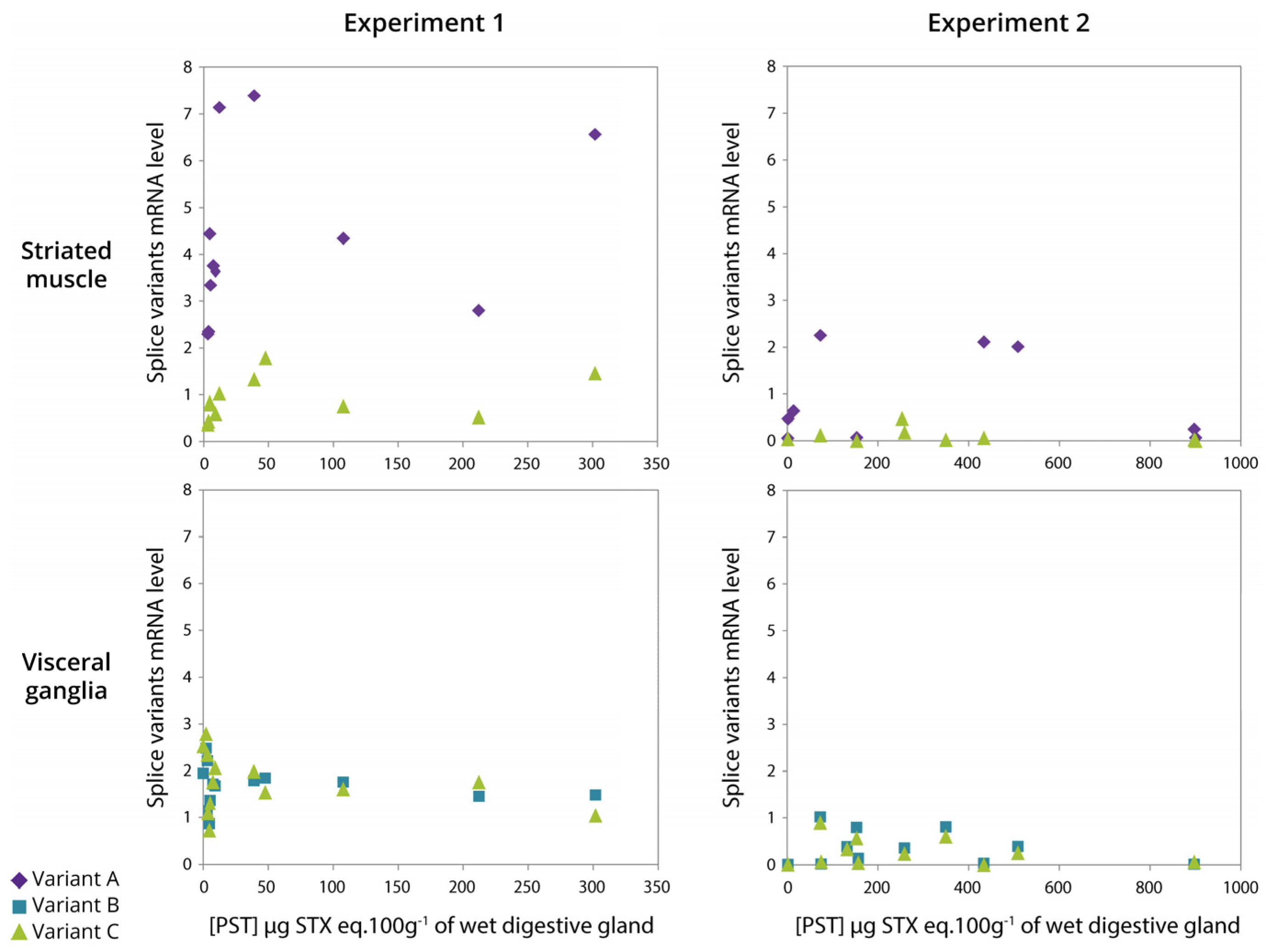
2.7. Relationship between Expression of CgNav1 α Subunit Splice Variants and PST Accumulation

PST accumulation in the digestive glands of oysters exposed to A. minutum, ranged from 2 to 302 (experiment 1) and from 2 to 900 (experiment 2) μg STX eq. 100 g−1 of wet digestive gland, the organ that accumulated the most (Figure 7). As a result, the toxin content varied by a factor 150 (experiment 1) and 450 (experiment 2) between oysters. According to the results presented in Figure 6, only variants A and C are represented for striated muscle, and variants B and C in visceral ganglia, in both experiments (Figure 7). In the striated muscle, no statistical significant correlation was observed between the expression of CgNav1 and toxin accumulation when the analyses were made on the full range of toxin accumulation values. However, CgNav1A and CgNav1C expression tended to increase according to PST accumulation for oysters with low toxin content (<100 μg STX eq. 100 g−1 of digestive gland). In visceral ganglia, a high inter-individual variability of the expression levels of variants B and C were observed for oysters that have very low toxin content. No correlation was observed between toxin content and expression in this tissue.

3. Discussion

3.1. Two Genes Encoding Two Types of Nav Channels (Nav1 and Nav2) in C. gigas

The mains objectives of our study were to characterize the voltage-gated sodium channel α subunit in the Pacific oyster C. gigas and explore a potential relationship between expression and PST bioaccumulation. In NCBI databases, two sequences are annotated as Nav genes (Nav9 and Nav5) for C. gigas [21]. Our results revealed that Nav9 and Nav5 cluster, respectively, with Nav1-type and Nav2-type genes and we, therefore, proposed to rename the C. gigas Nav genes CgNav1 (Nav9, EKC22630) and CgNav2 (Nav5, EKC21550). The sequence EKC22630 appeared incomplete, although it results from the oyster genome sequencing. This underlines the necessity to check different databases or to control candidate sequences by amplification and sequencing to obtain the most accurate reference sequence as highlighted by Rivière et al. [24].
Voltage-gated sodium channels share the amino acid sequence DEKA (for domains I, II, III and IV, respectively) responsible for the selectivity filter of the pore (between S5 and S6 in each domain) [14,25]. Our results showed that the DEKA sequence is found in the CgNav1 sequence as in the other Nav1-type proteins, but the protein CgNav2 presents a DEEA sequence characteristic of Nav2-type proteins [26,27]. The lysine amino acid in the domain III of Nav1 channels enhances the selectivity for sodium [28,29]. Conversely, the glutamic acid found in Nav2 is characteristic of calcium channels. Site-directed mutagenesis studies in the cockroach, Blattella germanica, highlighted the likelihood that glutamic acid in domain III of BSC1 gene plays a key role in the selectivity for calcium [30]. Similarly, in insects, the two genes para (DEKA) and DSC1 (DEEA), were initially classified as sodium channels [12]. Recently, functional studies demonstrated that the DSC1 gene could instead be a new type of voltage-gated cation channel [31]. A recent study proposed that the selectivity filter of choanoflagellates and metazoans (DEEA) is an intermediate between calcium channel (EEEE) and sodium channel (DEKA) and remains present in Nav2 of invertebrates [7]. These channels (DEEA) would be selective to both calcium and sodium ions [32]. Our results raise the issue of the nature and selectivity of CgNav2 channel in the Pacific oyster. CgNav2 could be a channel intermediate between the sodium and calcium channels. These uncertainties about the nature of CgNav2 led the study to focus only on CgNav1, a member of the voltage-gated sodium channels known to be the target of PST.

3.2. CgNav1 Genotype Could Confer a Certain Resistance of Oysters to PST

To investigate if the variability of PST accumulation between oysters is related to the existence of several forms of CgNav1 with different sensitivity to PST, two hypotheses were explored: (i) genetic polymorphism in PST binding sites of CgNav1 leads to different phenotypes of sensitivity to PST; (ii) alternative splicing of CgNav1 produces protein isoforms with different sensitivity to PST.
DNA polymorphism analysis in C. gigas revealed that the 10 amino acids of the PST binding sites were strictly monomorphic, despite the high nucleotid polymorphism of CgNav1, similar to the global SNP level described for this species [33]. Nav are encoded by genes highly conserved through evolution. This probably reflects the critical functional role of these proteins in the regulation of excitability [34], in particular in the most conserved pore region, the critical zone for the selection of ions and their flow [16,35]. In softshell clams, PST resistance is conferred by the substitution of the glutamic acid into aspartic acid in the PST binding site of domain II of Nav channel [4]. For this species, exposure to PST constitutes a strong selection pressure because PST causes mortalities of sensitive clams, leading to the increase in the resistant allele in populations regularly exposed to Alexandrium. In the Pacific oyster, at the same position, a glutamine is observed for CgNav1. Sensitivity studies of rat Nav1.2 revealed that mutation E945Q in domain II leads to resistance to STX and decreases sodium conductance [17,36]. As a result, oysters could have some resistance to STX attributable to the glutamine, which is consistent with the absence of mortalities observed in Pacific oyster populations during Alexandrium blooms. The high frequency of this CgNav1 “resistant” genotype in C. gigas populations could result from an ancestral polymorphism followed by selection under PST pressure in native Asian origins of the Pacific oyster, as the same genotype was found in the Japanese and French populations. This hypothesis needs to be confirmed by functional electrophysiological studies using heterologous expression of these genes in Xenopus oocytes as performed in insects [12].

3.3. CgNav1 is Spliced in Tissue-Specific Variants

The three variants (A, B and C) characterized for CgNav1 resulted from alternative splicing of the two exons 5 and 7, likely mutually exclusive because the complete form of the cDNA with exons 5 and 7 has never been detected. Our results showed that spliced exons were localised in the inter-domain region between domains I and II (ID I-II). This region is a common area of alternative splicing for voltage-gated sodium channels in many species, such as Drosophila melanogaster [37,38], Musca domestica [39], Bombyx mori [40], Cancer borealis [41], or in mammalian sodium channels [42,43]. In accordance with the role of Nav in excitable cells, CgNav1 mRNA appeared to be expressed in nerve cells of the central nervous system of oysters composed of visceral and cerebral ganglia, as well as in the peripheral nervous system composed of nerves innervating the tissues, such as branchial (at the base of the gills) or circumpallial (running along the mantle edge) nerves. Awad et al. specified in a study on rat that Nav1 mRNAs distribution generally corresponds to the localisation of the protein they encode [44]. As a result, CgNav1 mRNAs localization would translate CgNav1 channel distribution, even if further studies on protein are needed to confirm this pattern. The variant B was expressed in all the tissues; nerve cells of nerves or ganglia were stained by ISH, but not in the other tissues. The CgNav1B could be a form of CgNav1 specific to nerve cells. Conversely, the variant A was only expressed in muscles or in tissues with abundant muscular fibres: in the striated muscle, the mantle, and to a lesser extend in the smooth muscle and gills. Expression of CgNav1 observed in muscles of oysters is likely in relation to the function of Nav channels in neuromuscular communication. In fact, Nav channels are found in neuromuscular junctions and participate in the propagation of the action potential in the postsynaptic membrane, allowing the contraction of the muscle. As a result, CgNav1A and CgNav1B could be the specific form to neuromuscular junctions and nerve cells, respectively. The shortest form of CgNav1, the variant C seemed to present ubiquitous expression.

3.4. Potentially Different Pathways of Regulation Exist for CgNav1

The variant B of CgNav1 expressed in the nervous system of oysters encodes a larger protein than proteins translated from variants A and C. This variant results from the retention of the exon 7, encoding a larger ID I-II with 12 putative protein kinase A sites. The ID I-II is known to be an important region for protein regulation because of its richness in PKA phosphorylation sites. The alternative splicing of exons containing PKA sites allow conditional phosphorylation of the Nav channels. Smith and Goldin demonstrated in the rat that the ID I-II of Nav channel is longer in brain than in skeletal muscle, and that the brain channel had many phosphorylation sites involved in PKA signalling pathways [45]. Similarly, in oysters the results suggest that only variant B of CgNav1 could be modulated by PKA. It is also consistent with the presence of variant B in the nervous system and not in striated muscle. Accordingly, alternative splicing could be a mechanism involved in regulation of sodium channel expression in oysters. Phosphorylation of sodium channels by PKA and protein kinase C (PKC) has been shown to reduce the peak sodium current and modulates activation and inactivation phases [45]. A study in the cockroach identified two types of sodium current with two different signalling pathways, one of which was phosphorylation by PKA [46]. In parallel, in the same species, two current types were proposed to originate from the alternative splicing of one Nav gene [47]. Otherwise, this could suggest that variant B of CgNav1, exclusively present in the nervous system of the Pacific oyster, could be regulated by the PKA pathway, but variants A and C would be regulated by other pathways. In the same way, the different splice variants of CgNav1 could generate different current types. Functional studies of each form of CgNav1 would allow the study of electrophysiological properties of these channels.

3.5. The Level of PST Accumulation Would Be Independent of CgNav1 Transcription Level

The exposure of oysters to the PST-producer A. minutum, caused large individual variability of toxin accumulation in the digestive gland of oysters, as reported in a previous study [20]. The expression of the 3 variants of CgNav1 has been analysed and compared individually to toxin content. The variability of PST accumulation did not appeared correlated to Cg1 splice variant expression levels. However, in striated muscle, CgNav1A and CgNav1C expression tended to increase according to PST accumulation up to a threshold of 100 μg STX eq. 100 g−1 of wet digestive gland. This could suggests that when toxins bind to the channel and block the nerve impulses, the oyster has to compensate and produce more channels to maintain a sufficient flow of sodium for cell function. Studies revealed that treatment with Nav channel blockers (like PST) increases cell-surface expression of Nav channels [48]; however, in visceral ganglia, no correlation was observed between CgNav1 (B and C) expression and toxin content. The activity of CgNav1B could be regulated at post-translational level by PKA, as proposed in the previous section, rather than by modification of transcription rate. The CgNav1C, the shortest and less-expressed form, seemed to be regulated as CgNav1A in striated muscle but not in visceral ganglia and could have tissue-specific regulation. In these experiments, oyster Nav channels seem to have an activity-dependent regulation to optimise activity and avoid hyper-excitability. When the toxin content in digestive gland exceeded 100 μg·100 g−1, the CgNav1 mRNA synthesis in striated muscle did not increase further, possibly because of physiological disorders provoked by PST disturbing gene regulation processes. In oysters with the highest toxin content, ca. 900 μg STX eq. 100 g−1 of digestive gland, CgNav1 expression was near zero. This could correspond to the impossibility for oysters to maintain neuromuscular communication, leading to paralysis.
In conclusion, the alternative splicing of CgNav1 gene in Pacific oysters may be a critical mechanism allowing the production of CgNav1 channels adapted to different nervous functions. Specific regulation of CgNav1 isoforms may result in different channel properties. Given the absence of protein polymorphism at the PST binding site of specific variants in oysters accumulating the less or the more PST, we can rule out the initial hypothesis that different forms of CgNav1 could explain the inter-individual variability of the PST accumulation. Our results suggest that the blocking of Nav by PST in oysters could trigger the activation of regulatory pathways to modulate Nav channel expression. The protein sequence of CgNav1 would confer to all the forms of oyster Nav1 a relative resistance to STX. To validate this hypothesis, the specific sensitivity of each CgNav1 variant to PST needs to be investigated, using, for example, heterologous expression in Xenopus oocytes and an electrophysiological approach. Accordingly, in Pacific oysters the quantity of PST accumulated would result from individual variability in ecophysiological capacities such as filtration, detoxification or even biological rhythms rather than differential sensitivity of Nav channel.

4. Materials and Methods

4.1. Phylogenetic Analyses of the Voltage-Gated Sodium Channel α Subunit of Crassostrea gigas

Amino acid sequences alignment was based upon a multiple alignment method using MAFFT 7 [49]. Alignments were refined to select reliably-aligned positions by using Gblocks version 0.91b [50]. The substitution model LG (+I +G +F) used in this study was selected using ProtTest 3.4.2 [51]. The maximum likelihood phylogenetic tree was constructed using PhyML 3.0 [52], and tree robustness was assessed with 100 bootstrap replications. Tree visualization was performed using FigTree v1.4.2 [53]. Sequences used for the analysis are presented in Table 4.
The oyster Nav2 was not studied for further experiments as its sequence suggested no selectivity for sodium ions only, meaning that it may not be a true Nav channel (see explanation in the result section).

4.2. Biological Material

4.2.1. Crassostrea gigas Oysters

For polymorphism analyses (Section 4.6), a study was conducted on 4 populations of C. gigas. Three French populations located on the west coast: Bay of Brest, North Brittany; Larmor Baden, South Brittany; Ile de Ré, Charente-Maritime, and regularly exposed to toxic bloom of PST (n = 50 per population) and one Japanese population (n = 20) located in the Bay of Sendai, known to be exposed to toxic blooms of PST for many years [67]. Gills were sampled from oysters and stored in ethanol for DNA extraction. For the characterization of Nav cDNA sequences (Section 4.7) and expression analysis (Section 4.8 and Section 4.9), wild oysters were sampled in the Bay of Brest (Brittany, France). Nine different tissues were dissected from each individual: mantle, gills, heart, smooth muscle, striated muscle, labial palps, visceral ganglia, gonad, and digestive gland. Immediately after dissection, tissues were placed in RNA later solution (Invitrogen, Carlsbad, CA, USA) and stored at −80 °C until RNA extraction. For the study of CgNav1 variant expression in experiments 1 and 2 (Section 4.3), oysters were obtained from the experimental hatchery of Ifremer in La Tremblade (Charente-Maritime, France), and from a shellfish farmer in the Bay of Arcachon (Gironde, France), respectively. Immediately after dissection, tissues were placed in RNA later solution (Invitrogen, Carlsbad, CA, USA) and stored at −80 °C until RNA extraction.

4.2.2. Microalgae Cultures

The dinoflagellates Alexandrium minutum Halim (1960) strain Daoulas 1257 (isolated from Brest Bay, France) and strain AM89BM (isolated from Morlaix Bay, France) were used for toxic algal exposure (experiment 1 and experiment 2, respectively), and the non-toxic dinoflagellate Heterocapsa triquetra (Ehrenberg) Stein, strain HT99PZ (isolated from Penzé Bay, France) was used as a control. Both dinoflagellate cultures were grown in L1 medium [68] at 16 °C with a light/dark cycle of 12:12 h and were harvested during exponential growth phase. Algal cell densities were determined by counts using Nageotte counting chamber (PolyLabo, France) under a light microscope.

4.3. Experimental Design for Oyster Exposure to PST

To test the possible relationship between CgNav1 expression and PST accumulation, oysters were exposed to the toxic A. minutum or the non-toxic dinoflagellate Heterocapsa triquetra similar in size and shape to A. minutum. Both experiments were set up in two different phases: an acclimation period of 7 days to the non-toxic dinoflagellate followed by an exposure period of 4 or 6 days to the toxic dinoflagellate species or to the control. Each tank was supplied with microalgae using a peristaltic pump. Central air-lifts were used to homogenize microalgal concentration and water in each tank.

4.3.1. Experiment 1

Oysters were placed randomly in 18 L replicated tanks with 12 oysters per tank. During the acclimation period, all oysters were fed with a continuous flow of 19 L·day−1 of seawater with H. triquetra (106 cells·L−1), then oysters were separated into two groups, exposed for 4 days to a continuous flow of 19 L·day−1 of seawater with A. minutum strain Daoulas 1257 (3.106 cells·L−1; n = 60, 5 replicates of 12 individuals) or H. triquetra (106 cells·L−1; n = 36, 3 replicates of 12 individuals). At the end of the exposure period, digestive glands of oysters were dissected, weighed, frozen, and stored in liquid nitrogen until toxin analyses. Visceral ganglia and striated muscle also were dissected and stored in RNA later solution (Invitrogen, Carlsbad, CA, USA) at −80 °C until mRNA expression analyses.

4.3.2. Experiment 2

Oysters were distributed randomly into six tanks, with 29–30 oysters per tank. During the acclimation period, oysters were fed with a continuous flow of 144 L·day−1 of seawater with H. triquetra (105 cells·L−1). Then oysters were separated into two groups exposed for 6 days to a continuous flow of 144 L·day−1 of seawater with A. minutum strain AM89BM (105 cells·L−1; n = 88; 3 replicates) or H. triquetra (105 cells·L−1; n = 88; 3 replicates). At the end of the exposure period, digestive glands, visceral ganglia, and striated muscle were sampled as in experiment 1.

4.4. Toxin Quantification by Liquid Chromatography/Fluorescence Detection

To extract the PST, 5 mL of 0.1 N hydrochloric acid were added, and the samples were mixed with a high-speed homogenizer (15,000 rpm) for 2 min. The pH was adjusted between 2.0 and 4.0, then the samples were centrifuged at 4200× g for 10 min at 4 °C. The supernatants were filtered on 10-kDa polyethersulfone (PES) filters, and the toxin content was analyzed using the liquid chromatography with fluorescence detection (LC/FD) PSP toxin analyses method of Van de Riet [69]. The toxins GTX, dc-GTX, dc-STX and STX were separated using a reverse chromatography column (Zorbax Bonus RP, 3.5 μM, 4.6 mm × 150 mm, Agilent Technologies, Massy, France) with a flow rate of 0.8 mL·min−1. The eluent pH and/or column temperature were optimized to separate dc-GTX3/GTX5/dc-GTX-2 and C1/C2. The toxin concentrations were determined using certified standards provided by CNRC (Halifax, NS, Canada).

4.5. DNA and RNA Extractions and cDNA Synthesis

Genomic DNA was extracted from oyster gills with the DNeasy Blood and Tissue kit (Qiagen, Germantown, MD, USA) according to the manufacturer’s instructions. The concentration and purity of DNA were analysed with a Nanodrop 8000 spectrophotometer (Thermo Scientific, Waltham, MA, USA). Total RNA was extracted using TRI Reagent® (Sigma-Aldrich, St. Louis, MO, USA) following manufacturer’s instructions. Samples were treated with RTS DNase™ kit (MO BIO Laboratories, Germantown, MD, USA) to avoid genomic DNA contamination. The concentration and purity of all RNA were estimated with a Nanodrop 8000 spectrophotometer (Thermo Scientific, Waltham, MA, USA). RNA integrity was assessed by electrophoresis on agarose gel. cDNA synthesis was performed using 1 μg of total RNA primed with an Oligo(dT)18 and reverse-transcribed into first strand cDNA with the RevertAid H minus First Strand cDNA Synthesis kit (Fermentas, York, UK).

4.6. Single Nucleotid Polymorphism of C. gigas Nav1 α Subunit Gene

The DNA sequence of voltage-gated sodium channel α subunit (Nav1) of C. gigas was obtained from Zhang et al. [21]. This sequence (CGI_10001852) was first used as a reference sequence to design primers for DNA amplifications. The single nucleotid polymorphism of PST binding region was analysed by PCR amplification and sequencing (Figure 1). The region including the P segment of each domain of the Nav1, corresponding to the zone targeted by PST, was amplified by PCR with specific primer pairs (Table 5). Amplifications were performed in 25 μL of final reaction mixture. Each reaction contained 250 ng of DNA, 1.5 mM MgCl2, 200 μM dNTPs, 0.1 μM of each primer, 5 μL of Buffer 5× and 1.5 U polymerase GoTaq Flexi DNA (Promega, Madison, WI, USA). Cycling conditions were 2 min at 95 °C, 40 cycles of denaturation step for 45 s at 95 °C, annealing step for 45 s at 60 °C and elongation step for 45 s at 72 °C and a final step for 5 min at 72 °C. PCR products were verified by electrophoresis on agarose gel before sequencing (Sanger ABI 3730xl, GATC Biotech, Cologne, Germany). Chromatograms were checked and corrected by hand if needed and aligned to locate the SNP polymorphic site. A variation in the sequence was considered as a SNP only when its occurrence was above the threshold of 5% of the total number of oysters sampled. Nucleotide sequences then were translated into amino acid sequences to identify synonymous or non-synonymous mutations.

4.7. Amplification and Sequencing of the cDNA of CgNav1 α Subunit

The full cDNA sequence was amplified step by step using the set of primers described in Table 5. The fragments “a” to “h” amplified to cover the full cDNA sequence are represented in Figure 1. A consensus sequence of the cDNA of CgNav1 α subunit has been obtained from sequencing data using the Geneious software version 7.0.6 [70]. The fragments were amplified in 5 oysters and 9 tissues: striated and smooth muscles, mantle, visceral ganglia, heart, digestive gland, gills, gonad and labial palps. Amplification was performed in 50 μL of final reaction mixture. Each reaction contained 2 μL of cDNA, 1.5 mM MgCl2, 200 μM dNTPs, 0.1 μM of each primer (Table 5), 10 μL of Buffer 5× and 1.5 U polymerase GoTaq Flexi DNA (Promega, Madison, WI, USA). Cycling conditions were 2 min at 95 °C, 40 cycles of denaturation step for 45 s at 95 °C, annealing step for 45 s at 60 °C and elongation step for 4 min at 72 °C and a final step for 5 min at 72 °C. Specificity of amplification were checked on agarose electrophoresis and cloned into pCR4-TOPO vector with the TOPO TA cloning kit (Invitrogen) before sending to sequencing (Sanger ABI 3730xl). The characterization of splice variants was performed using PCR. Specific primers were designed to encompass the cytoplasmic loop linking domain I and domain II corresponding to fragment “c” which varies between variants.

4.8. Localization of CgNav1 α Subunit mRNA Expression by In Situ Hybridization

The tissue localization of CgNav1 mRNA was studied by in situ hybridization (ISH). For each individual, 4 transversal slices of 3 mm thickness were performed to study all sampled tissues: anterior region of the body (labial palps); anterior-middle region (digestive gland, gills, mantle, gonad); posterior-middle region (muscles, heart); posterior region (muscles, visceral ganglia). Samples were fixed immediately in Davidson’s fixative for 24 h at 4 °C, and then in ethanol 70% at 4 °C until inclusion, in RNAse free conditions. Tissues were dehydrated and embedded in paraffin wax using classical histological protocol according to Howard et al. [71], then were sectioned to 5 μm thickness with a microtome. Sections were mounted on glass slides treated with polylysine.
Specific riboprobes were designed from the cDNA sequence of C. gigas Nav9 (CGI_10001852). A 643 bp fragment, designed from the cDNA sequence (CGI_10001852) of CgNav1, was used as the template to synthesize riboprobes specific to all variants of CgNav1 (Table 3). The amplified sequence was cloned into pCR4-TOPO vector with the TOPO TA cloning kit (Invitrogen, Carlsbad, CA, USA). Sense and antisense riboprobes were synthesized by in vitro transcription from linearized plasmids (MEGAscript kit, Ambion, Austin, TX, USA). Digoxigenin (DIG) labeling was performed by the incorporation of modified nucleotides UTP-DIG with the DIG RNA labeling kit (Roche Diagnostics GmbH, Mannheim, Germany).
The in situ hybridization protocol was adapted from Santerre et al. [72]. Briefly, tissue sample sections were treated with 10 μg/mL proteinase K (Sigma-Aldrich, St. Louis, MO, USA) at 37 °C for 10 min, post-fixed with 4% paraformaldehyde in PBS for 7 min and pre-hybridized with yeast tRNA for 1 h 30 min at 50 °C. Hybridization was then performed by incubating tissue sections with DIG-labeled sense or antisense riboprobes in hybridization buffer (50% formamide, 4× saline-sodium citrate buffer (SSC), 2 mM EDTA, 1% Denhardt’s solution, 10% dextran sulfate and 0.5 mg·mL−1 of yeast tRNA) for 16 h at 55 °C. After extensive washes with 50% formamide, 2× SSC at 45 °C, 2X SSC and 1× SSC at 37 °C, revelation was performed by incubating sections with anti-DIG antibody coupled to alkaline phosphatase (Roche Diagnostics GmbH, Mannheim, Germany) for 2 h at room temperature. Sections were then incubated in detection buffer (0.1 M Tris-HCl, 0.1 M NaCl) for 2–4 h in darkness. The mounting between slide and cover slip was performed with Canada balsam (Sigma-Aldrich, St. Louis, MO, USA). Sections were examined and photographed using an optical microscope (Leica DM-IRB, Nussloch, Germany). Hybridization with sense riboprobes was used as negative control.

4.9. Expression of CgNav1 mRNA by Real-Time PCR

Expression of CgNav1 transcripts was studied by real-time PCR in 8 tissues of 6 oysters. The specificity of primers was validated by sequencing of PCR product and analysis of the melting profile. Each amplification was performed using LightCycler 480 (Roche Diagnostics GmbH, Mannheim, Germany) in 10 μL final reaction mixture containing 5 μL of SYBR Green I Master Mix (2×) (Thermo Fisher Scientific, Waltham, MA, USA), 0.5 μL of each primer (10 μM) for Nav or reference genes GAPDH and EF1α (Table 3), 1 μL of cDNA, RNA DNase treated (negative control) or water (blank control) and 3 μL of free-RNase water. Cycling conditions were 10 min at 95 °C, 40 cycles of denaturation step for 10 s at 95 °C, annealing step for 20 s at 57 °C and elongation step for 15 s at 72 °C. All PCR reactions were run in triplicate and crossing point (Cp) values used were the mean of this replicates. The efficiency of amplification was calculated from the slope of the standard curve constructed with five serial dilutions of a pool of cDNA (calibrator) from striated muscle and visceral ganglia in equal proportion. The expression stability of the reference genes (EF1α, GAPDH and Actin) was tested using NormFinder (Version 0.953) algorithm as an Excel add-in. The best stability value (0.003) was obtained for the combination of EF1α and GAPDH genes. As a result, CgNav1 transcript levels were normalised using the geometric mean of GAPDH and EF1α as reference genes and quantified with the E-method using the LightCycler 480 software 1.5.1.62 (Roche Diagnostics GmbH, Mannheim, Germany). mRNA levels of splice variants were expressed in relative quantity (Qr) compared to geometric mean of reference genes.

4.10. Data Analysis

BLAST searches were performed online at NCBI BLAST [73]. Multiple sequence alignments were carried out in Clustal Omega [74]. Exon-intron structure of sequences was assessed by aligning mRNA sequences to genomic sequences with the program MGAlign [75]. Donor and acceptor sites were predicted using a splice site predictor, NNSPLICE version 0.9 [76]. The deduced amino acid sequences were calculated by the ExPASy Proteomics Server [77]. Analyses of the predicted 2D structure of protein isoform were performed with the HCA program (Hydrophobic Cluster Analysis) from Mobyle portal [78]. Phosphorylation sites were predicted by NetPhos 3.1 server [79] and protein kinase A (PKA) and protein kinase C (PKC) sites were predicted with KinasePhos 2.0 server [80] and pkaPS server [81]. All statistical analyses were executed with R 3.2.2 [82]. The validation of the optimal reference gene for the normalization of real-time PCR data was performed using the NormFinder version 0.953 algorithm as an Excel add-in. Log (natural) transformed Cp values were used as input data. The analysis of the reference genes stability was completed by multiple-comparison test with Tukey’s HSD method. Comparisons of mRNA levels (relative to geometric mean of EF1α and GAPDH mRNA) between splice variants were performed with a Kruskal-Wallis test, followed by a non-parametric post-hoc with the Wilcoxon signed rank test. Pearson’s correlation was applied to assess relation between toxins accumulation and Nav mRNA expression. No correlation was applied when samples are less than three.

Acknowledgments

The authors are grateful to Sylvie Lapègue, Lionel Degremont and Serge Heurtebise for providing oysters, to Luca Bargeloni for phylogenetic analysis pipeline, to Arnaud Huvet for his help in oysters sampling in experiment 2, to Véronique Savar, Korian Lhaute and Georges Rovillon for their technical assistance in toxins analysis, and to Maëva Leitwen, Antoine Beuzit and Anne-Laure Cassone for technical assistance in molecular analysis. Authors thank Gary Wikfors for his help with editing the English and his advice on the manuscript. This study was carried out with the financial support of the National Research Agency (ANR) ‘‘ACCUTOX’’ project 13-CESA-0019 (2013–2017), the LabexMer axis 6 “Regitox” project, and the Brittany Region and the University of Occidental Brittany (UBO) for funding the PhD of Floriane Boullot.

Author Contributions

F.B., H.H., L.P., M.P., D.T., C.F., P.S. and P.B. designed the experiments; F.B., L.P., A.B., J.C., N.D. and Z.A. performed the experiments; F.B., A.B., J.C., Z.A. and C.F. analysed the data; F.B. and C.F. wrote the paper.

Conflicts of Interest

The authors declare no conflict of interest.

References

  1. Van Dolah, F.M. Marine algal toxins: Origins, health effects, and their increased occurrence. Environ. Health Perspect. 2000, 108, 133–141. [Google Scholar] [CrossRef] [PubMed]
  2. Manfrin, C.; De Moro, G.; Torboli, V.; Venier, P.; Pallavicini, A.; Gerdol, M. Physiological and molecular responses of bivalves to toxic dinoflagellates. Invertebr. Surviv. J. 2012, 9, 184–199. [Google Scholar]
  3. Sellner, K.G.; Doucette, G.J.; Kirkpatrick, G.J. Harmful algal blooms: Causes, impacts and detection. J. Ind. Microbiol. Biotechnol. 2003, 30, 383–406. [Google Scholar] [CrossRef] [PubMed]
  4. Bricelj, V.M.; Connell, L.; Konoki, K.; MacQuarrie, S.P.; Scheuer, T.; Catterall, W.A.; Trainer, V.L. Sodium channel mutation leading to saxitoxin resistance in clams increases risk of PSP. Nature 2005, 434, 763–767. [Google Scholar] [CrossRef] [PubMed]
  5. Catterall, W.A. From ionic currents to molecular mechanisms: The structure and function of voltage-gated sodium channels. Neuron 2000, 26, 13–25. [Google Scholar] [CrossRef]
  6. Cestèle, S.; Catterall, W.A. Molecular mechanisms of neurotoxin action on voltage-gated sodium channels. Biochimie 2000, 82, 883–892. [Google Scholar] [CrossRef]
  7. Zakon, H.H. Adaptive evolution of voltage-gated sodium channels: The first 800 million years. Proc. Natl. Acad. Sci. USA 2012, 109, 10619–10625. [Google Scholar] [CrossRef] [PubMed]
  8. Catterall, W.A. Structure and function of voltage-gated ion channels. Annu. Rev. Biochem. 1995, 64, 493–531. [Google Scholar] [CrossRef] [PubMed]
  9. Chopra, S.S.; Watanabe, H.; Zhong, T.P.; Roden, D.M. Molecular cloning and analysis of zebrafish voltage-gated sodium channel beta subunit genes: Implications for the evolution of electrical signaling in vertebrates. BMC Evol. Biol. 2007, 7, 113. [Google Scholar] [CrossRef] [PubMed]
  10. Feng, G.; Deak, P.; Chopra, M.; Hall, L.M. Cloning and functional analysis of TipE, a novel membrane protein that enhances drosophila para sodium channel function. Cell 1995, 82, 1001–1011. [Google Scholar] [CrossRef]
  11. Gurnett, C.A.; Campbell, K.P. Transmembrane auxiliary subunits of voltage-dependent ion channels. J. Biol. Chem. 1996, 271, 27975–27978. [Google Scholar] [CrossRef] [PubMed]
  12. Dong, K. Insect sodium channels and insecticide resistance. Invertebr. Neurosci. 2007, 7, 17–30. [Google Scholar] [CrossRef] [PubMed]
  13. Yu, F.H.; Catterall, W.A. Overview of the voltage-gated sodium channel family. Genome Biol. 2003, 4, 1–7. [Google Scholar]
  14. Anderson, P.A.V.; Roberts-Misterly, J.; Greenberg, R.M. The evolution of voltage-gated sodium channels: Were algal toxins involved? Harmful Algae 2005, 4, 95–107. [Google Scholar] [CrossRef]
  15. Al-Sabi, A.; McArthur, J.; Ostroumov, V.; French, R.J. Marine toxins that target voltage-gated sodium channels. Mar. Drugs 2006, 4, 157–192. [Google Scholar] [CrossRef]
  16. Marban, E.; Yamagishi, T.; Tomaselli, G.F. Structure and function of voltage-gated sodium channels. J. Physiol. 1998, 508, 647–657. [Google Scholar] [CrossRef] [PubMed]
  17. Terlau, H.; Heinemann, S.H.; Stühmer, W.; Pusch, M.; Conti, F.; Imoto, K.; Numa, S. Mapping the site of block by tetrodotoxin and saxitoxin of sodium channel II. FEBS Lett. 1991, 293, 93–96. [Google Scholar] [CrossRef]
  18. Bricelj, V.M.; MacQuarrie, S.; Twarog, B.M.; Trainer, V.L. Characterization of sensitivity to PSP toxins in North American populations of the softshell clam Mya arenaria. In Harmful Algae 2002; Steidinger, K.A., Landsberg, J.H., Tomas, C.R., Vargo, G.A., Eds.; Florida Institute of Oceanography and Intergovernmental Oceanographic Commission of UNESCO: Paris, France, 2004; pp. 172–174. [Google Scholar]
  19. Tan, J.; Liu, Z.; Nomura, Y.; Goldin, A.L.; Dong, K. Alternative splicing on an insect sodium channel gene generates pharmacologically distinct sodium channels. J. Neurosci. 2002, 22, 5300–5309. [Google Scholar] [PubMed]
  20. Haberkorn, H.; Tran, D.; Massabuau, J.C.; Ciret, P.; Savar, V.; Soudant, P. Relationship between valve activity, microalgae concentration in the water and toxin accumulation in the digestive gland of the Pacific oyster Crassostrea gigas exposed to Alexandrium minutum. Mar. Pollut. Bull. 2011, 62, 1191–1197. [Google Scholar] [CrossRef] [PubMed] [Green Version]
  21. Zhang, G.; Fang, X.; Guo, X.; Li, L.; Luo, R.; Xu, F.; Yang, P.; Zhang, L.; Wang, X.; Qi, H.; et al. The oyster genome reveals stress adaptation and complexity of shell formation. Nature 2012, 490, 49–54. [Google Scholar] [CrossRef] [PubMed]
  22. Morrison, C.M. The histology and ultrastructure of the adductor muscle of the Eastern oyster Crassostrea virginica. Am. Malacol. Bull. 1993, 10, 25–38. [Google Scholar]
  23. Sussarellu, R.; Fabioux, C.; Le Moullac, G.; Fleury, E.; Moraga, D. Transcriptomic response of the Pacific oyster Crassostrea gigas to hypoxia. Mar. Genom. 2010, 3, 133–143. [Google Scholar] [CrossRef] [PubMed]
  24. Riviere, G.; Klopp, C.; Ibouniyamine, N.; Huvet, A.; Boudry, P.; Favrel, P. GigaTON: An extensive publicly searchable database providing a new reference transcriptome in the pacific oyster Crassostrea gigas. BMC Bioinform. 2015, 16, 401. [Google Scholar] [CrossRef] [PubMed] [Green Version]
  25. Heinemann, S.H.; Terlau, H.; Stuhmer, W.; Imoto, K.; Numa, S. Calcium channel characteristics conferred on the sodium channel by single mutations. Nature 1992, 356, 441–443. [Google Scholar] [CrossRef] [PubMed]
  26. Gur Barzilai, M.; Reitzel, A.M.; Kraus, J.E.M.; Gordon, D.; Technau, U.; Gurevitz, M.; Moran, Y. Convergent Evolution of Sodium Ion Selectivity in Metazoan Neuronal Signaling. Cell Rep. 2012, 2, 242–248. [Google Scholar] [CrossRef] [PubMed]
  27. Liebeskind, B.J.; Hillis, D.M.; Zakon, H.H. Evolution of sodium channels predates the origin of nervous systems in animals. Proc. Natl. Acad. Sci. USA 2011, 108, 9154–9159. [Google Scholar] [CrossRef] [PubMed]
  28. Lipkind, G.M.; Fozzard, H.A. Voltage-gated Na+ channel selectivity: The role of the conserved domain III lysine residue. J. Gen. Physiol. 2008, 131, 523–529. [Google Scholar] [CrossRef] [PubMed]
  29. Schlief, T.; Schönherr, R.; Imoto, K.; Heinemann, H.S. Pore properties of rat brain II sodium channels mutated in the selectivity filter domain. Eur. Biophys. J. 1996, 25, 75–91. [Google Scholar] [CrossRef] [PubMed]
  30. Zhou, W.; Chung, I.; Liu, Z.; Goldin, A.L.; Dong, K. A voltage-gated calcium-selective channel encoded by a sodium channel-like gene. Neuron 2004, 42, 101–112. [Google Scholar] [CrossRef]
  31. Zhang, T.; Liu, Z.; Song, W.; Du, Y.; Dong, K. Molecular characterization and functional expression of the DSC1 channel. Insect Biochem. Mol. Biol. 2011, 41, 451–458. [Google Scholar] [CrossRef] [PubMed]
  32. Dong, K.; Du, Y.; Rinkevich, F.; Wang, L.; Xu, P. The Drosophila Sodium Channel 1 (DSC1): The founding member of a new family of voltage-gated cation channels. Pestic. Biochem. Physiol. 2015, 120, 36–39. [Google Scholar] [CrossRef] [PubMed]
  33. Sauvage, C.; Bierne, N.; Lapegue, S.; Boudry, P. Single Nucleotide polymorphisms and their relationship to codon usage bias in the Pacific oyster Crassostrea gigas. Gene 2007, 406, 13–22. [Google Scholar] [CrossRef] [PubMed]
  34. Goldin, A.L. Evolution of voltage-gated Na+ channels. J. Exp. Biol. 2002, 205, 575–584. [Google Scholar] [PubMed]
  35. Jost, M.C.; Hillis, D.M.; Lu, Y.; Kyle, J.W.; Fozzard, H.A.; Zakon, H.H. Toxin-resistant sodium channels: Parallel adaptive evolution across a complete gene family. Mol. Biol. Evol. 2008, 25, 1016–1024. [Google Scholar] [CrossRef] [PubMed]
  36. Kontis, K.J.; Goldin, A.L. Site-directed mutagenesis of the putative pore region of the rat IIA sodium channel. Mol. Pharmacol. 1993, 43, 635–644. [Google Scholar] [PubMed]
  37. Thackeray, J.R.; Ganetzky, B. Developmentally regulated alternative splicing generates a complex array of Drosophila para sodium channel isoforms. J. Neurosci. 1994, 14, 2569–2578. [Google Scholar] [PubMed]
  38. Lin, W.H.; Wright, D.E.; Muraro, N.I.; Baines, R.A. Alternative splicing in the voltage-gated sodium channel DmNav regulates activation, inactivation, and persistent current. J. Neurophysiol. 2009, 102, 1994–2006. [Google Scholar] [CrossRef] [PubMed]
  39. Lee, S.H.; Ingles, P.J.; Knipple, D.C.; Soderlund, D.M. Developmental regulation of alternative exon usage in the house fly Vssc1 sodium channel gene. Invertebr. Neurosci. 2002, 4, 125–133. [Google Scholar] [CrossRef] [PubMed]
  40. Shao, Y.M.; Dong, K.; Tang, Z.H.; Zhang, C.X. Molecular characterization of a sodium channel gene from the Silkworm Bombyx mori. Insect Biochem. Mol. Biol. 2009, 39, 145–151. [Google Scholar] [CrossRef] [PubMed]
  41. Dai, A.; Temporal, S.; Schulz, D.J. Cell-specific patterns of alternative splicing of voltage-gated ion channels in single identified neurons. Neuroscience 2010, 168, 118–129. [Google Scholar] [CrossRef] [PubMed]
  42. Schaller, K.L.; Krzemien, D.M.; McKenna, N.M.; Caldwell, J.H. Alternatively spliced sodium channel transcripts in brain and muscle. J. Neurosci. 1992, 12, 1370–1381. [Google Scholar] [PubMed]
  43. Plummer, N.W.; Galt, J.; Jones, J.M.; Burgess, D.l.; Sprunger, L.K.; Kohrman, D.C.; Meisler, M.H. Exon organization, coding sequence, physical mapping, and polymorphic intragenic markers for the human neuronal sodium channel gene SCN8A. Genomics 1998, 54, 287–296. [Google Scholar] [CrossRef] [PubMed]
  44. Awad, S.S.; Lightowlers, R.N.; Young, C.; Chrzanowska-Lightowlers, Z.M.; Lømo, T.; Slater, C.R. Sodium channel mRNAs at the neuromuscular junction: Distinct patterns of accumulation and effects of muscle activity. J. Neurosci. 2001, 21, 8456–8463. [Google Scholar] [PubMed]
  45. Smith, R.D.; Goldin, A.L. Phosphorylation of brain sodium channels in the I-II linker modulates channel function in Xenopus oocytes. J. Neurosci. 1996, 16, 1965–1974. [Google Scholar] [PubMed]
  46. Lavialle-Defaix, C.; Gautier, H.; Defaix, A.; Lapied, B.; Grolleau, F. Differential regulation of two distinct voltage-dependent sodium currents by group III metabotropic glutamate receptor activation in insect pacemaker neurons. J. Neurophysiol. 2006, 96, 2437–2450. [Google Scholar] [CrossRef] [PubMed]
  47. Zhao, X.; Ikeda, T.; Salgado, V.L.; Yeh, J.Z.; Narahashi, T. Block of two subtypes of sodium channels in coackroch neurons by indoxacarb insecticides. Neuro Toxicol. 2005, 26, 455–465. [Google Scholar]
  48. Shiraishi, S.; Yokoo, H.; Yanagita, T.; Kobayashi, H.; Minami, S.-I.; Saitoh, T.; Takasaki, M.; Wada, A. Differential effects of bupivacaine enantiomers, ropivacaine and lidocaine on up-regulation of cell surface voltage-dependent sodium channels in adrenal chromaffin cells. Brain Res. 2003, 966, 175–184. [Google Scholar] [CrossRef]
  49. Katoh, K.; Standley, D.M. MAFFT multiple sequence alignment software version 7: Improvements in performance and usability. Mol. Biol. Evol. 2013, 30, 772–780. [Google Scholar] [CrossRef] [PubMed]
  50. Castresana, J. Selection of conserved blocks from multiple alignments for their use in phylogenetic analysis. Mol. Biol. Evol. 2000, 17, 540–552. [Google Scholar] [CrossRef] [PubMed]
  51. Darriba, D.; Taboada, G.L.; Doallo, R.; Posada, D. ProtTest 3: Fast selection of best-fit models of protein evolution. Bioinformatics 2011, 27, 1164–1165. [Google Scholar] [CrossRef] [PubMed]
  52. Guindon, S.; Dufayard, J.-F.; Lefort, V.; Anisimova, M.; Hordijk, W.; Gascuel, O. New algorithms and methods to estimate maximum-likelihood phylogenies: Assessing the performance of PhyML 3.0. Syst. Biol. 2010, 59, 307–321. [Google Scholar] [CrossRef] [PubMed]
  53. Rambaut, A. Tree Figure Drawing Tool, version 1.4.2; Institute of Evolutionary Biology, University of Edinburgh: Edinburgh, UK, 2007. [Google Scholar]
  54. Anderson, P.A.; Holman, M.A.; Greenberg, R.M. Deduced amino acid sequence of a putative sodium channel from the scyphozoan jellyfish Cyanea capillata. Proc. Natl. Acad. Sci. USA 1993, 90, 7419–7423. [Google Scholar] [CrossRef] [PubMed]
  55. Spafford, J.D.; Spencer, A.N.; Gallin, W.J. Genomic organization of a voltage-gated Na+ channel in a hydrozoan jellyfish: Insights into the evolution of voltage-gated Na+ channel genes. Recept. Channels 1999, 6, 493–506. [Google Scholar] [PubMed]
  56. White, G.B.; Pfahnl, A.; Haddock, S.; Lamers, S.; Greenberg, R.M.; Anderson, P.A. Structure of a putative sodium channel from the sea anemone Aiptasia pallida. Invertebr. Neurosci. 1998, 3, 317–326. [Google Scholar] [CrossRef]
  57. Liu, Z.; Chung, I.; Dong, K. Alternative splicing of the BSC1 gene generates tissue-specific isoforms in the German cockroach. Insect Biochem. Mol. Biol. 2001, 31, 703–713. [Google Scholar] [CrossRef]
  58. Zhan, S.; Merlin, C.; Boore, J.L.; Reppert, S.M. The monarch butterfly genome yields insights into long-distance migration. Cell 2011, 147, 1171–1185. [Google Scholar] [CrossRef] [PubMed]
  59. Salkoff, L.; Butler, A.; Wei, A.; Scavarda, N.; Giffen, K.; Ifune, C.; Goodman, R.; Mandel, G. Genomic organization and deduced amino acid sequence of a putative sodium channel gene in Drosophila. Science 1987, 237, 744–749. [Google Scholar] [CrossRef] [PubMed]
  60. Hirota, K.; Kaneko, Y.; Matsumoto, G.; Hanyu, Y. Cloning and distribution of a putative tetrodotoxin-resistant Na+ channel in newt retina. Zool. Sci. 1999, 16, 587–594. [Google Scholar] [CrossRef]
  61. Rosenthal, J.J.; Gilly, W.F. Amino acid sequence of a putative sodium channel expressed in the giant axon of the squid Loligo opalescens. Proc. Natl. Acad. Sci. USA 1993, 90, 10026–10030. [Google Scholar] [CrossRef] [PubMed]
  62. Dyer, J.R.; Johnston, W.L.; Castellucci, V.F.; Dunn, R.J. Cloning and tissue distribution of the Aplysia Na+ channel α-subunit cDNA. DNA Cell Biol. 1997, 16, 347–356. [Google Scholar] [CrossRef] [PubMed]
  63. Schulz, D.J.; Goaillard, J.-M.; Marder, E.E. Quantitative expression profiling of identified neurons reveals cell-specific constraints on highly variable levels of gene expression. Proc. Natl. Acad. Sci. USA 2007, 104, 13187–13191. [Google Scholar] [CrossRef] [PubMed]
  64. Dong, K. A single amino acid change in the para sodium channel protein is associated with knockdown-resistance (kdr) to pyrethroid insecticides in German cockroach. Insect Biochem. Mol. Biol. 1997, 27, 93–100. [Google Scholar] [CrossRef]
  65. Loughney, K.; Kreber, R.; Ganetzky, B. Molecular analysis of the para locus, a sodium channel gene in Drosophila. Cell 1989, 58, 1143–1154. [Google Scholar] [CrossRef]
  66. Chang, C.; Shen, W.K.; Wang, T.T.; Lin, Y.H.; Hsu, E.L.; Dai, S.M. A novel amino acid substitution in a voltage-gated sodium channel is associated with knockdown resistance to permethrin in Aedes aegypti. Insect Biochem. Mol. Biol. 2009, 39, 272–278. [Google Scholar] [CrossRef] [PubMed]
  67. Kamiyama, T.; Yamauchi, H.; Nagai, S.; Yamaguchi, M. Differences in abundance and distribution of Alexandrium cysts in Sendai Bay, northern Japan, before and after the tsunami caused by the Great East Japan Earthquake. J. Oceanogr. 2014, 70, 185–195. [Google Scholar] [CrossRef]
  68. Guillard, R.R.L.; Hargraves, P.E. Stichochrysis immobilis is a diatom, not a chrysophyte. Phycologia 1993, 32, 234–236. [Google Scholar] [CrossRef]
  69. Van de Riet, J.; Gibbs, R.S.; Muggah, P.M.; Rourke, W.A.; MacNeil, J.D.; Quilliam, M.A. Liquid Chromatography Post-Column Oxidation (PCOX) Method for the Determination of Paralytic Shellfish Toxins in Mussels, Clams, Oysters, and Scallops: Collaborative Study. J. AOAC Int. 2011, 94, 1154–1176. [Google Scholar] [PubMed]
  70. Kearse, M.; Moir, R.; Wilson, A.; Stones-Havas, S.; Cheung, M.; Sturrock, S.; Buxton, S.; Cooper, A.; Markowitz, S.; Duran, C.; et al. Geneious basic: An integrated and extendable desktop software platform for the organization and analysis of sequence data. Bioinformatics 2012, 28, 1647–1649. [Google Scholar] [CrossRef] [PubMed]
  71. Howard, D.W.; Lewis, E.J.; Keller, B.J.; Smith, C.S. Histological Techniques for Marine Bivalve Mollusks and Crustaceans, 2nd ed.NOAA: Oxford, MD, 2004; p. 218.
  72. Santerre, C.; Sourdaine, P.; Martinez, A.S. Expression of a natural antisense transcript of Cg-Foxl2 during the gonadic differentiation of the oyster Crassostrea gigas: First demonstration in the gonads of a lophotrochozoa species. Sex. Dev. Genet. Mol. Biol. Evol. Endocrinol. Embryol. Pathol. Sex Determ. Differ. 2012, 6, 210–221. [Google Scholar]
  73. Altschul, S.F.; Gish, W.; Miller, W.; Myers, E.W.; Lipman, D.J. Basic local alignment search tool. J. Mol. Biol. 1990, 215, 403–410. [Google Scholar] [CrossRef]
  74. Sievers, F.; Wilm, A.; Dineen, D.; Gibson, T.J.; Karplus, K.; Li, W.; Lopez, R.; McWilliam, H.; Remmert, M.; Söding, J.; et al. Fast, scalable generation of high-quality protein multiple sequence alignments using Clustal Omega. Mol. Syst. Biol. 2011, 7, 539. [Google Scholar] [CrossRef] [PubMed]
  75. Lee, B.T.K.; Tan, T.W.; Ranganathan, S. MGAlignIt: A web service for the alignment of mRNA/EST and genomic sequences. Nucleic Acids Res. 2003, 31, 3533–3536. [Google Scholar] [CrossRef] [PubMed]
  76. Reese, M.G.; Eeckman, F.H.; Kulp, D.; Haussler, D. Improved splice site detection in Genie. J. Comput. Biol. 1997, 4, 311–323. [Google Scholar] [CrossRef] [PubMed]
  77. Artimo, P.; Jonnalagedda, M.; Arnold, K.; Baratin, D.; Csardi, G.; de Castro, E.; Duvaud, S.; Flegel, V.; Fortier, A.; Gasteiger, E.; et al. ExPASy: SIB bioinformatics resource portal. Nucleic Acids Res. 2012, 40, W597–W603. [Google Scholar] [CrossRef] [PubMed]
  78. Callebaut, I.; Labesse, G.; Durand, P.; Poupon, A.; Canard, L.; Chomilier, J.; Henrissat, B.; Mornon, P.J. Deciphering protein sequence information through hydrophobic cluster analysis (HCA): Current status and perspectives. Cell. Mol. Life Sci. CMLS 1997, 53, 621–645. [Google Scholar] [CrossRef] [PubMed]
  79. Blom, N.; Sicheritz-Pontén, T.; Gupta, R.; Gammeltoft, S.; Brunak, S. Prediction of post-translational glycosylation and phosphorylation of proteins from the amino acid sequence. Proteomics 2004, 4, 1633–1649. [Google Scholar] [CrossRef] [PubMed]
  80. Wong, Y.-H.; Lee, T.-Y.; Liang, H.-K.; Huang, C.-M.; Wang, T.-Y.; Yang, Y.-H.; Chu, C.-H.; Huang, H.-D.; Ko, M.-T.; Hwang, J.-K. KinasePhos 2.0: A web server for identifying protein kinase-specific phosphorylation sites based on sequences and coupling patterns. Nucleic Acids Res. 2007, 35, W588–W594. [Google Scholar] [CrossRef] [PubMed]
  81. Neuberger, G.; Schneider, G.; Eisenhaber, F. pkaPS: Prediction of protein kinase A phosphorylation sites with the simplified kinase-substrate binding model. Biol. Direct 2007, 2, 1–23. [Google Scholar] [CrossRef] [PubMed]
  82. R Core Team. R: A Language and Environment for Statistical Computing; R Foundation for Statistical Computing: Vienna, Austria, 2015. [Google Scholar]
Figure 1. Representation of the Nav1 α subunit of C. gigas oysters. This channel is composed of four homologous domains (I–IV), each having six transmembrane segments (1–6). Fragments “a”, “b”, “c”, “d”, “e”, “f”, “g” and “h” were used to obtain the complete sequence of the Nav. Lines indicate the location of PCR amplicons relative to the channel structure. Red boxes indicate sequenced regions including paralytic shellfish toxin (PST) binding site used for the study of Nav genomic polymorphism.
Marinedrugs 15 00021 g001
Figure 2. Maximum likelihood phylogenetic tree of the voltage-gated sodium channel α subunit family. The ten amino acids of the selectivity filter, in each domain (I–IV), are represented and key amino acids for the selectivity in each domain (DKEA, DEEA, and DEKA) are highlighted and bold. The amino acid responsible of the sensitivity to STX is bold for CgNav2 and M. arenaria Nav and bold and underlined for CgNav1. The numbers indicate the bootstraps score for 100 replications, with stars indicating scores of 100%. Species used for the phylogenetic analysis and the associated accession number of the Nav sequence: Cyanea capillata (AAA75572), Polyorchis penicillatus (AAC09306), Aiptasia pallida (AAB96953), Capitella teleta Nav2 (JGI protein ID 134859), Crassostrea gigas Nav2 (EKC21550), Blattella germanica BSC1 (AAK01090), Danaus plexippus Nav2 (EHJ64356), Drosophila melanogaster DSC1 (ABF70206), Capitella teleta Nav1 (JGI protein ID 210954), Cynops pyrrhogaster (AAD17315), Doryteuthis opalescens (AAA16202A), Aplysia californica (NP_001191637), Mya arenaria (AAX14719), Crassostrea gigas Nav1 (EKC22630), Cancer borealis (ABL10360), Blattella germanica para (AAC47483), Danaus plexippus Nav1 (EHJ74501), Drosophila melanogaster para (AAB59195), Aedes aegypti (ACB37023), Culex pipiens (AGO33659), Culex quinquefasciatus (AGO33660) (See Table 4 for references).
Marinedrugs 15 00021 g002
Figure 3. Alignment of CgNav1 α subunit protein sequences: the reference sequence CGI_10001852, variant A, variant B and variant C. The four homologous domains are annotated I, II, III and IV. The transmembrane segments (S1–S6) are highlighted in black. The motif DEKA responsible of selectivity for sodium ions, in S5–S6 linker, is highlighted in black. The spliced exons are boxed with bold line. The alternate exon 15 is boxed with dotted lines. Stars indicate shared amino acids.
Marinedrugs 15 00021 g003aMarinedrugs 15 00021 g003b
Figure 4. Alternative splicing of the C. gigas Nav1 α subunit. This channel is composed of four homologous domains (I–IV), each having six transmembrane segments (1–6). Location of alternatively spliced exons is noted on the protein structure with boxes. Detail of the mRNA structure is provided, where two alternative exons were found (exons 5 and 7), resulting in 3 splice isoforms (variants A, B and C). Variants-specific primers were represented by arrows.
Marinedrugs 15 00021 g004
Figure 5. Tissue localization of the C. gigas Nav1 α subunit by in situ hybridization using DIG labelling. (A,B) nerve cells and nerve fibres constituting the visceral ganglion; (C) negative control for visceral ganglion; (D) cerebral ganglion located at the base of the labial palps; (E,F) branchial nerve at the base of the gills; (G) circumpallial nerve in the mantle edge; (H) neuromuscular junction in the mantle; (I) neuromuscular junctions and muscle fibres in striated muscle. Black arrow head corresponds to nerve cells (A, B, C, D, E, F, and G) and white arrow head to neuromuscular junctions (H and I) containing CgNav1 mRNA. ct: connective tissue, nf: nerve fibres, mf: muscle fibres, cg: cerebral ganglion, ep: epithelial cells of labial palps, bn: branchial nerve, gf: gills filaments, m: mantle, eg: epithelial cells of gills, em: epithelial cells of mantle, cn: circumpallial nerve.
Marinedrugs 15 00021 g005
Figure 6. Expression of CgNav1 α subunit splice variants A, B and C related to EF1α and GAPDH genes measured by real-time PCR in tissues of C. gigas oyster. Relative quantity of Nav transcripts was calculated according to the E-method and using the mean of the two reference genes (Roche). Letters show significant differences between expression patterns of splice variants within the tissue. Homogeneous groups share letters.
Marinedrugs 15 00021 g006
Figure 7. Relationship between CgNav1 splice variants mRNA levels in striated muscle and visceral ganglia and PST accumulation in digestive gland of C. gigas oyster experimentally exposed to Alexandrium minutum (Experiments 1 and 2).
Marinedrugs 15 00021 g007
Table 1. Analysis of the nucleotid polymorphism of regions including PST binding site for each domain (DI to DIV) of C. gigas Nav1 α subunit.
DomainsPopulationsNPePiLeLi∏e∏i∏t
DILOG15131173110.0090.0100.009
LB12121173110.0090.0600.007
RE20131173110.0090.0100.009
JAP13131173110.0090.0100.009
DIILOG485-136-0.037-0.037
LB465-136-0.037-0.037
RE425-136-0.037-0.037
JAP196-136-0.044-0.044
DIIILOG140714018800.0370.021
LB80614018800.0320.018
RE110514018800.0270.015
JAP70514018800.0270.015
DIVLOG264-301-0.013-0.013
LB255-301-0.017-0.017
RE255-301-0.017-0.017
JAP204-301-0.013-0.013
LOG: Logonna-Daoulas, LB: Larmor Baden, RE: St Clément des Baleines, JAP: Japan. N: number of oyster analysed, Pe: number of SNP in exon, Pi: number of SNP in intron, Le: exon length (bp), Li: intron length (bp), ∏e: number of SNP per coding sites (Pe/Le), ∏i: number of SNP per non-coding sites (Pi/Li), ∏t: total number of SNP per sites ((Pe + Pi)/(Le + Li)), dash: no data.
Table 2. Structural characteristics of splice variants of C. gigas Nav1 α subunit.
Name of SequencesTotal cDNA Size (bp)Total Predicted Protein Size (aa)Alternatively Spliced FragmentsSpliced cDNA Fragments Size (bp)Spliced Protein Fragments Size (aa)
CGI_1000185252051734///
Variant A55321844−exon 7−405−135
+exon 15+33+11
Variant B58771959−exon 5−27−9
Variant C55051835−exons 5 and 7−432−144
+exon 15+33+11
Base pairs, bp; aa, amino acids.
Table 3. Primers used for in situ hybridization and real-time PCR. Accession number: GAPDH, XM_011446602 [23], EF1α, AB122066.
Amplicon NamesPrimer NamesPrimer Sequences (5′–3′)Length (bp)
Primers used for the in situ hybridization
Exon 24CgNav9_e25FAGGCGGGTGTTATGTTCTTG20
CgNav9_e25RGCGGTATCTTCGTGAATGGT20
Primers used in splice variants real-time PCR
Variant ACgNav9_a5FCTCTTGTGCTCATTCCAGCA20
CgNav9_s7RGACCCATTTATTGACCCCTTCT22
Variant BCgNav9_s5FCGAAAGATTCAACAAACAATGCATG25
CgNav9_a7R1TTAAAGGTTGATGGTCAGCGTGATT25
Variant CCgNav9_s5FCGAAAGATTCAACAAACAATGCATG25
CgNav9_s7RGACCCATTTATTGACCCCTTCT22
Variant DCgNav9_a5FCTCTTGTGCTCATTCCAGCA20
CgNav9_a7R1TTAAAGGTTGATGGTCAGCGTGATT25
GAPDHqFw_GAPDHGGAGACAAGCGAAGCAGCAT20
qRev_GAPDHCACAAAATTGTCATTCAAGGCAAT24
EF1αqfElongNGATTGCCACACTGCTCACAT20
qrElongNAGCATCTCCGTTCTTGATGC20
Table 4. Protein sequences of Nav channel α subunit used for the phylogenetic tree construction.
Species NameNav NameCommon NameGenBank Accession NumberSize (aa)Reference
Cyanea capillataCcNavLion’s mane jellyfishAAA755721740[54]
Polyorchis penicillatusPpNavHydrozoan jellyfishAAC093061695[55]
Aiptasia pallidaApNavPale anemoneAAB969531810[56]
Capitella teletaCtNav2Polychaete wormJGI 134859 *1694
Crassostrea gigasCgNav2 (Nav5)Pacific oysterEKC215501986[21]
Blattella germanicaBgNav2 (BSC1)German cockroachAAK010902304[57]
Danaus plexippusDpNav2Monarch butterflyEHJ643561991[58]
Drosophila melanogasterDmNav2 (DSC1)Fruit flyABF702062409[59]
Cynops pyrrhogasterCpNavJapanese common newtAAD173152007[60]
Capitella teletaCtNav1Polychaete wormJGI 210954 *1690
Doryteuthis opalescensDoNavOpalescent inshore squidAAA162021784[61]
Aplysia californicaAcNav1California sea hareNP_0011916371993[62]
Mya arenariaMaNavSoftshell clamAAX147191435[4]
Crassostrea gigasCgNav1 (Nav9)Pacific oysterEKC226301734[21]
Cancer borealisCbNavJonah crabABL103601989[63]
Blattella germanicaBgNav1 (para)German cockroachAAC474832031[64]
Danaus plexippusDpNav1 (para)Monarch butterflyEHJ745012112[58]
Drosophila melanogasterDmNav1 (para)Fruit flyAAB591952131[65]
Aedes aegyptiAaNav1 (para)Yellow fever mosquitoACB370232140[66]
Culex pipiens pallensCpNav1 (para)Northern house mosquitoAGO336592043
Culex quinquefasciatusCqNav1 (para)Southern house mosquitoAGO336602052
* Predicted protein sequences from JGI Genome portal.
Table 5. Primers used for the amplification of PST binding site for domains I–IV and full length cDNA of CgNav1 α subunit in C. gigas oyster.
Amplicon NamesPrimer NamesPrimer Sequences (5′–3′)Length (bp)Amplicon Size (bp)
Primers used for segment P region amplification
DICgNav9_1fTGACACTCACACAAACCCAGA21491
CgNav9_1rAACGAGCCCAGCAGTATCAC20
DIICgNav9_2fTGTTCTTGCCATTGTGGTGT20214
CgNav9_2rAAAGAACGGGACACAGATCG20
DIIICgNav9_3f’2GGTGTGCCTCATTTTCTGGT20385
CgNav9_3r’2CTGCACCGATATTCTCAGCA20
DIVCgNav9_4fGACGTCATGGACCAATTCCT20353
CgNav9_4rTTACAACCCTCCTCGTTCGT20
Primers used for the amplification of full length cDNA
aCgNav9_TF2GCTGTGTACGACTAAAATGGAG22425
CgNav9_e1RACGCGCTGAATAATGGATG19
bCgNav9_Ch2FAGCCCCTTTAACCCACTCAG20863
CgNav9_1RAACGAGCCCAGCAGTATCAC20
cCgNav9_Ch2FAGCCCCTTTAACCCACTCAG201873
CgNav9_Ch4RCAAAAGCATCCAACACGATG20
dCgNav9_Ch5FAGCGACTACCTTCCTTTCGAG21984
CgNav9_2RsGCTTGGTTCTCTCTCGTTCG20
eCgNav9_Ch6FGGAAGATGGGTCAAAGTCAAAG221172
CgNav9_3r’2GCGTCATTCATTACTTCGATCC22
fCgNav9_Ch8FCCTGAATCTGTTCATCGGTGT21801
CgNav9_4RTTACAACCCTCCTCGTTCGT20
gCgNav9_Ch9FCACGTTCGGGATGAGTTTCT20867
CgNav9_e25RGCGGTATCTTCGTGAATGGT20
hCgNav9_Ch10FACTACGCCGCAAGGGTTAT19590
CgNav9_TRGGGTTGATAACAGTGGGTGAA21

