Identification of (Z)-2,3-Diphenylacrylonitrile as Anti-Cancer Molecule in Persian Gulf Sea Cucumber Holothuria parva


Abstract
:1. Introduction
2. Results
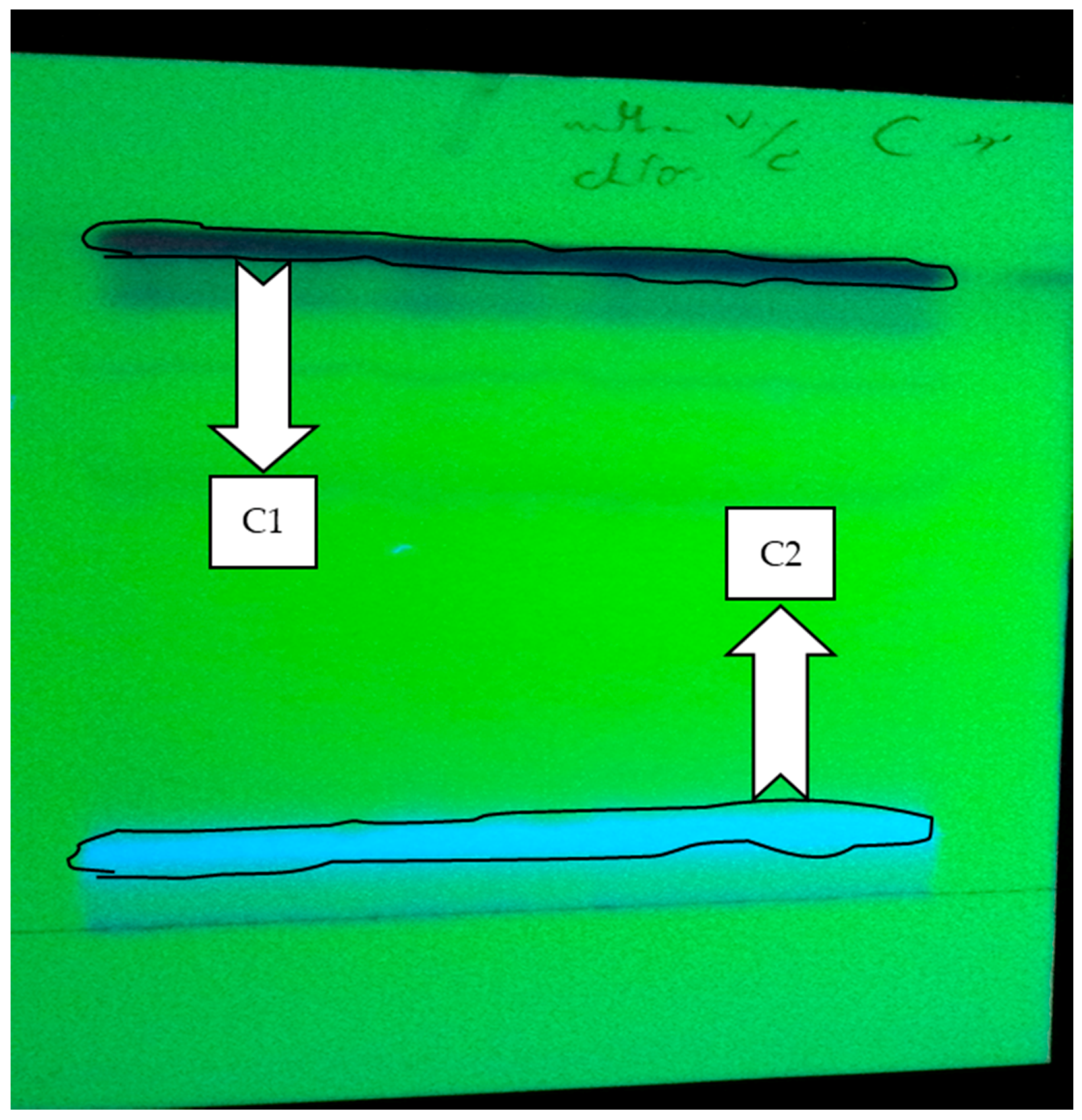
2.1. Fractionation
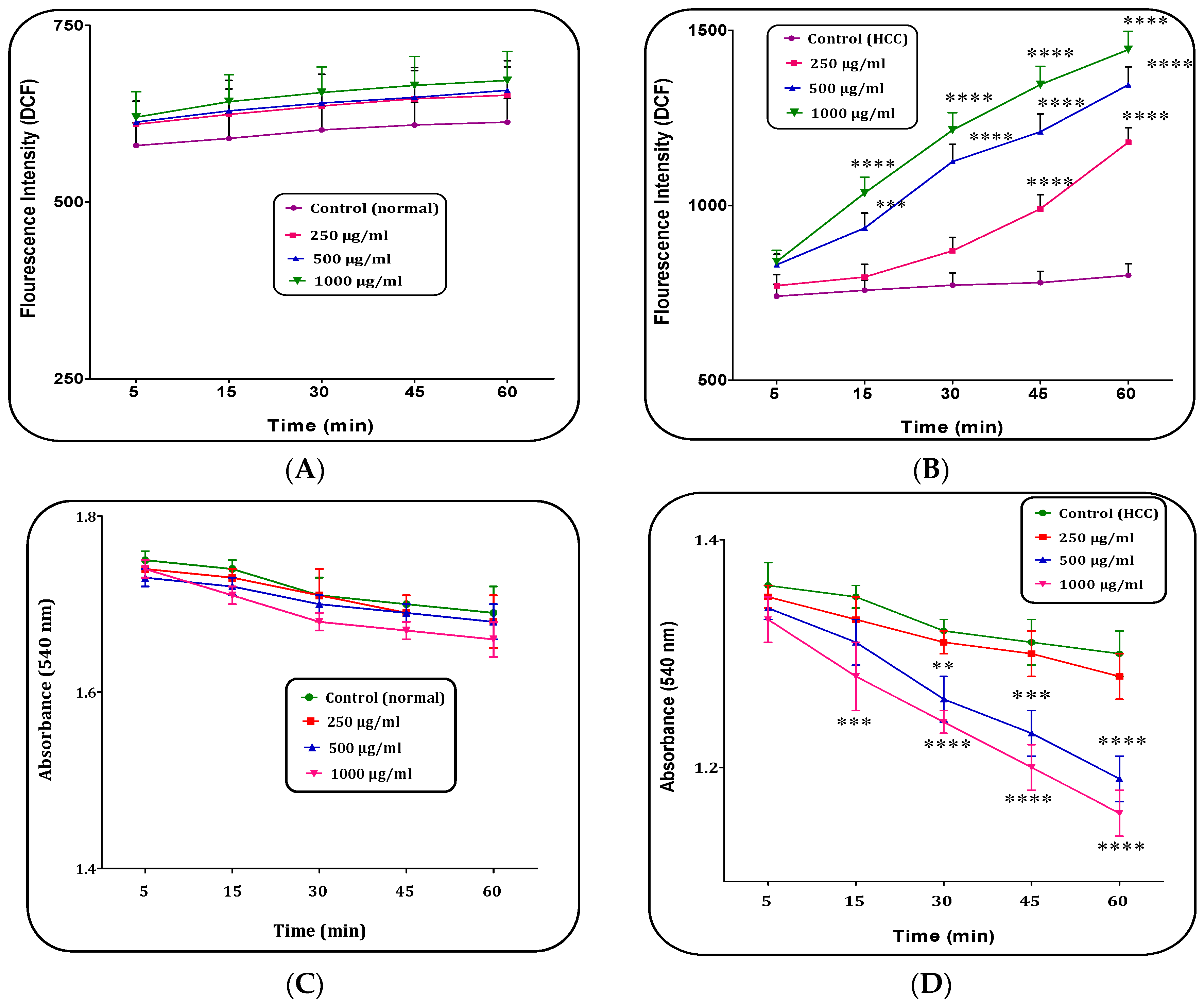
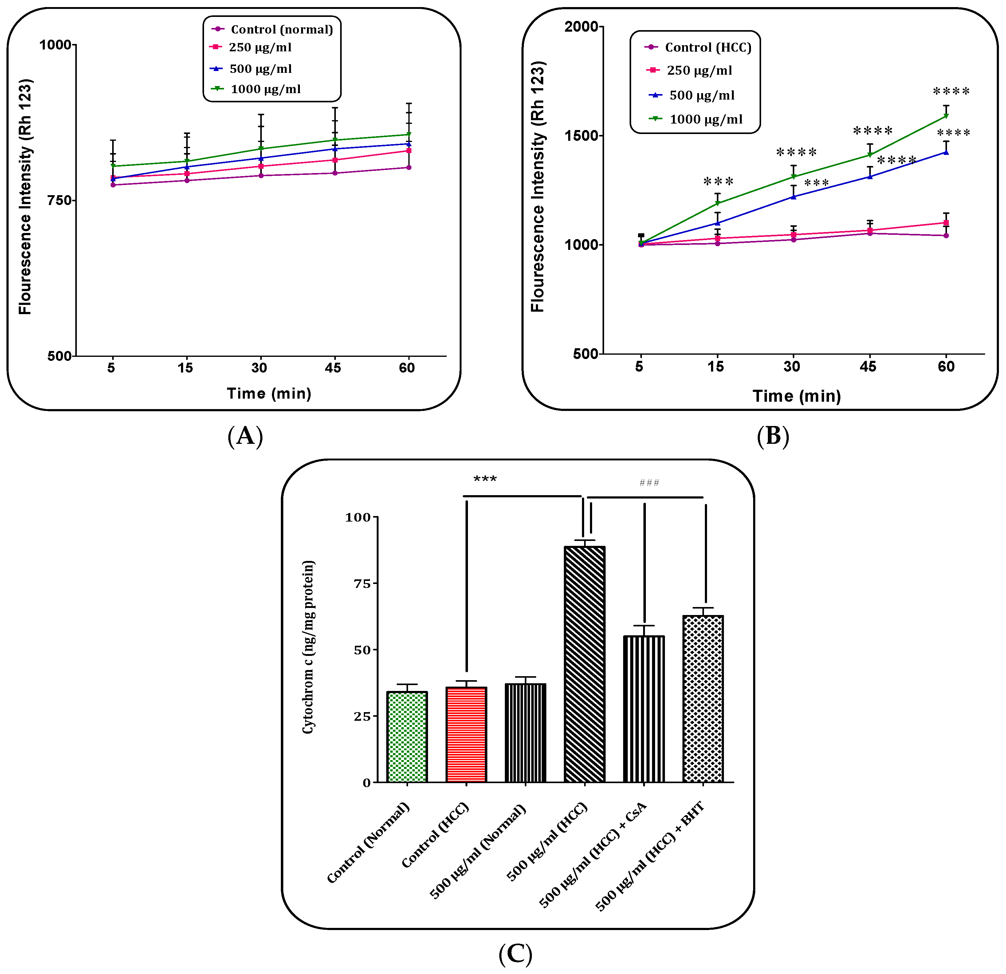
2.2. H. parva (C1 Sub-Fraction) Increased Mitochondrial ROS Level
2.3. H. parva (C1 Sub-Fraction) Induced Mitochondrial Swelling
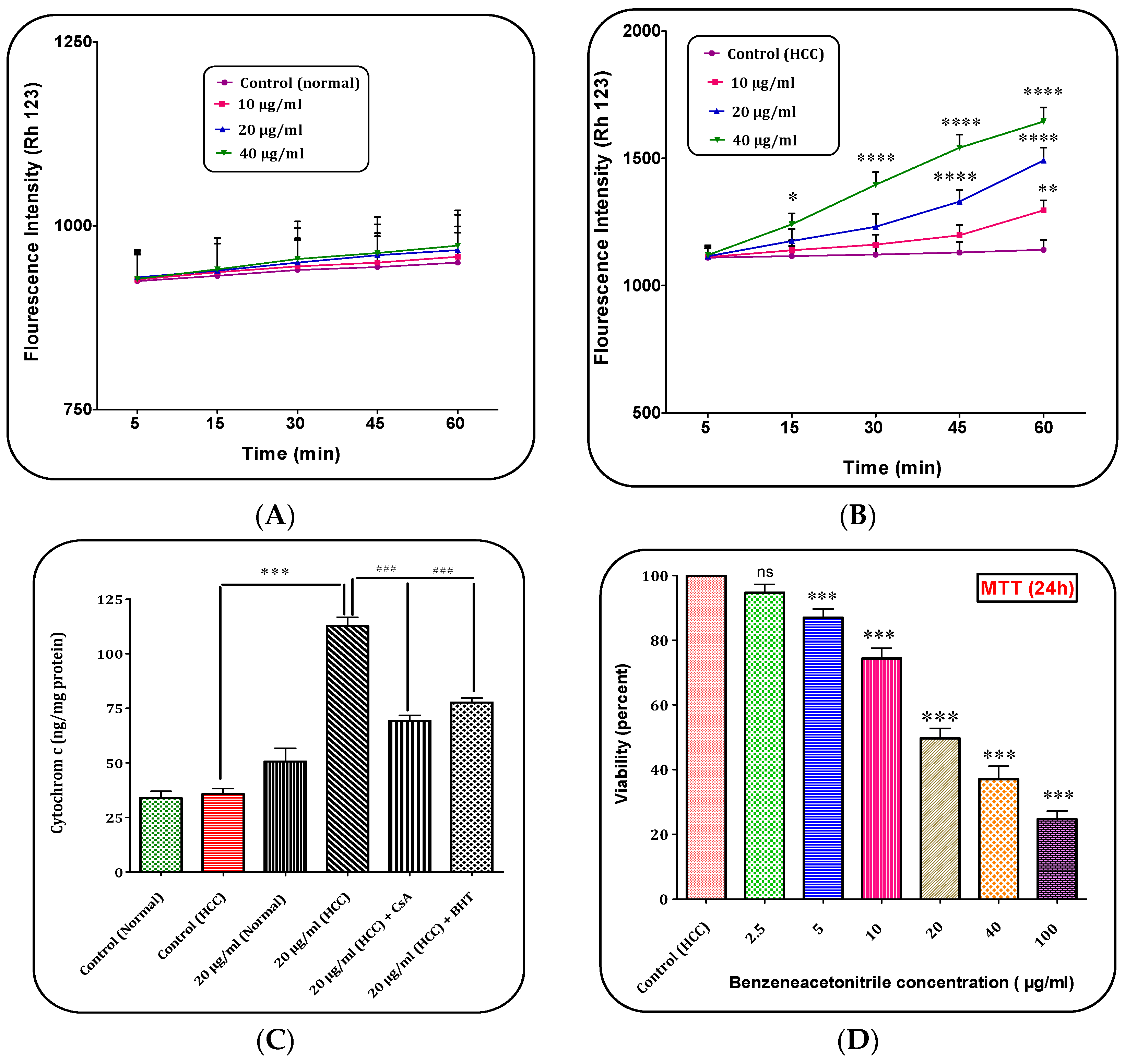
2.4. H. parva Extract (C1 Sub-Fraction) Induced Collapse on the Mitochondrial Membrane Potential (MMP)
2.5. H. parva Extract (C1 Sub-Fraction) Increased Cytochrome c Release
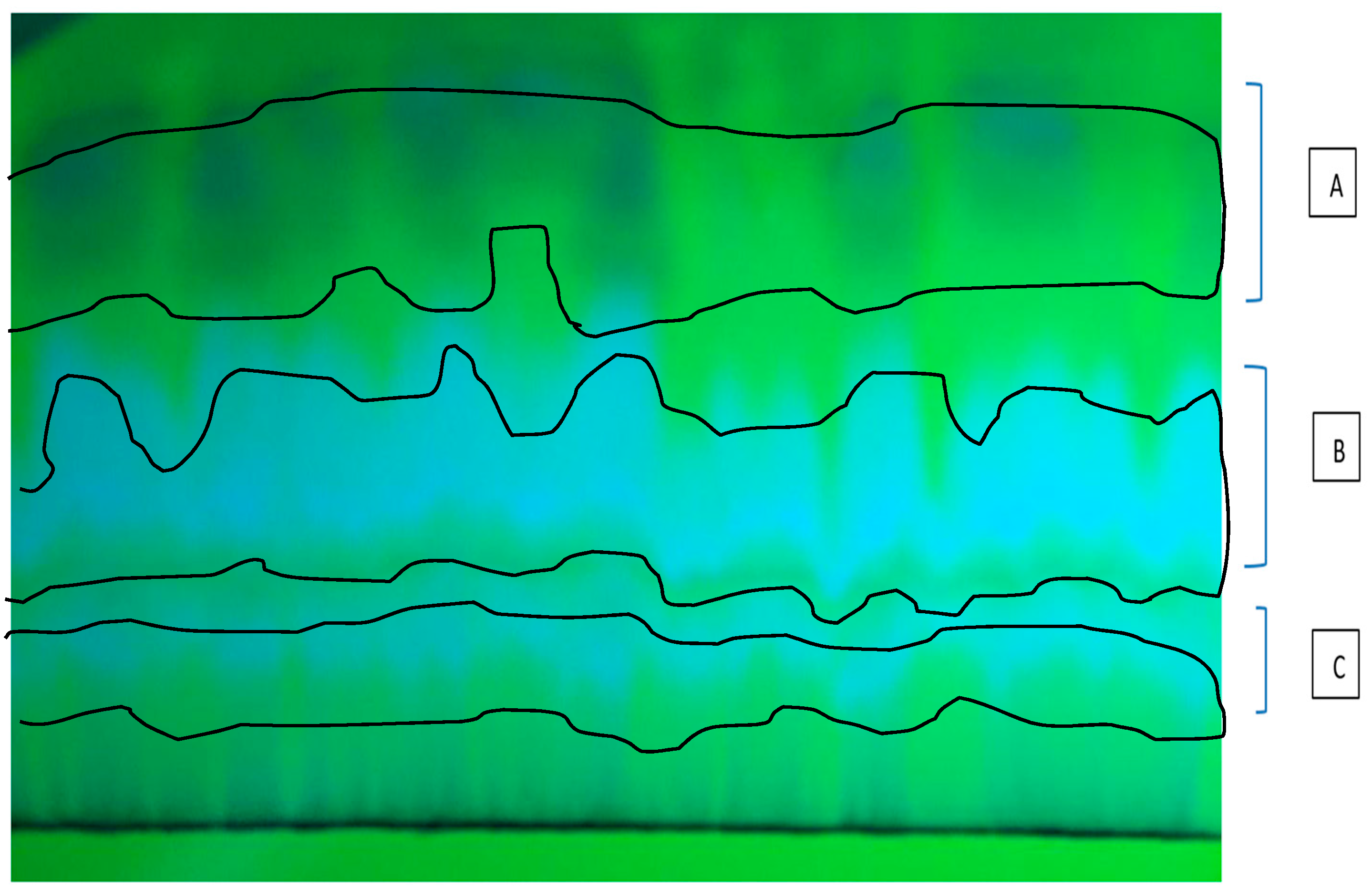
2.6. Determination of the Component in C1 Sub-Fraction of H. parva Methanolic Extract
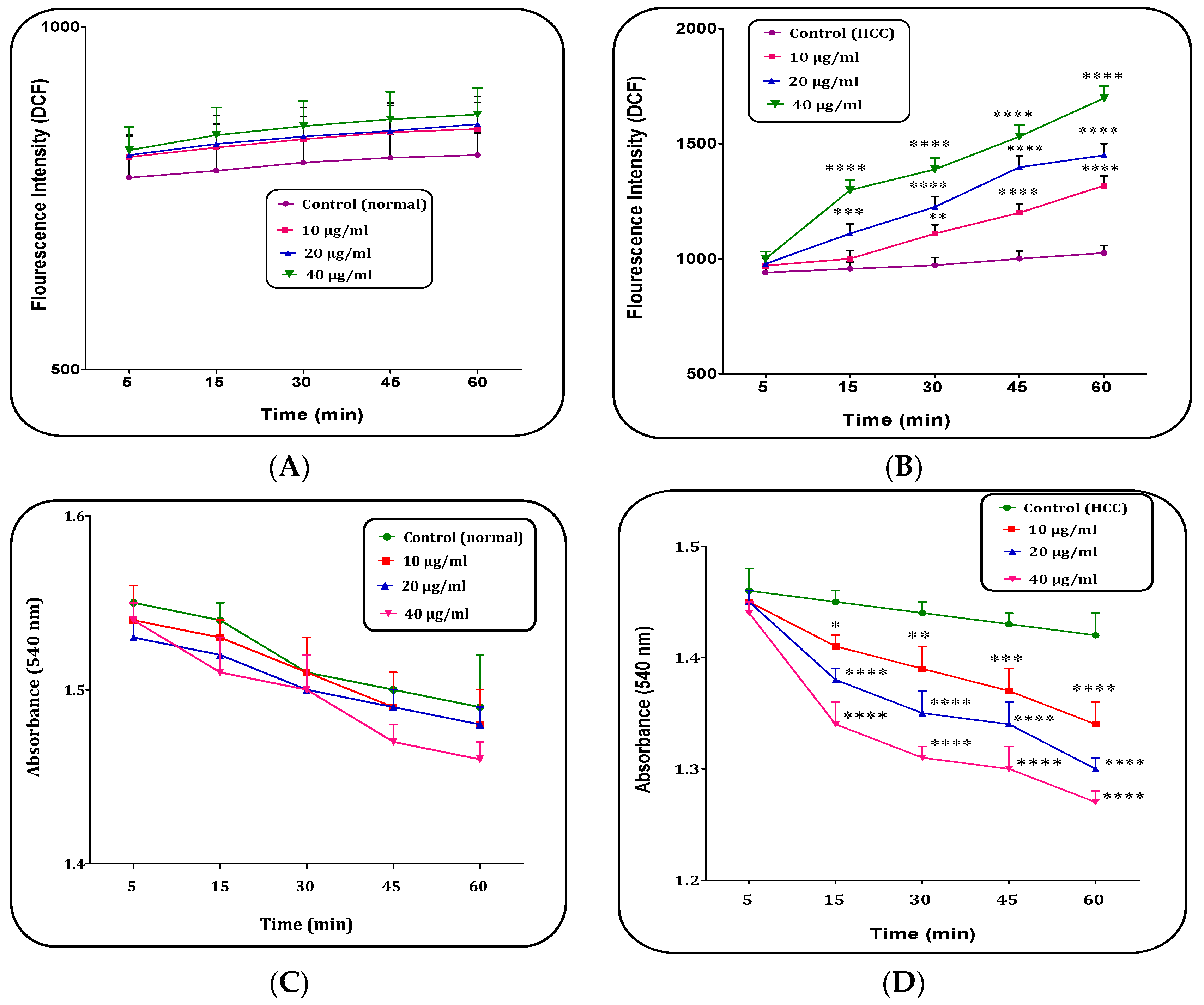
2.7. (Z)-2,3-Diphenylacrylonitrile Increased Mitochondrial ROS Level
2.8. (Z)-2,3-Diphenylacrylonitrile Induced Mitochondrial Swelling
2.9. (Z)-2,3-Diphenylacrylonitrile Induced Collapse on the Mitochondrial Membrane Potential (MMP)
2.10. (Z)-2,3-Diphenylacrylonitrile Increased Cytochrome c Release
2.11. (Z)-2,3-Diphenylacrylonitrile Decreased Cell Viability
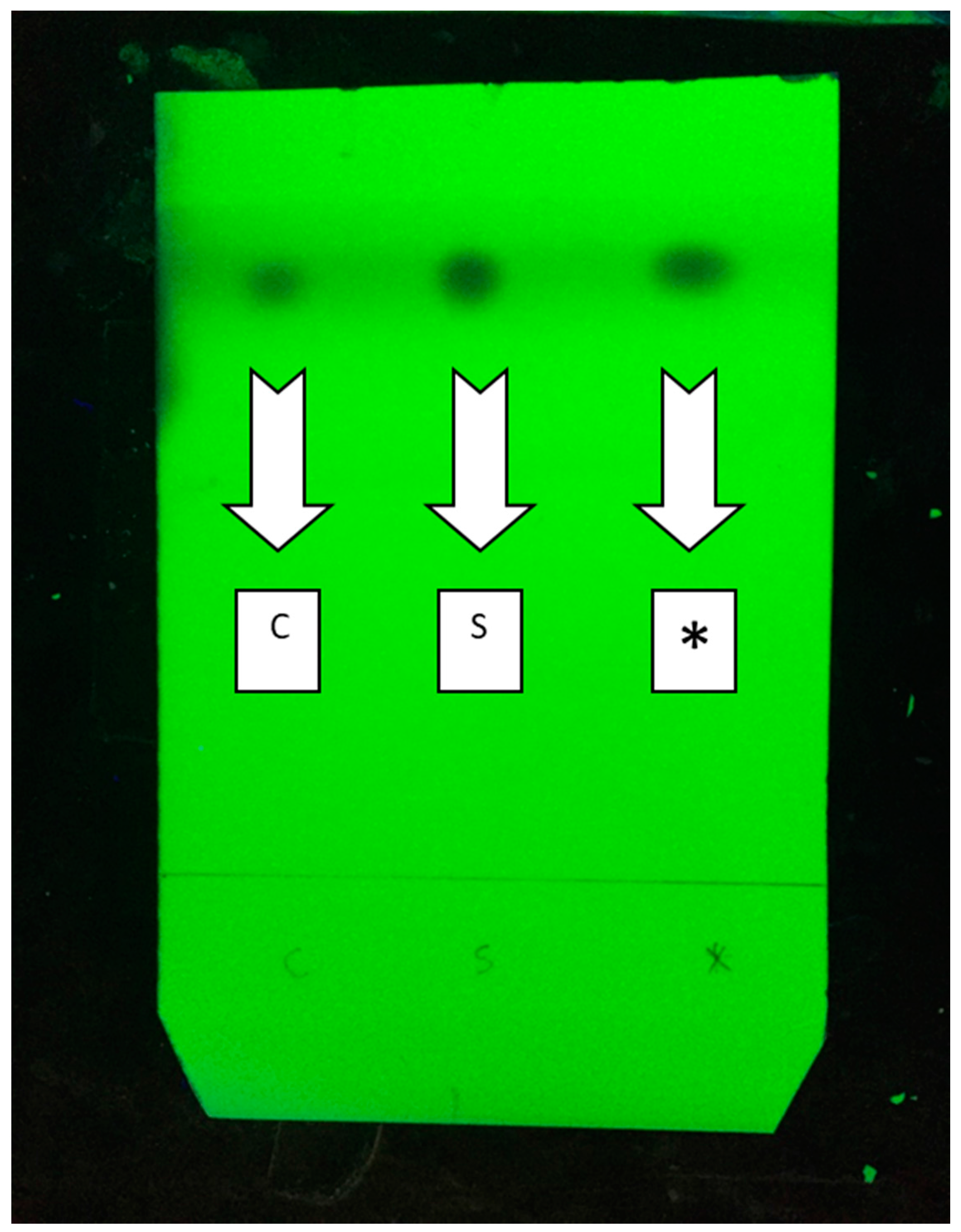
2.12. Mix Probe between Synthesized (Z)-2,3-Diphenylacrylonitrile and C1 Fraction
3. Discussion
4. Materials and Methods
4.1. Sampling and Extraction
4.2. Fractionation
4.3. Molecular Identification
4.4. General Procedure for Preparation of (Z)-2,3-Diphenylacrylonitrile
4.5. Animals
4.6. Experimental Design
4.7. Isolation the Mitochondria from Hepatocytes
4.8. Determination of Protein Concentration
4.9. Determination of Mitochondrial ROS
4.10. Determination of Mitochondrial Swelling
4.11. Determination of Mitochondrial Membrane Potential (MMP)
4.12. Determination of Cytochrome c Release
4.13. MTT Assay
4.14. Statistical Analysis
5. Conclusions
Acknowledgments
Author Contributions
Conflicts of Interest
References
- Chinembiri, T.N.; du Plessis, L.H.; Gerber, M.; Hamman, J.H.; du Plessis, J. Review of natural compounds for potential skin cancer treatment. Molecules 2014, 19, 11679–11721. [Google Scholar] [CrossRef] [PubMed] [Green Version]
- Rasool, M.; Rashid, S.; Arooj, M.; Ansari, S.A.; Khan, K.M.; Malik, A.; Naseer, M.I.; Zahid, S.; Manan, A.; Asif, M.; et al. New possibilities in hepatocellular carcinoma treatment. Anticancer Res. 2014, 34, 1563–1571. [Google Scholar] [PubMed]
- Forner, A.; Llovet, J.M.; Bruix, J. Hepatocellular carcinoma. Lancet 2012, 379, 1245–1255. [Google Scholar] [CrossRef]
- Seydi, E.; Motallebi, A.; Dastbaz, M.; Dehghan, S.; Salimi, A.; Nazemi, M.; Pourahmad, J. Selective Toxicity of Persian Gulf Sea Cucumber (Holothuria parva) and Sponge (Haliclona oculata) Methanolic Extracts on Liver Mitochondria Isolated from an Animal Model of Hepatocellular Carcinoma. Hepat. Mon. 2015, 15, e33073. [Google Scholar] [CrossRef] [PubMed]
- Sawadogo, W.R.; Schumacher, M.; Teiten, M.H.; Cerella, C.; Dicato, M.; Diederich, M. A survey of marine natural compounds and their derivatives with anti-cancer activity reported in 2011. Molecules 2013, 18, 3641–3673. [Google Scholar] [CrossRef] [PubMed]
- Mayer, A.M.; Rodriguez, A.D.; Berlinck, R.G.; Fusetani, N. Marine pharmacology in 2007–8: Marine compounds with antibacterial, anticoagulant, antifungal, anti-inflammatory, antimalarial, antiprotozoal, antituberculosis, and antiviral activities; affecting the immune and nervous system, and other miscellaneous mechanisms of action. Comp. Biochem. Physiol. C Toxicol. Pharmacol. 2011, 153, 191–222. [Google Scholar] [PubMed]
- Salimi, A.; Motallebi, A.; Ayatollahi, M.; Seydi, E.; Mohseni, A.R.; Nazemi, M.; Pourahmad, J. Selective toxicity of persian gulf sea cucumber Holothuria parva on human chronic lymphocytic leukemia b lymphocytes by direct mitochondrial targeting. Environ. Toxicol. 2017, 32, 1158–1169. [Google Scholar] [CrossRef] [PubMed]
- Janakiram, N.B.; Mohammed, A.; Rao, C.V. Sea Cucumbers Metabolites as Potent Anti-Cancer Agents. Mar. Drugs 2015, 13, 2909–2923. [Google Scholar] [CrossRef] [PubMed]
- Jayatilake, G.S.; Baker, B.J.; McClintock, J.B. Isolation and Identification of a Stilbene Derivative from the Antarctic Sponge Kirkpatrickia variolosa. J. Nat. Prod. 1995, 58, 1958–1960. [Google Scholar] [CrossRef]
- Cai, J.Z.; Tang, R.; Ye, G.F.; Qiu, S.X.; Zhang, N.L.; Hu, Y.J.; Shen, X.L. A Halogen-Containing Stilbene Derivative from the Leaves of Cajanus cajan that Induces Osteogenic Differentiation of Human Mesenchymal Stem Cells. Molecules 2015, 20, 10839–10847. [Google Scholar] [CrossRef] [PubMed]
- Alam, M.S.; Nam, Y.J.; Lee, D.U. Synthesis and evaluation of (Z)-2,3-diphenylacrylonitrile analogs as anti-cancer and anti-microbial agents. Eur. J. Med. Chem. 2013, 69, 790–797. [Google Scholar] [CrossRef] [PubMed]
- de Medina, P.; Casper, R.; Savouret, J.-F.; Poirot, M. Synthesis and Biological Properties of New Stilbene Derivatives of Resveratrol as New Selective Aryl Hydrocarbon Modulators. J. Med. Chem. 2005, 48, 287–291. [Google Scholar] [CrossRef] [PubMed]
- Roberti, M.; Pizzirani, D.; Simoni, D.; Rondanin, R.; Baruchello, R.; Bonora, C.; Buscemi, F.; Grimaudo, S.; Tolomeo, M. Synthesis and Biological Evaluation of Resveratrol and Analogues as Apoptosis-Inducing Agents. J. Med. Chem. 2003, 46, 3546–3554. [Google Scholar] [CrossRef] [PubMed]
- Kim, S.; Ko, H.; Park, J.E.; Jung, S.; Lee, S.K.; Chun, Y.-J. Design, Synthesis, and Discovery of Novel trans-Stilbene Analogues as Potent and Selective Human Cytochrome P450 1B1 Inhibitors. J. Med. Chem. 2002, 45, 160–164. [Google Scholar] [CrossRef] [PubMed]
- Taha, M.M.E.; Abdul, A.B.; Abdullah, R.; Ibrahim, T.A.T.; Abdelwahab, S.I.; Mohan, S. Potential chemoprevention of diethylnitrosamine-initiated and 2-acetylaminofluorene-promoted hepatocarcinogenesis by zerumbone from the rhizomes of the subtropical ginger (Zingiber zerumbet). Chem.-Biol. Interact. 2010, 186, 295–305. [Google Scholar] [CrossRef] [PubMed] [Green Version]
- Sadighara, M.; Amirsheardost, Z.; Minaiyan, M.; Hajhashemi, V.; Naserzadeh, P.; Salimi, A.; Seydi, E.; Pourahmad, J. Toxicity of Atorvastatin on Pancreas Mitochondria: A Justification for Increased Risk of Diabetes Mellitus. Basic Clin. Pharmacol. Toxicol. 2017, 120, 131–137. [Google Scholar] [CrossRef] [PubMed]
- Seydi, E.; Rasekh, H.R.; Salimi, A.; Mohsenifar, Z.; Pourahmad, J. Selective Toxicity of Apigenin on Cancerous Hepatocytes by Directly Targeting their Mitochondria. Anti-Cancer Agents Med. Chem. (Former. Curr. Med. Chem.-Anti-Cancer Agents) 2016, 16, 1576–1586. [Google Scholar] [CrossRef]
- Hosseini, M.J.; Shaki, F.; Ghazi-Khansari, M.; Pourahmad, J. Toxicity of copper on isolated liver mitochondria: Impairment at complexes I, II, and IV leads to increased ROS production. Cell Biochem Biophys. 2014, 70, 367–381. [Google Scholar] [CrossRef] [PubMed]
- Bradford, M.M. A rapid and sensitive method for the quantitation of microgram quantities of protein utilizing the principle of protein-dye binding. Anal. Biochem. 1976, 72, 248–254. [Google Scholar] [CrossRef]
- Pourahmad, J.; Eskandari, M.R.; Shakibaei, R.; Kamalinejad, M. A search for hepatoprotective activity of fruit extract of Mangifera indica L. against oxidative stress cytotoxicity. Plant Foods Hum. Nutr. 2010, 65, 83–89. [Google Scholar] [CrossRef] [PubMed]
- Pourahmad, J.; Eskandari, M.R.; Daraei, B. A comparison of hepatocyte cytotoxic mechanisms for thallium (I) and thallium (III). Environ. Toxicol. 2010, 25, 456–467. [Google Scholar] [CrossRef] [PubMed]
- Liang, C.; Xia, J.; Lei, D.; Li, X.; Yao, Q.; Gao, J. Synthesis, in vitro and in vivo antitumor activity of symmetrical bis-Schiff base derivatives of isatin. Eur. J. Med. Chem. 2014, 74, 742–750. [Google Scholar] [CrossRef] [PubMed]
- Seydi, E.; Rasekh, H.R.; Salimi, A.; Mohsenifar, Z.; Pourahmad, J. Myricetin selectively induces apoptosis on cancerous hepatocytes by directly targeting their mitochondria. Basic Clin. Pharmacol. Toxicol. 2016, 119, 249–258. [Google Scholar] [CrossRef] [PubMed]


© 2017 by the authors. Licensee MDPI, Basel, Switzerland. This article is an open access article distributed under the terms and conditions of the Creative Commons Attribution (CC BY) license (http://creativecommons.org/licenses/by/4.0/).
Share and Cite
Amidi, S.; Hashemi, Z.; Motallebi, A.; Nazemi, M.; Farrokhpayam, H.; Seydi, E.; Pourahmad, J. Identification of (Z)-2,3-Diphenylacrylonitrile as Anti-Cancer Molecule in Persian Gulf Sea Cucumber Holothuria parva. Mar. Drugs 2017, 15, 314. https://doi.org/10.3390/md15100314
Amidi S, Hashemi Z, Motallebi A, Nazemi M, Farrokhpayam H, Seydi E, Pourahmad J. Identification of (Z)-2,3-Diphenylacrylonitrile as Anti-Cancer Molecule in Persian Gulf Sea Cucumber Holothuria parva. Marine Drugs. 2017; 15(10):314. https://doi.org/10.3390/md15100314
Chicago/Turabian StyleAmidi, Salimeh, Zahra Hashemi, Abbasali Motallebi, Melika Nazemi, Hoda Farrokhpayam, Enayatollah Seydi, and Jalal Pourahmad. 2017. "Identification of (Z)-2,3-Diphenylacrylonitrile as Anti-Cancer Molecule in Persian Gulf Sea Cucumber Holothuria parva" Marine Drugs 15, no. 10: 314. https://doi.org/10.3390/md15100314