α-Pyrone Polyketides from Streptomyces ambofaciens BI0048, an Endophytic Actinobacterial Strain Isolated from the Red Alga Laurencia glandulifera


Abstract
:1. Introduction
2. Results and Discussion
3. Materials and Methods
3.1. General Experimental Procedures
3.2. Biological Material
3.3. Fermentation, Extraction and Isolation
3.4. Evaluation of Antibacterial Activity
3.5. Evaluation of Cytotoxic Activity
4. Conclusions
Acknowledgments
Author Contributions
Conflicts of Interest
References
- Knight, V.; Sanglier, J.-J.; DiTullio, D.; Braccili, S.; Bonner, P.; Waters, J.; Hughes, D.; Zhang, L. Diversifying microbial natural products for drug discovery. Appl. Microbiol. Biotechnol. 2003, 62, 446–458. [Google Scholar] [CrossRef] [PubMed]
- Bérdy, J. Bioactive microbial metabolites. J. Antibiot. 2005, 58, 1–26. [Google Scholar] [CrossRef] [PubMed]
- Demain, A.L.; Sanchez, S. Microbial drug discovery: 80 years of progress. J. Antibiot. 2009, 62, 5–16. [Google Scholar] [CrossRef] [PubMed]
- Barka, E.A.; Vatsa, P.; Sanchez, L.; Gaveau-Vaillant, N.; Jacquard, C.; Klenk, H.-P.; Clément, C.; Ouhdouch, Y.; van Wezel, G.P. Taxonomy, physiology, and natural products of Actinobacteria. Microbiol. Mol. Biol. Rev. 2016, 80, 1–43. [Google Scholar] [CrossRef] [PubMed]
- Piel, J.; Hoang, K.; Moore, B.S. Natural metabolic diversity encoded by the enterocin biosynthesis gene cluster. J. Am. Chem. Soc. 2000, 122, 5415–5416. [Google Scholar] [CrossRef]
- Xiang, L.; Kalaitzis, J.A.; Nilsen, G.; Chen, L.; Moore, B.S. Mutational analysis of the enterocin Favorskii biosynthetic rearrangement. Org. Lett. 2002, 4, 957–960. [Google Scholar] [CrossRef] [PubMed]
- Miyairi, N.; Sakai, H.-I.; Konomi, T.; Imanaka, H. Enterocin, a new antibiotic-taxonomy, isolation and characterization. J. Antibiot. 1976, 29, 227–235. [Google Scholar] [CrossRef] [PubMed]
- Seto, H.; Sato, T.; Urano, S.; Uzawa, J.; Yonehara, H. Utilization of 13C-13C coupling in structural and biosynthetic studies. VII. The structure and biosynthesis of vulgamycin. Tetrahedron Lett. 1976, 17, 4367–4370. [Google Scholar] [CrossRef]
- Sitachitta, N.; Gadepalli, M.; Davidson, B.S. New α-pyrone-containing metabolites from a marine-derived actinomycete. Tetrahedron 1996, 52, 8073–8080. [Google Scholar] [CrossRef]
- Petersen, F.; Zähner, H.; Metzger, J.W.; Freund, S.; Hummel, R.-P. Germicidin, an autoregulative germination inhibitor of Streptomyces viridochromogenes NRRL B-1551. J. Antibiot. 1993, 46, 1126–1138. [Google Scholar] [CrossRef] [PubMed]
- Pouchert, C.J.; Behnke, J. The Aldrich Library of 13C and 1H FT NMR Spectra, 1st ed.; Aldrich Chemical Company, Inc.: Milwaukee, WI, USA, 1993; Volume 2, pp. 985, 1043, 1063. [Google Scholar]
- Cutignano, A.; Fontana, A.; Renzulli, L.; Cimino, G. Placidenes C-F, novel α-pyrone proprionates from the Mediterranean sacoglossan Placida dendritica. J. Nat. Prod. 2003, 66, 1399–1401. [Google Scholar] [CrossRef] [PubMed]
- McGlacken, G.P.; Fairlamb, I.J.S. 2-Pyrone natural products and mimetics: Isolation, characterization and biological activity. Nat. Prod. Rep. 2005, 22, 369–385. [Google Scholar] [CrossRef] [PubMed]
- Bhat, Z.S.; Rather, M.A.; Maqbool, M.; Lah, H.U.L.; Yousuf, S.K.; Ahmad, Z. α-Pyrones: Small molecules with versatile structural diversity reflected in multiple pharmacological activities—An update. Biomed. Pharmacother. 2017, 91, 265–277. [Google Scholar] [CrossRef] [PubMed]
- Kawashima, A.; Seto, H.; Kato, M.; Uchida, K.; Otake, N. Preparation of fluorinated antibiotics followed by 19F NMR spectroscopy. I. Fluorinated vulgamycins. J. Antibiot. 1985, 38, 1499–1505. [Google Scholar] [CrossRef] [PubMed]
- Babczinski, P.; Dorgerloh, M.; Löbberding, A.; Santel, H.-J.; Schmidt, R.R.; Schmitt, P.; Wünsche, C. Herbicidal activity and mode of action of vulgamycin. Pestic. Sci. 1991, 33, 439–446. [Google Scholar] [CrossRef]
- Xu, Z.; Ding, L.; Hertweck, C. A branched extender unit shared between two orthogonal polyketide pathways in an endophyte. Angew. Chem. Int. Ed. 2011, 50, 4667–4670. [Google Scholar] [CrossRef] [PubMed]
- Bugni, T.S.; Woolery, M.; Kauffman, C.A.; Jensen, P.R.; Fenical, W. Bohemamines from a marine-derived Streptomyces sp. J. Nat. Prod. 2006, 69, 1626–1628. [Google Scholar] [CrossRef] [PubMed]
- Dimou, Μ.; Ioannou, Ε.; Daskalaki, M.G.; Tziveleka, L.A.; Kampranis, S.C.; Roussis, V. Disulfides with anti-inflammatory activity from the brown alga Dictyopteris membranacea. J. Nat. Prod. 2016, 79, 584–589. [Google Scholar] [CrossRef] [PubMed]
- Mavrogonatou, E.; Eliades, T.; Eliades, G.; Kletsas, D. The effect of triethylene glycol dimethacrylate on p53-dependent G2 arrest in human gingival fibroblasts. Biomaterials 2010, 31, 8530–8538. [Google Scholar] [CrossRef] [PubMed]


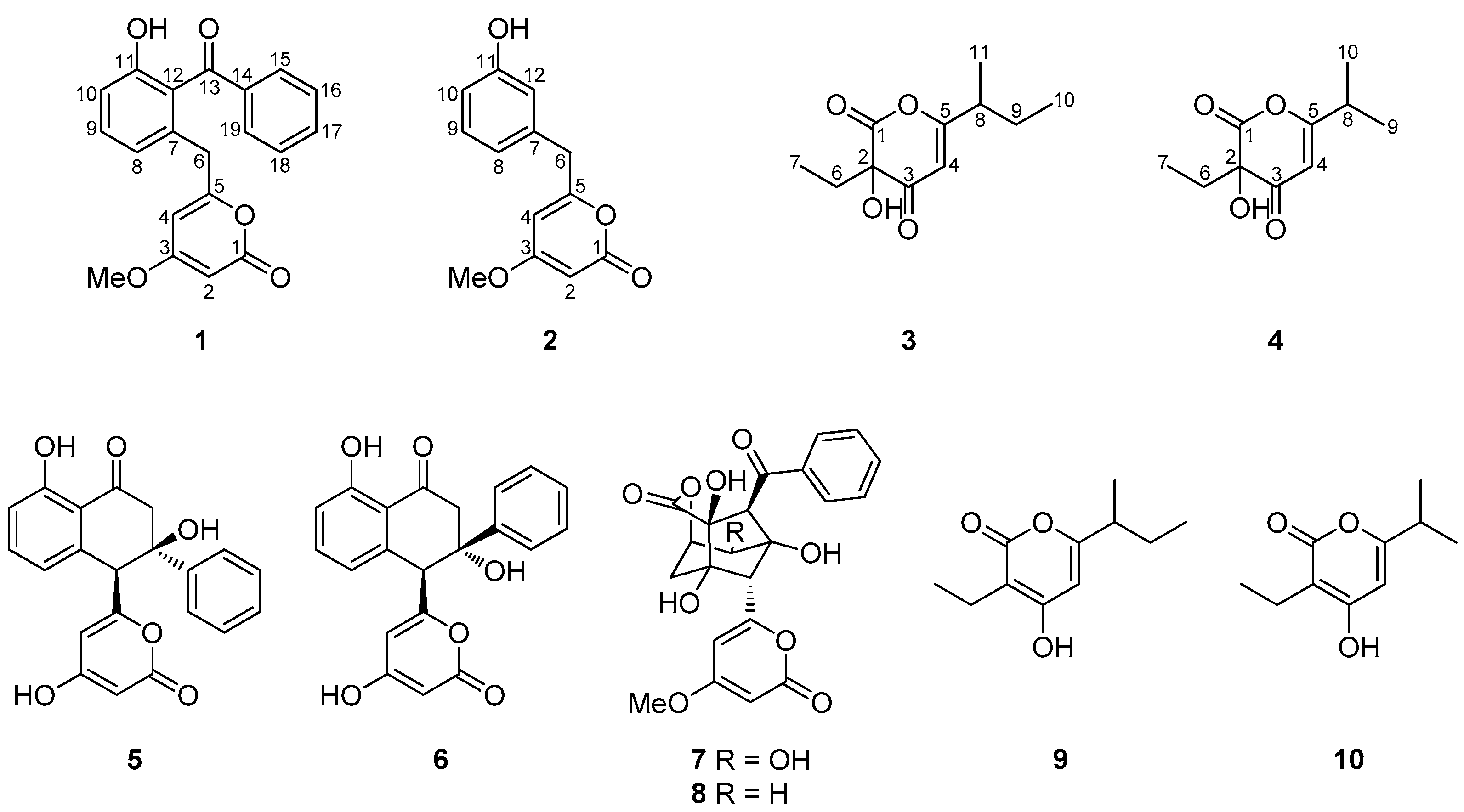
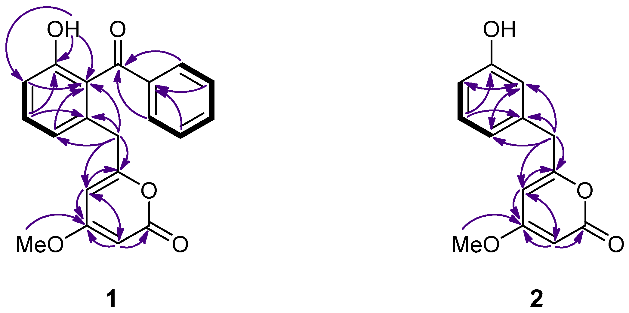
| Position | 1 | 2 | ||
|---|---|---|---|---|
| δC | δH (J in Hz) | δC | δH (J in Hz) | |
| 1 | 165.2, C 1 | - | 165.0, C 1 | - |
| 2 | 87.8, CH | 5.27, d (2.2) | 87.7, CH | 5.39, d (2.2) |
| 3 | 170.9, C | - | 171.3, C | - |
| 4 | 101.3, CH | 5.43, d (2.2) | 100.7, CH | 5.68, d (2.2) |
| 5 | 162.8, C | - | 164.2, C | - |
| 6 | 38.3, CH2 | 3.52, s | 39.7, CH2 | 3.67, s |
| 7 | 134.4, C 1 | - | 136.5, C | - |
| 8 | 123.2, CH | 6.87, d (7.5) | 121.5, CH | 6.78, brd (7.5) |
| 9 | 133.0, CH | 7.36, dd (8.2, 7.5) | 130.0, CH | 7.17, t (7.5) |
| 10 | 116.7, CH | 6.95, d (8.2) | 114.5, CH | 6.74, m |
| 11 | 157.0, C 1 | - | 156.0, C 1 | - |
| 12 | 123.7, C 1 | - | 116.2, CH | 6.73, d (1.0) |
| 13 | 199.2, C 1 | - | - | - |
| 14 | 138.2, C 1 | - | - | - |
| 15 | 129.1, CH | 7.67, d (8.3) | - | - |
| 16 | 128.9, CH | 7.43, dd (8.3, 7.4) | - | - |
| 17 | 133.6, CH | 7.57, t (7.4) | - | - |
| 18 | 128.9, CH | 7.43, dd (8.3, 7.4) | - | - |
| 19 | 129.1, CH | 7.67, d (8.3) | - | - |
| OMe | 55.8, CH3 | 3.69, s | 55.9, CH3 | 3.75, s |
| OH | - | 7.80, brs | - | - |
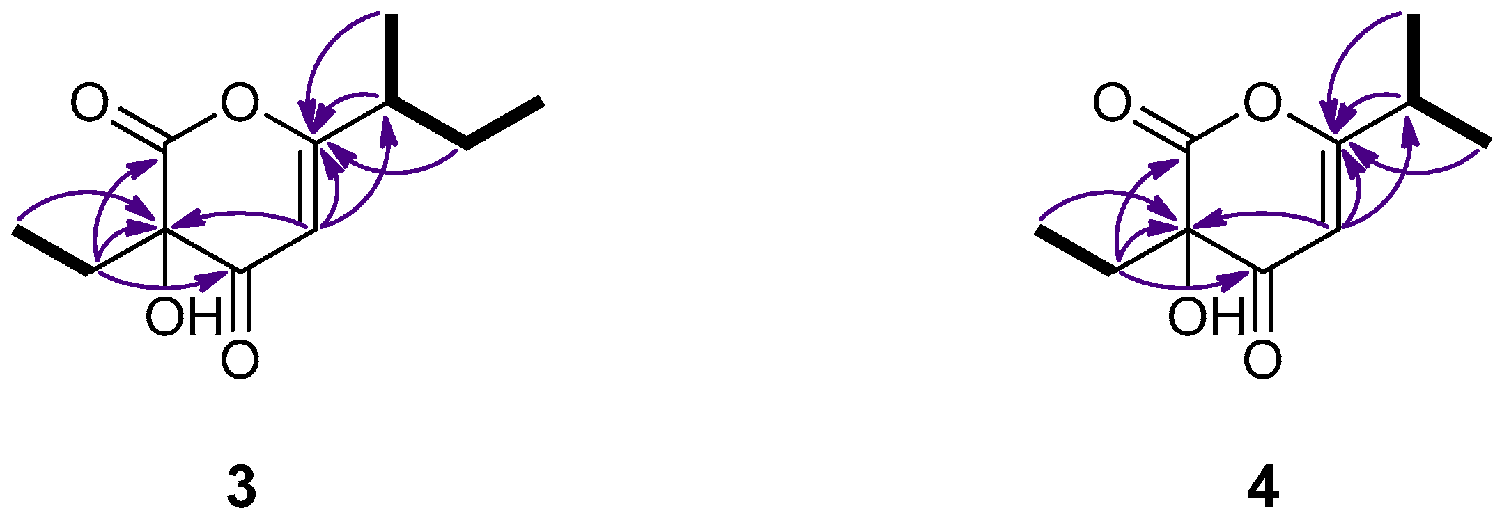
| Position | 3 | 4 | ||
|---|---|---|---|---|
| δC | δH (J in Hz) | δC | δH (J in Hz) | |
| 1 | 167.2, C 1 | - | 167.6, C | - |
| 2 | 91.7, C | - | 91.7, C | - |
| 3 | 191.2, C | - | 191.3, C | - |
| 4 | 104.6, CH | 5.68, s | 103.3, CH | 5.69, s |
| 5 | 175.3, C | - | 176.2, C | - |
| 6 | 30.7, CH2 | 1.98, q (7.6) | 30.7, CH2 | 1.98, q (7.6) |
| 7 | 7.3, CH3 | 0.97, t (7.6) | 7.3, CH3 | 0.96, t (7.6) |
| 8 | 40.2, CH | 2.41, m | 33.0, CH | 2.64, septet (6.8) |
| 9 | 26.5, CH2 | 1.68, m, 1.54, m | 19.2, CH3 | 1.22, d (6.8) |
| 10 | 11.4, CH3 | 0.92, t (7.4) | 19.3, CH3 | 1.23, d (6.8) |
| 11 | 17.1, CH3 | 1.20, d (6.9) | - | - |
| OH | - | 9.36, brs | - | 9.35, brs |
© 2017 by the authors. Licensee MDPI, Basel, Switzerland. This article is an open access article distributed under the terms and conditions of the Creative Commons Attribution (CC BY) license (http://creativecommons.org/licenses/by/4.0/).
Share and Cite
Rab, E.; Kekos, D.; Roussis, V.; Ioannou, E. α-Pyrone Polyketides from Streptomyces ambofaciens BI0048, an Endophytic Actinobacterial Strain Isolated from the Red Alga Laurencia glandulifera. Mar. Drugs 2017, 15, 389. https://doi.org/10.3390/md15120389
Rab E, Kekos D, Roussis V, Ioannou E. α-Pyrone Polyketides from Streptomyces ambofaciens BI0048, an Endophytic Actinobacterial Strain Isolated from the Red Alga Laurencia glandulifera. Marine Drugs. 2017; 15(12):389. https://doi.org/10.3390/md15120389
Chicago/Turabian StyleRab, Enikő, Dimitrios Kekos, Vassilios Roussis, and Efstathia Ioannou. 2017. "α-Pyrone Polyketides from Streptomyces ambofaciens BI0048, an Endophytic Actinobacterial Strain Isolated from the Red Alga Laurencia glandulifera" Marine Drugs 15, no. 12: 389. https://doi.org/10.3390/md15120389