
Bioactive Diphenyl Ethers and Isocoumarin Derivatives from a Gorgonian-Derived Fungus Phoma sp. (TA07-1)
by
 , and
, and
Ting Shi
1,2 ,
,
Jun Qi
1,2,
Chang-Lun Shao
1,2,
Dong-Lin Zhao
1,2,
Xue-Mei Hou
1,2 and
Chang-Yun Wang
1,2,3,* 1
Key Laboratory of Marine Drugs, School of Medicine and Pharmacy, Ocean University of China, the Ministry of Education of China, Qingdao 266003, China
2
Laboratory for Marine Drugs and Bioproducts, Qingdao National Laboratory for Marine Science and Technology, Qingdao 266071, China
3
Institute of Evolution & Marine Biodiversity, Ocean University of China, Qingdao 266003, China
*
Author to whom correspondence should be addressed.
Mar. Drugs 2017, 15(6), 146; https://doi.org/10.3390/md15060146
Submission received: 25 March 2017
/
Revised: 11 May 2017
/
Accepted: 23 May 2017
/
Published: 25 May 2017
(This article belongs to the Special Issue Marine Bioactive Natural Product Studies in Asia)
Abstract
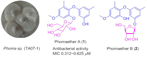
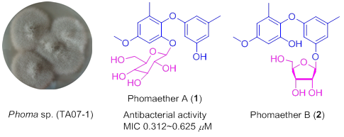
:Three new diphenyl ether derivatives—phomaethers A–C (1–3) and five known compounds—including a diphenyl ether analog, 2,3′-dihydroxy-4-methoxy-5′,6-dimethyl diphenyl ether (4); and four isocoumarin derivatives, diaportinol (5), desmethyldiaportinol (6), citreoisocoumarinol (7), and citreoisocoumarin (8)—were isolated from a gorgonian-derived fungus Phoma sp. (TA07-1). Their structures were elucidated by extensive spectroscopic investigation. The absolute configurations of 1 and 2 were determined by acid hydrolysis reactions. It was the first report to discover the diphenyl glycoside derivatives from coral-derived fungi. Compounds 1, 3, and 4 showed selective strong antibacterial activity against five pathogenic bacteria with the minimum inhibiting concentration (MIC) values and minimum bactericidal concentration (MBC) values between 0.156 and 10.0 μM.
1. Introduction
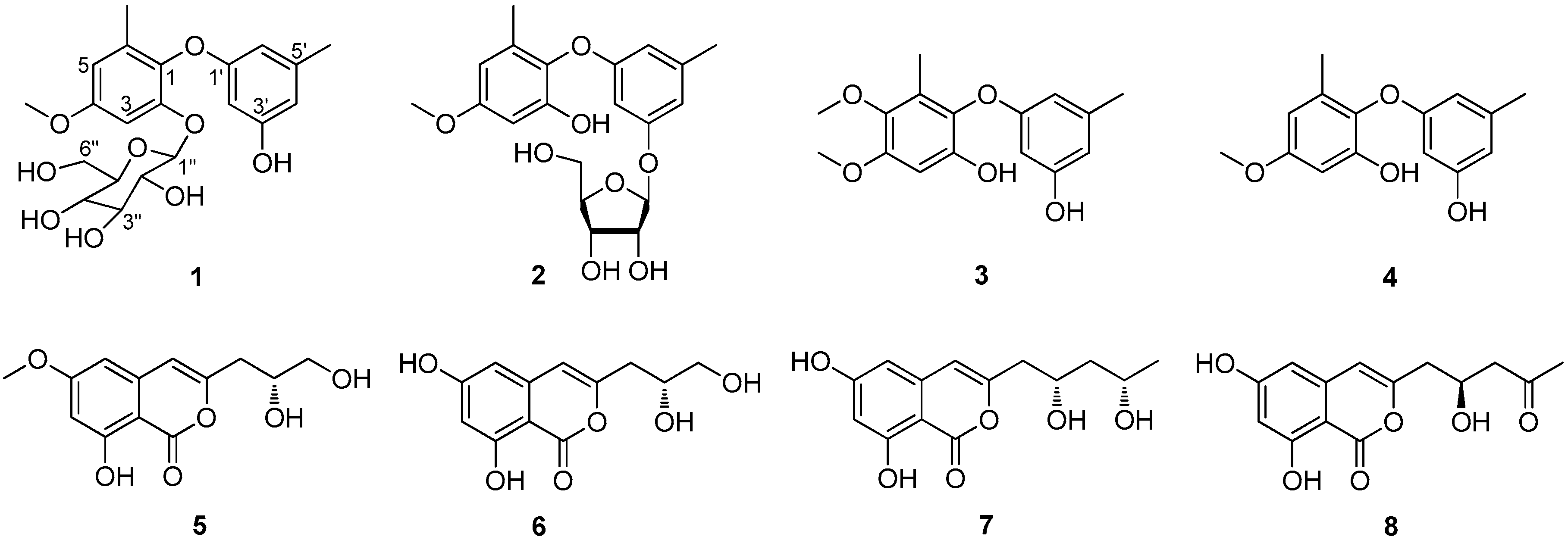
Marine microorganisms, especially marine fungi, have got more and more attention in recent years, as their outstanding abilities to produce bioactive compounds [1,2,3]. Among marine fungi, coral-derived fungi have played an important role in discovering pharmaceutical useful compounds [4]. Marine-derived Phoma sp. has been found to produce many novel and bioactive secondary metabolites such as cytotoxic epoxyphomalin A [5] and phomazine B [6], antifouling (+)-flavipucine [7], and antibacterial phomalevones A–C [8]. During our ongoing investigation for new bioactive compounds from gorgonian-derived fungi in the South China Sea [9,10,11,12,13], the fungal strain Phoma sp. (TA07-1) isolated from gorgonian Dichotella gemmacea attracted our attention, because its EtOAc extract of the fermentation showed significant antibacterial activity towards two Gram-positive bacteria, Staphylococcus albus and S. aureus, and three Gram-negative bacteria Escherichia coli, Vibrio parahaemolyticus, and V. anguillarum. Bioassay-guided separation led to the isolation of four diphenyl ether derivatives, including three new compounds, phomaethers A–C (1–3), and 2,3′-dihydroxy-4-methoxy-5′,6-dimethyl diphenyl ether (4) [14], together with four known isocoumarin derivatives, diaportinol (5) [15], desmethyldiaportinol (6) [16], citreoisocoumarinol (7) [17], and citreoisocoumarin (8) [17] (Figure 1). Herein we report the isolation, structure elucidation, and bioactivities of these compounds.
2. Results and Discussion
Phomaether A (1) was isolated as a colorless, amorphous powder. The molecular formula of C21H26O9 was determined by HRESIMS that displayed the [M + Na]+ peak at m/z 445.1474 (calcd. for C21H26O9Na, 445.1469) indicating nine degrees of unsaturation. The 1H NMR spectrum (Table 1) displayed five aromatic proton signals at δH 6.69 (1H, d, J = 2.9 Hz), 6.51 (1H, d, J = 2.9 Hz), 6.18 (1H, brs), 6.08 (1H, brs) and 5.93 (1H, brs), two methyls at δH 2.13 (3H, s) and 2.02 (3H, s), one methoxyl at δH 3.73 (3H, s), and a hydroxyl at δH 9.27 (1H, brs). The 13C NMR (Table 1) showed 12 aromatic carbon signals at δC 159.3, 158.2, 156.3, 150.9, 139.5, 135.2, 132.4, 109.4, 108.5, 106.6, 100.82, and 99.2; two methyls at δC 21.2, 16.2; and a methoxyl at δC 55.2. The NMR spectral feature indicated that 1 was a diphenyl ether derivative and very similar to 2,3′-dihydroxy-4-methoxy-5′,6-dimethyl diphenyl ether (4) [14]. The difference between these two compounds was the presence of a hexose residue in 1. The signals of the hexose residue in 1H NMR and 13C NMR displayed five oxymethines (δH 4.82, δC 100.76; δH 3.30, δC 77.3; δH 3.20, δC 76.8; δH 3.06, δC 73.2; δH 3.08, δC 69.8), one oxymethylene (δH 3.68, 3.40, δC 60.8), and four hydroxyls (δH 5.04, 5.04, 4.68, 4.60), and the hexose was determined as glucopyranose comparing to the NMR data with those of flavonoid glycoside, 4′-demethylleucomin-7-O-β-d-glucopyranoside [18]. The correlation from H-1″ to C-2 in HMBC (Figure 2) indicated the glucosyl was linked to C-2. The relative configuration of glucopyranose in 1 was determined by the 1H-1H coupling constants. The large coupling constant between H-1″ and H-2″ (J = 7.8 Hz) indicated a β-configuration of the glucopyranose [19]. The configuration of the glucopyranose was determined as d-glucopyranose by comparing the rotation of its acid hydrolysate ( +48.0 (c 0.04, H2O)) with that of the standard d-glucopyranose ( +54.0 (c 0.15, H2O)). Accordingly, 1 was determined as 2-O-β-d-glucopyranose-3′-hydroxy-4-methoxy-5′,6-dimethyl diphenyl ether, and was named phomaether A.
Phomaether B (2) was isolated as a light brown, amorphous powder. The molecular formula was assigned as C20H24O8 (nine degrees of unsaturation) by its HRESIMS data. Detailed inspection of the NMR data (Table 1) of 2 with those of 1 revealed that these two compounds were very similar, except for the sugar moiety. The sugar residue in 2 was defined as a ribose by comparing the NMR data (Table 1) with those of naphthyl ribofuranoside, isotorachrysone-6-O-α-d-ribofuranoside [20], chromene glycoside, and sterin A [21]. The key HMBC correlation from H-1″ to C-3′ (Figure 2) established the connection between the ribose and diphenyl ether moiety. The coupling constant of anomeric proton H-1″ (J = 4.5 Hz) in 2 was found to close to that in methyl-α-d-ribofuranoside (J = 4.3 Hz) [22], indicating an α-ribose in 2. The α-ribose was determined as d-configuration by comparing the optical rotation data of the acid hydrolysate of 2 with that of the standard d-ribose ( −23.0 (c 0.03, H2O) vs. −38.7 (c 0.10, H2O)). From above, 2 was determined as 2-hydroxy-3′-O-α-d-ribofuranoside-4-methoxy-5′,6-dimethyl diphenyl ether, and named phomaether B.
A literature survey revealed that the diphenyl glycoside derivatives were rare in marine natural products. To the best of our knowledge, a diphenyl glycoside was found from a sponge-derived fungus Metarhizium anisopliae [23]. In present study, the diphenyl glycoside derivatives were reported for the first time isolated from coral-derived fungi.
Phomaether C (3) was obtained as a colorless powder with a molecular formula C16H18O5, requiring eight degrees of unsaturation. The NMR data (Table 1) indicated that 3 is very similar to 2,3′-dihydroxy-4-methoxy-5′,6-dimethyl diphenyl ether (4) [14]. The only difference was the presence of an additional methoxyl in 3. The correlation from 5-OMe to C-5 in HMBC (Figure 2) indicated that the additional methoxyl was anchored at C-5. Thus, 3 was determined as 2,3′-dihydroxy-4,5-dimethoxy-5′,6-dimethyl diphenyl ether, and named phomaether C.
The structures of 4, 5, 6, 7, and 8 were determined as 2,3′-dihydroxy-4-methoxy-5′,6-dimethyl diphenyl ether [14], diaportinol [15], desmethyldiaportinol [16], citreoisocoumarinol [17], citreoisocoumarin [17], respectively, by comparing their NMR data with those in the literature.
All the isolated compounds (1–8) were evaluated for their antibacterial activity against a panel of pathogenic bacteria, including two Gram-positive bacteria, S. albus and S. aureus, and three Gram-negative bacteria E. coli, V. parahaemolyticus, and V. anguillarum (Table 2). Compound 1 exhibited remarkable antibacterial activity against S. albus, S. aureus, E. coli, and V. parahaemolyticus with MIC values ranging from 0.312 to 0.625 μM and MBC values from 0.625 to 2.50 μM. Compound 3 showed strong antibacterial activity to S. albus, S. aureus, and E. coli with MIC values ranging from 0.312 to 1.25 μM and MBC values from 0.625 to 5.00 μM. It was notable that compound 4 showed strong antibacterial activity to all of the tested pathogenic bacteria, with MIC and MBC values ranging from 0.156 to 5.00 μM.
Compounds 1–8 were also tested for their lethality to the brine shrimp, Artemia salina. Compounds 1, 3, and 4 showed moderate lethality to the brine shrimp A. salina with the LC50 values ranging from 14.01 ± 0.36 to 37.33 ± 0.26 μg/mL.
3. Experimental Section
3.1. General Experimental Procedures
Optical rotations were measured on a JASCO P-1020 digital polarimeter (JASCO Corporation, Tokyo, Japan). UV spectra were recorded using a Milton Roy spectrophotometer (Milton Roy, New York, NY, USA). IR spectra were recorded on a Nicolet-Nexus-470 spectrophotometer using KBr pellets (Thermo Electron, Waltham, MA, USA). NMR spectra were recorded on a JEOL Eclips-600 spectrometer (JEOL, Tokyo, Japan) at 600 MHz for 1H and 150 MHz for 13C in CD3OD or DMSO-d6. Chemical shifts δ were recorded in ppm, using TMS as internal standard. ESIMS and HRESIMS spectra were measured on a Micromass Q-TOF spectrophotometer (Waters Corp., Manchester, UK) and a Thermo Scientific LTQ Orbitrap XL spectrometer (Thermo Fisher Scientific, Bremen, Germany), respectively. HPLC separation was performed using a Hitachi LA-2000 prep-HPLC system (Hitachi High Technologies, Tokyo, Japan) coupled with a Hitachi L-2455 photodiodearray detector (Hitachi High Technologies, Tokyo, Japan). A Kromasil C18 semi-preparative HPLC column (250 × 10 mm, 5 μm) (Eka Nobel, Bohus, Sweden) was used. Silica gel (200–300 mesh; Qingdao Marine Chemical Group Co., Qingdao, China) and Sephadex LH-20 (Amersham Biosciences Inc., Piscataway, NJ, USA) were used for column chromatography. Precoated silica gel GF254 plates (Yantai Zifu Chemical Group Co., Yantai, China) were used for thin layer chromatography (TLC).
3.2. Fungal Materials
The fungus Phoma sp. (TA07-1) was isolated from a piece of fresh tissue from the inner part of the gorgonian Dichotella gemmacea (GX-WZ-2008003-4), collected from Weizhou coral reef in the South China Sea in September 2008. The strain was deposited in the Key Laboratory of Marine Drugs, the Ministry of Education of China, School of Medicine and Pharmacy, Ocean University of China, Qingdao, China, with the GenBank (NCBI) accession number KY556682.
3.3. Extraction and Isolation
The fungal strain Phoma sp. (TA07-1) was fermented in a rice medium in 50 Erlenmeyer flasks (500 mL) at 28 °C for four weeks. Each flask contained rice (Liaoyang City Jiapin Rice Co., LTD., Liaoyang, China) 80 g, water 120 mL and sea salt (Qingdao Salt Industry Co., LTD., Qingdao, China) 2.0 g. The cultivated solid medium was extracted repeatedly with EtOAc (3 × 300 mL for each flask). The combined EtOAc layer was evaporated to dryness under reduced pressure to afford a residue (10.0 g). The residue (10.0 g) was subjected to vacuum liquid chromatography (VLC) on silica gel using step gradient elution with EtOAc–petroleum ether (PE) (0–100%) and then with MeOH–EtOAc (0–100%) to afford nine fractions (Fr. 1–Fr. 9). Fr. 4 was first isolated by column chromatography (CC) on silica gel eluted with PE–EtOAc (v/v, 8:2), then subjected to Sephadex LH-20 CC with PE–CHCl3–MeOH (v/v/v, 2:1:1), and further purified by using semi-preparative HPLC on an ODS column (Kromasil C18, 250 × 10 mm, 5 μm, 2 mL/min) eluted with 65% MeOH–H2O to give compound 4 (30.0 mg). Fr. 5 was separated on silica gel CC eluting with PE–EtOAc (v/v, 7:3), then isolated on Sephadex LH-20 CC with PE–CHCl3–MeOH (v/v/v, 2:1:1), and further purified on HPLC eluted with 60% MeOH–H2O to obtain compound 3 (5.0 mg). Fr. 8 was eluted with CHCl3–MeOH (v/v, 15:1) on silica gel CC, then eluted with CHCl3–MeOH (v/v, 1:1) on Sephadex LH-20 CC, and further purified on HPLC with 40% MeOH–H2O for 6 (6.0 mg), 7 (7.0 mg) and 8 (5.0 mg), 45% MeOH–H2O for 1 (5.0 mg) and 5 (6.0 mg), and 55% MeOH–H2O for 2 (3.5 mg).
Phomaether A (1): colorless, amorphous powder; −2.1 (c 0.40, MeOH); UV (MeOH) λmax: 202, 228, 278 nm; IR (KBr) νmax 3470, 2965, 1565, 1475, 1320, 1165, 980 cm−1; 1H and 13C NMR data, see Table 1; ESIMS m/z 445.1 [M + Na]+, 867.2 [2M + Na]+; HRESIMS m/z 445.1474 [M + Na]+ (calcd. for C21H26O9Na, 445.1469).
Phomaether B (2): light brown, amorphous powder; +8.0 (c 0.20, MeOH); UV (MeOH) λmax: 204, 227, 278 nm; IR (KBr) νmax 3312, 2956, 1566, 1477, 1332, 1167, 970 cm−1; 1H and 13C NMR data, see Table 1; ESIMS m/z 415.1 [M + Na]+, 807.1 [2M + Na]+; HRESIMS m/z 415.1370 [M + Na]+ (calcd. for C20H24O8Na, 415.1363).
Phomaether C (3): colorless powder; UV (MeOH) λmax: 207, 226, 281 nm; IR (KBr) νmax 3422, 3310, 2944, 1578, 1465, 1298, 1165, 975 cm−1; 1H and 13C NMR data, see Table 1; ESIMS m/z 290.9 [M + H]+, 603.0 [2M + Na]+; HRESIMS m/z 291.1168 [M + H]+ (calcd. for C16H19O5, 291.1227).
3.4. Methanolysis of Compound 1
Compound 1 (2.0 mg) was dissolved in 5% HCl–MeOH (5 mL) and refluxed at boiled temperature for 2 h. The reaction mixture was neutralized and evaporated to give the residue. Then, the residue was extracted by 50% EtOAc–H2O to obtain aglycone. The configuration of d-glucoside was determined by comparing its rotation with that of the authentic sample.
3.5. Methanolysis of Compound 2
The aglycone of compound 2 was obtained by the same method for that of compound 1. The configuration of d-ribose was determined by comparing its rotation with that of a standard sample.
3.6. Biological Assays
The antibacterial activity of compounds was evaluated by the conventional broth dilution assay [24,25]. The MICs were tested in 96-well microtiter plates, and the concentrations of the compounds were serial double dilution which were certain in each well. The MICs were determined as the lowest concentrations at which no growth was observed. The MBCs were determined by transferring approximately 0.0015 mL from each well of the microtiter plate with the MIC 2000 inoculator to a petri dish (15 by 150 mm) containing solid LB culture. The plates were incubated at 35 °C for 48 h. The MBCs were read as the lowest concentrations of compounds that prevented growth of more than one colony on subculture. The test range of compounds 1–8 was 0.039–20.0 μM and the test range of positive control was 0.010–10.0 μM. Five bacterial strains S. albus (ATCC 23361), S. aureus (ATCC 27154), E. coli (ATCC 25922), V. parahaemolyticus (ATCC 17802), and V. anguillarum (ATCC 19109) were used, and ciprofloxacin was used as a positive control.
The lethality to the brine shrimp A. salina was tested according to the method in literature [26]. The brine shrimp A. salina eggs (Tianjin Red Sun Aquaculture Co., LTD., Tianjin, China) were incubated in nature seawater from Yellow Sea in Qingdao, China and oxygenated with an aquarium pump at 25 °C for 48 h. The nauplii of brine shrimp were separated from the eggs in small beakers containing sea water. The test compounds were dissolved in DMSO and the serial diluted concentration ranges of the compounds 1–8 were 0.625–100 μg/mL. In 24-well microplates, 15–20 brine shrimp in each well were incubated with the test compounds for 24 h. The lethality rates were observed and the LC50 values were calculated by Probit analysis.
4. Conclusions
In summary, three new diphenyl ether derivatives—phomaethers A–C (1–3) together with five known compounds—2,3′-dihydroxy-4-methoxy-5′,6-dimethyl diphenyl ether (4), diaportinol (5), desmethyldiaportinol (6), citreoisocoumarinol (7), and citreoisocoumarin (8)—were isolated from a gorgonian-derived Phoma sp. fungus collected from the South China Sea. It was the first example of diphenyl glycoside derivatives obtained from coral-derived fungi. Diphenyl ether derivatives 1, 3, and 4 showed strong antibacterial activity, suggesting that they might have potential to be developed as antibacterial agents.
Supplementary Materials
The NMR and MS spectra of 1, 2, and 3 are available on line at www.mdpi.com/1660-3397/15/6/146/s1 in Figures S1–S21.
Acknowledgments
This work was supported by the National Natural Science Foundation of China (No. U1406402), The Scientific and Technological Innovation Project Financially Supported by Qingdao National Laboratory for Marine Science and Technology (No. 2015ASKJ02), The Ocean Public Welfare Program, State Oceanic Administration of China (201405038), and the Taishan Scholars Program, China.
Author Contributions
T. Shi performed data analysis and manuscript preparation; J. Qi performed fermentation, extraction, and isolation; C.-L. Shao collected the coral specimen; D.-L. Zhao and X.-M. Hou modified the manuscript. C.-Y. Wang was the project leader, organizing and guiding the experiments and manuscript writing.
Conflicts of Interest
The authors declare no conflict of interest.
References
- Zhou, S.L.; Wang, M.; Feng, Q.; Lin, Y.Y.; Zhao, H.E. A study on biological activity of marine fungi from different habitats in coastal regions. SpringerPlus 2016, 5, 1966–1973. [Google Scholar] [CrossRef] [PubMed]
- Kobayashi, J.I. Search for new bioactive marine natural products and application to drug development. Chem. Pharm. Bull. 2016, 64, 1079–1083. [Google Scholar] [CrossRef] [PubMed]
- Jha, R.K.; Xu, Z.R. Biomedical compounds from marine organisms. Mar. Drugs 2004, 2, 123–146. [Google Scholar] [CrossRef]
- Hou, X.M.; Xu, R.F.; Gu, Y.C.; Wang, C.Y.; Shao, C.L. Biological and chemical diversity of coral-derived microorganisms. Curr. Med. Chem. 2015, 22, 3707–3762. [Google Scholar] [CrossRef] [PubMed]
- Mohamed, I.E.; Gross, H.; Pontius, A.; Kehraus, S.; Krick, A.; Kelter, G.; Maier, A.; Fiebig, H.H.; König, G.M. Epoxyphomalin A and B, prenylated polyketides with potent cytotoxicity from the marine-derived fungus Phoma sp. Org. Lett. 2009, 11, 5014–5017. [Google Scholar] [CrossRef] [PubMed]
- Kong, F.D.; Wang, Y.; Liu, P.P.; Dong, T.H.; Zhu, W.M. Thiodiketopiperazines from the marine-derived fungus Phoma sp. OUCMDZ-1847. J. Nat. Prod. 2014, 77, 132–137. [Google Scholar] [CrossRef] [PubMed]
- Loesgen, S.; Bruhn, T.; Meindl, K.; Dix, I.; Schulz, B.; Zeeck, A.; Bringmann, G. (+)-Flavipucine, the missing member of the pyridione epoxide family of fungal antibiotics. Eur. J. Org. Chem. 2011, 2011, 5156–5162. [Google Scholar] [CrossRef]
- Shim, S.H.; Baltrusaitis, J.; Gloer, J.B.; Wicklow, D.T. Phomalevones A–C: Dimeric and pseudodimeric polyketides from a fungicolous Hawaiian isolate of Phoma sp. (Cucurbitariaceae). J. Nat. Prod. 2011, 74, 395–401. [Google Scholar] [CrossRef] [PubMed]
- Chen, M.; Shao, C.L.; Meng, H.; She, Z.G.; Wang, C.Y. Anti-respiratory syncytial virus prenylated dihydroquinolone derivatives from the gorgonian-derived fungus Aspergillus sp. XS-20090B15. J. Nat. Prod. 2014, 77, 2720–2724. [Google Scholar] [CrossRef] [PubMed]
- Zhao, D.L.; Shao, C.L.; Zhang, Q.; Wang, K.L.; Guan, F.F.; Shi, T.; Wang, C.Y. Azaphilone and diphenyl ether derivatives from a gorgonian-derived strain of the fungus Penicillium pinophilum. J. Nat. Prod. 2015, 78, 2310–2314. [Google Scholar] [CrossRef] [PubMed]
- Cao, F.; Yang, Q.; Shao, C.L.; Kong, C.J.; Zheng, J.J.; Liu, Y.F.; Wang, C.Y. Bioactive 7-oxabicyclic [6.3.0] lactam and 12-membered macrolides from a gorgonian-derived Cladosporium sp. fungus. Mar. Drugs 2015, 13, 4171–4178. [Google Scholar] [CrossRef] [PubMed]
- Shao, C.L.; Xu, R.F.; Wei, M.Y.; She, Z.G.; Wang, C.Y. Structure and absolute configuration of fumiquinazoline L, an alkaloid from a gorgonian-derived Scopulariopsis sp. fungus. J. Nat. Prod. 2013, 76, 779–782. [Google Scholar] [CrossRef] [PubMed]
- Sun, X.P.; Cao, F.; Shao, C.L.; Chen, M.; Liu, H.J.; Zheng, C.J.; Wang, C.Y. Subergorgiaols A–L, 9, 10-secosteroids from the South China Sea gorgonian Subergorgia rubra. Steroids 2015, 94, 7–14. [Google Scholar] [CrossRef] [PubMed]
- Weber, H.A.; Gloer, J.B. Interference competition among natural fungal competitors: An antifungal metabolite from the coprophilous fungus Preussia fleischhakii. J. Nat. Prod. 1988, 51, 879–883. [Google Scholar] [CrossRef]
- Larsen, T.O.; Breinholt, J. Dichlorodiaportin, diaportinol, and diaportinic acid: Three novel isocoumarins from Penicillium nalgiovense. J. Nat. Prod. 1999, 62, 1182–1184. [Google Scholar] [CrossRef] [PubMed]
- Aly, A.H.; Edrada-Ebel, R.; Wray, V.; Müller, W.E.G.; Kozytska, S.; Hentschel, U.; Proksch, P.; Ebel, R. Bioactive metabolites from the endophytic fungus Ampelomyces sp. isolated from the medicinal plant Urospermum picroides. Phytochemistry 2008, 69, 1716–1725. [Google Scholar] [CrossRef] [PubMed]
- Lai, S.; Shizuri, Y.; Yamamura, S.; Kawai, K.; Furukawa, H. Three new phenolic metalolites from Penicillium species. Heterocycles 1991, 32, 297–305. [Google Scholar]
- Zhang, H.; Yang, F.; Qi, J.; Song, X.C.; Hu, Z.F.; Zhu, D.N.; Yu, B.Y. Homoisoflavonoids from the fibrous roots of Polygonatum odoratum with glucose uptake-stimulatory activity in 3T3-L1 adipocytes. J. Nat. Prod. 2010, 73, 548–552. [Google Scholar] [CrossRef] [PubMed]
- Kornsakulkarn, J.; Thongpanchang, C.; Lapanun, S.; Srichomthong, K. Isocoumarin glucosides from the scale insect fungus Torrubiella tenuis BCC 12732. J. Nat. Prod. 2009, 72, 1341–1343. [Google Scholar] [CrossRef] [PubMed]
- Du, L.; Zhu, T.J.; Liu, H.B.; Fang, Y.C.; Zhu, W.M.; Gu, Q.Q. Cytotoxic polyketides from a marine-derived fungus Aspergillus glaucus. J. Nat. Prod. 2008, 71, 1837–1842. [Google Scholar] [CrossRef] [PubMed]
- Yun, B.S.; Cho, Y.; Lee, I.K.; Cho, S.M.; Lee, T.H.; Yoo, I.D. Sterins A and B, new antioxidative compounds from Stereum hirsutum. J. Antibiot. 2002, 55, 208–210. [Google Scholar] [CrossRef] [PubMed]
- Serianni, A.S.; Barker, R. [13C]-Enriched tetroses and tetrofuranosides: An evaluation of the relationship between NMR parameters and furanosyl ring conformation. J. Org. Chem. 1984, 49, 3292–3300. [Google Scholar] [CrossRef]
- Kong, X.L.; Ma, X.H.; Xie, Y.Y.; Cai, S.X.; Zhu, T.J.; Gu, Q.Q.; Li, D.H. Aromatic polyketides from a sponge-derived fungus Metarhizium anisopliae mxh-99 and their antitubercular activities. Arch. Pharm. Res. 2013, 36, 739–744. [Google Scholar] [CrossRef] [PubMed]
- Appendino, G.; Gibbons, S.; Giana, A.; Pagani, A.; Grassi, G.; Stavri, M.; Smith, E.; Rahman, M.M. Antibacterial cannabinoids from Cannabis sativa: A structure-activity study. J. Nat. Prod. 2008, 71, 1427–1430. [Google Scholar] [CrossRef] [PubMed]
- Kirven, L.A.; Thornsberry, C. Minimum bactericidal concentration of sulfamethoxazole-trimethoprim for Haemophilus influenzae: Correlation with prophylaxis. Antimicrob. Agents Chemother. 1978, 14, 731–736. [Google Scholar] [CrossRef] [PubMed]
- Solis, P.N.; Wright, C.W.; Anderson, M.M.; Gupta, M.P.; Phillipson, J.D. A microwell cytotoxicity assay using Artemia salina (brine shrimp). Planta Med. 1993, 59, 250–252. [Google Scholar] [CrossRef] [PubMed]
Figure 1.
Structures of compounds 1–8.

Figure 2.
1H-1H COSY ( ![Marinedrugs 15 00146 i001]() ) and HMBC (
) and HMBC ( ![Marinedrugs 15 00146 i002]() ) correlations for compounds 1–3.
) correlations for compounds 1–3.
 ) and HMBC (
  ) correlations for compounds 1–3.

Table 1.
NMR spectroscopic data (600/150 MHz) for compounds 1–3.
| Position | 1, δC Type | 1, δH Mult. (J in Hz) | 2, δC Type | 2, δH Mult. (J in Hz) | 3, δC Type | 3, δH Mult. (J in Hz) |
|---|---|---|---|---|---|---|
| 1 | 135.2 C | 135.6 C | 134.7 C | |||
| 2 | 150.9 C | 151.8 C | 147.5 C | |||
| 3 | 100.82 CH | 6.69, d (2.9) | 101.4 CH | 6.35, d (2.9) | 100.0 CH | 6.49, s |
| 4 | 156.3 C | 158.7 C | 151.7 C | |||
| 5 | 108.5 CH | 6.51, d (2.8) | 107.8 CH | 6.31, d (2.9) | 141.3 C | |
| 6 | 132.4 C | 134.0 C | 127.2 C | |||
| 1′ | 159.3 C | 160.7 C | 160.8 C | |||
| 2′ | 99.2 CH | 5.93, brs | 102.6 CH | 6.39, brs | 100.3 CH | 6.02, brs |
| 3′ | 158.2 C | 159.8 C | 159.4 C | |||
| 4′ | 109.4 CH | 6.18, brs | 111.7 CH | 6.60, brs | 110.4 CH | 6.24, brs |
| 5′ | 139.5 C | 141.3 C | 141.4 C | |||
| 6′ | 106.6 CH | 6.08, brs | 110.4 CH | 6.29, brs | 108.1 CH | 6.15, brs |
| 1″ | 100.76 CH | 4.82, d (7.8) | 102.3 CH | 5.55, d (4.5) | ||
| 2″ | 73.2 CH | 3.06, m | 73.4 CH | 4.13, dd (6.4, 4.5) | ||
| 3″ | 76.8 CH | 3.20, dd (8.9, 8.8) | 71.2 CH | 4.06, dd (6.5, 3.2) | ||
| 4″ | 69.8 CH | 3.08, m | 87.5 CH | 4.10, dt (3.5, 3.5) | ||
| 5″ | 77.3 CH | 3.30, ddd (8.4, 6.4, 2.0) | 63.2 CH2 | 3.69, dd (12.1, 3.4) | ||
| 3.63, dd (12.2, 3.8) | ||||||
| 6″ | 60.8 CH2 | 3.68, d (11.0) | ||||
| 3.40, d (11.0) | ||||||
| 4-OMe | 55.2 OCH3 | 3.73, s | 55.8 OCH3 | 3.74, s | 56.4 OCH3 | 3.81, s |
| 5-OMe | 61.0 OCH3 | 3.70, s | ||||
| 6-Me | 16.2 CH3 | 2.02, s | 16.5 CH3 | 2.03, s | 9.8 CH3 | 1.98, s |
| 3′-OH | 9.27, brs | |||||
| 5′-Me | 21.2 CH3 | 2.13, s | 21.8 CH3 | 2.24, s | 21.6 CH3 | 2.18, s |
| 2″-OH | 5.04, brs | |||||
| 3″-OH | 5.04, brs | |||||
| 4″-OH | 4.68, d (5.2) | |||||
| 6″-OH | 4.60, brs |
Compound 1 was measured in DMSO-d6, Compounds 2 and 3 were measured in CD3OD.
Table 2.
Antibacterial activity of compounds 1, 3, and 4.
| Compounds | MIC/MBC (μM) | Test Ranges | ||||
|---|---|---|---|---|---|---|
| S. albus | S. aureus | E. coli | V. parahaemolyticus | V. anguillarum | ||
| 1 | 0.312/0.625 | 0.625/0.625 | 0.625/1.25 | 0.625/2.50 | –/– | 0.039–20.0 |
| 3 | 0.625/1.25 | 0.312/0.625 | 1.25/5.00 | –/– | 10.0/– | 0.039–20.0 |
| 4 | 0.312/0.312 | 0.156/0.312 | 0.156/0.156 | 0.312/0.312 | 2.50/5.00 | 0.039–20.0 |
| Ciprofloxacin | 0.312/0.312 | 0.156/0.156 | 0.156/0.156 | 0.156/0.156 | 0.156/0.156 | 0.010–10.0 |
“–” means no activity; each experiment of the activity bioassays has been repeated three times and the results were same.
© 2017 by the authors. Licensee MDPI, Basel, Switzerland. This article is an open access article distributed under the terms and conditions of the Creative Commons Attribution (CC BY) license (http://creativecommons.org/licenses/by/4.0/).
Share and Cite
MDPI and ACS Style
Shi, T.; Qi, J.; Shao, C.-L.; Zhao, D.-L.; Hou, X.-M.; Wang, C.-Y. Bioactive Diphenyl Ethers and Isocoumarin Derivatives from a Gorgonian-Derived Fungus Phoma sp. (TA07-1). Mar. Drugs 2017, 15, 146. https://doi.org/10.3390/md15060146
AMA Style
Shi T, Qi J, Shao C-L, Zhao D-L, Hou X-M, Wang C-Y. Bioactive Diphenyl Ethers and Isocoumarin Derivatives from a Gorgonian-Derived Fungus Phoma sp. (TA07-1). Marine Drugs. 2017; 15(6):146. https://doi.org/10.3390/md15060146
Chicago/Turabian StyleShi, Ting, Jun Qi, Chang-Lun Shao, Dong-Lin Zhao, Xue-Mei Hou, and Chang-Yun Wang. 2017. "Bioactive Diphenyl Ethers and Isocoumarin Derivatives from a Gorgonian-Derived Fungus Phoma sp. (TA07-1)" Marine Drugs 15, no. 6: 146. https://doi.org/10.3390/md15060146
Note that from the first issue of 2016, this journal uses article numbers instead of page numbers. See further details here.