Communication

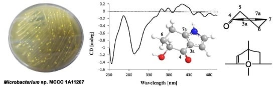
Microindolinone A, a Novel 4,5,6,7-Tetrahydroindole, from the Deep-Sea-Derived Actinomycete Microbacterium sp. MCCC 1A11207

1
State Key Laboratory Breeding Base of Marine Genetic Resources, Key Laboratory of Marine Genetic Resources, Third Institute of Oceanography, State Oceanic Administration, 184 Daxue Road, Xiamen 361005, China
2
Fujian Key Laboratory of Marine Genetic Resources, South China Sea Bio-Resource Exploitation and Utilization Collaborative Innovation Center, Third Institute of Oceanography, State Oceanic Administration, 184 Daxue Road, Xiamen 361005, China
*
Author to whom correspondence should be addressed.
Mar. Drugs 2017, 15(7), 230; https://doi.org/10.3390/md15070230
Submission received: 25 May 2017 / Revised: 30 June 2017 / Accepted: 14 July 2017 / Published: 19 July 2017
(This article belongs to the Special Issue Natural Products from Marine Actinomycetes)

Abstract

:
A novel indole, microindolinone A (1), was isolated from a deep-sea-derived actinomycete Microbacterium sp. MCCC 1A11207, together with 18 known compounds (219). By detailed analysis of the 1H, 13C, HSQC, COSY, HMBC, high resolution electron spray ionization mass spectrum (HRESIMS), and circular dichroism (CD) data, the absolute configuration of 1 was elucidated as 5R-hydroxy-4,5,6,7-tetrahydroindole-4-one. It is noteworthy that 1 is the second example of a saturated indole isolated from nature.

Graphical Abstract

1. Introduction

Actinomycetes are Gram-positive bacteria known for their ability to produce structurally novel secondary metabolites with various biological activities [1]. The best-known compound is salinosporamide A [2,3]. Very recently, the representative examples included pyrazolofluostatins and aminorifamycins isolated from marine Micromonospora species [4,5].
The genus Microbacterium of the Microbacteriaceae family was first proposed by Orla-Jensen in 1919. Up to now, there are 97 species reported from diverse habitats including land, ocean, air, blood, etc. However, only four compounds were reported from this genus [6,7]. In our current research for novel compounds from deep-sea-derived microorganisms [8,9,10], the actinomycete Microbacterium sp. MCCC 1A11207, isolated from southwestern Indian Ocean sediment, was subjected to a systematic chemical examination. Consequently, one new and 18 known compounds were obtained. Herein, we report the isolation, structural elucidation, and bioactivities of these compounds.

2. Results and Discussion

Microbacterium sp. MCCC 1A11207 was cultured in a 50 L fermentor containing 30 L A3 medium for 10 days. Then, the fermentation broth was centrifuged. The supernatant was extracted with EtOAc and the mycelium was extracted with MeOH. They were concentrated and combined to give the crude extract (17 g). By repeated column chromatography (CC) over silica gel, octadecylsilyl (ODS), and Sephadex LH-20, 19 compounds were obtained (Figure 1).

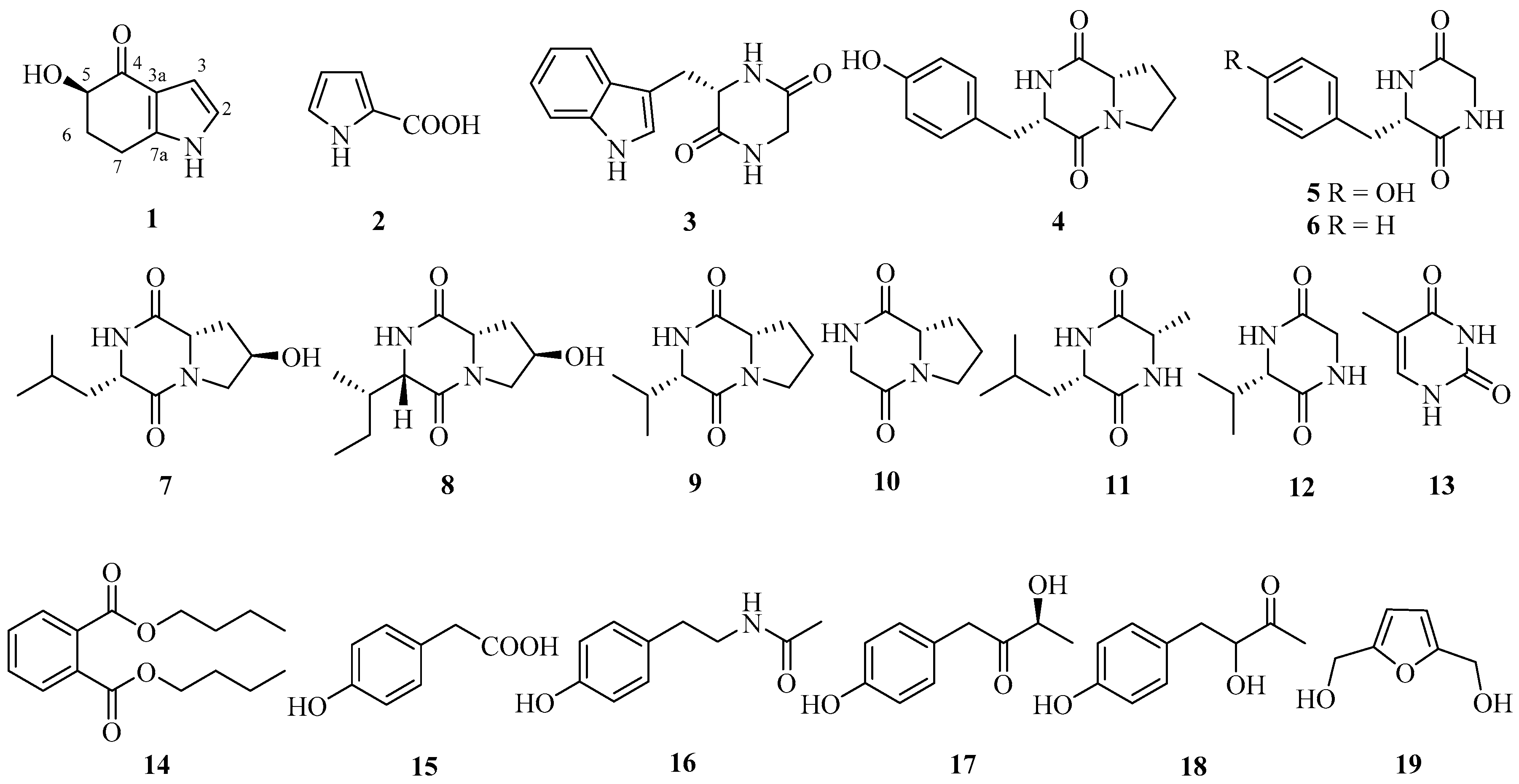
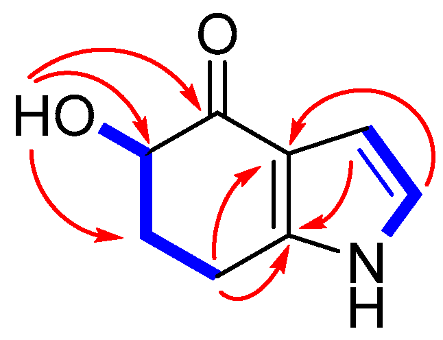
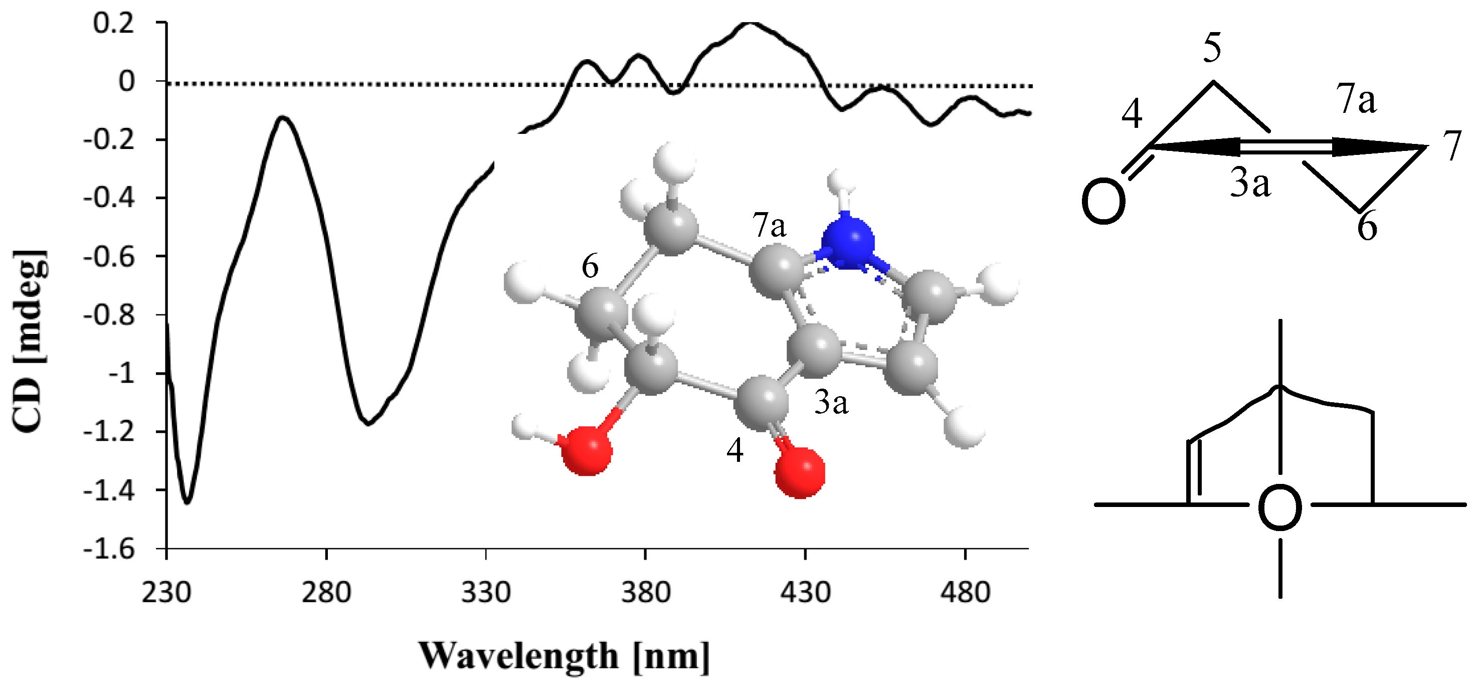
2.1. Structure Elucidation

Microindolinone A (1) was isolated as a colorless oil. The molecular formula was established as C8H9NO2 on the basis of a sodium adduct ion peak at m/z 174.0525 [M + Na]+ in its HRESIMS, requiring five degrees of unsaturation. The 1H-NMR spectrum (Figure S1) exhibited two exchangeable protons at δH 11.3 (1H, brs, 1-NH) and 4.98 (1H, d, J = 3.8 Hz, 5-OH), one oxygenated sp3 (δH 4.05, ddd, J = 11.6, 4.5, 3.8 Hz, H-5) and two sp2 [(δH 6.74, dd, J = 2.9, 2.4 Hz, H-2) and (δH 6.25, dd, J = 2.9, 2.2 Hz, H-3)] methines, together with two methylenes. The 13C attached proton test (APT)-NMR spectrum (Figure S2) showed eight resonance signals involving three non-protonated carbons at δC 194.1 (C-4), 143.4 (C-7a), and 118.4 (C-3a), three methines (δC 120.3/C-2, 105.2/C-3, and 72.6/C-5), and two methylenes at δC 33.0 (C-6) and 21.3 (C-7) (Table 1). In the 1H–1H COSY spectrum (Figure S4), two fragments of NH-1/C-2/C-3 and OH-5/C-5/C-6/C-7 were determined on the basis of COSY correlations of NH-1(δH 11.3)/H-2 (δH 6.74)/H-3 (δH 6.25), and 5-OH (δH 4.98)/H-5 (δH 4.05)/H2-6 (δH 1.87, m; 2.20, m)/H2-7 (δH 2.83, m) (Figure 2). These two fragments can be connected by a α,β-unsaturation ketone unit on the basis of HMBC cross-peaks (Figure S5) from H-2 to C-3/C-3a, H-3 to C-7a, OH-5 to C-4/C-5/C-6, and H2-7 to C-3a/C-5/C-6/C-7a (Figure 2), which established the planar structure of 1 as 5-hydroxy-4,5,6,7-tetrahydroindole-4-one.
The large coupling constant of H-5 and H-6a (JH-5/H-6a = 11.6 Hz) indicated H-5 as axial-orientation. In the CD spectrum, the negative Cotton effect (Δε296 −0.35) induced by n-π* electronic transition revealed the R-orientation of the 5-hydroxyl group on the basis of the octant rule (Figure 3) [11,12]. Therefore, the absolute configuration of 1 was determined as 5R-hydroxy-4,5,6,7-tetrahydroindole-4-one, and named microindolinone A. Surprisingly, although indoles occur broadly in nature [13,14,15], the saturated ones were seldom discovered. As a matter of fact, the only one reported was 6,7-dihydroxy-4,5,6,7-tetrahydroindole-4-one from Nocardia sp. (a soil-derived actinomycete) [16], and microindolinone A (1) was the second example. It is noteworthy that the absolute configuration of this saturated indole was determined for the first time.
By comparing the 1H-, 13C-NMR, MS, and optical rotation (OR) data with those reported in the literature, 18 known compounds were identified as pyrrole-2-carboxylic acid (2) [17], cyclo(l-Trp-Gly) (3) [18], cyclo-l-Tyr-l-Pro (4) [19], cyclo(l-Trp-Gly) (5) [20], cyclo(l-Phe-Gly) (6) [21], cyclo[l-(4-hydroxyprolinyl)-l-leucine] (7) [22,23], cyclo[l-(4-hydroxyprolinyl)-l-isoleucine] (8) [24], cyclo-(l-Pro-l-Val) (9) [25], cyclo-(l-Pro-Gly) (10) [25], cyclo-(l-Leu-l-Ala) (11) [26], cyclo-(l-Val-Gly) (12) [27], 5-methyluracil (13) [26], dibutyl phthalate (14) [28], 4-hydroxyphenylacetic acid (15) [29], N-(4-hydroxyphenyl)-acetamide (16) [30], (S)-3-hydroxy-4-(4-hydroxyphenyl)butan-2-one (17) [31], 3-hydroxy-4-(4-dihydroxyphenyl)-2-butanone (18) [32], and (5-hydroxymethyl-furan-2-yl)-methanol (19) [33]. Surprisingly, these 19 compounds were all firstly isolated from Microbacterium species.

2.2. Anti-Proliferative Activity of 1 against RBL-2H3 Cells

Microindolinone A (1) was tested for anti-proliferative activity against RBL-2H3 cells. Fortunately, it did not show significant cytotoxicity, even under the highest concentration of 20 µg/mL (Table 2).

2.3. Anti-Allergic Activity of 1

Microindolinone A (1) was further subjected to anti-allergic bioactivity on IgE-mediated rat mast RBL-2H3 cells. However, it did not show any positive effects under the concentration of 20 μg/mL, while the positive control loratadine exhibited a significant inhibition rate of 37% (Table 3).

3. Materials and Methods

3.1. General Experimental Procedures

HRESIMS spectra were obtained from a Xevo G2 Q-TOF mass spectrometer (Waters). Optical rotations were conducted by a Rudolph IV Autopol automatic polarimeter. NMR spectra were recorded on a Bruker 400 MHz spectrometer. Materials for column chromatography were silica gel (Qingdao Marine Chemistry Co. Ltd., Qingdao, China), ODS (50 μm, Daiso, Japan), and Sephadex LH-20 (Amersham Pharmacia Biotech AB, Uppsala, Sweden). Pre-coated silica gel plates were used for thin-layer chromatography (TLC) analysis.

3.2. Bacterial Material

The strain MCCC 1A11207 was isolated from a deep-sea sediment of the southwestern Indian Ocean (−1603 m) in 2014. By comparison of its 16S rRNA gene sequence with those of validly published names from the GenBank database via the BLAST program, the strain showed the highest similarity (98.03%) to Microbacterium amylolyticum N5T. Therefore, it was identified to be Microbacterium sp. MCCC 1A11207. A voucher strain of the actinomycete was deposited in the Marine Culture Collection of China with the accession number of MCCC 1A11207.

3.3. Cultivation and Extraction

The strain was cultured on the 2216E medium at 28 °C for 3 days and the colony was inoculated to 250 mL Erlenmeyer flasks containing 50 mL A3 medium compositing with 15 g bacterial peptone, 5 g soybean peptone, 15 g soluble starch, 30 g marine salt, and 1 L tap water, and then was cultured in a rotary shaker with 180 rpm at 28 °C for 3 days as the spores’ medium. The large-scale fermentation was performed by a 50 L fermentor containing 30 L A3 medium with the 5% seed culture, and the fermentation continued at 28 °C with 180 rpm for 10 days. Then, the fermentation broth was centrifuged (16,000 rpm) to get supernatant and mycelium. The supernatant was extracted with EtOAc three times, and then concentrated under reduced pressure to provide the crude extract A. The mycelium was extracted with MeOH twice. After removing MeOH, the residue was re-extracted with EtOAc three times to get extract B under reduced vacuum. Extracts A and B were combined to give the total crude extract.

3.4. Isolation and Purification

The total crude extract (17 g) was subjected to column chromatography (CC) on ODS, eluting with a gradient MeOH–H2O (5:95→100:0) to give four fractions (Fr.1–Fr.4). Fraction Fr.2 (92 mg) was first subjected to Sephadex LH-20 CC eluting with MeOH, and then by CC over silica gel using CHCl3–MeOH (100:1) to provide 2 (6.8 mg). Fraction Fr.3 (283 mg) was separated by CC over Sephadex LH-20 (MeOH) to get five subfractions (Fr.3.1–Fr.3.5). Subfraction Fr.3.1 was purified by CC on silica gel eluting with petroleum ether (PE)–acetone (2:1) to get 14 (11.3 mg). Compounds 12 (9.2 mg) and 19 (2.3 mg) were isolated from subfraction Fr.3.2 by CC over silica gel (CHCl3–MeOH, 20:1), while 5 (2.1 mg) and 13 (23.0 mg) were obtained from subfraction Fr.3.5 (CHCl3–MeOH, 6:1). Compound 10 (38.0 mg) was isolated from Fr.3.3 using recrystallization in MeOH. Subfraction Fr.3.4 was subjected to CC over silica gel eluting with PE–acetone (3:1) to get 1 (1.1 mg). Fraction Fr.4 (380 mg) was fractionated by CC on Sephadex LH-20 (MeOH) to obtain five subfractions (Fr.4.1–Fr.4.5). Subfraction Fr.4.1 was subjected to CC over silica gel (PE–acetone, 2:1) to get two subfractions Fr.4.1.1 and Fr.4.1.2. Subfraction Fr.4.1.1 was purified by CC over silica gel (PE–acetone, 2:1) to get 11 (1.8 mg). Fr.4.1.2 was subjected to MPLC using gradient MeOH–H2O (5→30%) to get 7 (8.0 mg), 8 (3.8 mg), and 9 (8.7 mg). Fr.4.2 was purified by CC over silica gel (CHCl3–MeOH, 6:1) to provide 16 (38.2 mg). Fr.4.3 was subjected to CC on silica gel (CHCl3–MeOH, 20:1) to get two subfractions Fr.4.3.1 and Fr.4.3.2. Subfraction Fr.4.3.1 was further purified by Prep. TLC (PE–EtOAc, 1:1) to get 17 (1.2 mg) and 18 (1.4 mg), while compounds 4 (12.1 mg) and 6 (9.8 mg) were isolated from Fr.4.3.2 by CC over silica gel (PE–EtOAc, 1:1). Fr.4.4 and Fr.4.5 were purified by CC on silica gel eluting with PE–EtOAc (1:1) and PE–EtOAc (2:1) to get 15 (8.6 mg) and 3 (2.3 mg), respectively.
Microindolinone A (1): Colorless oil; [ α ] D 25 +2.5 (c 0.11, MeOH); CD (CHCl3) λmax (Δε) 237 (−0.42), 268 (−0.04), 296 (−0.35); 1H- and 13C-NMR data, see Table 1; HRESIMS (positive) m/z 174.0525, calcd. for C8H9NO3Na+ 174.0531.

3.5. Anti-Proliferative Assay

According to previously reported protocols [34], the cytotoxicity test was carried out using the MTT assay on RBL-2H3 cells. In brief, RBL-2H3 cells were seeded into 96-well cell culture plates. Then, six different concentrations of 1, ranging from 0.625 to 20 µg/mL, were added. After 24 h, the cells were treated with 20 µL MTT solution. The cytotoxicity was quantified by measuring the absorbance at 570 nm. The cell viability was calculated using the following equation: Cell viability (%) = [(As − Av)/(Ac − Av) × 100%, where As is the absorbance of the sample, Av is the absorbance of the vehicle, and Ac is the absorbance of the control.

3.6. Anti-Allergic Test

The anti-allergic activity, indexed by the β-hexosaminidase release, was measured for the efficiency of the RBL-2H3 cell degranulation inhibition rate using IgE-mediated mast cell allergic reaction [8,35]. In short, RBL-2H3 cells were seeded into 96-well cell culture plates (1 × 10⁵ cells/well) to incubate with dinitrophenol (DNP) specific IgE overnight. IgE-sensitized RBL-2H3 cells were pre-treated with compound 1 (20 μg/mL) for 1 h and stimulated with dinitrophenyl-bovine serum albumin (DNP-BSA) (500 ng/mL). The negative control group was added to 200 μL phosphate-buffered saline (PBS) buffer. The β-hexosaminidase activity was quantified by measuring the fluorescence intensity of the hydrolyzed substrate in a fluorometer. The degranulation efficiency was calculated using the following formula: Degranulation efficiency (%) = Fsup/(Fsup + Flys) × 100%, where Fsup is the fluorescence value of the supernatant and Flys is the fluorescence value of cell lysates. The inhibition rate was calculated based on the following formula: Inhibition rate (%) = (Positive − Sample)/(Positive − Negative) × 100%, where Positive is the degranulation efficiency of the DNP-BSA stimulated group, Sample is the degranulation efficiency of the sample group, and Negative is the degranulation efficiency of the vehicle group.

3.7. Statistical Analysis

Anti-proliferative and anti-allergic experiments were conducted three times. Results are presented as means ± SD. One-way analysis of variance (one-way ANOVA) comparison tests of SPSS was used to evaluate the statistical significances of the differences between experimental groups. Differences were considered statistically significant for p < 0.05 using Duncan’s multiple range tests between groups.

4. Conclusions

From the deep-sea-derived rare actinomycete Microbacterium sp. MCCC 1A11207, 19 secondary metabolites were isolated and identified. The new compound, microindolinone A (1), was determined as 5R-hydroxy-4,5,6,7-tetrahydroindole-4-one. It was the second example of the tetrahydroindole found in nature. Its absolute configuration was determined for the first time.

Supplementary Materials

The following documents are available online at www.mdpi.com/1660-3397/15/7/230/s1.

Acknowledgments

The work was supported by the Science & Technology Research Program of Fujian Province, China (2017Y0060) and the National Natural Science Foundation of China (41676130, 41606185 and 21372233). We thank Qingmei Liu and Guangming Liu of the Jimei University for biological tests.

Author Contributions

Xian-Wen Yang designed the project; Siwen Niu, Ting-Ting Zhou and Chun-Lan Xie performed experiments; Gai-Yun Zhang isolated and identified the actinomycete; Siwen Niu and Xian-Wen Yang analyzed the data and wrote the paper, while critical revision of the publication was performed by all authors.

Conflicts of Interest

The authors declare no conflict of interest.

References

  1. Diminic, J.; Starcevic, A.; Lisfi, M.; Baranasic, D.; Gacesa, R.; Hranueli, D.; Long, P.F.; Cullum, J.; Zucko, J. Evolutionary concepts in natural products discovery: What actinomycetes have taught us. J. Ind. Microbiol. Biotechnol. 2014, 41, 211–217. [Google Scholar] [CrossRef] [PubMed]
  2. Feling, R.H.; Buchanan, G.O.; Mincer, T.J.; Kauffman, C.A.; Jensen, P.R.; Fenical, W. Salinosporamide A: A highly cytotoxic proteasome inhibitor from a novel microbial source, a marine bacterium of the new genus salinospora. Angew. Chem. Int. Ed. Engl. 2003, 42, 355–357. [Google Scholar] [CrossRef] [PubMed]
  3. Jensen, P.R.; Moore, B.S.; Fenical, W. The marine actinomycete genus Salinispora: A model organism for secondary metabolite discovery. Nat. Prod. Rep. 2015, 32, 738–751. [Google Scholar] [CrossRef] [PubMed]
  4. Zhang, W.; Yang, C.; Huang, C.; Zhang, L.; Zhang, H.; Zhang, Q.; Yuan, C.S.; Zhu, Y.; Zhang, C. Pyrazolofluostatins A–C, pyrazole-fused benzo[a]fluorenes from South China Sea-derived Micromonospora rosaria SCSIO N160. Org. Lett. 2017, 19, 592–595. [Google Scholar] [CrossRef] [PubMed]
  5. Williams, D.E.; Dalisay, D.S.; Chen, J.; Polishchuck, E.A.; Patrick, B.O.; Narula, G.; Ko, M.; Av-Gay, Y.; Li, H.; Magarvey, N.; et al. Aminorifamycins and sporalactams produced in culture by a Micromonospora sp. isolated from a Northeastern-Pacific marine sediment are potent antibiotics. Org. Lett. 2017, 19, 766–769. [Google Scholar] [CrossRef] [PubMed]
  6. Gao, S.; Huang, R.; Zhu, S.; Li, H.; Zheng, G. Identification and characterization of a novel (+)-γ-lactamase from Microbacterium hydrocarbonoxydans. Appl. Microbiol. Biotechnol. 2016, 100, 9543–9553. [Google Scholar] [CrossRef] [PubMed]
  7. Liu, D.; Lin, H.; Proksch, P.; Tang, X.; Shao, Z.; Lin, W. Microbacterins A and B, new peptaibols from the deep sea actinomycete Microbacterium sediminis sp. nov. YLB-01(T). Org. Lett. 2015, 17, 1220–1223. [Google Scholar] [CrossRef] [PubMed]
  8. Xie, C.L.; Liu, Q.; Xia, J.M.; Gao, Y.; Yang, Q.; Shao, Z.Z.; Liu, G.; Yang, X.W. Anti-allergic compounds from the deep-sea-derived actinomycete Nesterenkonia flava MCCC 1K00610. Mar. Drugs 2017, 15, 71. [Google Scholar] [CrossRef] [PubMed]
  9. Niu, S.; Si, L.; Liu, D.; Zhou, A.; Zhang, Z.; Shao, Z.; Wang, S.; Zhang, L.; Zhou, D.; Lin, W. Spiromastilactones: A new class of influenza virus inhibitors from deep-sea fungus. Eur. J. Med. Chem. 2016, 108, 229–244. [Google Scholar] [CrossRef] [PubMed]
  10. Yang, X.W.; Peng, K.; Liu, Z.; Zhang, G.Y.; Li, J.; Wang, N.; Steinmetz, A.; Liu, Y. Strepsesquitriol, a rearranged zizaane-type sesquiterpenoid from the deep-sea-derived actinomycete Streptomyces sp. SCSIO 10355. J. Nat. Prod. 2013, 76, 2360–2363. [Google Scholar] [CrossRef] [PubMed]
  11. Sun, Y.; Tian, L.; Huang, J.; Ma, H.Y.; Zheng, Z.; Lv, A.L.; Yasukawa, K.; Pei, Y.H. Trichodermatides A–D, novel polyketides from the marine-derived fungus Trichoderma reesei. Org. Lett. 2008, 10, 393–396. [Google Scholar] [CrossRef] [PubMed]
  12. Kirk, D.N. The chiroptical properties of carbonyl compounds. Tetrahedron 1986, 42, 777–818. [Google Scholar] [CrossRef]
  13. Netz, N.; Opatz, T. Marine indole alkaloids. Mar. Drugs 2015, 13, 4814–4914. [Google Scholar] [CrossRef] [PubMed]
  14. Ishikura, M.; Abe, T.; Choshi, T.; Hibino, S. Simple indole alkaloids and those with a non-rearranged monoterpenoid unit. Nat. Prod. Rep. 2013, 30, 694–752. [Google Scholar] [CrossRef] [PubMed]
  15. Gupta, L.; Talwar, A.; Chauhan, P.M. Bis and tris indole alkaloids from marine organisms: new leads for drug discovery. Curr. Med. Chem. 2007, 14, 1789–1803. [Google Scholar] [CrossRef] [PubMed]
  16. Henne, P.; Zeeck, A.; Grabley, S.; Thiericke, R. Secondary metabolites by chemical screening.35. 6,7-Dihydroxy-4,5,6,7-tetrahydroindole-4-one, a new type of indole-derivative from Nocardia sp. Nat. Prod. Lett. 1997, 10, 43–47. [Google Scholar] [CrossRef]
  17. Dietera, A.; Hamm, A.; Fiedler, H.P.; Goodfellow, M.; Muller, W.E.; Brun, R.; Beil, W.; Bringmann, G. Pyrocoll, an antibiotic, antiparasitic and antitumor compound produced by a novel alkaliphilic Streptomyces strain. J. Antibiot. 2003, 56, 639–646. [Google Scholar] [CrossRef] [PubMed]
  18. Deslauriers, R.; Grzonka, Z.; Schaumburg, K.; Shiba, T.; Walter, R. Carbon-13 nuclear magnetic resonance studies of the conformations of cyclic dipeptides. J. Am. Chem. Soc. 1975, 97, 5093–5100. [Google Scholar] [CrossRef] [PubMed]
  19. Thajudeen, H.; Park, K.; Moon, S.-S.; Hong, I.S. An efficient green synthesis of proline-based cyclic dipeptides under water-mediated catalyst-free conditions. Tetrahedron Lett. 2010, 51, 1303–1305. [Google Scholar] [CrossRef]
  20. Mollica, A.; Costante, R.; Fiorito, S.; Genovese, S.; Stefanucci, A.; Mathieu, V.; Kiss, R.; Epifano, F. Synthesis and anti-cancer activity of naturally occurring 2,5-diketopiperazines. Fitoterapia 2014, 98, 91–97. [Google Scholar] [CrossRef] [PubMed]
  21. Nakao, M.; Toriuchi, Y.; Fukayama, S.; Sano, S. Synthesis and conformational characterization of diketopiperazines bearing a benzyl moiety. Chem. Lett. 2014, 43, 340–342. [Google Scholar] [CrossRef]
  22. Ienaga, K.; Nakamura, K.; Goto, T. Bioactive compounds produced in animal tissues. I. Two diketopiperidine plant-growth regulators containing hydroxyproline isolated from rabbit skin tissue extract. Tetrahedron Lett. 1987, 28, 1285–1286. [Google Scholar] [CrossRef]
  23. Cronan, J.M., Jr.; Davidson, T.R.; Singleton, F.L.; Colwell, R.R.; Cardellina, J.H., II. Plant growth promoters isolated from a marine bacterium associated with Palythoa sp. Nat. Prod. Lett. 1998, 11, 271–278. [Google Scholar] [CrossRef]
  24. Maurer, G.; Kiechel, J.R. Ergopeptide Alkaloids. Patent DE2805977A1, 1978. [Google Scholar]
  25. Hendea, D.; Laschat, S.; Baro, A.; Frey, W. Diastereoselective alkylation of a proline-derived bicyclic lactim ether. Helv. Chim. Acta 2006, 89, 1894–1909. [Google Scholar] [CrossRef]
  26. Ding, Z.G.; Zhao, J.Y.; Yang, P.W.; Li, M.G.; Huang, R.; Cui, X.L.; Wen, M.L. 1H and 13C NMR assignments of eight nitrogen containing compounds from Nocardia alba sp.nov (YIM 30243T). Magn. Reson. Chem. 2009, 47, 366–370. [Google Scholar] [CrossRef] [PubMed]
  27. Lankiewicz, L.; Nyasse, B.; Fransson, B.; Grehn, L.; Ragnarsson, U. Synthesis of amino acid derivatives substituted in the backbone with stable isotopes for application in peptide synthesis. J. Chem. Soc. Perkin Trans. 1 1994, 17, 2503–2510. [Google Scholar] [CrossRef]
  28. McNulty, J.; Nair, J.J.; Cheekoori, S.; Larichev, V.; Capretta, A.; Robertson, A.J. Scope and mechanistic insights into the use of tetradecyl(trihexyl)phosphonium bistriflimide: A remarkably selective ionic liquid solvent for substitution reactions. Chem. A Eur. J. 2006, 12, 9314–9322. [Google Scholar] [CrossRef] [PubMed]
  29. Milne, J.E.; Storz, T.; Colyer, J.T.; Thiel, O.R.; Dilmeghani Seran, M.; Larsen, R.D.; Murry, J.A. Iodide-catalyzed reductions: Development of a synthesis of phenylacetic acids. J. Org. Chem. 2011, 76, 9519–9524. [Google Scholar] [CrossRef] [PubMed]
  30. Lin, Z.J.; Lu, X.M.; Zhu, T.J.; Fang, Y.C.; Gu, Q.Q.; Zhu, W. GPR12 Selections of the metabolites from an endophytic Streptomyces sp. asociated with Cistanches deserticola. Arch. Pharmacal Res. 2008, 31, 1108–1114. [Google Scholar] [CrossRef] [PubMed]
  31. Peng, X.P.; Wang, Y.; Liu, P.P.; Hong, K.; Chen, H.; Yin, X.; Zhu, W.M. Aromatic compounds from the halotolerant fungal strain of Wallemia sebi PXP-89 in a hypersaline medium. Arch. Pharmacal Res. 2011, 34, 907–912. [Google Scholar] [CrossRef] [PubMed]
  32. Kennedy, M.L. Phytochemical profile of the stems of Aeonium lindleyi. Rev. Bras. Farmacogn. 2012, 22, 676–679. [Google Scholar] [CrossRef]
  33. Goswami, S.; Dey, S.; Jana, S. Design and synthesis of a unique ditopic macrocyclic fluorescent receptor containing furan ring as a spacer for the recognition of dicarboxylic acids. Tetrahedron 2008, 64, 6358–6363. [Google Scholar] [CrossRef]
  34. Yang, X.W.; Zeng, H.W.; Liu, X.H.; Li, S.M.; Xu, W.; Shen, Y.H.; Zhang, C.; Zhang, W.D. Anti-inflammatory and anti-tumour effects of Abies georgei extracts. J. Pharm. Pharmacol. 2008, 60, 937–941. [Google Scholar] [CrossRef] [PubMed]
  35. Liu, Q.; Wang, Y.; Cao, M.; Pan, T.; Yang, Y.; Mao, H.; Sun, L.; Liu, G. Anti-allergic activity of R-phycocyanin from Porphyra haitanensis in antigen-sensitized mice and mast cells. Int. Immunopharmacol. 2015, 25, 465–473. [Google Scholar] [CrossRef] [PubMed]
Figure 1. Compounds isolated from Microbacterium sp. MCCC 1A11207.
Marinedrugs 15 00230 g001
Figure 2. Key 1H–1H COSY (bold) and HMBC (arrow) correlations of 1.
Marinedrugs 15 00230 g002
Figure 3. The CD spectrum in CHCl3 and octant projection of compound 1.
Marinedrugs 15 00230 g003
Table 1. The 1H (400 MHz) and 13C (100 MHz) NMR spectroscopic data for 1 in DMSO-d6.
PositionδC, TypeδH (J in Hz)
1 11.3, brs
2120.3, CH 6.74, dd (2.9, 2.4)
3105.2, CH 6.25, dd (2.9, 2.2)
3a118.4, C
4194.1, C
572.6, CH 4.05, ddd (11.6, 4.5, 3.8)
633.0, CH21.87, m; 2.20, m
721.3, CH32.83, m
7a143.4, C
5-OH 4.98, d (3.8)
Table 2. Anti-proliferative activity of 1 against RBL-2H3 cells (n = 3, means ± SD).
Concentrations (µg/mL)Cell Viability (%)
2091 ± 10
1093 ± 1.4
590 ± 10
2.593 ± 12
1.2594 ± 12
0.62599 ± 14
Table 3. The Anti-allergic activity of 1 against RBL-2H3 cells (n = 3, means ± SD).
CompoundConcentration (μg/mL)Inhibition Rate (%)
120−1.4 ± 0.8
Loratadine2037 ± 5.3

