Article

Tetrocarcin Q, a New Spirotetronate with a Unique Glycosyl Group from a Marine-Derived Actinomycete Micromonospora carbonacea LS276

State Key Laboratory of Bioactive Substance and Function of Natural Medicines; Key Laboratory of Biosynthesis of Natural Products of National Health and Family Planning Commission, Institute of Materia Medica, Chinese Academy of Medical Sciences and Peking Union Medical College, 1 Xian Nong Tan Street, Beijing 100050, China
*
Author to whom correspondence should be addressed.
Mar. Drugs 2018, 16(2), 74; https://doi.org/10.3390/md16020074
Submission received: 7 December 2017 / Revised: 7 February 2018 / Accepted: 12 February 2018 / Published: 24 February 2018
(This article belongs to the Special Issue Natural Products from Coral Reef Organisms)

Abstract

:
A new spirotetronate glycoside tetrocarcin Q (1) and six known analogues tetrocarcin A (2), AC6H (3), tetrocarcin N (4), tetrocarcin H (5), arisostatin A (6), and tetrocarcin F1 (7) were isolated from the fermentation broth of the marine-derived actinomycete Micromonospora carbonacea LS276. Their chemical structures were established on the basis of 1D- and 2D-NMR spectroscopy, as well as HR-ESI-MS analysis. The absolute configurations of their stereogenic carbons were determined by circular dichroism (CD) analysis. Compound 1 possesses 2-deoxy-allose, which is a unique sugar type at the C-9 position. This type has not been found in the previously reported spirotetronate glycosides. Compound 1 displayed moderate antibacterial activity against Bacillus subitlis ATCC 63501 with minimum inhibitory concentration (MIC) value of 12.5 μM.

1. Introduction

The spirotetronate family displays complicated chemical structures, potent bioactivities, and significant pharmacological potential [1]. This family features an unusual macrolide that contains a characteristic tetronic acid (spiro-linked to a cyclohexene ring) conjugated with a trans-decalin system. The structure is also linked with two sugar side chains, one of which is d-tetronitrose (NS), while the other comprises l-digitoxoses (DG) and l-amicetoses (AM) [2,3,4]. In terms of biological profile, the spirotetronate exhibits broad biological activities, including antibacterial, antitumor, antiviral, and antimalarial effects [5,6,7,8,9,10]. A representative of this group is the tetrocarcins, including tetrocarcins A–P, AC6H, arisostatins A and B which were isolated from Micromonospora bacteria [4,5,11,12,13,14]. Various studies reported that tetrocarcin A and its analogues had the antibiotic activity against several Gram-positive bacteria as well as anticancer activity [6,15,16]. Therefore, our objective is to discover new tetronolides with antibacterial activity, provide more information for the structure-activity relationship, as well as the possibility to improve their potential applications.
The ethyl acetate extract of the fermentation broth from the marine-derived M. carbonacea LS276 [17] showed antibacterial activity (Figure 1a). Bioassay-guided fractionation of the extract and further purification allowed for the isolation of seven spirotetronate glycosides (17). Among them, tetrocarcin Q (1) has a unique oligosaccharide chain at the C-9 position, which is different from other known spirotetronate glycosides. A major constituent, tetrocarcin A (2), was found to be the main active component of this strain (Figure 1b,c). Herein, we report the isolation, structure elucidation, and biological activities of these compounds.

2. Results and Discussion

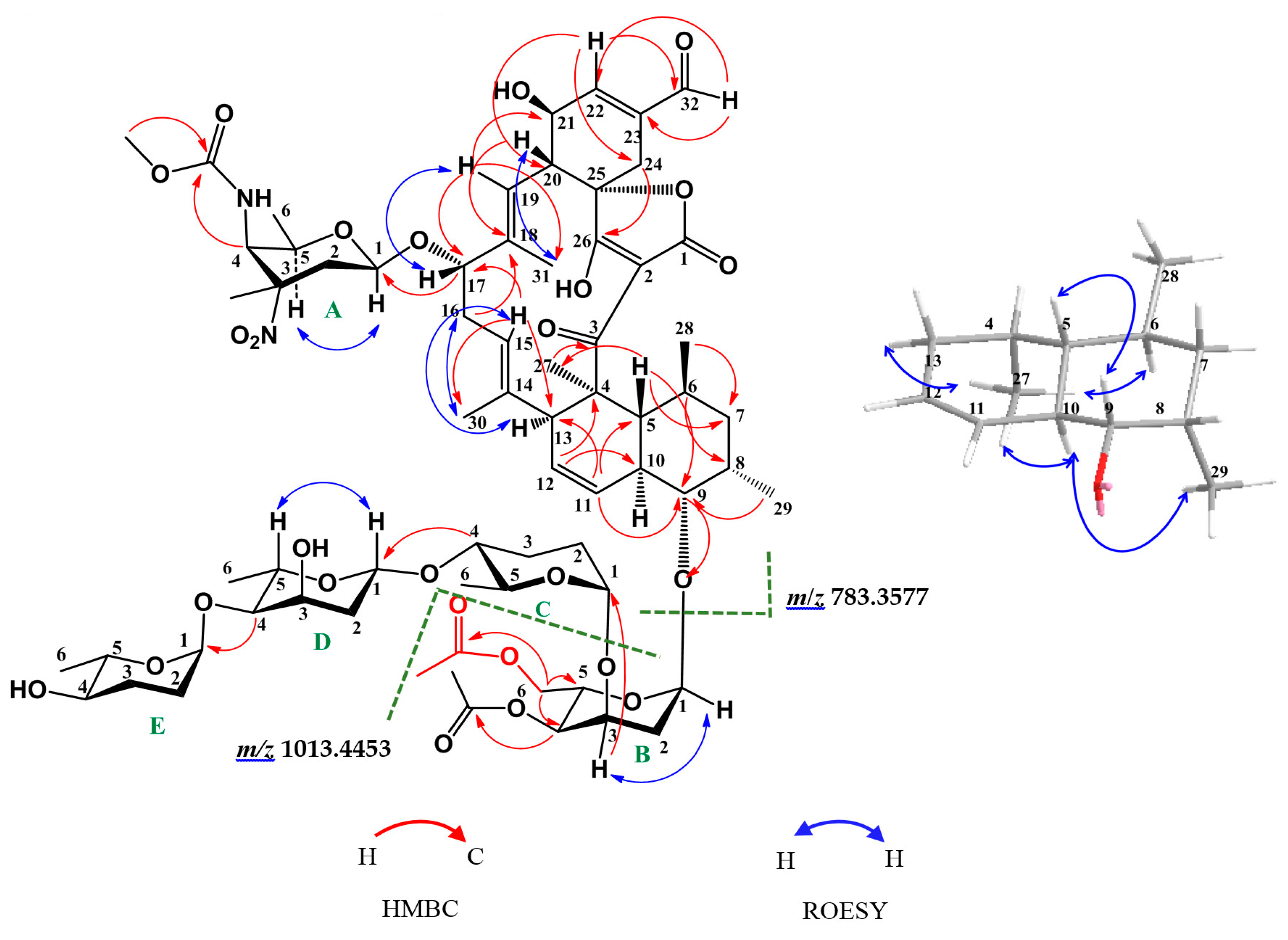
2.1. Structure Elucidation of Compounds

Compound 1 was obtained as a white powder. Its molecular formula C69H98N2O26 was determined by the (+)-HR-ESI-MS peak at m/z 1393.6260 [M + Na]+, indicating 22 degrees of unsaturation. The 1H NMR spectrum of 1 (Table 1, Supplementary Figure S2) displayed one aldehydic proton at δH 9.58 (s, H-32), five olenic protons (δH: 5.74 (d, J = 10.2 Hz, H-11), 5.42 (m, H-12), 5.16 (m, H-15), 5.21 (d, J = 10.2 Hz, H-19), 6.92 (s, H-22)), five glycosyl anomeric protons (δH: 4.44 (dd, J = 9.6, 1.8 Hz, H-A-1), 4.92 (d, J = 4.8 Hz, H-B-1), 4.88 (brd, J = 3.0 Hz, H-C-1), 4.90 (dd, J = 9.6, 1.8 Hz, H-D-1), 4.91 (brs, H-E-1)), one methoxy group at δH 3.71 (s, H-A4-NHCOOCH3), six methyl singlets (δH: 2.08 (H-B4-OCOCH3), 2.07 (H-B6-OCOCH3), 1.63 (H-27), 1.60 (H-A3-CH3), 1.53 (H-31), 1.34 (H-30)), and six methyl doublets (δH: 1.32(d, J = 6.0 Hz, H-D-6), 1.23 (d, J = 6.0 Hz, H-E-6), 1.16 (d, J = 6.6 Hz, H-C-6), 1.15 (d, J = 6.6 Hz, H-A-6), 1.09 (d, J = 7.2 Hz, H-29), 0.64 (d, J = 6.0 Hz, H-28)). The 13C NMR (Table 1, Supplementary Figure S3) and Heteronuclear Single Quantum Coherence (HSQC) (Supplementary Figure S4) spectra revealed 69 carbon signals, including seven carbonyls or keto-enolic carbons (δC: 206.4, 201.5, 192.6, 170.9, 170.2, 166.7, 157.4), nine olefinic carbons (δC: 149.6, 141.6, 136.5, 136.1, 126.2, 126.1, 123.1, 118.3, 100.9), five sugar anomeric carbons (δC: 99.5, 98.9, 96.5, 92.7, 92.0), one methoxyl (δC: 53.0), 12 methyls (δC: 25.4, 22.1, 21.0, 20.9, 19.0, 18.2, 17.8, 17.1, 16.3, 15.2, 14.5, 14.1). Comprehensive analysis of the 1H-1H Homonuclear chemical shift Correlation Spectroscopy (COSY) (Supplementary Figure S6), HSQC (Supplementary Figure S4) and Heteronuclear Multiple Bond Correlation (HMBC) (Supplementary Figure S5) spectra of 1, indicated the presence of a spiroteronate skeleton, a tetronitrose (NS), and a tetrasaccharide, which is similar to tetrocarcin A (2). The difference between 1 and tetrocarcin A (2) is that the 6-methyl group (δH 1.13 (3H, d, J = 6.6 Hz); δC 17.6) of one digitoxose unit (sugar B) in tetrocarcin A (2) is replaced by a 6-oxymethylene (δH 4.32 (1H, dd, J = 12.0, 5.4 Hz) and 4.12 (1H, dd, J = 12.0, 1.8 Hz); δC 63.4) and an acetyl group (δH 2.07 (3H, s); δC 21.0, 170.9) (Table 1, Supplementary Figure S10, Supplementary Table 1). The HMBC correlations of 6-oxymethylene protons (δH 4.32 and 4.12) to the ester carbonyl (δC 170.9, B6-OCOCH3) and two oxygenated-carbons (δC 69.5, B-4 and δC 64.5, B-5) confirmed that the first sugar (sugar B) is 4,6-O-diacetyl-2-deoxysugar, which was supported by the (+)-HR-ESI-MS fragments (m/z 1013.4453 and 783.3577) corresponding to the ion of a subunit consisting of the spiroteronate skeleton with NS (sugar A) and 4,6-O-diacetyl-sugar (sugar B), and a subunit of the spiroteronate skeleton with NS (Figure 2, Supplementary Figure S1). On the basis of the above information, all protons and carbon resonances were assigned and the planar structure of 1 was established.
The relative configuration of 1 was the same as those of the previously reported tetrocarcins, based on the similarity of their NMR spectral data of the aglycone, which was further supported by key coupling constants in 1H NMR spectrum and the correlations observed in the Rotating Frame Overhauser Effect Spectroscopy (ROESY) experiments (Figure 2, Supplementary Figure S7). The Z-configuration of the Δ11,12 double bond was assigned on the basis of the coupling constant JH-11/H-12 = 10.2 Hz. The ROESY correlations of H-13/H-15, H-16/H-30, H-17/H-19, and H-31/H-20 indicated that the E-configurations of the both double bonds Δ14,15 and Δ18,19. In addition, the ROESY correlations of H-13/H-27, H-27/H-10, H-27/H-6, and H-29/H-10 revealed that these protons were on the same side of the decalin ring, whereas the ROESY correlations of H-9/H-5 indicated that they were on the other side of the ring. The absolute configurations of the stereogenic carbons in the aglycone of 1 were the same as those of tetrocarcin A (2), since they displayed similar circular dichroism (CD) curves, which showed a negative Cotton effect at 224 nm and a positive Cotton effect at 264 and 300 nm [4] (Supplementary Figure S8). The relative configurations of sugars A-E were determined as β, α, α, β, and α-orientations by the coupling constants of the anomeric protons (δH: 4.44 (dd, J = 9.6, 1.8 Hz, H-A-1), 4.92 (d, J = 4.8 Hz, H-B-1), 4.88 (brd, J = 3.0 Hz, H-C-1), 4.90 (dd, J = 9.6, 1.8 Hz, H-D-1), 4.91 (brs, H-E-1)), which was confirmed by the ROESY correlations (H-A-1/H-A-5, H-B-1/H-B-3, H-D-1/H-D-5).
Compounds 27 were also obtained as white amorphous powders, and they were identified as tetrocarcin A (2), AC6H (3), tetrocarcin N (4), tetrocarcin H (5), arisostatin A (6), and tetrocarcin F1 (7) by comparison of their spectral data (MS, 1H, 13C NMR, specific rotation) with those reported in the literature [4,5,12,13,14].

2.2. Biological Assays

All of the isolated compounds were evaluated for their antibacterial activity against Bacillus subitlis ATCC 63501, Staphylococcus aureus ATCC 29213, Staphylococcus epidermidis ATCC 12228, Enterococcus faecalis ATCC 29212, Pseudomonas aeruginosa ATCC 27853, and Escherichia coli ATCC 25922. Except for 7, the other six compounds exhibited antibacterial activity against B. subtilis with minimum inhibitory concentration (MIC) from <0.048 μM to 50 μM, with 2 and 6 showing strong antibacterial activity (Table 2). The MIC values of 27 were found to be similar to those previously reported [4,9,11,14,18].
Compound 7 exhibited no activity, indicating that the oligosaccharide chain is required for the antibacterial activity. Compound 3 was at least 10-fold less active than 2, suggesting that the NO2-sugar is also important for the antibacterial activity. Compounds 4 and 5 were less active than 2, 3 and 6, inferring that the aldehyde group at C-23 is also essential for the activity. These results are all in accordance with the previously reported structure-activity relationship [1,4,9,11,14].
Compound 1 displayed a moderate antibacterial activity with MIC value of 12.5 μM, which was less active than 2, implying that 6-CH3 of sugar B in the oligosaccharide chain at C-9 plays a key role in the antibacterial activity (Table 2).
Compounds 17 were evaluated by MTT method for their in vitro antitumor activity against five human cancer cell lines including: human non-small cell lung cancer cell (A549), human gastric cancer cell (BGC823), human colonic carcinoma cell (HCT116), human hepatoma cell (HepG2), human glioblastoma multiform cell (U87MG). In addition to moderate activity against the other four cell lines with the IC50 values ranging from 5.33 μM to 19.7 μM, 2 and 6 exhibited the most potent antitumor activity against U87MG cell line with IC50 values of 0.50 μM and 2.42 μM, respectively (Table 3). The other compounds were considered to be weakly active or inactive (IC50 > 10 μM).
The structure-activity relationship of the seven compounds on the human tumor cell lines A549, BGC823, HCT116, HepG2 and U87G was very similar to that obtained from antibacterial assay against B. subitlis (Table 2 and Table 3). The most active compounds were 2 and 6, which was in agreement with the previous studies [9]. The activities of 3, 4 and 5 were decreased, suggesting the modification of the tetronolide skeleton have influence on the in vitro antitumor activity in some extent. The lack of activity of 7 implies that the sugar moiety at C-9 position could play an important role in the antitumor activity, which was also in agreement with the previous structure-activity relationship study [15]. Compound 1 showed no or weak in vitro antitumor activity, suggesting that the deoxy sugar analogue may also influence the antitumor activity.

3. Materials and Methods

3.1. General

Optical rotations were measured on a JASCO P-2000 digital polarimeter (JASCO Corporation, Tokyo, Japan). Circular dichroism (CD) spectrum was recorded using a JASCO J-815 CD spectro polarimeters (JASCO Corporation, Tokyo, Japan). 1H and 13C NMR, and 2D NMR spectra were obtained at 600 and 150 MHz, using a Bruker AVANCE 600-III spectrometer (Bruker Biospin Group, Karlsruhe, Germany) in chloroform-d with TMS as an internal reference. HR-ESI-MS data were measured using an Agilent 1100 LC/MSD Trap SL LC/MS/MS spectrometer (Agilent Technologies, Santa Clara, CA, USA). Semipreparative HPLC was performed by an Agilent 1200 HPLC system (Agilent Technologies, Santa Clara, CA, USA) using a Shiseido Capcell Pak C18 column (5 μm, 10 × 250 mm). Column chromatography was performed with RP-18 (40–60 μm, GE healthcare, Fairfield, CT, USA) and Sephadex LH-20 (18–110 μm, GE healthcare, Fairfield, CT, USA).

3.2. Bacterial Material and Fermentation

The strain LS276 was isolated from a sponge Gelliodes carnosa collected from Ling shui Bay, Hainan Province of China near Xincun Harbor (18°24′5.49″ N, 109°59′37.76″ E), in August 2007 [17]. It was identified as M. carbonacea based on the morphology and 16S rRNA gene sequence analysis. The DNA sequence was deposited in GenBank (Accession No. FJ937935.1). The strain LS276 was first cultivated on agar plates (medium: starch 40.0 g; glucose 0.5 g; peptone 5.0 g; soybean powder 5.0 g; CaCO3 1.0 g; K2HPO4 0.5 g; MgSO4 0.5 g; agar 10.0 g; distilled water 1 L; pH 7.0–7.2) at 28 °C for five days. Then, the mycelia were inoculated into 500-mL Erlenmeyer flasks, each containing 100 mL of liquid medium (composed of starch 40.0 g; glucose 0.5 g; peptone 5.0 g; soybean powder 5.0 g; CaCO3 1.0 g; K2HPO4 0.5 g; MgSO4 0.5 g; distilled water 1 L; pH 7.0–7.2). The flasks were incubated at 28 °C on a rotary shaker (200 rpm) for three days. Seed culture (10 mL) was transferred into two hundred 500-mL Erlenmeyer flasks each containing 100 mL of fermentation medium (composed of 10.0 g of starch; 20.0 g of glucose; 5.0 g of soybean powder; 1.0 g of KNO3; 0.5 g of NaCl; 0.5 g of K2HPO4; 0.01 g of MgSO4 in 1 L of distilled H2O) and incubated at 28 °C on a rotary shaker (200 rpm) for nine days.

3.3. Extraction and Isolation

The culture broth (20 L) was repeatedly extracted with ethyl acetate (v/v 1:3, three times) by ultrasound, and the organic solvent was evaporated to dryness under a vacuum to afford the crude extract (4.0 g). The crude extract was first subjected to Sephadex LH-20 chromatography (3 × 60 cm, 100 g) using CH3OH (each 20 mL) as eluent and afforded six primary Fractions (Frs) 1–6. Fr.3 (2.0 g) was separated via semipreparative HPLC using 65% CH3CN in H2O, flow rate 2 mL/min as eluent to give Fr.3.1 to Fr.3.6. Fr.3.1 was further purified by semipreparative HPLC with a linear gradient of CH3CN–0.02%CH3COOH/H2O (60% to 100%, 35 min) to provide compounds 1 (2.4 mg, tR = 10.0 min), 4 (12.6 mg, tR = 10.8 min) and 5 (9.4 mg, tR = 10.4 min). Fr.3.2 was further purified by semipreparative HPLC with a linear gradient of CH3CN–0.02%CH3COOH/H2O (65% to 72%, 40 min) to yield compound 2 (46.7 mg, tR = 16.7 min). Fr.3.5 was further purified by semipreparative HPLC with a linear gradient of CH3CN–0.02%CH3COOH/H2O (65% to 72%, 40 min) to yield compound 6 (2.6 mg, tR = 18.9 min). Fr.5. (300 mg) was purified by semipreparative HPLC afforded compound 3 (3.6 mg, linear gradient of 20–70% CH3CN in H2O for 50 min, flow rate 2 mL/min, tR = 28.2 min). Purification of the Fr.4. (100 mg) by semipreparative HPLC provided compound 7 (7.8 mg, linear gradient of 20–70% CH3CN in H2O for 50 min, flow rate 2 mL/min, tR = 37.3 min).
Tetrocarcin Q (1). White amorphous powder; [ α ] D 20 −80.9 (c 0.05, MeOH); UV (MeOH) λmax (log ε) 203 (4.13), 242 (3.02), and 274 (4.07) nm; CD (c 0.5 (w/v)%, MeOH) 224 (−22.6), 264 (6.00), 343 (−0.85) nm; 1H NMR (CDCl3, 600 MHz) and 13C NMR (CDCl3, 150 MHz) data, Table 1; (+)-HR-ESI-MS m/z 1393.6260 [M + Na]+ (calcd. for C69H98N2O26Na, 1393.6270).

3.4. Biological Assays

Antibacterial and in vitro antitumor assays were performed for the isolated compounds with the purity of >90% by HPLC.

3.4.1. Antibacterial Activity

The tested bacteria used in this study were as follows: B. subitlis ATCC 63501, S. aureus ATCC 29213, S. epidermidis ATCC 12228, E. faecalis ATCC 29212, P. aeruginosa ATCC 27853, and E. coli ATCC 25922, MIC values against the six bacterial strains were measured by using the 96-well plate-based assay [19]. The bacterial strains cultured in respective medium were collected at OD600 of 0.3–0.5, then further diluted to OD600 of 5 × 10⁻⁴. Aliquots of this suspension (100 μL) were placed into a 96-well plate. The tested compounds were added into the bacteria suspensions to give the desired concentration. The wells containing the same number of cells but no compounds were set as control groups. The positive control was ampicillin. The cultures were then added respective solutions and further incubated at 37 °C for 18 h. The plate was then read using a microplate reader at 600 nm. Each concentration had triplicate values, and the whole experiment was carried out at three times and the MIC value was determined by taking the average of triplicate OD600 values for each concentration and plotting it against concentration. The MIC value was determined, as the point in the curve where the OD600 is similar to that of control without bacteria.

3.4.2. Antitumor Activity

The in vitro antitumor activity (represented by IC50 values) of the isolated compounds against five tumor cell lines, including A549, BGC823, HCT116, HepG2 and U87MG, was determined by MTT method as reported [20,21], and the dose-response curves were fitted with Sigma plot.

4. Conclusions

In summary, seven spirotetronate glycosides were isolated and characterized from the marine-derived M. carbonacea LS276. Among them, tetrocarcin Q (1) is a new compound. It is worth mentioning that the sugar B of compound 1 is 6-O-acetylated, while the other sugars in the previous spirotetronate glycosides are 6-deoxy sugars (DG and AM), which expanded the structural variability of such spirotetronate glycosides. We propose that the glycosyltransferase, especially TcaT3 [2], might recognize other sugar donors, just having a preference for digitoxose to biosynthesize its “natural and suitable” products. Another possibility is that the glycosyltransferase had accidentally evolved, which could identify other sugar donors. Efforts are underway to verify our inference through the in vitro enzymatic reaction. Thus, this study provides a new idea for the future biosynthesis of the novel and potential spirotetronate glycosides.

Supplementary Materials

The following are available online at https://www.mdpi.com/1660-3397/16/2/74/s1, Figure S1: The (+)-HRESIMS spectrum of tetrocarcin Q (1); Figure S2: The 1H NMR spectrum of tetrocarcin Q (1); Figure S3: The 13C NMR spectrum of tetrocarcin Q (1); Figure S4: The HSQC spectrum of tetrocarcin Q (1); Figure S5: The HMBC spectrum of tetrocarcin Q (1); Figure S6: The 1H-1H COSY spectrum of tetrocarcin Q (1); Figure S7: The ROESY spectrum of tetrocarcin Q (1); Figure S8: The CD spectrum of tetrocarcin Q (1); Figure S9: The CD spectra of compounds 17; Figure S10: The 13C NMR difference spectra of tetrocarcin Q (1) and tetrocarcin A (2); Table S1: The 1H and 13C NMR different data for tetrocarcin Q (1) and tetrocarcin A (2).

Acknowledgments

This work was financially supported by grants from the National Natural Science Foundation of China (grant number 81402846) and CAMS Innovation Fund for Medical Sciences (CIFMS) (2016-I2M-2-002). We are grateful to the Department of Instrumental Analysis of our Institute for measurement of the NMR and MS data. We thank Li Li’s lab, Department of Synthetic Medicinal Chemistry in our Institute for measurement of the CD data. We acknowledge the research Team of Yan Li, Department of Pharmacology of our Institute for the in vitro antitumor bioassays.

Author Contributions

Ting Gong designed and performed the experiments, and drafted the manuscript; Xin Zhen and Xing-Lun Li helped to isolate the compounds; Jing-Jing Chen contributed the antibacterial bioassays; Tian-Jiao Chen and Jin-Ling Yang helped to analyze the data; Ping Zhu conceived, designed and supervised the study and revised the manuscript. All authors approved the final manuscript.

Conflicts of Interest

The authors declare no conflict of interest.

References

  1. Lacoske, M.H.; Theodorakis, E.A. Spirotetronate polyketides as leads in drug discovery. J. Nat. Prod. 2015, 78, 562–575. [Google Scholar] [CrossRef] [PubMed]
  2. Fang, J.; Zhang, Y.; Huang, L.; Jia, X.; Zhang, Q.; Zhang, X.; Tang, G.; Liu, W. Cloning and characterization of the tetrocarcin A gene cluster from Micromonospora chalcea NRRL 11289 reveals a highly conserved strategy for tetronate biosynthesis in spirotetronate antibiotics. J. Bacteriol. 2008, 190, 6014–6025. [Google Scholar] [CrossRef] [PubMed]
  3. Pan, H.Q.; Zhang, S.Y.; Wang, N.; Li, Z.L.; Hua, H.M.; Hu, J.C.; Wang, S.J. New spirotetronate antibiotics, lobophorins H and I, from a South China Sea-derived Streptomyces sp. 12A35. Mar. Drugs 2013, 11, 3891–3901. [Google Scholar] [CrossRef] [PubMed]
  4. Tan, Y.; Hu, Y.; Wang, Q.; Zhou, H.; Wang, Y.; Gan, M. Tetrocarcins N and O, glycosidic spirotetronates from a marine-derived Micromonospora sp. identified by PCR-based screening. RSC Adv. 2016, 6, 91773–91778. [Google Scholar] [CrossRef]
  5. Tamaoki, T.; Kasai, M.; Shirahata, K.; Ohkubo, S.; Morimoto, M.; Mineura, K.; Ishii, S.; Tomita, F. Tetrocarcins, novel antitumor antibiotics. II. Isolation, characterization and antitumor activity. J. Antibiot. 1980, 33, 946–950. [Google Scholar] [CrossRef] [PubMed]
  6. Morimoto, M.; Fukui, M.; Ohkubo, S.; Tamaoki, T.; Tomita, F. Tetrocarcins, new antitumor antibiotics. 3. Antitumor activity of tetrocarcin A. J. Antibiot. 1982, 35, 1033–1037. [Google Scholar] [CrossRef] [PubMed]
  7. Ohtomo, M.; Yamazaki, K.; Ito, S.; Shimura, K.; Shimizu, S.; Minami, T.; Fujinaga, T.; Shimada, K. Effects of tetrocarcin-A on bovine theileriosis in Japan. Nihon Juigaku Zasshi Jpn. J. Vet. Sci. 1985, 47, 581–587. [Google Scholar] [CrossRef]
  8. Namikawa, K.; Sakuma, Y.; Sunaga, F.; Kanno, Y. Characteristics of tetrocarcin-A compared with other anti-piroplasmotic drugs. Nihon Juigaku Zasshi Jpn. J. Vet. Sci. 1988, 50, 605–612. [Google Scholar] [CrossRef]
  9. Furumai, T.; Takagi, K.; Igarashi, Y.; Saito, N.; Oki, T. Arisostatins A and B, new members of tetrocarcin class of antibiotics from Micromonospora sp. TP-A0316. I. Taxonomy, fermentation, isolation and biological properties. J. Antibiot. 2000, 53, 227–232. [Google Scholar] [CrossRef] [PubMed]
  10. Nakashima, T.; Miura, M.; Hara, M. Tetrocarcin A inhibits mitochondrial functions of Bcl-2 and suppresses its anti-apoptotic activity. Cancer Res. 2000, 60, 1229–1235. [Google Scholar] [PubMed]
  11. Tamaoki, T.; Kasai, M.; Shirahata, K.; Tomita, F. Tetrocarcins E1, E2, F and F-1, new antibiotics. Fermentation, isolation and characterization. J. Antibiot. 1982, 35, 979–984. [Google Scholar] [CrossRef] [PubMed]
  12. Shimotohno, K.W.; Endo, T.; Furihata, K. Antibiotic AC6H, a new component of tetrocarcin group antibiotics. J. Antibiot. 1993, 46, 682–686. [Google Scholar] [CrossRef] [PubMed]
  13. Igarashi, Y.; Takagi, K.; Kan, Y.; Fujii, K.; Harada, K.; Furumai, T.; Oki, T. Arisostatins A and B, new members of tetrocarcin class of antibiotics from Micromonospora sp. TP-A0316. II. Structure determination. J. Antibiot. 2000, 53, 233–240. [Google Scholar] [CrossRef] [PubMed]
  14. Gui, C.; Zhang, S.; Zhu, X.; Ding, W.; Huang, H.; Gu, Y.C.; Duan, Y.; Ju, J. Antimicrobial spirotetronate metabolites from marine-derived Micromonospora harpali SCSIO GJ089. J. Nat. Prod. 2017, 80, 1594–1603. [Google Scholar] [CrossRef] [PubMed]
  15. Kaneko, M.; Nakashima, T.; Uosaki, Y.; Hara, M.; Ikeda, S.; Kanda, Y. Synthesis of tetrocarcin derivatives with specific inhibitory activity towards Bcl-2 functions. Bioorg. Med. Chem. Lett. 2001, 11, 887–890. [Google Scholar] [CrossRef]
  16. Hara, T.; Omura-Minamisawa, M.; Chao, C.; Nakagami, Y.; Ito, M.; Inoue, T. Bcl-2 inhibitors potentiate the cytotoxic effects of radiation in Bcl-2 overexpressing radioresistant tumor cells. Int. J. Radiat. Oncol. 2005, 61, 517–528. [Google Scholar] [CrossRef] [PubMed]
  17. Li, C.Q.; Liu, W.C.; Zhu, P.; Yang, J.L.; Cheng, K.D. Phylogenetic diversity of bacteria associated with the marine sponge Gelliodes carnosa collected from the Hainan Island coastal waters of the South China Sea. Microb. Ecol. 2011, 62, 800–812. [Google Scholar] [CrossRef] [PubMed]
  18. Kobinata, K.; Uramoto, M.; Mizuno, T.; Isono, K. A new antibiotic, antlermicin A. J. Antibiot. 1980, 33, 244–246. [Google Scholar] [CrossRef] [PubMed]
  19. Zhang, Q.; Wang, Q.; Xu, S.; Zuo, L.; You, X.; Hu, H.Y. Aminoglycoside-based novel probes for bacterial diagnostic and therapeutic applications. Chem. Commun. 2017, 53, 1366–1369. [Google Scholar] [CrossRef] [PubMed]
  20. Carmichael, J.; DeGraff, W.G.; Gazdar, A.F.; Minna, J.D.; Mitchell, J.B. Evaluation of a tetrazolium-based semiautomated colorimetric assay: Assessment of chemosensitivity testing. Cancer Res. 1987, 47, 936–942. [Google Scholar] [PubMed]
  21. Zhen, X.; Gong, T.; Liu, F.; Zhang, P.C.; Zhou, W.Q.; Li, Y.; Zhu, P. A new analogue of echinomycin and a new cyclic dipeptide from a marine-derived Streptomyces sp. LS298. Mar. Drugs 2015, 13, 6947–6961. [Google Scholar] [CrossRef] [PubMed]
Figure 1. The bioassay and the HPLC fingerprint of the organic extracts, and the structures of the isolated compounds. (a) The antibacterial activity of the organic extracts (i: the ethyl acetate extract; ii: the methanol extract) against B. subitlis ATCC 63501 using paper disk method (5 mg/piece); (b) The HPLC fingerprint of the ethyl acetate extract, and the peaks of the tetrocarcins were marked in red box based on their UV spectra. Note: the peak numbers represent the structure numbers; (c) The chemical structures of 17 from M. carbonacea LS276. Note: the sugar types marked in green color include NS (tetronitrose), DG (digitoxose), 2-Deoxy-All (2-deoxy-allose), and AM (amicetose).
Marinedrugs 16 00074 g001
Figure 2. Key HMBC (red arrows) and ROESY (blue arrows) correlations of tetrocarcin Q (1).
Marinedrugs 16 00074 g002
Table 1. The 1H (600 MHz) and 13C NMR (150 MHz) data of tetrocarcin Q (1) in CDCl3. Underline: the NMR data (3.71, 53.0) referred to the group CH3 in underline, while 157.4 referred to CO in underline.
No.δH Mult. (J in Hz)δCNo.δH Mult. (J in Hz)δC
Spiroteronate Skeleton
1-166.7174.28, brs78.0
2-100.918-141.6
3-206.4195.21, d (10.2)118.3
4-51.3203.06, t (9.6)45.0
52.07, m43.4214.85, m69.2
61.37, m31.3226.92, s149.6
71.46, m; 1.60, m41.623-136.5
82.20, m34.5242.56, m; 2.83, dt (2.5,18.9)29.8
93.49, dd (5.1, 10.5)84.825-84.1
102.10, t (9.8)38.526-201.5
115.74, d (10.2)126.1271.63, s15.2
125.42, m126.2280.64, d (6.0)22.1
133.28, m54.3291.09, d (7.2)14.1
14-136.1301.34, s14.5
155.16, m123.1311.53, s16.2
162.28, m; 1.59, m30.8329.58, s192.6
Sugars
A-14.44, dd (9.6, 1.8)96.5C-14.88, brd (3.0)92.7
A-22.72, brd (9.6); 1.64, m36.1C-21.88, m; 1.75, m29.6
A-3-91.6C-32.03, m; 1.97, m26.4
A-44.36, dd (10.2, 2.4)53.8C-43.21, td (9.6, 4.8)81.3
A-4-NH5.07, d (10.2) C-53.70, m68.1
A-53.48, m69.4C-61.16, d (6.6)18.2
A-61.15, d (6.6)17.1D-14.90, dd (9.6, 1.8)99.5
A3-CH31.60, s25.4D-22.15, dt (14.4, 1.8);1.67, m37.1
A4-NHCOOCH33.71, s53.0D-34.25, m64.0
A4-NHCOOCH3-157.4D-43.46, dd (9.6, 3.0)75.3
B-14.92, d (4.8)98.9D-53.85, dq (9.6, 6.0)67.9
B-22.24, dd (14.4, 3.0); 1.79, m31.2D-61.32, d (6.0)19.0
B-34.23, m66.5E-14.91, brs92.0
B-44.83, dd (10.5, 3.0)69.5E-21.83, 2H, m29.8
B-54.50, m64.6E-31.90, m; 1.74, m27.5
B-64.32, dd(12.0, 5.4);63.5E-43.30, td (9.6, 4.8)71.8
4.12, dd (12.0, 1.8)
B4-OCOCH32.08, s20.9E-53.63, dq (9.6, 6.0)70.4
B4-OCOCH3-170.2E-61.23, d (6.0)17.8
B6-OCOCH32.07, s21.0---
B6-OCOCH3-170.9---
Table 2. Minimum inhibitory concentrations (MICs) (μM) for B. subitlis ATCC 63501 of compounds 17.
CompoundsMICs (μM)
1234567Ampicillin
B. subitlis ATCC 6350112.5<0.0480.51.562500.048>4003.125
Table 3. In vitro antitumor activity (IC50, μM) of compounds 17.
CompoundsIC50 (μM)
A549BGC823HCT116HepG2U87 MG
1>50.028.332.449.313.3
25.717.455.9718.20.50
319.225.428.2>50.011.0
427.127.427.3>50.021.3
5>50.0>50.0>50.0>50.044.7
65.3319.76.5318.82.42
7>50.0>50.0>50.0>50.0>50.0
paclitaxel a0.0010.010.010.07-
gefitinib b----8.30
a Positive control used in A549, BGC823, HCT116 and HepG2 cell lines; b Positive control used in U87MG cell line.

